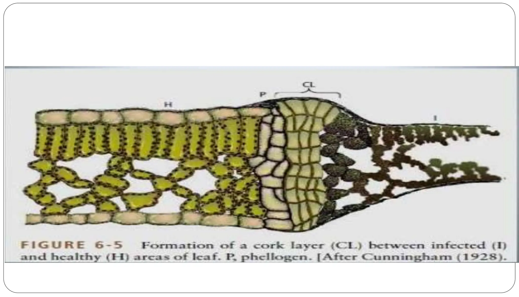
HOW PLANTS DEFEND THEMSELVES AGAINST PATHOGENS
Presented by-
Abhisek Tripathy
04PPT/PHD/17
DEFENCE MECHANISMS IN PLANTS
In general, plants defend themselves against pathogens
by a combination of weapons from two arsenals:
1) Structural characteristics that act as physical
barriers and inhibit the pathogen from gaining
entrance and spreading through the plant and
2) Biochemical reactions that take place in the cells
and tissues of the plant and produce substances that
are either toxic to the pathogen or create conditions
that inhibit growth of the pathogen in the plant.
 Two types
A. Pre formed or pre existing defense structures
B. Post infectional or induced defense structures
 Two types
A. Pre formed or pre existing biochemical
defense
B. Post infectional or induced biochemical
defense
1. Structural defence mechanisms
 A. Pre formed or pre existing defense structures
 The first line of defense of a plant against pathogens is its surface,which the
pathogen must adhere to and penetrate if it is to cause infection.
 Some structural defense are present in the plant even before the pathogen
comes in contact with the plant.
 Such structure include the amount and quality of wax and cuticle that cover
the epidermal cells, the structure of the epidermal cell walls, the size,
location and shapes of stomata and lenticels and
 The presence of tissues made of thick walled cells that hinder the advance
of pathogen on the plant.
 A thick mat of hairs on a plant surface may also exert a similar water-
repelling effect and may reduce infection.
 Eg. The stem rust of wheat can enter only when stomata are
open. Thus, some wheat varieties, in which the stomata open
late in the day are resistant because the germ tubes of spores
germinating in the night dew dessicate due to evaporation of the
dew before the stomata begin to open (fuctional resistance).
 Eg. The structure of stomata, e.g. ,a very narrow entrance and
broad, elevated guard cells may also confer resistance to some
varieties of mandarin against X. a. pv. Citri.
 Also, the xylem, bundle sheath, and cells of the leaf veins
effectively block the spread of some fungal, bacterial and
nematode pathogens that cause various “angular” leaf spots
because of their spread only into areas between, but not across,
veins.
B. Post infectional or induced defense structures
 Some of the defense structures formed involve the
cytoplasm of the cells under attack, and the cytoplasmic
defense reaction.
 Others invove the walls of invaded cells and are called cell
wall defense structures.
 Still others involve tissues ahead of the pathogen (deeper
into the plant) and are called histological defense
structures.
 Finally, the death of the invaded cell may protect the plant
from further invasion. This is called necrotic or
1. Cytoplasmic defense reaction
 The plant cell cytoplasm surrounds the clump of hyphae and the
plant cell nucleus is stretched to the point where it breaks in two.
 In some cells, the cytoplasmic reaction is overcome and the
protoplast dissappears while fungal growth increases.
 In some of the invaded cells, however the cytoplasm and nucleus
enlarge. The cytoplasm becomes granular and dense, and various
particles or structures appear in it.
 Finally, the mycelium of the pathogen disintegrates and the invasion
stops.
2. Cell wall defense structures
 The outer layer of cell wall of parenchyma cells coming in contact
with incompatible bacteria swells and produces an amorphous,
fimbrillar material that surrounds and traps the bacteria and
prevents them from multiplying.
 Cells walls thicken in response to several pathogens by producing
what appears to be a cellulosic materials. This material, however, is
often infused with phenoloic substances that are cross-linked and
further increase its resistance to penetration.
 Callose papillae are deposited on the inner side of cell walls in
response to invasion by fungal pathogens.
 Papillae seem to be produced by cells within minutes after
wounding and within 2 to 3 hours after inoculation with
microorganisms.
 Although the main function of papillae seems to be repair of
cellular damage, sometimes, especially if papillae are
present before inoculation, they also seem to prevent the
pathogen from subsequently penetrating the cell.
 In some cases, hyphal tips of fungi penetrating a cell wall
and growing into the cell lumen are enveloped by cellulosic
(callose) materials that later become infused with phenolic
substances and form a sheath or lignituber around the hypha
(Fig. 6-4).
3. Histological Defense Structures
A. Formation of Cork Layers
 Infection by fungi or bacteria, and even by some viruses and nematodes, frequently induces
plants to form several layers of cork cells beyond the point of infection apparently as a result of
stimulation of the host cells by substances secreted by the pathogen.
 The cork layers inhibit further invasion by the pathogen beyond the initial lesion and also block
the spread of any toxic substances that the pathogen may secrete.
 Furthermore, cork layers stop the flow of nutrients and water from the healthy to the infected area
and deprive the pathogen of nourishment.
 The dead tissues, including the pathogen, are thus delimited by the cork layers and may remain in
place, forming necrotic lesions (spots) that are remarkably uniform in size and shape for a
particular host–pathogen combination.
 In some host–pathogen combinations the necrotic tissues are pushed outward by the underlying
healthy tissues and form scabs that may be sloughed off, thus removing the pathogen from the
host completely.
 Eg. Cypress trees canker caused by the fungus Seiridium cardinale.
B. Formation of Abscission layers
 Abscission layers are formed on young, active leaves of stone fruit
trees after infection by any of several fungi, bacteria, or viruses.
 An abscission layer consists of a gap formed between two circular
layers of leaf cells surrounding the locus of infection.
 Upon infection, the middle lamella between these two layers of
cells is dissolved throughout the thickness of the leaf, completely
cutting off the central area of the infection from the rest of the leaf
(Fig. 6-7).
 Gradually, this area shrivels, dies, and sloughs off, carrying with it
the pathogen.
 Thus, the plant, by discarding the infected area along with a few
yet uninfected cells, protects the rest of the leaf tissue from being
invaded by the pathogen and from becoming affected by the toxic
C. Formation of Tyloses
 Tyloses are overgrowths of the protoplast of adjacent
living parenchymatous cells, which protrude into xylem
vessels through pits (Fig. 6-8).
 Tyloses have cellulosic walls and may, by their size and
numbers, clog the vessel completely.
 In some varieties of plants, tyloses form abundantly and
quickly ahead of the pathogen, while the pathogen is still
in the young roots, and block further advance of the
pathogen.
 The plants of these varieties remain free of and therefore
D. Deposition of Gums
 Various types of gums are produced by many plants
around lesions after infection by pathogens or injury.
 Gum secretion is most common in stone fruit trees but
occurs in most plants.
 The defensive role of gums stems from the fact that they
are deposited quickly in the intercellular spaces and
within the cells surrounding the locus of infection, thus
forming an impenetrable barrier that completely encloses
the pathogen.
 The pathogen then becomes isolated, starves, and
E. Necrotic Structural Defense Reaction through
the Hypersensitive Response
 In many host–pathogen combinations, as soon as the pathogen
establishes contact with the cell, the nucleus moves toward the
invading pathogen and soon disintegrates.
 At the same time, brown, resin-like granules form in the
cytoplasm, first around the point of penetration of the pathogen
and then throughout the cytoplasm.
 As the browning discoloration of the plant cell cytoplasm
continues and death sets in, the invading hypha begins to
degenerate (Fig. 6-9).
 In most cases the hypha does not grow out of such cells, and
further invasion is stopped.
 In bacterial infections of leaves, the hypersensitive response
results in the destruction of all cellular membranes of cells in
contact with bacteria, which is followed by desiccation and
 Apparently, the necrotic tissue not only isolates the
parasite from the living substance on which it
depends for its nutrition and, thereby, results in its
starvation and death,
 but, more importantly, it signifies the concentration
of numerous biochemical cell responses and
antimicrobial substances that neutralize the
pathogen.
 The faster the host cell dies after invasion, the
more resistant to infection the plant seems to be.
2. Biochemical defense mechanisms
 A. Pre formed or pre existing biochemical defense
 A particular pathogen will not infect certain plant varieties
even though no structural barriers of any kind seem to be
present or to form in these varieties.
 Similarly, in resistant varieties, the rate of disease
development oon slows down, and finally, in the absence of
structural defenses, the disease is completely checked.
 These examples suggest that defense mechanisms of a
chemical rather than a structural nature are responsible for
the resistance to infection exhibited by plants against certain
pathogens.
1. Inhibitors Released by the Plant in Its
Environment
 Plants exude a variety of substances through the surface of
their aboveground parts as well as through the surface of their
roots (Fungitoxic exudates)
 In the case of onion smudge, caused by the fungus
Colletotrichum circinans, resistant varieties generally have red
scales and contain, in addition to the red pigments, the
phenolic compounds protocatechuic acid and catechol.
 Both fungitoxic exudates and inhibition of infection are
missing in white-scaled, susceptible onion varieties (Fig. 6-2).
2. Inhibitors Present in Plant Cells before
Infection
 Some plants are resistant to diseases caused by certain
pathogens because of one or more inhibitory antimicrobial
compounds, known as phytoanticipins, which are present in the
cell before infection.
 Several phenolic compounds, tannins, and some fatty acid-
like compounds such as dienes, which are present in high
concentrations in cells of young fruits, leaves, or seeds, have
been proposed as responsible for the resistance of young tissues
to pathogenic microorganisms such as Botrytis.
 For example, increased 9-hexadecanoic acid in cutin monomers in
transgenic tomato plants led to resistance of such plants to
3. Lack of Recognition between Host and
Pathogen
 Plants of a species or variety may not become infected by a
pathogen if their surface cells lack specific recognition
factors (specific molecules or structures) that can be
recognized by the pathogen.
 If the pathogen does not recognize the plant as one of its
host plants, it may not become attached to the plant or may
notproduce infection substances, such as enzymes, or
structures, such as appressoria, penetration pegs, and
haustoria, necessary for the establishment of infection.
4. Lack of Host Receptors and Sensitive Sites for
Toxins
 In host–pathogen combinations in which the pathogen
(usually a fungus) produces a host-specific toxin, the toxin,
which is responsible for the symptoms, is thought to attach to
and react with specific receptors or sensitive sites in the cell.
 Only plants that have such sensitive receptors or sites
become diseased. Plants of other varieties or species that
lack such receptors or sites remain resistant to the toxin and
develop no symptoms.
5. Lack of Essential Substances for the
Pathogen
 Species or varieties of plants that for some reason do not produce one of
the substances essential for the survival of an obligate parasite, or for
development of infection by any parasite, would be resistant to the
pathogen that requires it.
 Thus, for Rhizoctonia to infect a plant it needs to obtain from the plant a
substance necessary for formation of a hyphal cushion from which the
fungus sends into the plant its penetration hyphae.
 In plants in which this substance is apparently lacking, cushions do not
form, infection does not occur, and the plants are resistant.
 The fungus does not normally form hyphal cushions in pure cultures but
forms them when extracts from a susceptible but not a resistant plant are
added to the culture.
 Eg. bacterial soft rot of potatoes, causedby Erwinia carotovora var.
atroseptica, is less severe on potatoes with low-reducing sugar content
B. Post infectional or induced biochemical
defense
 Defense through Production of Secondary Metabolites:
Phenolics
 Phytoalexins
 Phytoalexins are toxic antimicrobial substances produced in
appreciable amounts in plants only after stimulation by various
types of phytopathogenic microorganisms or by chemical and
mechanical injury.
 Phytoalexins are produced by healthy cells adjacent to localized
damaged and necrotic cells in response to materials diffusing
from the damaged cells.
 Phytoalexins accumulate around both resistant and susceptible
necrotic tissues. Resistance occurs when one or more
phytoalexins reach a concentration sufficient to restrict pathogen
development.
 Most known phytoalexins are toxic to and inhibit the growth of
fungi pathogenic to plants, but some are also toxic to bacteria,
 The chemical structures of phytoalexins produced by
plants of a family are usually quite similar;
 e.g., in most legumes, phytoalexins are isoflavonoids, and
in the Solanaceae they are terpenoids.
 Some of the better studied phytoalexins include
phaseollin in bean (Fig. 6-21); pisatin in pea; glyceollin in
soybean, alfalfa, and clover; rishitin in potato; gossypol in
cotton; and capsidiol in pepper.
2. Production of Antimicrobial Substances in
Attacked Host Cells
Pathogenesis-Related Proteins
 Pathogenesis-related proteins, often called PR proteins, are a
structurally diverse group of plant proteins that are toxic to
invading fungal pathogens.
 They are widely distributed in plants in trace amounts, but are
produced in much greater concentration following pathogen
attack or stress.
 PR proteins exist in plant cells intracellularly and also in the
intercellular spaces, particularly in the cell walls of different
 The several groups of PR proteins have been classified according to their
function, serological relationship, amino acid sequence, molecular weight, and
certain other properties.
 PR proteins are either extremely acidic or extremely basic and therefore are
highly soluble and reactive.
 At least 14 families of PR proteins are recognized.
 The better known PR proteins are PR1 proteins (antioomycete and antifungal),
PR2 (b-1,3-glucanases), PR3 (chitinases), PR4 proteins (antifungal), PR6
(proteinase inhibitors), thaumatine-like proteins, defensins, thionins, lysozymes,
osmotinlike proteins, lipoxygenases, cysteine-rich proteins, glycine-rich proteins,
proteinases, chitosanases, and peroxidases.
3.Immunization of plants against pathogens
Defense through Plantibodies
 Antibodies, encoded by animal genes but produced in and by the plant, are called
plantibodies.
 It has already been shown that transgenic plants producing plantibodies against coat
proteins of viruses, e.g., artichoke mottle crinkle virus, to which they are susceptible,
can defend themselves and show some resistance to infection by these viruses.
 Plants such as tobacco, potato, and pea have been shown to be good producers of
antibody for pharmaceutical purposes.
 Plants have been shown to produce functional antibodies that can be used to increase
the resistance of plants against specific pathogens.
 Eg. 1. Plantibodies to tobacco mosaic virus in tobacco
decreased infectivity of the virus by 90%
 Eg. 2. Plantibodies to beet necrotic yellow vein virus,
also in tobacco, provides a partial protection against
the virus in the early stages of infection and against
development of symptoms later on;
 Eg. 3. Plantibodies to stolbur phytoplasma and to corn
stunt spiroplasma, also in tobacco, which remained
free from infection for more than two months.
4. DETOXIFICATION OF PATHOGEN TOXINS
BY PLANTS
 In at least some of the diseases in which the pathogen produces a
toxin, resistance to disease is apparently the same as resistance
to the toxin.
 Detoxification of at least some toxins, e.g., HC toxin and
pyricularin, produced by the fungi Cochliobolus carbonum and
Magnaporthe grisea, respectively, is known to occur in plants
and may play a role in disease resistance.
 Some of these toxins appear to be metabolized more rapidly by
resistant varieties or are combined with other substances and form
less toxic or nontoxic compounds.
 The amount of the nontoxic compound formed is often
proportional to the disease resistance of the variety.
 Resistant plants and nonhosts are not affected by the specific
toxins produced by Cochliobolus, Periconia, and Alternaria.
5) Disruption of host cell membrane:
 Some structural and permeability changes in host cell membrane play a role in
defense.
Most common are -
 Release of molecules in signal transduction around cell and systemically.
 Release and accumulation of reactive oxygen 'radicals' and lipoxygenase enzyme.
 Activation of phenol oxidases and oxidation of phenolic due to loss of comport
meutalisation.
6) Oxidative burst and release of Nitric oxide (NO):
 Rapid generation of activated oxygen radicals, superoxide (O2-),
 Hydrogen peroxide (H2O2), Hydroxylradical (OH) released by multisubunit
NADPH enzymes complex of cell membrane.
 Hydroperoxidation of membrane phospholipids to lipid hydroperoxides.
 Disrupt cell membrane and induce cell colapse and death.
7) Strengthening of host cell wall resistance:
 Accumulation of defense related substances in cell wall. e.g.
Callose, glycoproteins rich in hydroxy proline, phenolics
(lignin, suberin) and mineral like silicon and calcium. Form
complex polymer and cross link with one-another forming
insoluble cell wall structures.
8) Production of anti-microbials in attacked cells:
Common phenolics-
 Produced and accumulated after infection in a resistant variety.
 Chlorogenic acid, caffeic acid and ferulic acid
 Combined effect, rather than individual.
CONCLUSIONS
1) Plants defened to pathogens at pre-germination and
germination stage with structural defense (pre-existing and
induced).
2)When plants fails to defense pathogen with structural
defense the biochemical defense mechanism is activated.
3) When pathogen elicitors reach to the host receptors, signal
transduction takes place and a cascade of several structural
and bio-chemical responses starts.
4)Plant defense is not the result of a single mechanism but
the cumulative effect of more than one mechanisms.
5)All the defense exhibited by the plants are governed by it's
References
• Agrios, G.N. (1988). Plant Pathology. Academic Press, New
York, pp. 803.
• Gareth Jones, D. (1989). Plant Pathology, Principles and
Practices. Aditya Books, New Delhi, pp. 191.
• Singh, R.S. (1984). Introduction to Principles of Plant
Pathology. Oxford and IBH Publishing Co., New Delhi, pp.
534.
• Walker, J.C. (1969). Plant Pathology. Mc Graw-Hill, Inc. New
York, (Indian Edition), pp. 818.
THANK YOU

defensemechanismsinplants-180308104711.pptx

  • 1.
    HOW PLANTS DEFENDTHEMSELVES AGAINST PATHOGENS Presented by- Abhisek Tripathy 04PPT/PHD/17 DEFENCE MECHANISMS IN PLANTS
  • 2.
    In general, plantsdefend themselves against pathogens by a combination of weapons from two arsenals: 1) Structural characteristics that act as physical barriers and inhibit the pathogen from gaining entrance and spreading through the plant and 2) Biochemical reactions that take place in the cells and tissues of the plant and produce substances that are either toxic to the pathogen or create conditions that inhibit growth of the pathogen in the plant.
  • 3.
     Two types A.Pre formed or pre existing defense structures B. Post infectional or induced defense structures
  • 4.
     Two types A.Pre formed or pre existing biochemical defense B. Post infectional or induced biochemical defense
  • 5.
    1. Structural defencemechanisms  A. Pre formed or pre existing defense structures  The first line of defense of a plant against pathogens is its surface,which the pathogen must adhere to and penetrate if it is to cause infection.  Some structural defense are present in the plant even before the pathogen comes in contact with the plant.  Such structure include the amount and quality of wax and cuticle that cover the epidermal cells, the structure of the epidermal cell walls, the size, location and shapes of stomata and lenticels and  The presence of tissues made of thick walled cells that hinder the advance of pathogen on the plant.  A thick mat of hairs on a plant surface may also exert a similar water- repelling effect and may reduce infection.
  • 6.
     Eg. Thestem rust of wheat can enter only when stomata are open. Thus, some wheat varieties, in which the stomata open late in the day are resistant because the germ tubes of spores germinating in the night dew dessicate due to evaporation of the dew before the stomata begin to open (fuctional resistance).  Eg. The structure of stomata, e.g. ,a very narrow entrance and broad, elevated guard cells may also confer resistance to some varieties of mandarin against X. a. pv. Citri.  Also, the xylem, bundle sheath, and cells of the leaf veins effectively block the spread of some fungal, bacterial and nematode pathogens that cause various “angular” leaf spots because of their spread only into areas between, but not across, veins.
  • 7.
    B. Post infectionalor induced defense structures  Some of the defense structures formed involve the cytoplasm of the cells under attack, and the cytoplasmic defense reaction.  Others invove the walls of invaded cells and are called cell wall defense structures.  Still others involve tissues ahead of the pathogen (deeper into the plant) and are called histological defense structures.  Finally, the death of the invaded cell may protect the plant from further invasion. This is called necrotic or
  • 8.
    1. Cytoplasmic defensereaction  The plant cell cytoplasm surrounds the clump of hyphae and the plant cell nucleus is stretched to the point where it breaks in two.  In some cells, the cytoplasmic reaction is overcome and the protoplast dissappears while fungal growth increases.  In some of the invaded cells, however the cytoplasm and nucleus enlarge. The cytoplasm becomes granular and dense, and various particles or structures appear in it.  Finally, the mycelium of the pathogen disintegrates and the invasion stops.
  • 9.
    2. Cell walldefense structures  The outer layer of cell wall of parenchyma cells coming in contact with incompatible bacteria swells and produces an amorphous, fimbrillar material that surrounds and traps the bacteria and prevents them from multiplying.  Cells walls thicken in response to several pathogens by producing what appears to be a cellulosic materials. This material, however, is often infused with phenoloic substances that are cross-linked and further increase its resistance to penetration.  Callose papillae are deposited on the inner side of cell walls in response to invasion by fungal pathogens.
  • 10.
     Papillae seemto be produced by cells within minutes after wounding and within 2 to 3 hours after inoculation with microorganisms.  Although the main function of papillae seems to be repair of cellular damage, sometimes, especially if papillae are present before inoculation, they also seem to prevent the pathogen from subsequently penetrating the cell.  In some cases, hyphal tips of fungi penetrating a cell wall and growing into the cell lumen are enveloped by cellulosic (callose) materials that later become infused with phenolic substances and form a sheath or lignituber around the hypha (Fig. 6-4).
  • 12.
    3. Histological DefenseStructures A. Formation of Cork Layers  Infection by fungi or bacteria, and even by some viruses and nematodes, frequently induces plants to form several layers of cork cells beyond the point of infection apparently as a result of stimulation of the host cells by substances secreted by the pathogen.  The cork layers inhibit further invasion by the pathogen beyond the initial lesion and also block the spread of any toxic substances that the pathogen may secrete.  Furthermore, cork layers stop the flow of nutrients and water from the healthy to the infected area and deprive the pathogen of nourishment.  The dead tissues, including the pathogen, are thus delimited by the cork layers and may remain in place, forming necrotic lesions (spots) that are remarkably uniform in size and shape for a particular host–pathogen combination.  In some host–pathogen combinations the necrotic tissues are pushed outward by the underlying healthy tissues and form scabs that may be sloughed off, thus removing the pathogen from the host completely.  Eg. Cypress trees canker caused by the fungus Seiridium cardinale.
  • 15.
    B. Formation ofAbscission layers  Abscission layers are formed on young, active leaves of stone fruit trees after infection by any of several fungi, bacteria, or viruses.  An abscission layer consists of a gap formed between two circular layers of leaf cells surrounding the locus of infection.  Upon infection, the middle lamella between these two layers of cells is dissolved throughout the thickness of the leaf, completely cutting off the central area of the infection from the rest of the leaf (Fig. 6-7).  Gradually, this area shrivels, dies, and sloughs off, carrying with it the pathogen.  Thus, the plant, by discarding the infected area along with a few yet uninfected cells, protects the rest of the leaf tissue from being invaded by the pathogen and from becoming affected by the toxic
  • 17.
    C. Formation ofTyloses  Tyloses are overgrowths of the protoplast of adjacent living parenchymatous cells, which protrude into xylem vessels through pits (Fig. 6-8).  Tyloses have cellulosic walls and may, by their size and numbers, clog the vessel completely.  In some varieties of plants, tyloses form abundantly and quickly ahead of the pathogen, while the pathogen is still in the young roots, and block further advance of the pathogen.  The plants of these varieties remain free of and therefore
  • 19.
    D. Deposition ofGums  Various types of gums are produced by many plants around lesions after infection by pathogens or injury.  Gum secretion is most common in stone fruit trees but occurs in most plants.  The defensive role of gums stems from the fact that they are deposited quickly in the intercellular spaces and within the cells surrounding the locus of infection, thus forming an impenetrable barrier that completely encloses the pathogen.  The pathogen then becomes isolated, starves, and
  • 20.
    E. Necrotic StructuralDefense Reaction through the Hypersensitive Response  In many host–pathogen combinations, as soon as the pathogen establishes contact with the cell, the nucleus moves toward the invading pathogen and soon disintegrates.  At the same time, brown, resin-like granules form in the cytoplasm, first around the point of penetration of the pathogen and then throughout the cytoplasm.  As the browning discoloration of the plant cell cytoplasm continues and death sets in, the invading hypha begins to degenerate (Fig. 6-9).  In most cases the hypha does not grow out of such cells, and further invasion is stopped.  In bacterial infections of leaves, the hypersensitive response results in the destruction of all cellular membranes of cells in contact with bacteria, which is followed by desiccation and
  • 23.
     Apparently, thenecrotic tissue not only isolates the parasite from the living substance on which it depends for its nutrition and, thereby, results in its starvation and death,  but, more importantly, it signifies the concentration of numerous biochemical cell responses and antimicrobial substances that neutralize the pathogen.  The faster the host cell dies after invasion, the more resistant to infection the plant seems to be.
  • 24.
    2. Biochemical defensemechanisms  A. Pre formed or pre existing biochemical defense  A particular pathogen will not infect certain plant varieties even though no structural barriers of any kind seem to be present or to form in these varieties.  Similarly, in resistant varieties, the rate of disease development oon slows down, and finally, in the absence of structural defenses, the disease is completely checked.  These examples suggest that defense mechanisms of a chemical rather than a structural nature are responsible for the resistance to infection exhibited by plants against certain pathogens.
  • 25.
    1. Inhibitors Releasedby the Plant in Its Environment  Plants exude a variety of substances through the surface of their aboveground parts as well as through the surface of their roots (Fungitoxic exudates)  In the case of onion smudge, caused by the fungus Colletotrichum circinans, resistant varieties generally have red scales and contain, in addition to the red pigments, the phenolic compounds protocatechuic acid and catechol.  Both fungitoxic exudates and inhibition of infection are missing in white-scaled, susceptible onion varieties (Fig. 6-2).
  • 27.
    2. Inhibitors Presentin Plant Cells before Infection  Some plants are resistant to diseases caused by certain pathogens because of one or more inhibitory antimicrobial compounds, known as phytoanticipins, which are present in the cell before infection.  Several phenolic compounds, tannins, and some fatty acid- like compounds such as dienes, which are present in high concentrations in cells of young fruits, leaves, or seeds, have been proposed as responsible for the resistance of young tissues to pathogenic microorganisms such as Botrytis.  For example, increased 9-hexadecanoic acid in cutin monomers in transgenic tomato plants led to resistance of such plants to
  • 28.
    3. Lack ofRecognition between Host and Pathogen  Plants of a species or variety may not become infected by a pathogen if their surface cells lack specific recognition factors (specific molecules or structures) that can be recognized by the pathogen.  If the pathogen does not recognize the plant as one of its host plants, it may not become attached to the plant or may notproduce infection substances, such as enzymes, or structures, such as appressoria, penetration pegs, and haustoria, necessary for the establishment of infection.
  • 29.
    4. Lack ofHost Receptors and Sensitive Sites for Toxins  In host–pathogen combinations in which the pathogen (usually a fungus) produces a host-specific toxin, the toxin, which is responsible for the symptoms, is thought to attach to and react with specific receptors or sensitive sites in the cell.  Only plants that have such sensitive receptors or sites become diseased. Plants of other varieties or species that lack such receptors or sites remain resistant to the toxin and develop no symptoms.
  • 30.
    5. Lack ofEssential Substances for the Pathogen  Species or varieties of plants that for some reason do not produce one of the substances essential for the survival of an obligate parasite, or for development of infection by any parasite, would be resistant to the pathogen that requires it.  Thus, for Rhizoctonia to infect a plant it needs to obtain from the plant a substance necessary for formation of a hyphal cushion from which the fungus sends into the plant its penetration hyphae.  In plants in which this substance is apparently lacking, cushions do not form, infection does not occur, and the plants are resistant.  The fungus does not normally form hyphal cushions in pure cultures but forms them when extracts from a susceptible but not a resistant plant are added to the culture.  Eg. bacterial soft rot of potatoes, causedby Erwinia carotovora var. atroseptica, is less severe on potatoes with low-reducing sugar content
  • 31.
    B. Post infectionalor induced biochemical defense  Defense through Production of Secondary Metabolites: Phenolics  Phytoalexins
  • 32.
     Phytoalexins aretoxic antimicrobial substances produced in appreciable amounts in plants only after stimulation by various types of phytopathogenic microorganisms or by chemical and mechanical injury.  Phytoalexins are produced by healthy cells adjacent to localized damaged and necrotic cells in response to materials diffusing from the damaged cells.  Phytoalexins accumulate around both resistant and susceptible necrotic tissues. Resistance occurs when one or more phytoalexins reach a concentration sufficient to restrict pathogen development.  Most known phytoalexins are toxic to and inhibit the growth of fungi pathogenic to plants, but some are also toxic to bacteria,
  • 33.
     The chemicalstructures of phytoalexins produced by plants of a family are usually quite similar;  e.g., in most legumes, phytoalexins are isoflavonoids, and in the Solanaceae they are terpenoids.  Some of the better studied phytoalexins include phaseollin in bean (Fig. 6-21); pisatin in pea; glyceollin in soybean, alfalfa, and clover; rishitin in potato; gossypol in cotton; and capsidiol in pepper.
  • 34.
    2. Production ofAntimicrobial Substances in Attacked Host Cells Pathogenesis-Related Proteins  Pathogenesis-related proteins, often called PR proteins, are a structurally diverse group of plant proteins that are toxic to invading fungal pathogens.  They are widely distributed in plants in trace amounts, but are produced in much greater concentration following pathogen attack or stress.  PR proteins exist in plant cells intracellularly and also in the intercellular spaces, particularly in the cell walls of different
  • 35.
     The severalgroups of PR proteins have been classified according to their function, serological relationship, amino acid sequence, molecular weight, and certain other properties.  PR proteins are either extremely acidic or extremely basic and therefore are highly soluble and reactive.  At least 14 families of PR proteins are recognized.  The better known PR proteins are PR1 proteins (antioomycete and antifungal), PR2 (b-1,3-glucanases), PR3 (chitinases), PR4 proteins (antifungal), PR6 (proteinase inhibitors), thaumatine-like proteins, defensins, thionins, lysozymes, osmotinlike proteins, lipoxygenases, cysteine-rich proteins, glycine-rich proteins, proteinases, chitosanases, and peroxidases.
  • 36.
    3.Immunization of plantsagainst pathogens Defense through Plantibodies  Antibodies, encoded by animal genes but produced in and by the plant, are called plantibodies.  It has already been shown that transgenic plants producing plantibodies against coat proteins of viruses, e.g., artichoke mottle crinkle virus, to which they are susceptible, can defend themselves and show some resistance to infection by these viruses.  Plants such as tobacco, potato, and pea have been shown to be good producers of antibody for pharmaceutical purposes.  Plants have been shown to produce functional antibodies that can be used to increase the resistance of plants against specific pathogens.
  • 37.
     Eg. 1.Plantibodies to tobacco mosaic virus in tobacco decreased infectivity of the virus by 90%  Eg. 2. Plantibodies to beet necrotic yellow vein virus, also in tobacco, provides a partial protection against the virus in the early stages of infection and against development of symptoms later on;  Eg. 3. Plantibodies to stolbur phytoplasma and to corn stunt spiroplasma, also in tobacco, which remained free from infection for more than two months.
  • 38.
    4. DETOXIFICATION OFPATHOGEN TOXINS BY PLANTS  In at least some of the diseases in which the pathogen produces a toxin, resistance to disease is apparently the same as resistance to the toxin.  Detoxification of at least some toxins, e.g., HC toxin and pyricularin, produced by the fungi Cochliobolus carbonum and Magnaporthe grisea, respectively, is known to occur in plants and may play a role in disease resistance.  Some of these toxins appear to be metabolized more rapidly by resistant varieties or are combined with other substances and form less toxic or nontoxic compounds.  The amount of the nontoxic compound formed is often proportional to the disease resistance of the variety.  Resistant plants and nonhosts are not affected by the specific toxins produced by Cochliobolus, Periconia, and Alternaria.
  • 39.
    5) Disruption ofhost cell membrane:  Some structural and permeability changes in host cell membrane play a role in defense. Most common are -  Release of molecules in signal transduction around cell and systemically.  Release and accumulation of reactive oxygen 'radicals' and lipoxygenase enzyme.  Activation of phenol oxidases and oxidation of phenolic due to loss of comport meutalisation. 6) Oxidative burst and release of Nitric oxide (NO):  Rapid generation of activated oxygen radicals, superoxide (O2-),  Hydrogen peroxide (H2O2), Hydroxylradical (OH) released by multisubunit NADPH enzymes complex of cell membrane.  Hydroperoxidation of membrane phospholipids to lipid hydroperoxides.  Disrupt cell membrane and induce cell colapse and death.
  • 40.
    7) Strengthening ofhost cell wall resistance:  Accumulation of defense related substances in cell wall. e.g. Callose, glycoproteins rich in hydroxy proline, phenolics (lignin, suberin) and mineral like silicon and calcium. Form complex polymer and cross link with one-another forming insoluble cell wall structures. 8) Production of anti-microbials in attacked cells: Common phenolics-  Produced and accumulated after infection in a resistant variety.  Chlorogenic acid, caffeic acid and ferulic acid  Combined effect, rather than individual.
  • 41.
    CONCLUSIONS 1) Plants defenedto pathogens at pre-germination and germination stage with structural defense (pre-existing and induced). 2)When plants fails to defense pathogen with structural defense the biochemical defense mechanism is activated. 3) When pathogen elicitors reach to the host receptors, signal transduction takes place and a cascade of several structural and bio-chemical responses starts. 4)Plant defense is not the result of a single mechanism but the cumulative effect of more than one mechanisms. 5)All the defense exhibited by the plants are governed by it's
  • 42.
    References • Agrios, G.N.(1988). Plant Pathology. Academic Press, New York, pp. 803. • Gareth Jones, D. (1989). Plant Pathology, Principles and Practices. Aditya Books, New Delhi, pp. 191. • Singh, R.S. (1984). Introduction to Principles of Plant Pathology. Oxford and IBH Publishing Co., New Delhi, pp. 534. • Walker, J.C. (1969). Plant Pathology. Mc Graw-Hill, Inc. New York, (Indian Edition), pp. 818.
  • 43.