SlideShare a Scribd company logo
ANATOMY OF
PARAPHARYNGEAL SPACE
AND ITS DISEASE
GUEST----- HOD AND PROFF DR KAVITA
SACHDEVA MADAM
SPEAKER-----DR JOSHNA THAKUR
PARAPHARYNGEAL SPACE
(lateral pharyngeal space, pharyngomaxillary space)
 It is classically described as an inverted pyramidal shaped potential space
.
 Bounderies Base lies superiorly at the skull base and comprises the
sphenoid and petrous portion of temporal bone .
 Apex ----inferiorly at the level of greater cornu hyoid bone.
 Medial surface---comprises superior pharyngeal constrictor muscle
,buccopharyngeal membrane and the pharynx.
 Lateral suface-----medial pterygoid muscle,the ramus of mandible,deep
lobe of parotid gland and fascia of post. Belly of diagastric.
 Posterior surface----bordered by carotid sheath
FASCIAL CONDENSATION
 Two fascial condensation are present in parapharyngeal
space
 1. Aponeurosis of Zuckerkandl and Testut which divide the
paraphyrngeal space into 2 compartment by joining the
styloid process to the tensor veli palatini.
 2. Second condensation of cervical fascia form a band which
extend from styloid process to angle and post. Border of
mandible and form a stylomandibular tunnel
 Tumors of deep lobe parotid gland can extend into
parapharyngeal space through this tunnel giving rise to a
dumbbell shaped tumour.
…
Compartment of parapharyngeal space
COMPARTMENT VESSELS NERVES Others
PRESTYLOID 1 Maxillary artery
2 Ascending
pharyngeal artery
1 Auriculotempora
l nerve
2 Lingual nerve
3 Inferior alveolar
nerve.
Fat,connective
tissue,
Deep aspect of
parotid gland
Lymph nodes
POSTSTYLOID 1 ICA
2 Internal jugular
vein
1 Cranial nerve
9th,10th,11th,12th,
2 Cervical
sympathetic chain.
Lymph node of
glomus bodies
CLINICAL SIGNIFICANCE
 Radiological features-----
 Displacement pattern of fat and internal carotid artery within the space
will aid in the localization of lesion.
 In prestyloid space lesion---mc associated with deep lobe of parotid
gland and will deflect the carotid sheath and paraphyrngeal fat
posterolaterally and may have a rim of fat anteromedially
 In poststyloid space lesion---- include neuroendocrine origin arising
from carotid sheath as carotid body tumors or vagal schwannomas or
neuroma of sympathetic chain.Lesion displaces parapharyngeal space
fat and internal carotid artery anteromedially.
TUMOURS OF
PARAPHARYNGEAL SPACE
PRIMARY TUMOUR METASTATIC LESIONS MISCELLANEOUS
1 salivary gland tumour(benign
and malignant
SCC of nasopharynx lipoma
2Neurogenic neoplasm
vagal schwannoma
sympathetic chain Sch.
vagal paragangliomas
neurofibromas
SCC of oral cavity sarcoma
3 Vascular and Lymphatic
lesios ----- ---carotid artery
aneurysm brachial cleft
cyst --Hemangioma --
-A V malformation
--Lymphatic malformation
Carcinoma of oropharynx lymphoma
Carcinoma of thyroid gland teratoma
Sign and Symptoms
1 Neck mass
2 Oropharngeal mass
3 Dyspnea
4 Cranial nerve deficit
5 Dysphgia or odynophagia may present.
 6 Unilateral Conductive hearing loss due to ET blockage
 7 Hoarseness of voice due to vagus nerve involvement
 8 Horner syndrom due to sympathetic chain involvement
 9 Rhinolalia clausia due to oropharyngeal component.
 10 Trismus due to pterygoid muscle involvement.
 11 Cranial nerve deficits
 12 symptoms of catecholamines excess
…
 13. tumor involve prestyloid area
 displace lateral pharyngeal wall and tonsil medially.
 lump behind the angle of mandible.
 14. tumour involve poststyloid area
 fusiform appearance of tumour eg nerve sheath tumour.
 Intracranial extension via carotid canal ,jugular foramen,foramen ovale.
....
...
EXAMINATION
 Detailed head and neck examination.
 Cranial nerve examination.
 Bimanual palpation
 Bruit / thrill
 Carotid tumour moves side to side but fixed when moved up and down.
 Vitals ----blood pressure ,pulse rate,
Salivary gland neoplasms
 Neoplasms of salivary gland origin are located in the prestyloid parapharyngeal
space (PPS) and account for 40-50% of PPS lesions.
 Salivary neoplasms may arise from the deep lobe of the parotid gland, ectopic
salivary rests, or minor salivary glands of the lateral pharyngeal wall.
 The most common prestyloid PPS lesion is pleomorphic adenoma, which
represents 45-64% of salivary neoplasms in the PPS.
 Common benign neoplasms include pleomorphic adenomas, monomorphic
adenomas, Warthin tumors, and oncocytomas.
 Malignant neoplasms include adenoid cystic carcinomas, mucoepidermoid
carcinomas, adenocarcinomas, and acinic cell carcinomas. Approximately 20% of
all salivary lesions in the PPS are malignant, with carcinoma ex pleomorphic
adenoma and adenoid cystic carcinoma being the most frequently reported.
SCHWANNOMAS
 Neurilemmomas, or schwannomas, arise from any nerve surrounded by Schwann
cells and fibroblast supporting the nerve.
 Present in patients between 30 nd 70 yrs of age.
 In the PPS, the most common sites of origin are the vagus nerve and the
sympathetic chain.
 They grow slowly and rarely cause palsy of the nerve of origin.
 Nerve paraesthesia common in these.
 Neurilemmomas are encapsulated and histologically distinct from the nerve itself.
 Treatment is by enucleation, and preservation of the nerve of origin is usually
possible; however, every patient should be cautioned about the possibility of
postoperative paralysis
PARAGANGLIOMAS
 Paragangliomas are benign vascular neoplasms that arise from the paraganglia or extra-
adrenal neural crest tissue.
 Paraganglia function as chemoreceptors and are associated with the carotid body, the jugular
bulb, and the vagus nerve in the poststyloid PPS.
 Highly vascular tumor.
 Carotid body tumors, glomus jugulare, and glomus vagale are slow-growing paragangliomas
that may not produce symptoms
 may cause cranial nerve (CN) deficits, bone erosion, or intracranial extension as they increase
in size.
 These are mobile in lateral direction not in cephalocaudal direction.
 Approximately 2% of head and neck paragangliomas secrete catecholamines and may cause
paroxysmal symptoms of catecholamine excess.
..
 Ten percent of paragangliomas are multiple and associated
with paraganglioma at other locations.
 Ten percent of paragangliomas are hereditary, associated with
a familial paraganglioma syndrome. ( genetic origin related to SHD sucinyl
dehydroge gene locus)
 Head and Neck paraganglioma may also be associated with MEN 2a and MEN 2b
syndromes
 Hypertension and flushing are suggestive of either a secreting
paraganglioma or an associated pheochromocytoma.
 Malignant paragangliomas occur in less than 5-13.5% of patients
and are associated with rapid growth and development of metastatic
disease
 Carotid para ganglioma mobile in lateral direction but not in
cephalocaudal direction , which differentiated it from lymph
node.(fontaine sign)
CAROTID BODY TUMOUR
 Carotid body tumour, also known as a chemodectoma or carotid body
paraganglioma, are highly vascular tumors arising from the paraganglion cells of the
carotid body. These tumors are located at the site of the carotid bifurcation.
 Shamblin classification helps in predicting the prognosis and difficulties for surgical
resection
 Shamblin classification
 Shamblin, et al. in 1971 classified carotid body tumors in relation to the carotid vessels.
In his classification
 Group I – includes localized tumors not involving the carotid vessels.
 Group II – tumors partially surround carotid vessels or is adherent to them.
 Group III – defined as larger tumors encasing the carotid vessels.
 As carotid body tumors become larger in size they get more adherent to the vessels.
However, Shamblin classification had some limitations because it does not reflect how
deep tumor infiltrates through the wall of carotid vessels, which decides if it is possible to
preserve the carotid vessels.
…
 Modified Shamblin classification
 Luna-Ortiz, et al. in 2006 proposed modifications to Shamblin classification, to improve the
prediction of difficulties and complications.
 They introduced a class IIIa, representing the old class III, and a class IIIb which includes
tumors of any class (I, II, or III) where there is infiltration of the vessel wall and not just
circumferential encasement.
..
 Need for vascular resection and reconstruction was significantly higher among
class IIIb tumors.
.
NEUROFIBROMA
 Neurofibromas, in contrast, are unencapsulated and intimately involved with the
nerve of origin.
 They may occur as a manifestation of the neurofibromatosis-1 (NF-1) syndrome.
 a disease in which the incidence of malignant transformation is increased.
 Cystic degeneration is common.
 The nerve of origin is usually sacrificed during excision to ensure complete
removal of the neoplasm.
METASTASIS TO THE PARAPHARYNGEAL
SPACE
 In parapharyngeal space lymph nodes are present which drain the mucosa of the
upper aerodigestive tract and viscera of the neck .
 The most common metastasis to the parapharngeal space is from nasopharyngeal
cancer .
 Nodes may also involved in tonsil and tongue base cancers and maxillary sinus
cancer.
 Other primary sites include thyroid and parotid glands.
 Space may be directly invaded by malignancy from nasopharnx, tonsil, retromolar
trigone, palate and tongue base.
Lymphoreticular lesions
 Lymphoreticular lesions make up 10-15% of PPS lesions. [3, 22] The lymphatics of
the PPS may be involved primarily or secondarily by carcinoma, or they may be
involved by infectious or inflammatory processes. Lymphoma is the most common
malignant lymphoid process, but metastases from thyroid cancer, osteogenic
sarcoma, squamous cell carcinoma, renal cell carcinoma, hypernephroma, and
meningioma may also appear as PPS masses. The most common lymphoreticular
lesions are lymphomas and metastases
INVESTIGATION
 Fine slice CT and MRI provides the detail reqired.
 MR Angiography and Carotid angiography provide information regarding vascularity and
relationship to the neurovascular structures.
 FNAC
 BIOPSY ( EXCISIONAL , INCISIONAL—FROZEN SECTION,TRANSORAL FNAC IS
SAFE AND INTRAORAL INCISIONAL BIOPSY MUST NOT BE CARRIED OUT.)
 BALLON OCCLUSION TEST
 24 HOURS VMA(VANILLYLMANDELIC ACID) ---measures amount of VMA in urine over24
hr to detect excess epinephrine and norepinephrine.
 PLASMA CATECHOLAMINES LEVELS.
 METAIODOBENZLGUANIDINE SCAN. (MIBG) ---radioactive material injected intravenous
,which is takeup by tumour cells and this is sccaned by a scanner to locate and confirm
the presence of tumour.
..
 In imaging (MRI) paraganglioma gives salt and pepper appearance
 LYRE SIGN (splaying of internal and external carotid arteries) in vascular carotid body tumor.
 CT Scan shows evidence of bony details and erosion by these tumours.
 MRI Scan show soft tissue extension of these tumours including intracranial.extension.
 MRI Scan with gadolinium also show involvement of nerves and perineural spread..
.
.
.
.
.
PATIENT
SURGEON
Comobidies
Unwilling
age
Experience
Equipment
Postop care
Natural
history
Histological
nature
resectibility
PRE OPERATION
 Pre op counselling
 Preop embolization----------24 hrs prior to surgery, tumor >3cm with obvious
feeding vessels
 Alpha blockers, Beta blockers------functioning paraganglioma.
SURGICAL APPROACHES FOR
PARAPHARYNGEAL TUMOURS
SURGICAL APPROACHES
1 TRANSCERVICAL APPROACH
 It is the most commonly utilized approach.used in about 46% of cases.
 It is ideal for small benign tumors independent of deep lobe involvement of parotid gland.
 For poststyloid and prestyloid space tumours.
 Incision given 2 finger(5 cm) breadth below mandible.
 Submandibular triangle exposed (if needed)
 Facial nerve branches preserved.
 Facial artery ligated and divided.
 Submandibular gland retracted or excised.
 Digastric tendon divided
 Mandibulectomy done (if needed)
Transcervical approach
TRANSCERVICAL TRANSPAROTID
APPROACH
 Next most common approach. Used in 27% cases.
 For lesion involve deep lobe of parotid tumours, vascular tumours.
 Doubly modified blair incision or slight anterior extension (in front of ear) of cervical
incision i .
 Same procedure as in transcervical approach.
 Division of stylomandibular ligament ,styloglossus,stylohyoid muscle
 Deep parotid lobe tumours
MANDIBULOTOMY
 Transcervical and transcervical transparotid approaches can be combined with
a mandibular osteotomy.
 Better visual asses to superior aspect of parapharyngeal space and the skull
base.
 Inverted L OSTEOTOMY or double osteotomy above lingual and in front of the
mental foramen to protect the inferior alveolar nerve.
 For excision of infiltrating malignancy and multifocal recurrent benign tumours.
TRANSORAL ROBOTIC SURGERY
(TORS)
1 It has technical advantage ,it being the 3D high resolution image with magnification.
2 Scaled movement and tremor filtration allow for deldicated dissection around tumour
capsule ensuring resection with a margin.
3 Ideal for benign salivary gland tumour in prestyloid space.
4 Absolute contraindication tumour that is adherent or involving the carotid artery, vascular
tumor ,dumbbell tumor with significant involvement of deep lobe of parotid gland.
RADIOTHERAPY
 POST OP radiotherapy ---extensive malignant head and neck tumours
 Irresectable tumours.
 Control/ Arrest the growth of tumours.
 Nasopharyngeal tumour.
 Recurrent deep lobe of parotid tumour.
 Regional lymph node metastasis.
 Paraganglioma
CHEMOTHERAPY
 RHABDOMYOSARCOMA AND OTHER SARCOMAS
 NASOPHARYNGEAL TUMOUR.
 DOXORUBICIN---GLANDULAR TUMOUR
 CISPLATIN AND 5 FLUOROURACIL----SQUMOUS CELL TUMOURS
COMPLICATION
 VASCULAR INJURY
 CRANIAL NERVE INJURIES
 TUMOUR SPILLAGE AND RECURRENCE.
 FIRST BITE SYNDROME
 HEMATOMAS
 MARGINAL MANDIBULAR NERVE INJURY
Anatomy of parapharyngeal space and its tumours

More Related Content

What's hot

Cavity obliteration @ sayan
Cavity obliteration  @ sayanCavity obliteration  @ sayan
Cavity obliteration @ sayan
IPGMER
 
surgical approaches to frontal sinus ppt
surgical approaches to frontal sinus pptsurgical approaches to frontal sinus ppt
surgical approaches to frontal sinus ppt
Vaibhav Lahane
 
Frontal sinus surgical aproach
Frontal sinus surgical aproachFrontal sinus surgical aproach
Frontal sinus surgical aproach
Azadmeena7
 
Spaces of middle ear and their surgical importance
Spaces of middle ear  and their surgical importanceSpaces of middle ear  and their surgical importance
Spaces of middle ear and their surgical importance
Dr Soumya Singh
 
Temporal bone neoplasms
Temporal bone neoplasmsTemporal bone neoplasms
Temporal bone neoplasms
abhijeet89singh
 
Glomus Tumour and its Approaches
Glomus Tumour and its ApproachesGlomus Tumour and its Approaches
Glomus Tumour and its Approaches
Lady Hardinge Medical College
 
Facial nerve decompression
Facial nerve decompressionFacial nerve decompression
Facial nerve decompression
Mamoon Ameen
 
NASOPHARYNGEAL CARCINOMA
NASOPHARYNGEAL CARCINOMA NASOPHARYNGEAL CARCINOMA
NASOPHARYNGEAL CARCINOMA
Mamoon Ameen
 
Hadad.bassagasteguy flap
Hadad.bassagasteguy flap Hadad.bassagasteguy flap
Hadad.bassagasteguy flap
Chandra Veer Suryavanshi
 
Infratemporal fossa approaches
Infratemporal fossa approachesInfratemporal fossa approaches
Infratemporal fossa approaches
Md Roohia
 
SURGICAL ANATOMY OF DEEP NECK SPACES
SURGICAL ANATOMY OF DEEP NECK SPACESSURGICAL ANATOMY OF DEEP NECK SPACES
SURGICAL ANATOMY OF DEEP NECK SPACES
Ajay Manickam
 
Parapharyngeal tumor
Parapharyngeal tumorParapharyngeal tumor
Parapharyngeal tumor
Balasubramanian Thiagarajan
 
Retropharyngeal space
Retropharyngeal spaceRetropharyngeal space
Retropharyngeal space
Dr Safika Zaman
 
Tumours of hypopharynx
Tumours of hypopharynxTumours of hypopharynx
Tumours of hypopharynxVinay Bhat
 
Differential diagnosis of nasal mass
Differential diagnosis of nasal massDifferential diagnosis of nasal mass
Differential diagnosis of nasal mass
Sharath Chandra
 
Surgical anatomy of Infratemporal fossa. by Dr. Aditya Tiwari
Surgical anatomy of Infratemporal fossa. by Dr. Aditya TiwariSurgical anatomy of Infratemporal fossa. by Dr. Aditya Tiwari
Surgical anatomy of Infratemporal fossa. by Dr. Aditya Tiwari
Aditya Tiwari
 
Sinus tympani prof dr bikash
Sinus tympani prof dr bikashSinus tympani prof dr bikash
Sinus tympani prof dr bikash
Bikash Shrestha
 
Stroboscopy
StroboscopyStroboscopy
Stroboscopy
Sanjay Maharjan
 

What's hot (20)

Cavity obliteration @ sayan
Cavity obliteration  @ sayanCavity obliteration  @ sayan
Cavity obliteration @ sayan
 
surgical approaches to frontal sinus ppt
surgical approaches to frontal sinus pptsurgical approaches to frontal sinus ppt
surgical approaches to frontal sinus ppt
 
Frontal sinus surgical aproach
Frontal sinus surgical aproachFrontal sinus surgical aproach
Frontal sinus surgical aproach
 
Spaces of middle ear and their surgical importance
Spaces of middle ear  and their surgical importanceSpaces of middle ear  and their surgical importance
Spaces of middle ear and their surgical importance
 
Petrous apex and skull base
Petrous apex and skull basePetrous apex and skull base
Petrous apex and skull base
 
Temporal bone neoplasms
Temporal bone neoplasmsTemporal bone neoplasms
Temporal bone neoplasms
 
Glomus Tumour and its Approaches
Glomus Tumour and its ApproachesGlomus Tumour and its Approaches
Glomus Tumour and its Approaches
 
Facial nerve decompression
Facial nerve decompressionFacial nerve decompression
Facial nerve decompression
 
NASOPHARYNGEAL CARCINOMA
NASOPHARYNGEAL CARCINOMA NASOPHARYNGEAL CARCINOMA
NASOPHARYNGEAL CARCINOMA
 
Hadad.bassagasteguy flap
Hadad.bassagasteguy flap Hadad.bassagasteguy flap
Hadad.bassagasteguy flap
 
Infratemporal fossa approaches
Infratemporal fossa approachesInfratemporal fossa approaches
Infratemporal fossa approaches
 
SURGICAL ANATOMY OF DEEP NECK SPACES
SURGICAL ANATOMY OF DEEP NECK SPACESSURGICAL ANATOMY OF DEEP NECK SPACES
SURGICAL ANATOMY OF DEEP NECK SPACES
 
Parapharyngeal tumor
Parapharyngeal tumorParapharyngeal tumor
Parapharyngeal tumor
 
Retropharyngeal space
Retropharyngeal spaceRetropharyngeal space
Retropharyngeal space
 
Tumours of hypopharynx
Tumours of hypopharynxTumours of hypopharynx
Tumours of hypopharynx
 
Differential diagnosis of nasal mass
Differential diagnosis of nasal massDifferential diagnosis of nasal mass
Differential diagnosis of nasal mass
 
Petrous apex 360°
Petrous apex 360°Petrous apex 360°
Petrous apex 360°
 
Surgical anatomy of Infratemporal fossa. by Dr. Aditya Tiwari
Surgical anatomy of Infratemporal fossa. by Dr. Aditya TiwariSurgical anatomy of Infratemporal fossa. by Dr. Aditya Tiwari
Surgical anatomy of Infratemporal fossa. by Dr. Aditya Tiwari
 
Sinus tympani prof dr bikash
Sinus tympani prof dr bikashSinus tympani prof dr bikash
Sinus tympani prof dr bikash
 
Stroboscopy
StroboscopyStroboscopy
Stroboscopy
 

Similar to Anatomy of parapharyngeal space and its tumours

Parapharyngeal swellings
Parapharyngeal swellingsParapharyngeal swellings
Parapharyngeal swellings
Mai Hatem
 
Tumours of nasopharynx (2) itp class dr.davis - 03.06.16
Tumours of nasopharynx (2) itp class   dr.davis - 03.06.16Tumours of nasopharynx (2) itp class   dr.davis - 03.06.16
Tumours of nasopharynx (2) itp class dr.davis - 03.06.16
ophthalmgmcri
 
Sino-nasal malignancy
Sino-nasal malignancySino-nasal malignancy
Sino-nasal malignancy
Dr Safika Zaman
 
pharyngeal Tumors
pharyngeal Tumorspharyngeal Tumors
pharyngeal Tumors
AmeenaAjam1
 
Pre management salivary gland by dr pallavi jain
Pre management salivary gland by dr pallavi jainPre management salivary gland by dr pallavi jain
Pre management salivary gland by dr pallavi jain
Dr. Pallavi Jain
 
Unusual non epithelial tumors of head and neck
Unusual non epithelial tumors of head and neckUnusual non epithelial tumors of head and neck
Unusual non epithelial tumors of head and neck
DrAyush Garg
 
paragangliomas
paragangliomasparagangliomas
Central nervous system
Central nervous systemCentral nervous system
Central nervous system
Xayneb Zia
 
Glomus Tumours
Glomus TumoursGlomus Tumours
Glomus Tumours
Khalidmsayed
 
Study of parapharyngeal tumors
Study of parapharyngeal tumorsStudy of parapharyngeal tumors
Study of parapharyngeal tumors
Dr.Juveria Majeed
 
Meninges and related tumors
Meninges and related tumorsMeninges and related tumors
Meninges and related tumors
Bahoran Singh Rajput
 
Pathology of pharynx
Pathology of pharynxPathology of pharynx
Pathology of pharynxDilnia Qader
 
Benign brain tumours
Benign brain tumoursBenign brain tumours
Nasopharyngeal Carcinoma
Nasopharyngeal CarcinomaNasopharyngeal Carcinoma
Nasopharyngeal Carcinoma
Balasubramanian Thiagarajan
 
Salivary Gland Cytology case of adenoid cyst carcinoma
Salivary Gland Cytology case of adenoid cyst carcinoma Salivary Gland Cytology case of adenoid cyst carcinoma
Salivary Gland Cytology case of adenoid cyst carcinoma
SEJOJO PHAAROE
 
Problem oriented approach in pediatric radiology
Problem oriented approach in pediatric radiologyProblem oriented approach in pediatric radiology
Problem oriented approach in pediatric radiology
Ahmed Bahnassy
 
IMAGING OF SPINAL TUMORS
IMAGING OF SPINAL TUMORS IMAGING OF SPINAL TUMORS
IMAGING OF SPINAL TUMORS
aasrithakotha2
 

Similar to Anatomy of parapharyngeal space and its tumours (20)

Tumours of the parapharyngeal space
Tumours of the parapharyngeal spaceTumours of the parapharyngeal space
Tumours of the parapharyngeal space
 
Parapharyngeal swellings
Parapharyngeal swellingsParapharyngeal swellings
Parapharyngeal swellings
 
Tumours of nasopharynx (2) itp class dr.davis - 03.06.16
Tumours of nasopharynx (2) itp class   dr.davis - 03.06.16Tumours of nasopharynx (2) itp class   dr.davis - 03.06.16
Tumours of nasopharynx (2) itp class dr.davis - 03.06.16
 
Sino-nasal malignancy
Sino-nasal malignancySino-nasal malignancy
Sino-nasal malignancy
 
pharyngeal Tumors
pharyngeal Tumorspharyngeal Tumors
pharyngeal Tumors
 
Pre management salivary gland by dr pallavi jain
Pre management salivary gland by dr pallavi jainPre management salivary gland by dr pallavi jain
Pre management salivary gland by dr pallavi jain
 
Unusual non epithelial tumors of head and neck
Unusual non epithelial tumors of head and neckUnusual non epithelial tumors of head and neck
Unusual non epithelial tumors of head and neck
 
paragangliomas
paragangliomasparagangliomas
paragangliomas
 
Central nervous system
Central nervous systemCentral nervous system
Central nervous system
 
Glomus Tumours
Glomus TumoursGlomus Tumours
Glomus Tumours
 
caso 2
caso 2caso 2
caso 2
 
Study of parapharyngeal tumors
Study of parapharyngeal tumorsStudy of parapharyngeal tumors
Study of parapharyngeal tumors
 
Meninges and related tumors
Meninges and related tumorsMeninges and related tumors
Meninges and related tumors
 
Pathology of pharynx
Pathology of pharynxPathology of pharynx
Pathology of pharynx
 
Benign brain tumours
Benign brain tumoursBenign brain tumours
Benign brain tumours
 
Nasopharyngeal Carcinoma
Nasopharyngeal CarcinomaNasopharyngeal Carcinoma
Nasopharyngeal Carcinoma
 
Cases 12 fna 7
Cases 12 fna  7Cases 12 fna  7
Cases 12 fna 7
 
Salivary Gland Cytology case of adenoid cyst carcinoma
Salivary Gland Cytology case of adenoid cyst carcinoma Salivary Gland Cytology case of adenoid cyst carcinoma
Salivary Gland Cytology case of adenoid cyst carcinoma
 
Problem oriented approach in pediatric radiology
Problem oriented approach in pediatric radiologyProblem oriented approach in pediatric radiology
Problem oriented approach in pediatric radiology
 
IMAGING OF SPINAL TUMORS
IMAGING OF SPINAL TUMORS IMAGING OF SPINAL TUMORS
IMAGING OF SPINAL TUMORS
 

Recently uploaded

Triangles of Neck and Clinical Correlation by Dr. RIG.pptx
Triangles of Neck and Clinical Correlation by Dr. RIG.pptxTriangles of Neck and Clinical Correlation by Dr. RIG.pptx
Triangles of Neck and Clinical Correlation by Dr. RIG.pptx
Dr. Rabia Inam Gandapore
 
basicmodesofventilation2022-220313203758.pdf
basicmodesofventilation2022-220313203758.pdfbasicmodesofventilation2022-220313203758.pdf
basicmodesofventilation2022-220313203758.pdf
aljamhori teaching hospital
 
Superficial & Deep Fascia of the NECK.pptx
Superficial & Deep Fascia of the NECK.pptxSuperficial & Deep Fascia of the NECK.pptx
Superficial & Deep Fascia of the NECK.pptx
Dr. Rabia Inam Gandapore
 
THOA 2.ppt Human Organ Transplantation Act
THOA 2.ppt Human Organ Transplantation ActTHOA 2.ppt Human Organ Transplantation Act
THOA 2.ppt Human Organ Transplantation Act
DrSathishMS1
 
24 Upakrama.pptx class ppt useful in all
24 Upakrama.pptx class ppt useful in all24 Upakrama.pptx class ppt useful in all
24 Upakrama.pptx class ppt useful in all
DrSathishMS1
 
Flu Vaccine Alert in Bangalore Karnataka
Flu Vaccine Alert in Bangalore KarnatakaFlu Vaccine Alert in Bangalore Karnataka
Flu Vaccine Alert in Bangalore Karnataka
addon Scans
 
Report Back from SGO 2024: What’s the Latest in Cervical Cancer?
Report Back from SGO 2024: What’s the Latest in Cervical Cancer?Report Back from SGO 2024: What’s the Latest in Cervical Cancer?
Report Back from SGO 2024: What’s the Latest in Cervical Cancer?
bkling
 
Alcohol_Dr. Jeenal Mistry MD Pharmacology.pdf
Alcohol_Dr. Jeenal Mistry MD Pharmacology.pdfAlcohol_Dr. Jeenal Mistry MD Pharmacology.pdf
Alcohol_Dr. Jeenal Mistry MD Pharmacology.pdf
Dr Jeenal Mistry
 
ARTHROLOGY PPT NCISM SYLLABUS AYURVEDA STUDENTS
ARTHROLOGY PPT NCISM SYLLABUS AYURVEDA STUDENTSARTHROLOGY PPT NCISM SYLLABUS AYURVEDA STUDENTS
ARTHROLOGY PPT NCISM SYLLABUS AYURVEDA STUDENTS
Dr. Vinay Pareek
 
ANATOMY AND PHYSIOLOGY OF URINARY SYSTEM.pptx
ANATOMY AND PHYSIOLOGY OF URINARY SYSTEM.pptxANATOMY AND PHYSIOLOGY OF URINARY SYSTEM.pptx
ANATOMY AND PHYSIOLOGY OF URINARY SYSTEM.pptx
Swetaba Besh
 
BRACHYTHERAPY OVERVIEW AND APPLICATORS
BRACHYTHERAPY OVERVIEW  AND  APPLICATORSBRACHYTHERAPY OVERVIEW  AND  APPLICATORS
BRACHYTHERAPY OVERVIEW AND APPLICATORS
Krishan Murari
 
Surgical Site Infections, pathophysiology, and prevention.pptx
Surgical Site Infections, pathophysiology, and prevention.pptxSurgical Site Infections, pathophysiology, and prevention.pptx
Surgical Site Infections, pathophysiology, and prevention.pptx
jval Landero
 
Maxilla, Mandible & Hyoid Bone & Clinical Correlations by Dr. RIG.pptx
Maxilla, Mandible & Hyoid Bone & Clinical Correlations by Dr. RIG.pptxMaxilla, Mandible & Hyoid Bone & Clinical Correlations by Dr. RIG.pptx
Maxilla, Mandible & Hyoid Bone & Clinical Correlations by Dr. RIG.pptx
Dr. Rabia Inam Gandapore
 
KDIGO 2024 guidelines for diabetologists
KDIGO 2024 guidelines for diabetologistsKDIGO 2024 guidelines for diabetologists
KDIGO 2024 guidelines for diabetologists
د.محمود نجيب
 
Are There Any Natural Remedies To Treat Syphilis.pdf
Are There Any Natural Remedies To Treat Syphilis.pdfAre There Any Natural Remedies To Treat Syphilis.pdf
Are There Any Natural Remedies To Treat Syphilis.pdf
Little Cross Family Clinic
 
For Better Surat #ℂall #Girl Service ❤85270-49040❤ Surat #ℂall #Girls
For Better Surat #ℂall #Girl Service ❤85270-49040❤ Surat #ℂall #GirlsFor Better Surat #ℂall #Girl Service ❤85270-49040❤ Surat #ℂall #Girls
For Better Surat #ℂall #Girl Service ❤85270-49040❤ Surat #ℂall #Girls
Savita Shen $i11
 
MANAGEMENT OF ATRIOVENTRICULAR CONDUCTION BLOCK.pdf
MANAGEMENT OF ATRIOVENTRICULAR CONDUCTION BLOCK.pdfMANAGEMENT OF ATRIOVENTRICULAR CONDUCTION BLOCK.pdf
MANAGEMENT OF ATRIOVENTRICULAR CONDUCTION BLOCK.pdf
Jim Jacob Roy
 
Cervical & Brachial Plexus By Dr. RIG.pptx
Cervical & Brachial Plexus By Dr. RIG.pptxCervical & Brachial Plexus By Dr. RIG.pptx
Cervical & Brachial Plexus By Dr. RIG.pptx
Dr. Rabia Inam Gandapore
 
Ophthalmology Clinical Tests for OSCE exam
Ophthalmology Clinical Tests for OSCE examOphthalmology Clinical Tests for OSCE exam
Ophthalmology Clinical Tests for OSCE exam
KafrELShiekh University
 
Tom Selleck Health: A Comprehensive Look at the Iconic Actor’s Wellness Journey
Tom Selleck Health: A Comprehensive Look at the Iconic Actor’s Wellness JourneyTom Selleck Health: A Comprehensive Look at the Iconic Actor’s Wellness Journey
Tom Selleck Health: A Comprehensive Look at the Iconic Actor’s Wellness Journey
greendigital
 

Recently uploaded (20)

Triangles of Neck and Clinical Correlation by Dr. RIG.pptx
Triangles of Neck and Clinical Correlation by Dr. RIG.pptxTriangles of Neck and Clinical Correlation by Dr. RIG.pptx
Triangles of Neck and Clinical Correlation by Dr. RIG.pptx
 
basicmodesofventilation2022-220313203758.pdf
basicmodesofventilation2022-220313203758.pdfbasicmodesofventilation2022-220313203758.pdf
basicmodesofventilation2022-220313203758.pdf
 
Superficial & Deep Fascia of the NECK.pptx
Superficial & Deep Fascia of the NECK.pptxSuperficial & Deep Fascia of the NECK.pptx
Superficial & Deep Fascia of the NECK.pptx
 
THOA 2.ppt Human Organ Transplantation Act
THOA 2.ppt Human Organ Transplantation ActTHOA 2.ppt Human Organ Transplantation Act
THOA 2.ppt Human Organ Transplantation Act
 
24 Upakrama.pptx class ppt useful in all
24 Upakrama.pptx class ppt useful in all24 Upakrama.pptx class ppt useful in all
24 Upakrama.pptx class ppt useful in all
 
Flu Vaccine Alert in Bangalore Karnataka
Flu Vaccine Alert in Bangalore KarnatakaFlu Vaccine Alert in Bangalore Karnataka
Flu Vaccine Alert in Bangalore Karnataka
 
Report Back from SGO 2024: What’s the Latest in Cervical Cancer?
Report Back from SGO 2024: What’s the Latest in Cervical Cancer?Report Back from SGO 2024: What’s the Latest in Cervical Cancer?
Report Back from SGO 2024: What’s the Latest in Cervical Cancer?
 
Alcohol_Dr. Jeenal Mistry MD Pharmacology.pdf
Alcohol_Dr. Jeenal Mistry MD Pharmacology.pdfAlcohol_Dr. Jeenal Mistry MD Pharmacology.pdf
Alcohol_Dr. Jeenal Mistry MD Pharmacology.pdf
 
ARTHROLOGY PPT NCISM SYLLABUS AYURVEDA STUDENTS
ARTHROLOGY PPT NCISM SYLLABUS AYURVEDA STUDENTSARTHROLOGY PPT NCISM SYLLABUS AYURVEDA STUDENTS
ARTHROLOGY PPT NCISM SYLLABUS AYURVEDA STUDENTS
 
ANATOMY AND PHYSIOLOGY OF URINARY SYSTEM.pptx
ANATOMY AND PHYSIOLOGY OF URINARY SYSTEM.pptxANATOMY AND PHYSIOLOGY OF URINARY SYSTEM.pptx
ANATOMY AND PHYSIOLOGY OF URINARY SYSTEM.pptx
 
BRACHYTHERAPY OVERVIEW AND APPLICATORS
BRACHYTHERAPY OVERVIEW  AND  APPLICATORSBRACHYTHERAPY OVERVIEW  AND  APPLICATORS
BRACHYTHERAPY OVERVIEW AND APPLICATORS
 
Surgical Site Infections, pathophysiology, and prevention.pptx
Surgical Site Infections, pathophysiology, and prevention.pptxSurgical Site Infections, pathophysiology, and prevention.pptx
Surgical Site Infections, pathophysiology, and prevention.pptx
 
Maxilla, Mandible & Hyoid Bone & Clinical Correlations by Dr. RIG.pptx
Maxilla, Mandible & Hyoid Bone & Clinical Correlations by Dr. RIG.pptxMaxilla, Mandible & Hyoid Bone & Clinical Correlations by Dr. RIG.pptx
Maxilla, Mandible & Hyoid Bone & Clinical Correlations by Dr. RIG.pptx
 
KDIGO 2024 guidelines for diabetologists
KDIGO 2024 guidelines for diabetologistsKDIGO 2024 guidelines for diabetologists
KDIGO 2024 guidelines for diabetologists
 
Are There Any Natural Remedies To Treat Syphilis.pdf
Are There Any Natural Remedies To Treat Syphilis.pdfAre There Any Natural Remedies To Treat Syphilis.pdf
Are There Any Natural Remedies To Treat Syphilis.pdf
 
For Better Surat #ℂall #Girl Service ❤85270-49040❤ Surat #ℂall #Girls
For Better Surat #ℂall #Girl Service ❤85270-49040❤ Surat #ℂall #GirlsFor Better Surat #ℂall #Girl Service ❤85270-49040❤ Surat #ℂall #Girls
For Better Surat #ℂall #Girl Service ❤85270-49040❤ Surat #ℂall #Girls
 
MANAGEMENT OF ATRIOVENTRICULAR CONDUCTION BLOCK.pdf
MANAGEMENT OF ATRIOVENTRICULAR CONDUCTION BLOCK.pdfMANAGEMENT OF ATRIOVENTRICULAR CONDUCTION BLOCK.pdf
MANAGEMENT OF ATRIOVENTRICULAR CONDUCTION BLOCK.pdf
 
Cervical & Brachial Plexus By Dr. RIG.pptx
Cervical & Brachial Plexus By Dr. RIG.pptxCervical & Brachial Plexus By Dr. RIG.pptx
Cervical & Brachial Plexus By Dr. RIG.pptx
 
Ophthalmology Clinical Tests for OSCE exam
Ophthalmology Clinical Tests for OSCE examOphthalmology Clinical Tests for OSCE exam
Ophthalmology Clinical Tests for OSCE exam
 
Tom Selleck Health: A Comprehensive Look at the Iconic Actor’s Wellness Journey
Tom Selleck Health: A Comprehensive Look at the Iconic Actor’s Wellness JourneyTom Selleck Health: A Comprehensive Look at the Iconic Actor’s Wellness Journey
Tom Selleck Health: A Comprehensive Look at the Iconic Actor’s Wellness Journey
 

Anatomy of parapharyngeal space and its tumours

  • 1. ANATOMY OF PARAPHARYNGEAL SPACE AND ITS DISEASE GUEST----- HOD AND PROFF DR KAVITA SACHDEVA MADAM SPEAKER-----DR JOSHNA THAKUR
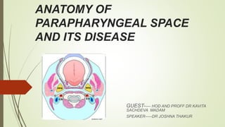
  • 2. PARAPHARYNGEAL SPACE (lateral pharyngeal space, pharyngomaxillary space)  It is classically described as an inverted pyramidal shaped potential space .  Bounderies Base lies superiorly at the skull base and comprises the sphenoid and petrous portion of temporal bone .  Apex ----inferiorly at the level of greater cornu hyoid bone.  Medial surface---comprises superior pharyngeal constrictor muscle ,buccopharyngeal membrane and the pharynx.  Lateral suface-----medial pterygoid muscle,the ramus of mandible,deep lobe of parotid gland and fascia of post. Belly of diagastric.  Posterior surface----bordered by carotid sheath
  • 3. FASCIAL CONDENSATION  Two fascial condensation are present in parapharyngeal space  1. Aponeurosis of Zuckerkandl and Testut which divide the paraphyrngeal space into 2 compartment by joining the styloid process to the tensor veli palatini.  2. Second condensation of cervical fascia form a band which extend from styloid process to angle and post. Border of mandible and form a stylomandibular tunnel  Tumors of deep lobe parotid gland can extend into parapharyngeal space through this tunnel giving rise to a dumbbell shaped tumour.
  • 4.
  • 5. Compartment of parapharyngeal space COMPARTMENT VESSELS NERVES Others PRESTYLOID 1 Maxillary artery 2 Ascending pharyngeal artery 1 Auriculotempora l nerve 2 Lingual nerve 3 Inferior alveolar nerve. Fat,connective tissue, Deep aspect of parotid gland Lymph nodes POSTSTYLOID 1 ICA 2 Internal jugular vein 1 Cranial nerve 9th,10th,11th,12th, 2 Cervical sympathetic chain. Lymph node of glomus bodies
  • 6. CLINICAL SIGNIFICANCE  Radiological features-----  Displacement pattern of fat and internal carotid artery within the space will aid in the localization of lesion.  In prestyloid space lesion---mc associated with deep lobe of parotid gland and will deflect the carotid sheath and paraphyrngeal fat posterolaterally and may have a rim of fat anteromedially  In poststyloid space lesion---- include neuroendocrine origin arising from carotid sheath as carotid body tumors or vagal schwannomas or neuroma of sympathetic chain.Lesion displaces parapharyngeal space fat and internal carotid artery anteromedially.
  • 8. PRIMARY TUMOUR METASTATIC LESIONS MISCELLANEOUS 1 salivary gland tumour(benign and malignant SCC of nasopharynx lipoma 2Neurogenic neoplasm vagal schwannoma sympathetic chain Sch. vagal paragangliomas neurofibromas SCC of oral cavity sarcoma 3 Vascular and Lymphatic lesios ----- ---carotid artery aneurysm brachial cleft cyst --Hemangioma -- -A V malformation --Lymphatic malformation Carcinoma of oropharynx lymphoma Carcinoma of thyroid gland teratoma
  • 9. Sign and Symptoms 1 Neck mass 2 Oropharngeal mass 3 Dyspnea 4 Cranial nerve deficit 5 Dysphgia or odynophagia may present.  6 Unilateral Conductive hearing loss due to ET blockage  7 Hoarseness of voice due to vagus nerve involvement  8 Horner syndrom due to sympathetic chain involvement  9 Rhinolalia clausia due to oropharyngeal component.  10 Trismus due to pterygoid muscle involvement.  11 Cranial nerve deficits  12 symptoms of catecholamines excess
  • 10. …  13. tumor involve prestyloid area  displace lateral pharyngeal wall and tonsil medially.  lump behind the angle of mandible.  14. tumour involve poststyloid area  fusiform appearance of tumour eg nerve sheath tumour.  Intracranial extension via carotid canal ,jugular foramen,foramen ovale.
  • 11. ....
  • 12. ...
  • 13. EXAMINATION  Detailed head and neck examination.  Cranial nerve examination.  Bimanual palpation  Bruit / thrill  Carotid tumour moves side to side but fixed when moved up and down.  Vitals ----blood pressure ,pulse rate,
  • 14. Salivary gland neoplasms  Neoplasms of salivary gland origin are located in the prestyloid parapharyngeal space (PPS) and account for 40-50% of PPS lesions.  Salivary neoplasms may arise from the deep lobe of the parotid gland, ectopic salivary rests, or minor salivary glands of the lateral pharyngeal wall.  The most common prestyloid PPS lesion is pleomorphic adenoma, which represents 45-64% of salivary neoplasms in the PPS.  Common benign neoplasms include pleomorphic adenomas, monomorphic adenomas, Warthin tumors, and oncocytomas.  Malignant neoplasms include adenoid cystic carcinomas, mucoepidermoid carcinomas, adenocarcinomas, and acinic cell carcinomas. Approximately 20% of all salivary lesions in the PPS are malignant, with carcinoma ex pleomorphic adenoma and adenoid cystic carcinoma being the most frequently reported.
  • 15. SCHWANNOMAS  Neurilemmomas, or schwannomas, arise from any nerve surrounded by Schwann cells and fibroblast supporting the nerve.  Present in patients between 30 nd 70 yrs of age.  In the PPS, the most common sites of origin are the vagus nerve and the sympathetic chain.  They grow slowly and rarely cause palsy of the nerve of origin.  Nerve paraesthesia common in these.  Neurilemmomas are encapsulated and histologically distinct from the nerve itself.  Treatment is by enucleation, and preservation of the nerve of origin is usually possible; however, every patient should be cautioned about the possibility of postoperative paralysis
  • 16. PARAGANGLIOMAS  Paragangliomas are benign vascular neoplasms that arise from the paraganglia or extra- adrenal neural crest tissue.  Paraganglia function as chemoreceptors and are associated with the carotid body, the jugular bulb, and the vagus nerve in the poststyloid PPS.  Highly vascular tumor.  Carotid body tumors, glomus jugulare, and glomus vagale are slow-growing paragangliomas that may not produce symptoms  may cause cranial nerve (CN) deficits, bone erosion, or intracranial extension as they increase in size.  These are mobile in lateral direction not in cephalocaudal direction.  Approximately 2% of head and neck paragangliomas secrete catecholamines and may cause paroxysmal symptoms of catecholamine excess.
  • 17. ..  Ten percent of paragangliomas are multiple and associated with paraganglioma at other locations.  Ten percent of paragangliomas are hereditary, associated with a familial paraganglioma syndrome. ( genetic origin related to SHD sucinyl dehydroge gene locus)  Head and Neck paraganglioma may also be associated with MEN 2a and MEN 2b syndromes  Hypertension and flushing are suggestive of either a secreting paraganglioma or an associated pheochromocytoma.  Malignant paragangliomas occur in less than 5-13.5% of patients and are associated with rapid growth and development of metastatic disease  Carotid para ganglioma mobile in lateral direction but not in cephalocaudal direction , which differentiated it from lymph node.(fontaine sign)
  • 18. CAROTID BODY TUMOUR  Carotid body tumour, also known as a chemodectoma or carotid body paraganglioma, are highly vascular tumors arising from the paraganglion cells of the carotid body. These tumors are located at the site of the carotid bifurcation.  Shamblin classification helps in predicting the prognosis and difficulties for surgical resection  Shamblin classification  Shamblin, et al. in 1971 classified carotid body tumors in relation to the carotid vessels. In his classification  Group I – includes localized tumors not involving the carotid vessels.  Group II – tumors partially surround carotid vessels or is adherent to them.  Group III – defined as larger tumors encasing the carotid vessels.  As carotid body tumors become larger in size they get more adherent to the vessels. However, Shamblin classification had some limitations because it does not reflect how deep tumor infiltrates through the wall of carotid vessels, which decides if it is possible to preserve the carotid vessels.
  • 19. …  Modified Shamblin classification  Luna-Ortiz, et al. in 2006 proposed modifications to Shamblin classification, to improve the prediction of difficulties and complications.  They introduced a class IIIa, representing the old class III, and a class IIIb which includes tumors of any class (I, II, or III) where there is infiltration of the vessel wall and not just circumferential encasement.
  • 20. ..  Need for vascular resection and reconstruction was significantly higher among class IIIb tumors.
  • 21. .
  • 22. NEUROFIBROMA  Neurofibromas, in contrast, are unencapsulated and intimately involved with the nerve of origin.  They may occur as a manifestation of the neurofibromatosis-1 (NF-1) syndrome.  a disease in which the incidence of malignant transformation is increased.  Cystic degeneration is common.  The nerve of origin is usually sacrificed during excision to ensure complete removal of the neoplasm.
  • 23. METASTASIS TO THE PARAPHARYNGEAL SPACE  In parapharyngeal space lymph nodes are present which drain the mucosa of the upper aerodigestive tract and viscera of the neck .  The most common metastasis to the parapharngeal space is from nasopharyngeal cancer .  Nodes may also involved in tonsil and tongue base cancers and maxillary sinus cancer.  Other primary sites include thyroid and parotid glands.  Space may be directly invaded by malignancy from nasopharnx, tonsil, retromolar trigone, palate and tongue base.
  • 24. Lymphoreticular lesions  Lymphoreticular lesions make up 10-15% of PPS lesions. [3, 22] The lymphatics of the PPS may be involved primarily or secondarily by carcinoma, or they may be involved by infectious or inflammatory processes. Lymphoma is the most common malignant lymphoid process, but metastases from thyroid cancer, osteogenic sarcoma, squamous cell carcinoma, renal cell carcinoma, hypernephroma, and meningioma may also appear as PPS masses. The most common lymphoreticular lesions are lymphomas and metastases
  • 25. INVESTIGATION  Fine slice CT and MRI provides the detail reqired.  MR Angiography and Carotid angiography provide information regarding vascularity and relationship to the neurovascular structures.  FNAC  BIOPSY ( EXCISIONAL , INCISIONAL—FROZEN SECTION,TRANSORAL FNAC IS SAFE AND INTRAORAL INCISIONAL BIOPSY MUST NOT BE CARRIED OUT.)  BALLON OCCLUSION TEST  24 HOURS VMA(VANILLYLMANDELIC ACID) ---measures amount of VMA in urine over24 hr to detect excess epinephrine and norepinephrine.  PLASMA CATECHOLAMINES LEVELS.  METAIODOBENZLGUANIDINE SCAN. (MIBG) ---radioactive material injected intravenous ,which is takeup by tumour cells and this is sccaned by a scanner to locate and confirm the presence of tumour.
  • 26. ..  In imaging (MRI) paraganglioma gives salt and pepper appearance  LYRE SIGN (splaying of internal and external carotid arteries) in vascular carotid body tumor.  CT Scan shows evidence of bony details and erosion by these tumours.  MRI Scan show soft tissue extension of these tumours including intracranial.extension.  MRI Scan with gadolinium also show involvement of nerves and perineural spread..
  • 27. .
  • 28. .
  • 29. .
  • 30. .
  • 31. .
  • 33. PRE OPERATION  Pre op counselling  Preop embolization----------24 hrs prior to surgery, tumor >3cm with obvious feeding vessels  Alpha blockers, Beta blockers------functioning paraganglioma.
  • 36. 1 TRANSCERVICAL APPROACH  It is the most commonly utilized approach.used in about 46% of cases.  It is ideal for small benign tumors independent of deep lobe involvement of parotid gland.  For poststyloid and prestyloid space tumours.  Incision given 2 finger(5 cm) breadth below mandible.  Submandibular triangle exposed (if needed)  Facial nerve branches preserved.  Facial artery ligated and divided.  Submandibular gland retracted or excised.  Digastric tendon divided  Mandibulectomy done (if needed)
  • 38. TRANSCERVICAL TRANSPAROTID APPROACH  Next most common approach. Used in 27% cases.  For lesion involve deep lobe of parotid tumours, vascular tumours.  Doubly modified blair incision or slight anterior extension (in front of ear) of cervical incision i .  Same procedure as in transcervical approach.  Division of stylomandibular ligament ,styloglossus,stylohyoid muscle  Deep parotid lobe tumours
  • 39. MANDIBULOTOMY  Transcervical and transcervical transparotid approaches can be combined with a mandibular osteotomy.  Better visual asses to superior aspect of parapharyngeal space and the skull base.  Inverted L OSTEOTOMY or double osteotomy above lingual and in front of the mental foramen to protect the inferior alveolar nerve.  For excision of infiltrating malignancy and multifocal recurrent benign tumours.
  • 40. TRANSORAL ROBOTIC SURGERY (TORS) 1 It has technical advantage ,it being the 3D high resolution image with magnification. 2 Scaled movement and tremor filtration allow for deldicated dissection around tumour capsule ensuring resection with a margin. 3 Ideal for benign salivary gland tumour in prestyloid space. 4 Absolute contraindication tumour that is adherent or involving the carotid artery, vascular tumor ,dumbbell tumor with significant involvement of deep lobe of parotid gland.
  • 41. RADIOTHERAPY  POST OP radiotherapy ---extensive malignant head and neck tumours  Irresectable tumours.  Control/ Arrest the growth of tumours.  Nasopharyngeal tumour.  Recurrent deep lobe of parotid tumour.  Regional lymph node metastasis.  Paraganglioma
  • 42. CHEMOTHERAPY  RHABDOMYOSARCOMA AND OTHER SARCOMAS  NASOPHARYNGEAL TUMOUR.  DOXORUBICIN---GLANDULAR TUMOUR  CISPLATIN AND 5 FLUOROURACIL----SQUMOUS CELL TUMOURS
  • 43. COMPLICATION  VASCULAR INJURY  CRANIAL NERVE INJURIES  TUMOUR SPILLAGE AND RECURRENCE.  FIRST BITE SYNDROME  HEMATOMAS  MARGINAL MANDIBULAR NERVE INJURY