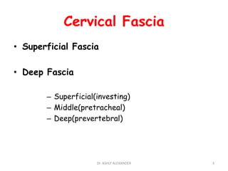
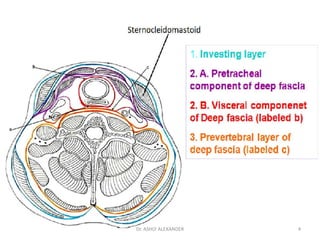
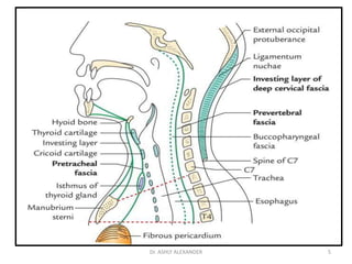
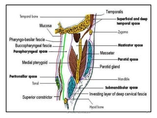
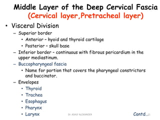
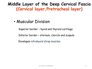
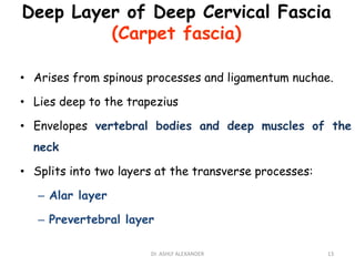
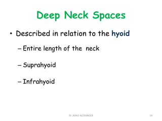
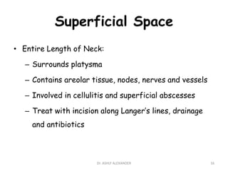
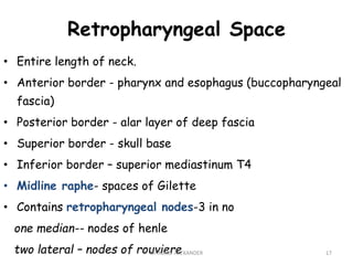
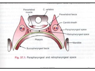
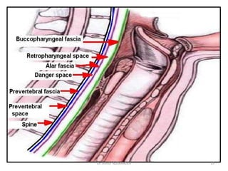
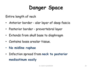
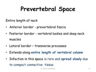
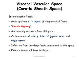
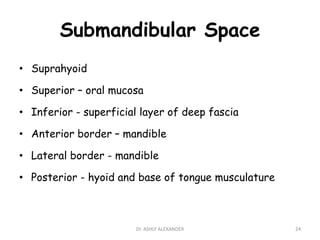
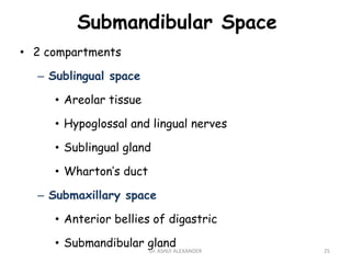
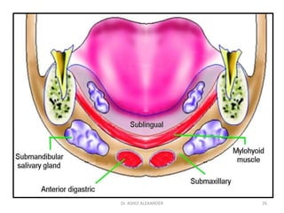
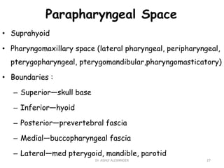
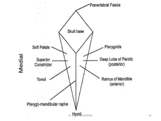
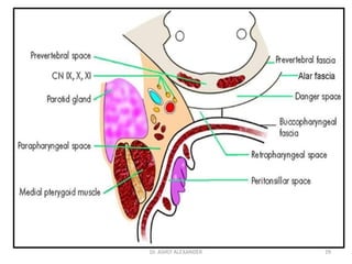
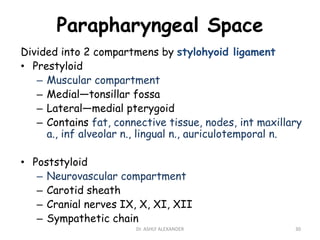
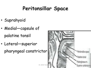
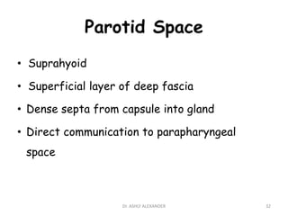
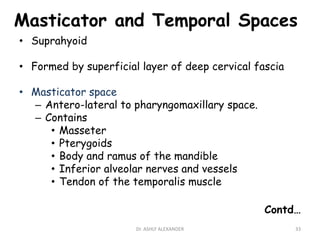
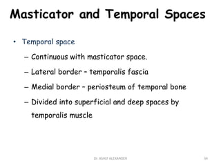
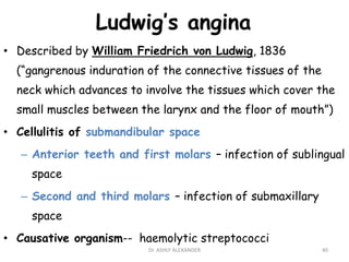
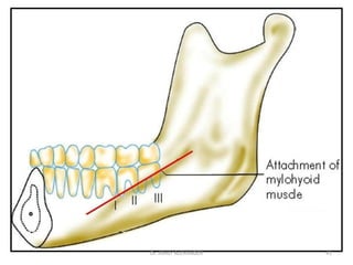
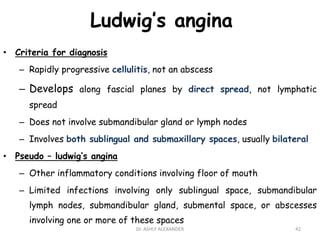
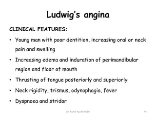
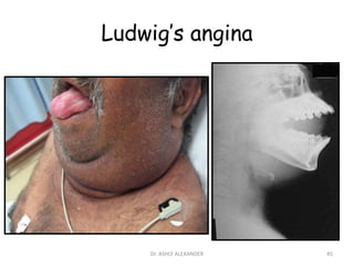
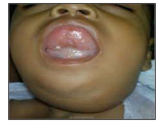
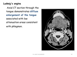
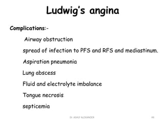
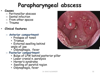
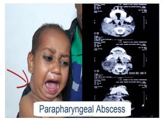
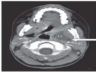
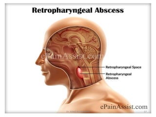
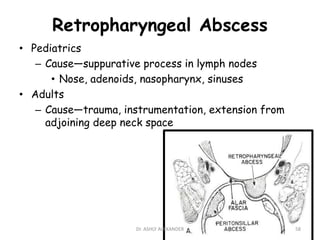
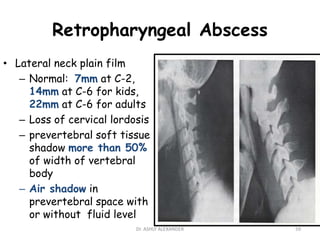
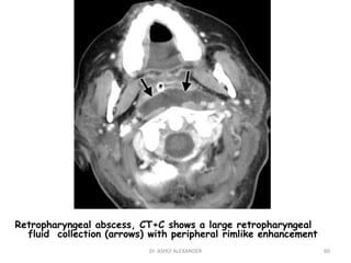
“Deep Neck Space
Infections”
Dr. Ashly Alexander
ENT RSO - 2nd Year
GMC Bhopal
• Anatomy of the Cervical Fascia
• Anatomy of the Deep Neck Spaces
• Deep Neck Space Infections
2
Dr. ASHLY ALEXANDER
Cervical Fascia
• Superficial Fascia
• Deep Fascia
– Superficial(investing)
– Middle(pretracheal)
– Deep(prevertebral)
3
Dr. ASHLY ALEXANDER
4
Dr. ASHLY ALEXANDER
5
Dr. ASHLY ALEXANDER
Superficial fascia
(Tela subcuta)
• Superior attachment – zygomatic process
• Inferior attachment – thorax, axilla.
• Similar to subcutaneous tissue
• Ensheathes platysma and muscles of facial
expression
• Marginal mandibular n. lies deep to it
6
Dr. ASHLY ALEXANDER
Deep Cervical Fascia
1) Superficial layer
2) Middle layer
3) Deep layer
7
Dr. ASHLY ALEXANDER
Superficial Layer of the Deep Cervical Fascia
(Enveloping,Investing,Anterior layer)
• Completely surrounds the neck from skull to chest
• Arises from spinous processes, ligamentum nuchae
• Superior border – nuchal line, skull base, zygoma,
mandible.
• Inferior border –scapula, clavicle and manubrium
• Splits at mandible and covers the masseter laterally
and the medial surface of the medial pterygoid.
Contd…
8
Dr. ASHLY ALEXANDER
9
Dr. ASHLY ALEXANDER
Superficial Layer of the Deep Cervical Fascia
(Enveloping,Investing,Anterior layer)
• Envelopes
– SCM
– Trapezius
– Submandibular
– Parotid
• Forms floor of submandibular space
• Create superficial sternal space (of Burn)
10
Dr. ASHLY ALEXANDER
Middle Layer of the Deep Cervical Fascia
(Cervical layer,Pretracheal layer)
• Visceral Division
– Superior border
• Anterior – hyoid and thyroid cartilage
• Posterior – skull base
– Inferior border – continuous with fibrous pericardium in the
upper mediastinum.
– Buccopharyngeal fascia
• Name for portion that covers the pharyngeal constrictors
and buccinator.
– Envelopes
• Thyroid
• Trachea
• Esophagus
• Pharynx
• Larynx Contd…11
Dr. ASHLY ALEXANDER
Middle Layer of the Deep Cervical Fascia
(Cervical layer,Pretracheal layer)
• Muscular Division
Superior border – hyoid and thyroid cartilage
Inferior border – sternum, clavicle and scapula
Envelopes infrahyoid strap muscles
12
Dr. ASHLY ALEXANDER
Deep Layer of Deep Cervical Fascia
(Carpet fascia)
• Arises from spinous processes and ligamentum nuchae.
• Lies deep to the trapezius
• Envelopes vertebral bodies and deep muscles of the
neck
• Splits into two layers at the transverse processes:
– Alar layer
– Prevertebral layer
13
Dr. ASHLY ALEXANDER
Deep Neck Spaces
• Described in relation to the hyoid
– Entire length of the neck
– Suprahyoid
– Infrahyoid
14
Dr. ASHLY ALEXANDER
Space Involving Entire Length
Of Neck
• Superficial space
• Retropharyngeal Space
• Danger Space
• Prevertebral Space
• Carotid Sheath Space
15
Dr. ASHLY ALEXANDER
Superficial Space
• Entire Length of Neck:
– Surrounds platysma
– Contains areolar tissue, nodes, nerves and vessels
– Involved in cellulitis and superficial abscesses
– Treat with incision along Langer’s lines, drainage
and antibiotics
16
Dr. ASHLY ALEXANDER
Retropharyngeal Space
• Entire length of neck.
• Anterior border - pharynx and esophagus (buccopharyngeal
fascia)
• Posterior border - alar layer of deep fascia
• Superior border - skull base
• Inferior border – superior mediastinum T4
• Midline raphe- spaces of Gilette
• Contains retropharyngeal nodes-3 in no
one median-- nodes of henle
two lateral – nodes of rouviere 17
Dr. ASHLY ALEXANDER
18
Dr. ASHLY ALEXANDER
19
Dr. ASHLY ALEXANDER
Danger Space
Entire length of neck
• Anterior border - alar layer of deep fascia
• Posterior border - prevertebral layer
• Extends from skull base to diaphragm
• Contains loose areolar tissue.
• No midline raphae
• Infection spread from neck to posterior
mediastinum easily
20
Dr. ASHLY ALEXANDER
Prevertebral Space
Entire length of neck
• Anterior border - prevertebral fascia
• Posterior border - vertebral bodies and deep neck
muscles
• Lateral border – transverse processes
• Extends along entire length of vertebral column
• Infection in this space is rare and spread slowly due
to compact connective tissue
21
Dr. ASHLY ALEXANDER
Visceral Vascular Space
(Carotid Sheath Space)
Entire length of neck
– Made up from all 3 layers of deep cervical fascia
– “Lincoln Highway”
– Anatomically separate from all layers
– Contains carotid artery, internal jugular vein, and
vagus nerve
– Infection from any deep fascia can spread to this space
– Extends from skull base to thorax.
22
Dr. ASHLY ALEXANDER
Space Limit To Above The Hyoid
Bone
• Submandibular Space
• Parapharyngeal Space
• Peritonsillar Space
• Parotid Space
• Masticator & Temporal Space
23
Dr. ASHLY ALEXANDER
Submandibular Space
• Suprahyoid
• Superior – oral mucosa
• Inferior - superficial layer of deep fascia
• Anterior border – mandible
• Lateral border - mandible
• Posterior - hyoid and base of tongue musculature
24
Dr. ASHLY ALEXANDER
Submandibular Space
• 2 compartments
– Sublingual space
• Areolar tissue
• Hypoglossal and lingual nerves
• Sublingual gland
• Wharton’s duct
– Submaxillary space
• Anterior bellies of digastric
• Submandibular gland
25
Dr. ASHLY ALEXANDER
26
Dr. ASHLY ALEXANDER
Parapharyngeal Space
• Suprahyoid
• Pharyngomaxillary space (lateral pharyngeal, peripharyngeal,
pterygopharyngeal, pterygomandibular,pharyngomasticatory)
• Boundaries :
– Superior—skull base
– Inferior—hyoid
– Posterior—prevertebral fascia
– Medial—buccopharyngeal fascia
– Lateral—med pterygoid, mandible, parotid
27
Dr. ASHLY ALEXANDER
28
Dr. ASHLY ALEXANDER
29
Dr. ASHLY ALEXANDER
Parapharyngeal Space
Divided into 2 compartmens by stylohyoid ligament
• Prestyloid
– Muscular compartment
– Medial—tonsillar fossa
– Lateral—medial pterygoid
– Contains fat, connective tissue, nodes, int maxillary
a., inf alveolar n., lingual n., auriculotemporal n.
• Poststyloid
– Neurovascular compartment
– Carotid sheath
– Cranial nerves IX, X, XI, XII
– Sympathetic chain
30
Dr. ASHLY ALEXANDER
Peritonsillar Space
• Suprahyoid
• Medial—capsule of
palatine tonsil
• Lateral—superior
pharyngeal constrictor
31
Dr. ASHLY ALEXANDER
Parotid Space
• Suprahyoid
• Superficial layer of deep fascia
• Dense septa from capsule into gland
• Direct communication to parapharyngeal
space
32
Dr. ASHLY ALEXANDER
Masticator and Temporal Spaces
• Suprahyoid
• Formed by superficial layer of deep cervical fascia
• Masticator space
– Antero-lateral to pharyngomaxillary space.
– Contains
• Masseter
• Pterygoids
• Body and ramus of the mandible
• Inferior alveolar nerves and vessels
• Tendon of the temporalis muscle
Contd…
33
Dr. ASHLY ALEXANDER
Masticator and Temporal Spaces
• Temporal space
– Continuous with masticator space.
– Lateral border – temporalis fascia
– Medial border – periosteum of temporal bone
– Divided into superficial and deep spaces by
temporalis muscle
34
Dr. ASHLY ALEXANDER
Infrahyoid
• Visceral Compartment
– Middle layer of deep fascia
– Contains thyroid, trachea, esophagus
– Extends from thyroid cartilage into superior mediastinum
2 spaces-
• Retrovisceral space {Retropharyngeal space}
– Extends along whole length of neck
• Pretracheal space
– Superiorly - attachment of strap muscles to
thyroid and hyoid
– Inferiorly - up to upper border of arch of aorta
35
Dr. ASHLY ALEXANDER
Deep Neck Space Infections
• Etiology/ pathogenesis of Infection
• Microbiology
• Clinical manifestations
• Some specific infections
• Complications
36
Dr. ASHLY ALEXANDER
Etiopathogenesis
• Deep neck space infeections have been recognised from the
time of Galen in 2nd century AD
• Preantibiotic era – 70% from infections of pharynx and
tonsils
• Present situation
– Dental infection (major source)
– Peritonsillar abscess
– Upper aerodigestive tract trauma
– Retropharyngeal lymphadenitis
– Pott’s disease
– Sialadenitis – submandibular, parotid
– From temporal bone- Bezold’s abscess, petrous apex
infections
– Congenital cysts and fistulas
– Intravenous drug abuse
37
Dr. ASHLY ALEXANDER
Microbiology
• Preantibiotic era – S. aureus
• Currently
– Aerobes – alpha hemolytic Streptococci, S. aureus
– Anaerobes – Fusobacterium, Bacteroides,
Peptostreptococcus, Veilonella
• Gram-negatives uncommon
• Almost always polymicrobial
38
Dr. ASHLY ALEXANDER
Clinical manifestations
• Pain
– Constant feature
– Indication of extension or resolution
– Exception – retropharyngeal abscess in children
• Fever
– Constant feature
– Initial spike, followed by elevated temperature
– Spiking temperatures- doubt septicemia/septic
thrombophlebitis of IJV/mediastinal extension
• Swelling
• Trismus and limitation of neck movements – depending
on site
• Progressive dysphagia and odynophagia
• Voice change
• Dyspnoea
• Chest pain 39
Dr. ASHLY ALEXANDER
Ludwig’s angina
• Described by William Friedrich von Ludwig, 1836
(“gangrenous induration of the connective tissues of the
neck which advances to involve the tissues which cover the
small muscles between the larynx and the floor of mouth”)
• Cellulitis of submandibular space
– Anterior teeth and first molars – infection of sublingual
space
– Second and third molars – infection of submaxillary
space
• Causative organism-- haemolytic streptococci
40
Dr. ASHLY ALEXANDER
41
Dr. ASHLY ALEXANDER
Ludwig’s angina
• Criteria for diagnosis
– Rapidly progressive cellulitis, not an abscess
– Develops along fascial planes by direct spread, not lymphatic
spread
– Does not involve submandibular gland or lymph nodes
– Involves both sublingual and submaxillary spaces, usually bilateral
• Pseudo – ludwig’s angina
– Other inflammatory conditions involving floor of mouth
– Limited infections involving only sublingual space, submandibular
lymph nodes, submandibular gland, submental space, or abscesses
involving one or more of these spaces
42
Dr. ASHLY ALEXANDER
Ludwig’s angina
ETIOLOGY:
• 75-80% dental cause
• Extraction of a diseased molar initiates infection
• Penetrating injury of the floor of mouth
• Mandibular fractures
43
Dr. ASHLY ALEXANDER
Ludwig’s angina
CLINICAL FEATURES:
• Young man with poor dentition, increasing oral or neck
pain and swelling
• Increasing edema and induration of perimandibular
region and floor of mouth
• Thrusting of tongue posteriorly and superiorly
• Neck rigidity, trismus, odynophagia, fever
• Dyspnoea and stridor
44
Dr. ASHLY ALEXANDER
Ludwig’s angina
45
Dr. ASHLY ALEXANDER
46
Dr. ASHLY ALEXANDER
Ludwig's angina
Axial CT section through the
tongue demonstrates diffuse
enlargement of the tongue
associated with low
attenuation areas consistent
with phlegmon.
47
Dr. ASHLY ALEXANDER
Ludwig’s angina
TREATMENT
• Early stage- IV antibiotics {penicillin + metronidazole},
extraction of the diseased tooth
• Late stage-
– Airway {tracheostomy }
– Surgery
• Horizontal incision with wide exposure
• Tissues have peculiar “salt pork appearance”,
with woody induration, watery edema, and little bleeding
• Gross purulence is rare
• Multiple drains/wound kept open 48
Dr. ASHLY ALEXANDER
Ludwig’s angina
Complications:-
Airway obstruction
spread of infection to PFS and RFS and mediastinum.
Aspiration pneumonia
Lung abscess
Fluid and electrolyte imbalance
Tongue necrosis
septicemia
49
Dr. ASHLY ALEXANDER
Parapharyngeal abscess
• Causes
– Peritonsillar abscess
– Dental infection
– From other spaces
– Trauma
• Clinical features
– Anterior compartment
• Prolapse of tonsil
• Trismus
• External swelling behind
angle of jaw
• Odynophagia, fever
– Posterior compartment
• Bulge of LPW behind posterior pillar
• Lower cranial n. paralysis
• Horner’s syndrome
• Swelling of parotid region
• Odynophagia, fever
50
Dr. ASHLY ALEXANDER
51
Dr. ASHLY ALEXANDER
52
Dr. ASHLY ALEXANDER
• Treatment
– IV antibiotics
– Correct dehydration
– Analgesics
– Surgery
• External approach
(access along the carotid sheath)
–Transverse submandibular incision
– Mosher’s T-shaped incision
53
Dr. ASHLY ALEXANDER
Complications:
• Rates of About 16%
• Carotid sheath involvement causing
Internal jugular vein thrombosis
Carotid artery thrombosis.
• Internal carotid artery pseudo aneurysm
presenting as Horner's syndrome.
• Acute pharyngeal perforation
• Mediastinitis; requiring urgent drainage.
• Descending necroting fasciitis of neck and
mediastinum;requiring widespread debridement.
• Upper airway obstruction
54
Dr. ASHLY ALEXANDER
Retropharyngeal Abscess
– 50% occur in patients 6-12 months of age
– 96% occur before 6 years of age
– Children - fever, irritability, lymphadenopathy,
torticollis, poor oral intake, sore throat, drooling
– Adults - pain, dysphagia, odynophagia, anorexia
– Dyspnea and respiratory distress
– Lateral posterior pharyngeal wall bulge
55
Dr. ASHLY ALEXANDER
Retropharyngeal Abscess
56
Dr. ASHLY ALEXANDER
57
Dr. ASHLY ALEXANDER
Retropharyngeal Abscess
• Pediatrics
– Cause—suppurative process in lymph nodes
• Nose, adenoids, nasopharynx, sinuses
• Adults
– Cause—trauma, instrumentation, extension from
adjoining deep neck space
58
Dr. ASHLY ALEXANDER
Retropharyngeal Abscess
• Lateral neck plain film
– Normal: 7mm at C-2,
14mm at C-6 for kids,
22mm at C-6 for adults
– Loss of cervical lordosis
– prevertebral soft tissue
shadow more than 50%
of width of vertebral
body
– Air shadow in
prevertebral space with
or without fluid level
59
Dr. ASHLY ALEXANDER
Retropharyngeal abscess, CT+C shows a large retropharyngeal
fluid collection (arrows) with peripheral rimlike enhancement
60
Dr. ASHLY ALEXANDER
Retropharyngeal Abscess
• Treatment
– IV antibiotics and fluid
replacement
– Surgical drainage
• Intraoral
• External –
tracheostomy +
anterior cervical
approach
61
Dr. ASHLY ALEXANDER
Chronic Retropharyngeal
abscess
• Common in adults
• Due to TB of cervical vertebra
• Abscess is formed posterior to prevertebral fascia
Clinical Features
• Initially asymptomatic
• Mild discomfort and sore throat
• O/E smooth bulging on Post Pharyngeal wall
• Without signs of inflammation
62
Dr. ASHLY ALEXANDER
Tuberculosis of cervical
Spine with
retropharyngeal
abscess
63
Dr. ASHLY ALEXANDER
Chronic Retropharyngeal
abscess
Diagnosis
• Clinical examination
• Blood Examination
• Sputum for AFB
• X-Ray cervical spine
• X-Ray Chest
Treatment
• Antitubercular drugs
• Aspiration of abscess
wide bore needle
• Large abscess drainage
through external root
64
Dr. ASHLY ALEXANDER
•Acute Retropharyngeal •Chronic Retropharyngeal
 Age 1-3 yrs
 Space of Gillette
 Pyogenic organisms
 Suppuration of LN
 Sudden onset
 Dysphagia
 Resp distress
 High fever
 Swelling one side midline
 Signs of inflammation
 I/D Oral route
Adults
Behind prevertebral fascia
Tubercular organisms
Cervical carries
Slow onset
No / mild dysphagia
Not common
Mild fever
Midline swelling
No Signs of inflammation
Anti TB & aspiration
Drainage through external
route
65
Dr. ASHLY ALEXANDER
Retropharyngeal abscess
Complications:- meningitis
haemorrhage
laryngeal spasm
septicemia
Metastatic abscess
Jugular vein thrombosis
Rupture with aspiration pneumonia
Pericardial tamponade
Mediastinitis
Acute hemiplegia of childhood
Spead in to other spaces
66
Dr. ASHLY ALEXANDER
Peritonsillar abscess (quinsy)
• Cause
-Local complication of tonsillar infection→ Lacunar type
-Infection→crypta magna→paratonsillar space
-Weber’ glands infection
-De novo
• Symptoms
– Fever with chills and rigor
– Odynophagia
– “Hot Potato” voice
– Halitosis
– Head tilted towards affected site
67
Dr. ASHLY ALEXANDER
68
Dr. ASHLY ALEXANDER
69
Dr. ASHLY ALEXANDER
Peritonsillar abscess (quinsy)
Signs
• Anxious facies and stiffly held head,↑ pulse &↑ temp
• Trismus
• Unilateral swelling over palate & ant pillar
• Uvula pushed to opposite site
• Tonsil displaced medially and downward
• Palate angry red, immobile, thick mucous
• JDLN enlarged & tender
70
Dr. ASHLY ALEXANDER
Peritonsillar abscess (quinsy)
Treatment :
• Hospitalization
• Correction of dehydration
• Systemic parentral broad spectrum antibiotics
• Incision and drainage
If no pus: parentral antibiotics heavy doses
If no response in 24 hrs : I & D
If pus present: I&D and supportive treatment
After 6weeks tonsillectomy (intermittent tonsillectomy)to
prevent recurrence
71
Dr. ASHLY ALEXANDER
• Anesthesia – local
• Position –semi sitting
• Incision :
 most prominent part or Halfway
between last molar and uvula
 Stab incision & dilatation by
st claire thomson Quinsy scissors
and suction of pus
Incision and Drainage
72
Dr. ASHLY ALEXANDER
Peritonsillar abscess (quinsy)
Complications :
•Mediastinitis
•Necrotizing fasciitis.
•Oedema of larynx
•Septicemia
•IJV thrombosis
•Pneumonitis or lung abscess
73
Dr. ASHLY ALEXANDER
Parotid space infections
Contents:-parotid gland
VII nerve
LN
ECA
retromandibular vein
Etiology:- post surgical cases
debilitated and dehydrated pt
drugs which decrease salivary flow
Infections of oral cavity
Severe otitis externa spreading thru fissure
of Santorini.
74
Dr. ASHLY ALEXANDER
Clinical features
• usually follow 5-7 day after surgery.
• marked swelling of jaw
• Pain and induration over parotid gland
• Congested stenson’s duct.
• No fluctuation d/to thick capsule.
Treatment
correct dehydration
improve oral hygine
IV antibiotics
I&D:
Modified Blair’s incision
75
Dr. ASHLY ALEXANDER
76
Dr. ASHLY ALEXANDER
Masticator-Temporal Space
infection
• Cause
– Odontogenic
– Trauma
• Superficial compartment
– Extensive facial swelling
– Severe trismus
– Pain
• Deep compartment
– Trismus
– Pain
– Dysphagia and
odynophagia
– Intraoral swelling in RMT
area
•Treatment
IV antibiotics
Surgery
Intraoral
Along inner
margin of
mandibular ramus
in RMT area
External
Horizontal
incision, 2-3cm
beneath angle of
mandible
77
Dr. ASHLY ALEXANDER
78
Dr. ASHLY ALEXANDER
Complications
• Internal Jugular Vein Thrombophlebitis
(Lemierre’s syndrome)
– Fusobacterium necrophorum
– High fever with chills and rigor
– Swelling and pain along SCM
– Bacteremia, septic embolization, dural sinus
thrombosis
– IV drug abusers
– Treatment
• IV antibiotics
• Anticoagulation - controversial
• Ligation and excision
79
Dr. ASHLY ALEXANDER
Complications
• Mediastinitis
– Mortality of 40%
– Increasing dyspnea, chest pain
– CXR - widened mediastinum
– Treatment
• EARLY RECOGNITION AND INTERVENTION
• Aggressive IV antibiotic therapy
• Surgical drainage
– Transcervical approach
– Chest tube / thoracotomy
80
Dr. ASHLY ALEXANDER
Complications
• Cranial nerve deficits
• Necrotising cervical fasciitis
• Osteomyelitis
• Grisel syndrome ( inflammatory
torticollis causing cervical vertebral
subluxation )
81
Dr. ASHLY ALEXANDER
Special considerations
• Airway protection
– Observation
– Intubation
• Direct laryngoscopy: risk of rupture and
aspiration
• Flexible fiberoptic
– Tracheostomy
• Safest
• Abscess may overlie trachea
• Distorted anatomy and tissue planes
82
Dr. ASHLY ALEXANDER
Special considerations
• Image-guided Aspiration
– Patient selection
• Smaller abscesses, limited extension,
uniloculated
– Advantages
• Early specimen collection
• Reduced expense
• Avoidance of neck scar
83
Dr. ASHLY ALEXANDER
84
Dr. ASHLY ALEXANDER

deepneckspaceinfections-dr-190619164411.pdf

  • 1.
    “Deep Neck Space Infections” Dr.Ashly Alexander ENT RSO - 2nd Year GMC Bhopal
  • 2.
    • Anatomy ofthe Cervical Fascia • Anatomy of the Deep Neck Spaces • Deep Neck Space Infections 2 Dr. ASHLY ALEXANDER
  • 3.
    Cervical Fascia • SuperficialFascia • Deep Fascia – Superficial(investing) – Middle(pretracheal) – Deep(prevertebral) 3 Dr. ASHLY ALEXANDER
  • 4.
  • 5.
  • 6.
    Superficial fascia (Tela subcuta) •Superior attachment – zygomatic process • Inferior attachment – thorax, axilla. • Similar to subcutaneous tissue • Ensheathes platysma and muscles of facial expression • Marginal mandibular n. lies deep to it 6 Dr. ASHLY ALEXANDER
  • 7.
    Deep Cervical Fascia 1)Superficial layer 2) Middle layer 3) Deep layer 7 Dr. ASHLY ALEXANDER
  • 8.
    Superficial Layer ofthe Deep Cervical Fascia (Enveloping,Investing,Anterior layer) • Completely surrounds the neck from skull to chest • Arises from spinous processes, ligamentum nuchae • Superior border – nuchal line, skull base, zygoma, mandible. • Inferior border –scapula, clavicle and manubrium • Splits at mandible and covers the masseter laterally and the medial surface of the medial pterygoid. Contd… 8 Dr. ASHLY ALEXANDER
  • 9.
  • 10.
    Superficial Layer ofthe Deep Cervical Fascia (Enveloping,Investing,Anterior layer) • Envelopes – SCM – Trapezius – Submandibular – Parotid • Forms floor of submandibular space • Create superficial sternal space (of Burn) 10 Dr. ASHLY ALEXANDER
  • 11.
    Middle Layer ofthe Deep Cervical Fascia (Cervical layer,Pretracheal layer) • Visceral Division – Superior border • Anterior – hyoid and thyroid cartilage • Posterior – skull base – Inferior border – continuous with fibrous pericardium in the upper mediastinum. – Buccopharyngeal fascia • Name for portion that covers the pharyngeal constrictors and buccinator. – Envelopes • Thyroid • Trachea • Esophagus • Pharynx • Larynx Contd…11 Dr. ASHLY ALEXANDER
  • 12.
    Middle Layer ofthe Deep Cervical Fascia (Cervical layer,Pretracheal layer) • Muscular Division Superior border – hyoid and thyroid cartilage Inferior border – sternum, clavicle and scapula Envelopes infrahyoid strap muscles 12 Dr. ASHLY ALEXANDER
  • 13.
    Deep Layer ofDeep Cervical Fascia (Carpet fascia) • Arises from spinous processes and ligamentum nuchae. • Lies deep to the trapezius • Envelopes vertebral bodies and deep muscles of the neck • Splits into two layers at the transverse processes: – Alar layer – Prevertebral layer 13 Dr. ASHLY ALEXANDER
  • 14.
    Deep Neck Spaces •Described in relation to the hyoid – Entire length of the neck – Suprahyoid – Infrahyoid 14 Dr. ASHLY ALEXANDER
  • 15.
    Space Involving EntireLength Of Neck • Superficial space • Retropharyngeal Space • Danger Space • Prevertebral Space • Carotid Sheath Space 15 Dr. ASHLY ALEXANDER
  • 16.
    Superficial Space • EntireLength of Neck: – Surrounds platysma – Contains areolar tissue, nodes, nerves and vessels – Involved in cellulitis and superficial abscesses – Treat with incision along Langer’s lines, drainage and antibiotics 16 Dr. ASHLY ALEXANDER
  • 17.
    Retropharyngeal Space • Entirelength of neck. • Anterior border - pharynx and esophagus (buccopharyngeal fascia) • Posterior border - alar layer of deep fascia • Superior border - skull base • Inferior border – superior mediastinum T4 • Midline raphe- spaces of Gilette • Contains retropharyngeal nodes-3 in no one median-- nodes of henle two lateral – nodes of rouviere 17 Dr. ASHLY ALEXANDER
  • 18.
  • 19.
  • 20.
    Danger Space Entire lengthof neck • Anterior border - alar layer of deep fascia • Posterior border - prevertebral layer • Extends from skull base to diaphragm • Contains loose areolar tissue. • No midline raphae • Infection spread from neck to posterior mediastinum easily 20 Dr. ASHLY ALEXANDER
  • 21.
    Prevertebral Space Entire lengthof neck • Anterior border - prevertebral fascia • Posterior border - vertebral bodies and deep neck muscles • Lateral border – transverse processes • Extends along entire length of vertebral column • Infection in this space is rare and spread slowly due to compact connective tissue 21 Dr. ASHLY ALEXANDER
  • 22.
    Visceral Vascular Space (CarotidSheath Space) Entire length of neck – Made up from all 3 layers of deep cervical fascia – “Lincoln Highway” – Anatomically separate from all layers – Contains carotid artery, internal jugular vein, and vagus nerve – Infection from any deep fascia can spread to this space – Extends from skull base to thorax. 22 Dr. ASHLY ALEXANDER
  • 23.
    Space Limit ToAbove The Hyoid Bone • Submandibular Space • Parapharyngeal Space • Peritonsillar Space • Parotid Space • Masticator & Temporal Space 23 Dr. ASHLY ALEXANDER
  • 24.
    Submandibular Space • Suprahyoid •Superior – oral mucosa • Inferior - superficial layer of deep fascia • Anterior border – mandible • Lateral border - mandible • Posterior - hyoid and base of tongue musculature 24 Dr. ASHLY ALEXANDER
  • 25.
    Submandibular Space • 2compartments – Sublingual space • Areolar tissue • Hypoglossal and lingual nerves • Sublingual gland • Wharton’s duct – Submaxillary space • Anterior bellies of digastric • Submandibular gland 25 Dr. ASHLY ALEXANDER
  • 26.
  • 27.
    Parapharyngeal Space • Suprahyoid •Pharyngomaxillary space (lateral pharyngeal, peripharyngeal, pterygopharyngeal, pterygomandibular,pharyngomasticatory) • Boundaries : – Superior—skull base – Inferior—hyoid – Posterior—prevertebral fascia – Medial—buccopharyngeal fascia – Lateral—med pterygoid, mandible, parotid 27 Dr. ASHLY ALEXANDER
  • 28.
  • 29.
  • 30.
    Parapharyngeal Space Divided into2 compartmens by stylohyoid ligament • Prestyloid – Muscular compartment – Medial—tonsillar fossa – Lateral—medial pterygoid – Contains fat, connective tissue, nodes, int maxillary a., inf alveolar n., lingual n., auriculotemporal n. • Poststyloid – Neurovascular compartment – Carotid sheath – Cranial nerves IX, X, XI, XII – Sympathetic chain 30 Dr. ASHLY ALEXANDER
  • 31.
    Peritonsillar Space • Suprahyoid •Medial—capsule of palatine tonsil • Lateral—superior pharyngeal constrictor 31 Dr. ASHLY ALEXANDER
  • 32.
    Parotid Space • Suprahyoid •Superficial layer of deep fascia • Dense septa from capsule into gland • Direct communication to parapharyngeal space 32 Dr. ASHLY ALEXANDER
  • 33.
    Masticator and TemporalSpaces • Suprahyoid • Formed by superficial layer of deep cervical fascia • Masticator space – Antero-lateral to pharyngomaxillary space. – Contains • Masseter • Pterygoids • Body and ramus of the mandible • Inferior alveolar nerves and vessels • Tendon of the temporalis muscle Contd… 33 Dr. ASHLY ALEXANDER
  • 34.
    Masticator and TemporalSpaces • Temporal space – Continuous with masticator space. – Lateral border – temporalis fascia – Medial border – periosteum of temporal bone – Divided into superficial and deep spaces by temporalis muscle 34 Dr. ASHLY ALEXANDER
  • 35.
    Infrahyoid • Visceral Compartment –Middle layer of deep fascia – Contains thyroid, trachea, esophagus – Extends from thyroid cartilage into superior mediastinum 2 spaces- • Retrovisceral space {Retropharyngeal space} – Extends along whole length of neck • Pretracheal space – Superiorly - attachment of strap muscles to thyroid and hyoid – Inferiorly - up to upper border of arch of aorta 35 Dr. ASHLY ALEXANDER
  • 36.
    Deep Neck SpaceInfections • Etiology/ pathogenesis of Infection • Microbiology • Clinical manifestations • Some specific infections • Complications 36 Dr. ASHLY ALEXANDER
  • 37.
    Etiopathogenesis • Deep neckspace infeections have been recognised from the time of Galen in 2nd century AD • Preantibiotic era – 70% from infections of pharynx and tonsils • Present situation – Dental infection (major source) – Peritonsillar abscess – Upper aerodigestive tract trauma – Retropharyngeal lymphadenitis – Pott’s disease – Sialadenitis – submandibular, parotid – From temporal bone- Bezold’s abscess, petrous apex infections – Congenital cysts and fistulas – Intravenous drug abuse 37 Dr. ASHLY ALEXANDER
  • 38.
    Microbiology • Preantibiotic era– S. aureus • Currently – Aerobes – alpha hemolytic Streptococci, S. aureus – Anaerobes – Fusobacterium, Bacteroides, Peptostreptococcus, Veilonella • Gram-negatives uncommon • Almost always polymicrobial 38 Dr. ASHLY ALEXANDER
  • 39.
    Clinical manifestations • Pain –Constant feature – Indication of extension or resolution – Exception – retropharyngeal abscess in children • Fever – Constant feature – Initial spike, followed by elevated temperature – Spiking temperatures- doubt septicemia/septic thrombophlebitis of IJV/mediastinal extension • Swelling • Trismus and limitation of neck movements – depending on site • Progressive dysphagia and odynophagia • Voice change • Dyspnoea • Chest pain 39 Dr. ASHLY ALEXANDER
  • 40.
    Ludwig’s angina • Describedby William Friedrich von Ludwig, 1836 (“gangrenous induration of the connective tissues of the neck which advances to involve the tissues which cover the small muscles between the larynx and the floor of mouth”) • Cellulitis of submandibular space – Anterior teeth and first molars – infection of sublingual space – Second and third molars – infection of submaxillary space • Causative organism-- haemolytic streptococci 40 Dr. ASHLY ALEXANDER
  • 41.
  • 42.
    Ludwig’s angina • Criteriafor diagnosis – Rapidly progressive cellulitis, not an abscess – Develops along fascial planes by direct spread, not lymphatic spread – Does not involve submandibular gland or lymph nodes – Involves both sublingual and submaxillary spaces, usually bilateral • Pseudo – ludwig’s angina – Other inflammatory conditions involving floor of mouth – Limited infections involving only sublingual space, submandibular lymph nodes, submandibular gland, submental space, or abscesses involving one or more of these spaces 42 Dr. ASHLY ALEXANDER
  • 43.
    Ludwig’s angina ETIOLOGY: • 75-80%dental cause • Extraction of a diseased molar initiates infection • Penetrating injury of the floor of mouth • Mandibular fractures 43 Dr. ASHLY ALEXANDER
  • 44.
    Ludwig’s angina CLINICAL FEATURES: •Young man with poor dentition, increasing oral or neck pain and swelling • Increasing edema and induration of perimandibular region and floor of mouth • Thrusting of tongue posteriorly and superiorly • Neck rigidity, trismus, odynophagia, fever • Dyspnoea and stridor 44 Dr. ASHLY ALEXANDER
  • 45.
  • 46.
  • 47.
    Ludwig's angina Axial CTsection through the tongue demonstrates diffuse enlargement of the tongue associated with low attenuation areas consistent with phlegmon. 47 Dr. ASHLY ALEXANDER
  • 48.
    Ludwig’s angina TREATMENT • Earlystage- IV antibiotics {penicillin + metronidazole}, extraction of the diseased tooth • Late stage- – Airway {tracheostomy } – Surgery • Horizontal incision with wide exposure • Tissues have peculiar “salt pork appearance”, with woody induration, watery edema, and little bleeding • Gross purulence is rare • Multiple drains/wound kept open 48 Dr. ASHLY ALEXANDER
  • 49.
    Ludwig’s angina Complications:- Airway obstruction spreadof infection to PFS and RFS and mediastinum. Aspiration pneumonia Lung abscess Fluid and electrolyte imbalance Tongue necrosis septicemia 49 Dr. ASHLY ALEXANDER
  • 50.
    Parapharyngeal abscess • Causes –Peritonsillar abscess – Dental infection – From other spaces – Trauma • Clinical features – Anterior compartment • Prolapse of tonsil • Trismus • External swelling behind angle of jaw • Odynophagia, fever – Posterior compartment • Bulge of LPW behind posterior pillar • Lower cranial n. paralysis • Horner’s syndrome • Swelling of parotid region • Odynophagia, fever 50 Dr. ASHLY ALEXANDER
  • 51.
  • 52.
  • 53.
    • Treatment – IVantibiotics – Correct dehydration – Analgesics – Surgery • External approach (access along the carotid sheath) –Transverse submandibular incision – Mosher’s T-shaped incision 53 Dr. ASHLY ALEXANDER
  • 54.
    Complications: • Rates ofAbout 16% • Carotid sheath involvement causing Internal jugular vein thrombosis Carotid artery thrombosis. • Internal carotid artery pseudo aneurysm presenting as Horner's syndrome. • Acute pharyngeal perforation • Mediastinitis; requiring urgent drainage. • Descending necroting fasciitis of neck and mediastinum;requiring widespread debridement. • Upper airway obstruction 54 Dr. ASHLY ALEXANDER
  • 55.
    Retropharyngeal Abscess – 50%occur in patients 6-12 months of age – 96% occur before 6 years of age – Children - fever, irritability, lymphadenopathy, torticollis, poor oral intake, sore throat, drooling – Adults - pain, dysphagia, odynophagia, anorexia – Dyspnea and respiratory distress – Lateral posterior pharyngeal wall bulge 55 Dr. ASHLY ALEXANDER
  • 56.
  • 57.
  • 58.
    Retropharyngeal Abscess • Pediatrics –Cause—suppurative process in lymph nodes • Nose, adenoids, nasopharynx, sinuses • Adults – Cause—trauma, instrumentation, extension from adjoining deep neck space 58 Dr. ASHLY ALEXANDER
  • 59.
    Retropharyngeal Abscess • Lateralneck plain film – Normal: 7mm at C-2, 14mm at C-6 for kids, 22mm at C-6 for adults – Loss of cervical lordosis – prevertebral soft tissue shadow more than 50% of width of vertebral body – Air shadow in prevertebral space with or without fluid level 59 Dr. ASHLY ALEXANDER
  • 60.
    Retropharyngeal abscess, CT+Cshows a large retropharyngeal fluid collection (arrows) with peripheral rimlike enhancement 60 Dr. ASHLY ALEXANDER
  • 61.
    Retropharyngeal Abscess • Treatment –IV antibiotics and fluid replacement – Surgical drainage • Intraoral • External – tracheostomy + anterior cervical approach 61 Dr. ASHLY ALEXANDER
  • 62.
    Chronic Retropharyngeal abscess • Commonin adults • Due to TB of cervical vertebra • Abscess is formed posterior to prevertebral fascia Clinical Features • Initially asymptomatic • Mild discomfort and sore throat • O/E smooth bulging on Post Pharyngeal wall • Without signs of inflammation 62 Dr. ASHLY ALEXANDER
  • 63.
    Tuberculosis of cervical Spinewith retropharyngeal abscess 63 Dr. ASHLY ALEXANDER
  • 64.
    Chronic Retropharyngeal abscess Diagnosis • Clinicalexamination • Blood Examination • Sputum for AFB • X-Ray cervical spine • X-Ray Chest Treatment • Antitubercular drugs • Aspiration of abscess wide bore needle • Large abscess drainage through external root 64 Dr. ASHLY ALEXANDER
  • 65.
    •Acute Retropharyngeal •ChronicRetropharyngeal  Age 1-3 yrs  Space of Gillette  Pyogenic organisms  Suppuration of LN  Sudden onset  Dysphagia  Resp distress  High fever  Swelling one side midline  Signs of inflammation  I/D Oral route Adults Behind prevertebral fascia Tubercular organisms Cervical carries Slow onset No / mild dysphagia Not common Mild fever Midline swelling No Signs of inflammation Anti TB & aspiration Drainage through external route 65 Dr. ASHLY ALEXANDER
  • 66.
    Retropharyngeal abscess Complications:- meningitis haemorrhage laryngealspasm septicemia Metastatic abscess Jugular vein thrombosis Rupture with aspiration pneumonia Pericardial tamponade Mediastinitis Acute hemiplegia of childhood Spead in to other spaces 66 Dr. ASHLY ALEXANDER
  • 67.
    Peritonsillar abscess (quinsy) •Cause -Local complication of tonsillar infection→ Lacunar type -Infection→crypta magna→paratonsillar space -Weber’ glands infection -De novo • Symptoms – Fever with chills and rigor – Odynophagia – “Hot Potato” voice – Halitosis – Head tilted towards affected site 67 Dr. ASHLY ALEXANDER
  • 68.
  • 69.
  • 70.
    Peritonsillar abscess (quinsy) Signs •Anxious facies and stiffly held head,↑ pulse &↑ temp • Trismus • Unilateral swelling over palate & ant pillar • Uvula pushed to opposite site • Tonsil displaced medially and downward • Palate angry red, immobile, thick mucous • JDLN enlarged & tender 70 Dr. ASHLY ALEXANDER
  • 71.
    Peritonsillar abscess (quinsy) Treatment: • Hospitalization • Correction of dehydration • Systemic parentral broad spectrum antibiotics • Incision and drainage If no pus: parentral antibiotics heavy doses If no response in 24 hrs : I & D If pus present: I&D and supportive treatment After 6weeks tonsillectomy (intermittent tonsillectomy)to prevent recurrence 71 Dr. ASHLY ALEXANDER
  • 72.
    • Anesthesia –local • Position –semi sitting • Incision :  most prominent part or Halfway between last molar and uvula  Stab incision & dilatation by st claire thomson Quinsy scissors and suction of pus Incision and Drainage 72 Dr. ASHLY ALEXANDER
  • 73.
    Peritonsillar abscess (quinsy) Complications: •Mediastinitis •Necrotizing fasciitis. •Oedema of larynx •Septicemia •IJV thrombosis •Pneumonitis or lung abscess 73 Dr. ASHLY ALEXANDER
  • 74.
    Parotid space infections Contents:-parotidgland VII nerve LN ECA retromandibular vein Etiology:- post surgical cases debilitated and dehydrated pt drugs which decrease salivary flow Infections of oral cavity Severe otitis externa spreading thru fissure of Santorini. 74 Dr. ASHLY ALEXANDER
  • 75.
    Clinical features • usuallyfollow 5-7 day after surgery. • marked swelling of jaw • Pain and induration over parotid gland • Congested stenson’s duct. • No fluctuation d/to thick capsule. Treatment correct dehydration improve oral hygine IV antibiotics I&D: Modified Blair’s incision 75 Dr. ASHLY ALEXANDER
  • 76.
  • 77.
    Masticator-Temporal Space infection • Cause –Odontogenic – Trauma • Superficial compartment – Extensive facial swelling – Severe trismus – Pain • Deep compartment – Trismus – Pain – Dysphagia and odynophagia – Intraoral swelling in RMT area •Treatment IV antibiotics Surgery Intraoral Along inner margin of mandibular ramus in RMT area External Horizontal incision, 2-3cm beneath angle of mandible 77 Dr. ASHLY ALEXANDER
  • 78.
  • 79.
    Complications • Internal JugularVein Thrombophlebitis (Lemierre’s syndrome) – Fusobacterium necrophorum – High fever with chills and rigor – Swelling and pain along SCM – Bacteremia, septic embolization, dural sinus thrombosis – IV drug abusers – Treatment • IV antibiotics • Anticoagulation - controversial • Ligation and excision 79 Dr. ASHLY ALEXANDER
  • 80.
    Complications • Mediastinitis – Mortalityof 40% – Increasing dyspnea, chest pain – CXR - widened mediastinum – Treatment • EARLY RECOGNITION AND INTERVENTION • Aggressive IV antibiotic therapy • Surgical drainage – Transcervical approach – Chest tube / thoracotomy 80 Dr. ASHLY ALEXANDER
  • 81.
    Complications • Cranial nervedeficits • Necrotising cervical fasciitis • Osteomyelitis • Grisel syndrome ( inflammatory torticollis causing cervical vertebral subluxation ) 81 Dr. ASHLY ALEXANDER
  • 82.
    Special considerations • Airwayprotection – Observation – Intubation • Direct laryngoscopy: risk of rupture and aspiration • Flexible fiberoptic – Tracheostomy • Safest • Abscess may overlie trachea • Distorted anatomy and tissue planes 82 Dr. ASHLY ALEXANDER
  • 83.
    Special considerations • Image-guidedAspiration – Patient selection • Smaller abscesses, limited extension, uniloculated – Advantages • Early specimen collection • Reduced expense • Avoidance of neck scar 83 Dr. ASHLY ALEXANDER
  • 84.