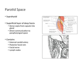
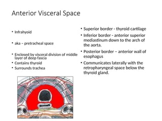
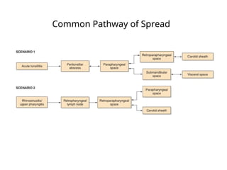
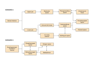
Deep Neck Space Infections
Dr. Hitender Basista
Assistant Professor
Dept. of Otorhinolaryngology
Cervical Fascia
• Superficial Layer
• Deep Layer
• Subdivisions not histologically
separate
• Superficial
• Enveloping layer
• Investing layer
• Middle
• Visceral fascia
• Prethyroid fascia
• Pretracheal fascia
• Deep
Superficial Layer
•Superior attachment –
zygomatic process
•Inferior attachment –
thorax, axilla.
•Similar to subcutaneous
tissue
•Ensheathes platysma and
muscles of facial
expression
Superficial Layer of the Deep Cervical Fascia
• Completely surrounds the neck.
• Arises from spinous processes.
• Superior border – nuchal line, skull
base, zygoma, mandible.
• Inferior border – chest and axilla
• Splits at mandible and covers the
masseter laterally and the medial
surface of the medial pterygoid.
Envelopes
• SCM
• Trapezius
• Submandibular
• Parotid
•Forms floor of submandibular
space
Middle Layer of the Deep Cervical Fascia
• Muscular Division
• Superior border – hyoid and thyroid
cartilage
• Inferior border – sternum, clavicle
and scapula
• Envelopes infrahyoid strap muscles
• Visceral Division
• Superior border
• Anterior – hyoid and thyroid
cartilage
• Posterior – skull base
• Inferior border – continuous with
fibrous pericardium in the upper
mediastinum.
• Buccopharyngeal fascia
• Name for portion that covers
the pharyngeal constrictors and
buccinator.
• Envelopes
• Thyroid
• Trachea
• Esophagus
• Pharynx
• Larynx
Deep Layer of Deep Cervical Fascia
•Arises from spinous processes and ligamentum
nuchae.
•Splits into two layers at the transverse processes:
• Alar layer
• Superior border – skull base
• Inferior border – upper mediastinum at T1-T2
• Prevertebral layer
• Superior border – skull base
• Inferior border – coccyx
• Envelopes vertebral bodies and deep muscles of the neck.
• Extends laterally as the axillary sheath.
Carotid Sheath
• Formed by all three layers of deep fascia
• Anatomically separate from all layers.
• Contains carotid artery, internal jugular vein, and vagus nerve
• Travels through pharyngomaxillary space.
• Extends from skull base to thorax.
Deep Neck Spaces
• Described in relation to the hyoid.
• Entire length of neck
• Superficial space
• Retropharyngeal
• Danger
• Prevertebral
• Vascular visceral
• Suprahyoid
• Submandibular
• Pharyngomaxillary (Parapharyngeal)
• Parotid
• Peritonsillar
• Temporal
• Masticator
• Infrahyoid
• Anterior visceral
Superficial Space
• Entire length of neck
• Surrounds platysma
• Contains areolar tissue,
nodes, nerves and vessels
• Subplatysmal Flaps
• Involved with cellulitis and
superficial abscesses
• Treat with incision along
Langer’s lines, drainage and
antibiotics
Retropharyngeal Space
• Entire length of neck.
• Anterior border - pharynx and
esophagus (buccopharyngeal fascia)
• Posterior border - alar layer of deep
fascia
• Superior border - skull base
• Inferior border – superior mediastinum
• Combines with buccopharyngeal fascia
at level of T1-T2
• Midline raphe connects superior
constrictor to the deep layer of deep
cervical fascia.
• Contains retropharyngeal nodes.
Danger Space
• Entire length of neck
• Anterior border - alar
layer of deep fascia
• Posterior border -
prevertebral layer
• Extends from skull
base to diaphragm
• Contains loose areolar
tissue.
Prevertebral Space
• Entire length of neck
• Anterior border -
prevertebral fascia
• Posterior border - vertebral
bodies and deep neck
muscles
• Lateral border – transverse
processes
• Extends along entire length
of vertebral column
Visceral Vascular Space
• Entire length of neck
• Carotid Sheath
• “Lincoln Highway”
• Lymphatic vessels can
receive drainage from
most of lymphatic vessels
in head and neck.
Submandibular Space
• Suprahyoid
• Superior – oral mucosa
• Inferior - superficial layer of
deep fascia
• Anterior border – mandible
• Lateral border - mandible
• Posterior - hyoid and base
of tongue musculature
• 2 compartments
• Sublingual space
• Areolar tissue
• Hypoglossal and lingual nerves
• Sublingual gland
• Wharton’s duct
• Submaxillary space
• Anterior bellies of digastrics
• Submental compartment
• Submaxillary compartments
• Submandibular gland
Submandibular Space
Submandibular Space
Parapharyngeal space
• Suprahyoid
• Superior—skull base
• Inferior—hyoid
• Anterior—ptyergomandibular
raphe
• Posterior—prevertebral fascia
• Medial—buccopharyngeal fascia
• Lateral—superficial layer of deep
fascia
• Prestyloid
• Muscular compartment
• Medial—tonsillar fossa
• Lateral—medial pterygoid
• Contains fat, connective tissue,
nodes
• Poststyloid
• Neurovascular compartment
• Carotid sheath
• Cranial nerves IX, X, XI, XII
• Sympathetic chain
• Stylopharyngeal aponeurosis of
Zuckerkandel and Testut
• Alar, buccopharyngeal and
stylomuscular fascia.
• Prevents infectious spread from
anterior to posterior.
• Communicates with several
deep neck spaces.
• Parotid
• Masticator
• Peritonsillar
• Submandibular
• Retropharyngeal
Peritonsillar Space
• Suprahyoid
• Medial—capsule of palatine
tonsil
• Lateral—superior
pharyngeal constrictor
• Superior—anterior tonsil
pillar
• Inferior—posterior tonsil
pillar
Masticator and Temporal Spaces
• Suprahyoid
• Formed by superficial layer of deep
cervical fascia
• Masticator space
• Antero-lateral to pharyngomaxillary
space.
• Contains
• Masseter
• Pterygoids
• Body and ramus of the mandible
• Inferior alveolar nerves and
vessels
• Tendon of the temporalis muscle
• Temporal space
• Continuous with masticator space.
• Lateral border – temporalis fascia
• Medial border – periosteum of
temporal bone
• Superficial and deep spaces divided
by temporalis muscle
Parotid Space
• Suprahyoid
• Superficial layer of deep fascia
• Dense septa from capsule into
gland
• Direct communication to
parapharyngeal space
• Contains
• External carotid artery
• Posterior facial vein
• Facial nerve
• Lymph nodes
Anterior Visceral Space
• Infrahyoid
• aka – pretracheal space
• Enclosed by visceral division of middle
layer of deep fascia
• Contains thyroid
• Surrounds trachea
• Superior border - thyroid cartilage
• Inferior border - anterior superior
mediastinum down to the arch of
the aorta.
• Posterior border – anterior wall of
esophagus
• Communicates laterally with the
retropharyngeal space below the
thyroid gland.
Common Pathway of Spread
Bacteriology
Aerobic Anaerobic
G (+) n % G (-) n % n %
Total 645
87.4
0 Total
13
7
18.5
6 Total
20
1 27.24
Strep sp. 229 31.03 Klebsiella sp. 90 12.20 Peptostreptococcus 43 5.83
Staph sp. 112 15.18 Neisseria sp. 20 2.71 Bacteroides sp. 50 6.78
B-hemolytic Strep 80 10.84 Acinebacter sp. 7 0.95 Unidentified 46 6.23
Strep viridans 71 9.62 Enterobacter sp. 7 0.95
Bacteroides
melaninogenicus 13 1.76
Staph aureus 57 7.72 Proteus sp. 4 0.54 Propionibacterium 9 1.22
Coagulase neg. Staph
sp. 55 7.45 E coli 3 0.41 Provotella sp. 7 0.95
Strep pneum 13 1.76 Citrobacter sp 2 0.27 Fusobacterium 7 0.95
Enterococcus 10 1.36 M. Catarrhalis 2 0.27 Bacteroidies fragilis 6 0.81
Mycobacterium tub.* 10 1.36 Pseudomonas sp. 1 0.14 Eubacterium 6 0.81
Micrococcus 8 1.08 H. Parainfluenza 1 0.14 Peptococcus 6 0.81
Diptheroids 7 0.95 H influenzae 1 0.14 Veillonella parvula 5 0.68
Bacillus sp. 6 0.81 Salmonella sp. 1 0.14 Clostridium sp. 4 0.54
Actinomycosis israelii 3 0.41 Lactobacillus 4 0.54
Bifidobacterium sp. 3 0.41
Polymicrobial 181
24.5
3 Sterile 71 9.62
Laboratory Evaluation
• Blood tests – CBC usually suggestive of leukocytosis
• Imaging studies – plain film radiology to identify dental sources of
infection, lateral neck films to identify suspected retropharyngeal
abscess.
• Presence of air fluid level or thickening of pre-vertebral tissue at C2
of more than 5 mm – indicates retropharyngeal infection.
• CT – remains the standard IOC for these infections
• USG – Non- invasive nature and lack of radiation - makes it an ideal
modality for pediatric patients. Limited use in case of significant
edema and less sensitive for non-lateral neck spaces.
Treatment
• Medical Management –
a.Airway Management
b.Fluid resuscitation
c.Antibiotic therapy
• Surgical Management- Main goal is to provide fluid or tissue sample
for culture and sensitivity.
Principles :
1.Limited antibiotic availability in pus filled space doe to poor
vascularity.
2.Treatment of facial space infection depends on open incision and
dependent drainage
3.Facial spaces are contiguous, hence infection spreads from one
region to another – making it necessary to open all primary and
secondary spaces followed by irrigation.
Peritonsillar abscess
 Peritonsillar abscess (Quinsy) is a collection of pus in the peritonsillar space
which lies between tonsil capsule and the superior constrictor muscle covered
by the buccopharyngeal fascia.
 Tonsillitis is usually the source of infection. The crypta magna gets infected and
sealed off. An intratonsillar abscess develops which subsequently bursts
through the tonsillar capsule into the peritonsillar space.
 It is a mixed infection of Streptococcus pyogenes, Staphylococcus aureus, and
anaerobic organisms.
Clinical features include: „
Age: Most patients are adults.
Children are rarely affected. Acute tonsillitis is more common in children. Side:
„
Usually, it is unilateral. „
Local features:
They are– Pain: Unilateral severe throat pain.
 Ipsilateral referred otalgia: Cranial nerve (CN) IX supplies tonsil, as well as ear.
Odynophagia may lead to drooling of saliva from the angle of mouth and
dehydration.
Hot potato voice: Muffled and thick speech.
Halitosis: Foul breath due to oral sepsis and poor hygiene.
 Trismus: Due to spasm of pterygoid muscles which are near to superior
constrictor muscle. „
General:
The general features due to septicemia, which is usually present in all deep space
neck infections include: Fever (up to 104° F) with chills and rigors General malaise,
body pain and headache Nausea and constipation.
Fig. showing Peritonsillar abscess
Management of Peritonsillar abscess
 Usually, antibiotics which are effective against streptococci, anaerobes and b-
lactamase-producing bacteria are used, and they include the following:
„ Ampicillin-sulbactam or cefoxitin or ceftriaxone. „ Combination of
clindamycin plus ciprofloxacin.
 Incision and drainage of abscess is indicated in cases of frank abscess
formation.
 Method: With the help of a guarded knife, a small stab incision is made at the
point of maximum bulge above the upper pole of tonsil, or the junction of
anterior pillar and base of uvula . A sinus- or artery-forcep is inserted to open
and drain the abscess. It may need to be repeated the following day to drain
any reaccumulation
Parapharyngeal space abscess
Etiology:
Oropharynx: Bursting of peritonsillar abscess, pharyngitis, tonsillitis
and adenoiditis „
Dental: Infections of usually lower last molars. „
Suppurative otitis media complications: Bezold’s abscess and
petrositis.
 Extensions: Infections of parotid, retropharyngeal and submaxillary
„
spaces. „
Injuries: Penetrating injuries of neck. „
Iatrogenic: Injection local anesthetic for tonsillectomy or mandibular
nerve block.
Clinical features of Parapharyngeal abscess
Common features:
• Fever
• Odynophagia
• Sore throat
• Torticollis (due to spasm of prevertebral muscles)
• Toxemia
Other features depend upon the compartment involved. They include the
following: „
• Anterior compartment:
• Prolapse of tonsil and tonsillar fossa
• Trismus due to spasm of medial pterygoid muscle
• Swelling behind the angle of jaw
• Odynophagia. „
Posterior compartment:
• Pharyngeal bulging behind the posterior pillar.
Cranial nerve palsies: CN 9, 10, 11 and 12 palsies will present with
dysphagia and hoarseness of voice, and ipsilateral nasal regurgitation and
ipsilateral palsies of palate, larynx and tongue.
Horner’s Syndrome due to the involvement of sympathetic chain
Management of Parapharyngeal abscess
Medical:
• Intravenous antibiotics to combat infection. „
Surgical drainage under general anesthesia:
oPreoperative tracheostomy is required in cases of marked trismus or airway
obstruction.
oA horizontal incision is made 2–3 cm below the angle of mandible. Abscess is
approached and drained with blunt dissection along the inner surface of
medial pterygoid muscle towards styloid process.
oA drain is usually inserted.
oTransoral drainage has the danger of injuring great vessels, and is avoided.
LUDWIG’S ANGINA
• It is the infection of the submandibular space
• Bacteriology - Infections involve both aerobes and anaerobes.
„ The most common causative microorganisms are a Hemolytic
streptococci, staphylococci and bacteroides groups. „ Rarely
Haemophilus influenzae, Escherichia coli and Pseudomonas are
noted.
• Sources of infection „
Dental infections account for 80% of the cases. „
Others: Submandibular sialadenitis, injuries of oral mucosa and
fractures of the mandible.
Clinical features :
•Presenting complaints are marked difficulty in swallowing,
odynophagia and varying degrees of trismus. „
•There occurs swelling in the floor of mouth.
•The tongue is pushed up and back if the infection happens to be
involving the sublingual space. „
•Once the infection spreads to the submaxillary and submental
spaces, the submandibular regions become swollen and tender and
feel woody hard.
•There is marked cellulitis of these areas. The frank abscess is
uncommon. „
•In advanced cases, airway is threatened. Tongue is progressively
pushed upwards and backwards. Laryngeal edema may ensue.
Treatment
• Systemic antibiotics and incision and drainage are the main lines of
management.
• Drainage material must be submitted for culture and sensitivity.
• Incision and drainage: Intraoral approach is used when abscess is
„
localized to sublingual space
• External approach is needed if swelling involves submaxillary space. –
Method: A transverse incision extending between angles of mandible,
is made. The vertical opening of midline musculature of tongue is
made with the help of hemostat. Usually, it is serous fluid and not
frank pus. It provides significant relief to the patient. „
• Tracheostomy must be considered if the airway is compromised.
Complications
• Spread of infection: The infection can spread to parapharyngeal and
retropharyngeal spaces and then to the mediastinum.
„
• Airway obstruction: Airway is compromised not only due to
laryngeal edema but also due to swelling and pushing back of the
tongue. „
• Septicemia and aspiration pneumonia need immediate attention.
Selected Complications of Deep Neck
Infections
• Vascular complications
1.Lemierre’s Syndrome – rare thrombophlebitis of IJV, usually Gram
negative bacilli
2.Cavernous Sinus Thrombosis – dreaded complication
3.Carotid Artery Pseudo-aneurysm or Rupture
• Mediasinitis
• Necrotizing Fasciitis
THANK YOU

deep neck infection EDITED.ppt and spread

  • 1.
    Deep Neck SpaceInfections Dr. Hitender Basista Assistant Professor Dept. of Otorhinolaryngology
  • 3.
    Cervical Fascia • SuperficialLayer • Deep Layer • Subdivisions not histologically separate • Superficial • Enveloping layer • Investing layer • Middle • Visceral fascia • Prethyroid fascia • Pretracheal fascia • Deep
  • 4.
    Superficial Layer •Superior attachment– zygomatic process •Inferior attachment – thorax, axilla. •Similar to subcutaneous tissue •Ensheathes platysma and muscles of facial expression
  • 5.
    Superficial Layer ofthe Deep Cervical Fascia • Completely surrounds the neck. • Arises from spinous processes. • Superior border – nuchal line, skull base, zygoma, mandible. • Inferior border – chest and axilla • Splits at mandible and covers the masseter laterally and the medial surface of the medial pterygoid. Envelopes • SCM • Trapezius • Submandibular • Parotid •Forms floor of submandibular space
  • 6.
    Middle Layer ofthe Deep Cervical Fascia • Muscular Division • Superior border – hyoid and thyroid cartilage • Inferior border – sternum, clavicle and scapula • Envelopes infrahyoid strap muscles • Visceral Division • Superior border • Anterior – hyoid and thyroid cartilage • Posterior – skull base • Inferior border – continuous with fibrous pericardium in the upper mediastinum. • Buccopharyngeal fascia • Name for portion that covers the pharyngeal constrictors and buccinator. • Envelopes • Thyroid • Trachea • Esophagus • Pharynx • Larynx
  • 7.
    Deep Layer ofDeep Cervical Fascia •Arises from spinous processes and ligamentum nuchae. •Splits into two layers at the transverse processes: • Alar layer • Superior border – skull base • Inferior border – upper mediastinum at T1-T2 • Prevertebral layer • Superior border – skull base • Inferior border – coccyx • Envelopes vertebral bodies and deep muscles of the neck. • Extends laterally as the axillary sheath.
  • 8.
    Carotid Sheath • Formedby all three layers of deep fascia • Anatomically separate from all layers. • Contains carotid artery, internal jugular vein, and vagus nerve • Travels through pharyngomaxillary space. • Extends from skull base to thorax.
  • 9.
    Deep Neck Spaces •Described in relation to the hyoid. • Entire length of neck • Superficial space • Retropharyngeal • Danger • Prevertebral • Vascular visceral • Suprahyoid • Submandibular • Pharyngomaxillary (Parapharyngeal) • Parotid • Peritonsillar • Temporal • Masticator • Infrahyoid • Anterior visceral
  • 10.
    Superficial Space • Entirelength of neck • Surrounds platysma • Contains areolar tissue, nodes, nerves and vessels • Subplatysmal Flaps • Involved with cellulitis and superficial abscesses • Treat with incision along Langer’s lines, drainage and antibiotics
  • 11.
    Retropharyngeal Space • Entirelength of neck. • Anterior border - pharynx and esophagus (buccopharyngeal fascia) • Posterior border - alar layer of deep fascia • Superior border - skull base • Inferior border – superior mediastinum • Combines with buccopharyngeal fascia at level of T1-T2 • Midline raphe connects superior constrictor to the deep layer of deep cervical fascia. • Contains retropharyngeal nodes.
  • 12.
    Danger Space • Entirelength of neck • Anterior border - alar layer of deep fascia • Posterior border - prevertebral layer • Extends from skull base to diaphragm • Contains loose areolar tissue.
  • 13.
    Prevertebral Space • Entirelength of neck • Anterior border - prevertebral fascia • Posterior border - vertebral bodies and deep neck muscles • Lateral border – transverse processes • Extends along entire length of vertebral column
  • 14.
    Visceral Vascular Space •Entire length of neck • Carotid Sheath • “Lincoln Highway” • Lymphatic vessels can receive drainage from most of lymphatic vessels in head and neck.
  • 15.
    Submandibular Space • Suprahyoid •Superior – oral mucosa • Inferior - superficial layer of deep fascia • Anterior border – mandible • Lateral border - mandible • Posterior - hyoid and base of tongue musculature • 2 compartments • Sublingual space • Areolar tissue • Hypoglossal and lingual nerves • Sublingual gland • Wharton’s duct • Submaxillary space • Anterior bellies of digastrics • Submental compartment • Submaxillary compartments • Submandibular gland
  • 16.
  • 17.
    Parapharyngeal space • Suprahyoid •Superior—skull base • Inferior—hyoid • Anterior—ptyergomandibular raphe • Posterior—prevertebral fascia • Medial—buccopharyngeal fascia • Lateral—superficial layer of deep fascia
  • 18.
    • Prestyloid • Muscularcompartment • Medial—tonsillar fossa • Lateral—medial pterygoid • Contains fat, connective tissue, nodes • Poststyloid • Neurovascular compartment • Carotid sheath • Cranial nerves IX, X, XI, XII • Sympathetic chain • Stylopharyngeal aponeurosis of Zuckerkandel and Testut • Alar, buccopharyngeal and stylomuscular fascia. • Prevents infectious spread from anterior to posterior.
  • 19.
    • Communicates withseveral deep neck spaces. • Parotid • Masticator • Peritonsillar • Submandibular • Retropharyngeal
  • 20.
    Peritonsillar Space • Suprahyoid •Medial—capsule of palatine tonsil • Lateral—superior pharyngeal constrictor • Superior—anterior tonsil pillar • Inferior—posterior tonsil pillar
  • 21.
    Masticator and TemporalSpaces • Suprahyoid • Formed by superficial layer of deep cervical fascia • Masticator space • Antero-lateral to pharyngomaxillary space. • Contains • Masseter • Pterygoids • Body and ramus of the mandible • Inferior alveolar nerves and vessels • Tendon of the temporalis muscle • Temporal space • Continuous with masticator space. • Lateral border – temporalis fascia • Medial border – periosteum of temporal bone • Superficial and deep spaces divided by temporalis muscle
  • 22.
    Parotid Space • Suprahyoid •Superficial layer of deep fascia • Dense septa from capsule into gland • Direct communication to parapharyngeal space • Contains • External carotid artery • Posterior facial vein • Facial nerve • Lymph nodes
  • 23.
    Anterior Visceral Space •Infrahyoid • aka – pretracheal space • Enclosed by visceral division of middle layer of deep fascia • Contains thyroid • Surrounds trachea • Superior border - thyroid cartilage • Inferior border - anterior superior mediastinum down to the arch of the aorta. • Posterior border – anterior wall of esophagus • Communicates laterally with the retropharyngeal space below the thyroid gland.
  • 24.
  • 26.
    Bacteriology Aerobic Anaerobic G (+)n % G (-) n % n % Total 645 87.4 0 Total 13 7 18.5 6 Total 20 1 27.24 Strep sp. 229 31.03 Klebsiella sp. 90 12.20 Peptostreptococcus 43 5.83 Staph sp. 112 15.18 Neisseria sp. 20 2.71 Bacteroides sp. 50 6.78 B-hemolytic Strep 80 10.84 Acinebacter sp. 7 0.95 Unidentified 46 6.23 Strep viridans 71 9.62 Enterobacter sp. 7 0.95 Bacteroides melaninogenicus 13 1.76 Staph aureus 57 7.72 Proteus sp. 4 0.54 Propionibacterium 9 1.22 Coagulase neg. Staph sp. 55 7.45 E coli 3 0.41 Provotella sp. 7 0.95 Strep pneum 13 1.76 Citrobacter sp 2 0.27 Fusobacterium 7 0.95 Enterococcus 10 1.36 M. Catarrhalis 2 0.27 Bacteroidies fragilis 6 0.81 Mycobacterium tub.* 10 1.36 Pseudomonas sp. 1 0.14 Eubacterium 6 0.81 Micrococcus 8 1.08 H. Parainfluenza 1 0.14 Peptococcus 6 0.81 Diptheroids 7 0.95 H influenzae 1 0.14 Veillonella parvula 5 0.68 Bacillus sp. 6 0.81 Salmonella sp. 1 0.14 Clostridium sp. 4 0.54 Actinomycosis israelii 3 0.41 Lactobacillus 4 0.54 Bifidobacterium sp. 3 0.41 Polymicrobial 181 24.5 3 Sterile 71 9.62
  • 27.
    Laboratory Evaluation • Bloodtests – CBC usually suggestive of leukocytosis • Imaging studies – plain film radiology to identify dental sources of infection, lateral neck films to identify suspected retropharyngeal abscess. • Presence of air fluid level or thickening of pre-vertebral tissue at C2 of more than 5 mm – indicates retropharyngeal infection. • CT – remains the standard IOC for these infections • USG – Non- invasive nature and lack of radiation - makes it an ideal modality for pediatric patients. Limited use in case of significant edema and less sensitive for non-lateral neck spaces.
  • 28.
    Treatment • Medical Management– a.Airway Management b.Fluid resuscitation c.Antibiotic therapy
  • 30.
    • Surgical Management-Main goal is to provide fluid or tissue sample for culture and sensitivity. Principles : 1.Limited antibiotic availability in pus filled space doe to poor vascularity. 2.Treatment of facial space infection depends on open incision and dependent drainage 3.Facial spaces are contiguous, hence infection spreads from one region to another – making it necessary to open all primary and secondary spaces followed by irrigation.
  • 31.
    Peritonsillar abscess  Peritonsillarabscess (Quinsy) is a collection of pus in the peritonsillar space which lies between tonsil capsule and the superior constrictor muscle covered by the buccopharyngeal fascia.  Tonsillitis is usually the source of infection. The crypta magna gets infected and sealed off. An intratonsillar abscess develops which subsequently bursts through the tonsillar capsule into the peritonsillar space.  It is a mixed infection of Streptococcus pyogenes, Staphylococcus aureus, and anaerobic organisms.
  • 32.
    Clinical features include:„ Age: Most patients are adults. Children are rarely affected. Acute tonsillitis is more common in children. Side: „ Usually, it is unilateral. „ Local features: They are– Pain: Unilateral severe throat pain.  Ipsilateral referred otalgia: Cranial nerve (CN) IX supplies tonsil, as well as ear. Odynophagia may lead to drooling of saliva from the angle of mouth and dehydration. Hot potato voice: Muffled and thick speech. Halitosis: Foul breath due to oral sepsis and poor hygiene.  Trismus: Due to spasm of pterygoid muscles which are near to superior constrictor muscle. „ General: The general features due to septicemia, which is usually present in all deep space neck infections include: Fever (up to 104° F) with chills and rigors General malaise, body pain and headache Nausea and constipation.
  • 33.
  • 34.
    Management of Peritonsillarabscess  Usually, antibiotics which are effective against streptococci, anaerobes and b- lactamase-producing bacteria are used, and they include the following: „ Ampicillin-sulbactam or cefoxitin or ceftriaxone. „ Combination of clindamycin plus ciprofloxacin.  Incision and drainage of abscess is indicated in cases of frank abscess formation.  Method: With the help of a guarded knife, a small stab incision is made at the point of maximum bulge above the upper pole of tonsil, or the junction of anterior pillar and base of uvula . A sinus- or artery-forcep is inserted to open and drain the abscess. It may need to be repeated the following day to drain any reaccumulation
  • 35.
    Parapharyngeal space abscess Etiology: Oropharynx:Bursting of peritonsillar abscess, pharyngitis, tonsillitis and adenoiditis „ Dental: Infections of usually lower last molars. „ Suppurative otitis media complications: Bezold’s abscess and petrositis.  Extensions: Infections of parotid, retropharyngeal and submaxillary „ spaces. „ Injuries: Penetrating injuries of neck. „ Iatrogenic: Injection local anesthetic for tonsillectomy or mandibular nerve block.
  • 36.
    Clinical features ofParapharyngeal abscess Common features: • Fever • Odynophagia • Sore throat • Torticollis (due to spasm of prevertebral muscles) • Toxemia Other features depend upon the compartment involved. They include the following: „ • Anterior compartment: • Prolapse of tonsil and tonsillar fossa • Trismus due to spasm of medial pterygoid muscle • Swelling behind the angle of jaw • Odynophagia. „ Posterior compartment: • Pharyngeal bulging behind the posterior pillar. Cranial nerve palsies: CN 9, 10, 11 and 12 palsies will present with dysphagia and hoarseness of voice, and ipsilateral nasal regurgitation and ipsilateral palsies of palate, larynx and tongue. Horner’s Syndrome due to the involvement of sympathetic chain
  • 37.
    Management of Parapharyngealabscess Medical: • Intravenous antibiotics to combat infection. „ Surgical drainage under general anesthesia: oPreoperative tracheostomy is required in cases of marked trismus or airway obstruction. oA horizontal incision is made 2–3 cm below the angle of mandible. Abscess is approached and drained with blunt dissection along the inner surface of medial pterygoid muscle towards styloid process. oA drain is usually inserted. oTransoral drainage has the danger of injuring great vessels, and is avoided.
  • 38.
    LUDWIG’S ANGINA • Itis the infection of the submandibular space • Bacteriology - Infections involve both aerobes and anaerobes. „ The most common causative microorganisms are a Hemolytic streptococci, staphylococci and bacteroides groups. „ Rarely Haemophilus influenzae, Escherichia coli and Pseudomonas are noted. • Sources of infection „ Dental infections account for 80% of the cases. „ Others: Submandibular sialadenitis, injuries of oral mucosa and fractures of the mandible.
  • 39.
    Clinical features : •Presentingcomplaints are marked difficulty in swallowing, odynophagia and varying degrees of trismus. „ •There occurs swelling in the floor of mouth. •The tongue is pushed up and back if the infection happens to be involving the sublingual space. „ •Once the infection spreads to the submaxillary and submental spaces, the submandibular regions become swollen and tender and feel woody hard. •There is marked cellulitis of these areas. The frank abscess is uncommon. „ •In advanced cases, airway is threatened. Tongue is progressively pushed upwards and backwards. Laryngeal edema may ensue.
  • 41.
    Treatment • Systemic antibioticsand incision and drainage are the main lines of management. • Drainage material must be submitted for culture and sensitivity. • Incision and drainage: Intraoral approach is used when abscess is „ localized to sublingual space • External approach is needed if swelling involves submaxillary space. – Method: A transverse incision extending between angles of mandible, is made. The vertical opening of midline musculature of tongue is made with the help of hemostat. Usually, it is serous fluid and not frank pus. It provides significant relief to the patient. „ • Tracheostomy must be considered if the airway is compromised.
  • 42.
    Complications • Spread ofinfection: The infection can spread to parapharyngeal and retropharyngeal spaces and then to the mediastinum. „ • Airway obstruction: Airway is compromised not only due to laryngeal edema but also due to swelling and pushing back of the tongue. „ • Septicemia and aspiration pneumonia need immediate attention.
  • 43.
    Selected Complications ofDeep Neck Infections • Vascular complications 1.Lemierre’s Syndrome – rare thrombophlebitis of IJV, usually Gram negative bacilli 2.Cavernous Sinus Thrombosis – dreaded complication 3.Carotid Artery Pseudo-aneurysm or Rupture • Mediasinitis • Necrotizing Fasciitis
  • 44.