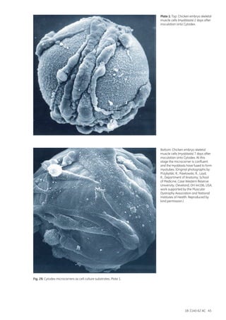
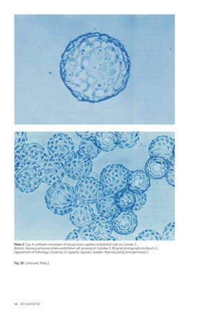
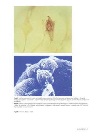
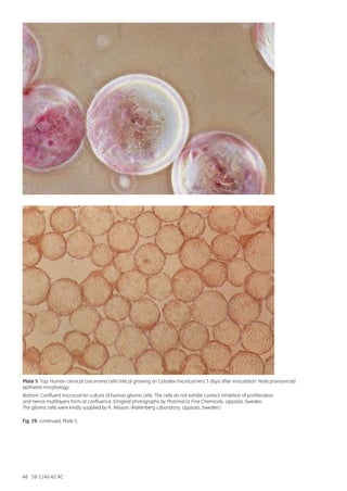
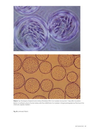
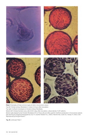
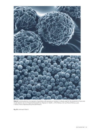
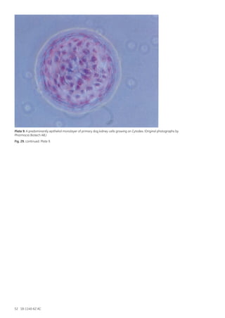
MicrocarrierCellCulture:PrinciplesandMethods
GE Healthcare
Life Sciences
imagination at work imagination at work
18-1140-62 AC  11/2013
GE, imagination at work, and GE monogram are trademarks of General Electric Company.
Ficoll-Paque, STREAMLINE, Ficoll, Sepharose, Sephadex, Cytodex, Cytopore, and Cytoline
are trademarks of GE Healthcare Ltd, a General Electric Company. GE and GE monogram
are trademarks of General Electric Company.
Dispase is a trademark of Worthington Biochemical Corp.
Perspex is a trademark of the Lucite International Ltd.
Pronase is a trademark of Calbiochem
Triton is a trademark of Union Carbide Chemicals and Plastics Co.
Teflon is a trademark of DuPont.
Sigmacote is a trademark of Sigma-Aldrich Co.
Siliclad is a trademark of Gelest Inc.
Fibra-Cel is a trademark of New Brunswick Scientific Co.
Tween is a trademark of ICI Americas Inc.
Cytopilot is a trademark of Vogelbusch GmbH
© 2005–2013 General Electric Company – All rights reserved.
First published Apr. 2005.
All goods and services are sold subject to the terms and conditions of sale of the company
within GE Healthcare which supplies them. A copy of these terms and conditions is available
on request. Contact your local GE Healthcare representative for the most current information.
GE Healthcare UK Limited Amersham Place
Little Chalfont
Buckinghamshire, HP7 9NA
UK
GE Healthcare Europe, GmbH
Munzinger Strasse 5
D-79111 Freiburg
Germany
GE Healthcare Bio-Sciences Corp.
800 Centennial Avenue, P.O. Box 1327
Piscataway, NJ 08855-1327
USA
GE Healthcare Japan Corporation
Sanken Bldg., 3-25-1, Hyakunincho
Shinjuku-ku, Tokyo 169-0073
Japan
For local office contact information,
please visit www.gelifesciences.com/contact
www.gelifesciences.com/
GE Healthcare Bio-Sciences AB
Björkgatan 30
751 84 Uppsala
Sweden
imagination at work
Microcarrier
Cell Culture
Principles and Methods
Handbooks from GE Healthcare Life Sciences
For more information refer to www.gelifesciences.com/handbooks
2-D Electrophoresis
Principles and Methods
GE Healthcare
Life Sciences 2-D Electrophoresis using
immobilized pH gradients
Principles and Methods
80-6429-60
Affinity Chromatography
Principles and Methods
GE Healthcare
Life Sciences Affinity Chromatography
Principles and Methods
18-1022-29
GE Healthcare
Life Sciences
ÄKTA™
Laboratory-scale
Chromatography Systems
Instrument Management Handbook
ÄKTA Laboratory-scale
Chromatography Systems
Instrument Management
Handbook
29-0108-31
GE Healthcare
Life Sciences
Antibody Purification
Handbook
Antibody Purification
Handbook
18-1037-46
GE Healthcare
Life Sciences
Biacore™ Assay Handbook
Biacore Assay
Handbook
29-0194-00
GE Healthcare
Life Sciences
Biacore
Sensor Surface Handbook
Biacore Sensor Surface
BR-1005-71
GE Healthcare
Life Sciences
Cell Separation Media
Methodology and applications
Cell Separation Media
Methodology and
Applications
18-1115-69
GE Healthcare
Life Sciences
Gel filtration
Principles and Methods
Gel Filtration
Principles and Methods
18-1022-18
GE Healthcare
Life Sciences
GST Gene
Fusion System
Handbook
GST Gene Fusion System
Handbook
18-1157-58
GE Healthcare
Life Sciences
High-throughput
Process Development
with PreDictor™
Plates
Principles and Methods
High-throughput Process
Development with
PreDictor Plates
Principles and Methods
28-9403-58
Hydrophobic Interaction
and Reversed Phase
Chromatography
Principles and Methods
GE Healthcare
Life Sciences Hydrophobic Interaction
and Reversed Phase
Chromatography
Principles and Methods
11-0012-69
GE Healthcare
Life Sciences
Imaging
Principles and Methods
Laser
CCD IRUV
IRUV
trans
epi
630 710520
W
460365
312
473 532 635 650 685 785
epi
Imaging
Principles and Methods
29-0203-01
GE Healthcare
Life Sciences
Ion Exchange
Chromatography
& Chromatofocusing
Principles and Methods
Ion Exchange
Chromatography and
Chromatofocusing
Principles and Methods
11-0004-21
GE Healthcare
Life Sciences
Isolation of
mononuclear cells
Methodology and applications
Isolation of
mononuclear cells
Methodology and
Applications
18-1152-69
GE Healthcare
Life Sciences
MicroCal™
Calorimeters
Achieving high quality data
MicroCal Calorimetry—
Achieving High Quality
Data
Handbook
29-0033-51
GE Healthcare
Life Sciences
Microcarrier
Cell Culture
Principles and Methods
Microcarrier Cell Culture
Principles and Methods
18-1140-62
imagination at work
GE Healthcare
Life Sciences
Multimodal
Chromatography
Handbook
Multimodal
Chromatography
Handbook
29-0548-08
GE Healthcare
Life Sciences
Nucleic Acid Sample
Preparation for
Downstream Analyses
Principles and Methods
Nucleic Acid Sample
Preparation for
Downstream Analyses
Principles and Methods
28-9624-00
GE Healthcare
Life Sciences
Protein Sample
Preparation
Handbook
Protein Sample
Preparation
Handbook
28-9887-41
GE Healthcare
Life Sciences
Purifying
Challenging Proteins
Principles and Methods
Purifying Challenging
Proteins
Principles and Methods
28-9095-31
GE Healthcare
Life Sciences
Recombinant
Protein Purification
Principles and Methods
Recombinant Protein
Purification Handbook
Principles and Methods
18-1142-75
GE Healthcare
Life Sciences
Spectrophotometry
Handbook
Spectrophotometry
Handbook
29-0331-82
GE Healthcare
Life Sciences
Strategies for
Protein Purification
Handbook
Strategies for Protein
Purification
Handbook
28-9833-31
GE Healthcare
Life Sciences
Western Blotting
Principles and Methods
Western Blotting
Principles and Methods
28-9998-97
Microcarrier
Cell Culture
Principles and Methods
Contents
1 Introduction............................................................................................................................................7
1.1 Reasons for using microcarriers.......................................................................................................................9
1.1.1 New opportunities and applications for animal cell culture................................................. 10
1.1.2 Increased production capacity........................................................................................................... 10
1.1.3 Separating cells from secreted product......................................................................................... 10
1.1.4 Improved control....................................................................................................................................... 11
1.1.5 Protection against physical and chemical stress....................................................................... 11
1.1.6 Reduced requirements for culture medium................................................................................. 12
1.1.7 Reduced requirements for labor........................................................................................................ 12
1.1.8 Lower risk of contamination................................................................................................................ 12
1.2 Aim of this book..................................................................................................................................................... 12
2 Microcarrier background ..................................................................................................................13
2.1 Adhesion (cell-cell, cell-surface)..................................................................................................................... 13
2.1.1 Adhesion to microcarriers..................................................................................................................... 17
2.2 Immobilization principles.................................................................................................................................. 19
2.3 Materials................................................................................................................................................................... 19
2.4 Size, shape and diffusion limits...................................................................................................................... 20
2.5 Specific density and sedimentation velocity............................................................................................ 21
2.6 Rigidity and shear force..................................................................................................................................... 22
2.7 Porosity...................................................................................................................................................................... 22
2.8 Cell observation..................................................................................................................................................... 23
3 Microcarrier technology.....................................................................................................................25
3.1 The development of microcarriers for animal cell culture................................................................ 25
3.2 Advantages of microcarriers........................................................................................................................... 27
3.3 Possible drawbacks............................................................................................................................................. 27
3.4 Scale-up considerations.................................................................................................................................... 28
3.5 Choice of supplier................................................................................................................................................. 29
4 Applications..........................................................................................................................................31
4.1 Vaccines.................................................................................................................................................................... 31
4.1.1 Production of viruses and vectors..................................................................................................... 32
4.2 Natural and recombinant proteins............................................................................................................... 34
4.2.1 Comparison of microcarriers in different reactors (packed bed or
fluidized bed reactor).......................................................................................................................................... 35
4.2.2 Long-term production with perfused macroporous microcarriers................................... 35
4.2.3 Process stability of perfusion with macroporous microcarriers......................................... 36
4.3 Monoclonal antibodies....................................................................................................................................... 37
4.3.1 Comparison of a hollow fiber reactor with a fluidized bed reactor................................... 38
4.4 Potential future applications........................................................................................................................... 38
4.5 Further applications............................................................................................................................................ 39
4.5.1 Production of large numbers of cells............................................................................................... 39
4.5.2 Studies on cell function, metabolism and differentiation....................................................... 40
4.5.3 Proteolytic enzyme-free subcultivation and cell transfer...................................................... 43
4.5.4 Microscopy................................................................................................................................................... 44
4.5.5 Harvesting mitotic cells.......................................................................................................................... 53
4.6 Which microcarrier to use?.............................................................................................................................. 54
4.6.1 Decision tree................................................................................................................................................ 54
4.6.2 Further comments on selecting Cytodex microcarriers......................................................... 57
5 Microcarrier products.........................................................................................................................59
5.1 Cytodex microcarriers........................................................................................................................................ 59
5.1.1 Cytodex 1...................................................................................................................................................... 59
5.1.2 Cytodex 3 ..................................................................................................................................................... 60
5.1.3 Cell types cultured on Cytodex microcarriers.............................................................................. 62
5.2 Cytopore microcarriers...................................................................................................................................... 65
5.2.1 Cell types cultured on Cytopore......................................................................................................... 66
6 Microcarrier culture equipment.......................................................................................................69
6.1 General outline of procedure.......................................................................................................................... 69
6.2 Requirements.......................................................................................................................................................... 70
6.2.1 Siliconizing culture vessels.................................................................................................................... 70
6.3 Unit process system............................................................................................................................................ 70
6.4 Small-scale equipment...................................................................................................................................... 71
6.4.1 Spinner and rod-stirred vessels.......................................................................................................... 71
6.4.2 Roller bottles................................................................................................................................................ 73
Method 1: Cytodex used to increase the yield from roller bottles using
standard roller techniques. ..................................................................................................................... 74
Method 2: Microcarriers used for “suspension” culture in roller bottles. ........................... 74
Method 3: Cells grown to confluence in rotating roller bottles using
standard techniques. ................................................................................................................................. 74
6.4.3 Rocking bottles shaker flasks.............................................................................................................. 75
6.4.4 Air-lift and fluid-lift culture systems................................................................................................. 76
6.4.5 Small-scale perfusion culture.............................................................................................................. 76
6.4.6 Dishes, tubes and multi-well trays.................................................................................................... 76
6.4.7 Bench-scale fermenters......................................................................................................................... 77
6.5 Large-scale equipment (culture vessels)................................................................................................... 78
6.5.1 Large-scale stirred tank microcarrier culture vessels.............................................................. 78
6.5.2 Packed beds................................................................................................................................................. 79
6.5.3 Fluidized beds............................................................................................................................................. 79
6.5.3.1 Fluidized bed with external circulation................................................................................ 79
6.5.3.2 Cytopilot – fluidized bed with internal circulation.......................................................... 80
6.5.3.3 Fluidization and fluidization velocity.................................................................................... 81
6.6 Retention systems................................................................................................................................................ 81
6.6.1 Spin filters..................................................................................................................................................... 81
6.6.2 Settlers........................................................................................................................................................... 82
6.6.3 Ultrasonic device....................................................................................................................................... 83
6.6.4 Hollow fibers................................................................................................................................................ 84
6.6.5 Centrifuges................................................................................................................................................... 85
7 Culture conditions...............................................................................................................................87
7.1 Media and components..................................................................................................................................... 87
7.1.1 Choice of culture medium .................................................................................................................... 87
7.1.1.1 General comments on components of culture media ................................................ 88
7.1.1.2 Practical aspects of culture media ....................................................................................... 89
7.1.1.3 Amino acids ..................................................................................................................................... 89
7.1.1.4 Nucleic acid precursors ............................................................................................................. 90
7.1.1.5 Carbon sources and lactate .................................................................................................... 90
7.1.1.6 Vitamins and choline .................................................................................................................. 91
7.1.1.7 Polymers ........................................................................................................................................... 91
7.1.1.8 The purpose of serum in culture media ............................................................................. 91
7.1.1.9 Choice and concentration of serum supplement ......................................................... 91
7.1.1.10 Reducing the serum concentration................................................................................... 93
7.1.1.11 Changing to another type of serum................................................................................... 94
7.1.1.12 Blending different sera ............................................................................................................ 94
7.1.1.13 Different sera for various stages of the culture cycle .............................................. 94
7.1.1.14 Variability of sera ....................................................................................................................... 95
7.1.2 Serum-free media .................................................................................................................................... 96
7.1.2.1 Additives............................................................................................................................................. 96
7.1.3 Protein-free media.................................................................................................................................... 97
7.2 Gas supply................................................................................................................................................................ 98
7.2.1 Gas supply and exchange in microcarrier cultures ................................................................. 98
7.2.2 Oxygen .......................................................................................................................................................... 99
7.2.3 Carbon dioxide.........................................................................................................................................100
7.2.4 Purity of gas supply ...............................................................................................................................101
7.3 Culture pH..............................................................................................................................................................101
7.3.1 pH optima for cell culture....................................................................................................................101
7.3.2 Buffers and the control of pH ...........................................................................................................103
7.3.3 Minimizing accumulation of lactate...............................................................................................104
7.4 Osmolarity .............................................................................................................................................................105
7.5 Redox potential....................................................................................................................................................105
7.6 Storing cells...........................................................................................................................................................105
7.7 Contamination.....................................................................................................................................................106
7.8 Stirring......................................................................................................................................................................107
7.9 Control and feeding strategies.....................................................................................................................107
8 Microcarriers in practice..................................................................................................................109
8.1 General considerations....................................................................................................................................109
8.2 Microcarrier concentrations..........................................................................................................................109
8.3 Inoculum.................................................................................................................................................................109
8.4 Preparing microcarriers for culture............................................................................................................112
8.4.1 Preparing for sterilization....................................................................................................................112
8.4.1.1 Cytodex............................................................................................................................................112
8.4.1.2 Cytopore..........................................................................................................................................112
8.4.1.3 Other sterilization methods ...................................................................................................112
8.4.2 Initiating a microcarrier culture ......................................................................................................113
8.4.2.1 Equilibration before inoculation ..........................................................................................113
8.4.2.2 Initial stirring .................................................................................................................................113
8.4.2.3 Concentration of microcarriers ...........................................................................................116
8.4.2.4 Inoculation density ....................................................................................................................117
8.4.2.5 Inoculum condition ....................................................................................................................118
8.4.2.6 Relationship between plating efficiency and culture procedure .........................119
8.4.3 Maintaining a microcarrier culture ................................................................................................121
8.4.3.1 Stirring speed ...............................................................................................................................121
8.4.3.2 Replenishment of culture medium......................................................................................123
8.4.3.3 Maintaining cultures at confluence ...................................................................................126
8.5 Cell quantification...............................................................................................................................................127
8.5.1 Direct observation by microscopy .................................................................................................128
8.5.2 Counting cells released after trypsinization...............................................................................128
8.5.3 Counting released nuclei ....................................................................................................................129
8.5.4 Fixing cells..................................................................................................................................................129
8.5.5 Staining cells..............................................................................................................................................130
8.5.5.1 Staining with Giemsa stain.....................................................................................................130
8.5.5.2 Staining with hematoxylin.......................................................................................................130
8.6 Scale-up..................................................................................................................................................................130
8.6.1 Harvesting cells........................................................................................................................................132
8.6.1.1 Chelating agents .........................................................................................................................133
8.6.1.2 Proteolytic enzymes...................................................................................................................133
8.6.1.3 Hypotonic treatment ................................................................................................................135
8.6.1.4 Cold treatment .............................................................................................................................135
8.6.1.5 Sonication.......................................................................................................................................135
8.6.1.6 Lignocaine for harvesting macrophages ........................................................................135
8.6.1.7 Modifications to harvesting procedures for large-scale cultures ........................136
8.6.1.8 Harvesting from macroporous microcarriers................................................................136
8.6.1.9 Separating detached cells from microcarriers .............................................................136
8.6.1.9.1 Differential sedimentation...........................................................................................136
8.6.1.9.2 Filtration...............................................................................................................................136
8.6.1.9.3 Density gradient centrifugation ...............................................................................137
8.6.1.9.4 Fluidized bed separation..............................................................................................137
8.6.1.9.5 Vibromixer...........................................................................................................................137
8.6.1.10 Measurement of cell viability...............................................................................................138
8.6.2 Sub-culturing techniques....................................................................................................................138
8.6.2.1 Colonization....................................................................................................................................138
8.6.2.2 Bead-to-bead transfer..............................................................................................................138
8.6.3 Suspension cells for inoculation.......................................................................................................140
8.6.4 Re-using microcarriers ........................................................................................................................140
8.6.5 Waste............................................................................................................................................................140
8.6.5.1 Dissolving microcarriers...........................................................................................................140
Cytodex...................................................................................................................................................140
Cytopore.................................................................................................................................................140
8.6.5.2 Incinerating microcarriers.......................................................................................................140
8.6.6 Documentation........................................................................................................................................141
8.6.7 Scale-up methods and process development needs.............................................................141
8.6.8 Integrated bioprocessing....................................................................................................................144
9 Production considerations...............................................................................................................145
9.1 Production economy.........................................................................................................................................145
9.1.1 Consumable cost comparison..........................................................................................................145
Culture surface............................................................................................................................................145
Serum and additives.................................................................................................................................145
9.1.2 Volumetric productivity comparison..............................................................................................146
9.1.2.1 Batch, fed-batch versus perfusion with macroporous microcarrier...................146
9.1.2.2 Roller bottle versus macroporous carriers (fluidized bed technology)...............147
9.1.2.3 Roller bottles versus Cytodex.................................................................................................148
9.1.3 SPF egg production versus Cytodex...............................................................................................148
9.1.4 Low cost production of rabies vaccine.........................................................................................148
9.1.5 Size of the production plant...............................................................................................................148
9.2 Product quality.....................................................................................................................................................149
9.3 Important developments................................................................................................................................149
10 Optimizing culture conditions.......................................................................................................151
11 Troubleshooting..............................................................................................................................153
11.1 Stirred microcarrier cultures.......................................................................................................................153
11.2 Fluidized bed.......................................................................................................................................................155
12 References........................................................................................................................................157
6 18-1140-62 AC
18-1140-62 AC 7
1 Introduction
The pressure in biotechnology production today is for greater speed, lower costs and more flexibility.
Ideally, a production unit should be compact (requires less investment) and modular (for use in
different production schemes) (Figure 1). Designs that allow the same plant to be used for different cell
types (bacteria, yeast, insect cells, plus suspension and anchorage-dependent animal cells) are thus
preferable. Animal cell culture is constantly being developed to increase unit productivity and thus
make the production costs of animal cell products more competitive.
Because animal cells have a relatively low productivity, large amounts of culture supernatant are
needed per clinical dose of final product. Extremely large volume cultures will be needed to produce
kilogram quantities of a therapeutic monoclonal antibody, for example.
Fig. 1A. Scheme of a production plant.
Fluidized bed
(Cytopilot™)
Stirred tank
(bacterial
configuration)
Stirred tank
(cell culture
configuration)
Airlift
fermenter
Stirred lift
fermenter
FermentationMedia preparation
TDC 3000DDC Control
Downstream
Decontamination Peripheral equipment
Filtration unitsChromatography CentrifugationHomogenizer
Sterile filtration
180 300 600 600 2000
200
7/14 40/80 150/300 600 2000
Buffer storage
180600 180
HQ water HQ steam
Compressed
air
UPS
Gas
N2O2 CO2
3 x 2500
(A)
(B)
Fig. 1B. Different reactor configurations. Courtesy of Polymun Scientific GmbH, Vienna, Austria.
8 18-1140-62 AC
Animal cell culture is currently operated at 15 000 liter scale in suspension cultures and 6 000 liter scale
in microcarrier cultures of anchorage-dependent cells (Figure 2). Both are large unit operations. As
the size of such operations increases, so does investment in resources and personnel. Cell culture
conditions are also more critical. Failures become far more costly.
Fig. 2. Influenza production plant (6000 liter vessel for cultivating Vero cells on Cytodex™). Courtesy of Baxter Biosciences.
The productivity of large-scale cell culture can be increased either by scaling up to larger volumes with
cell densities of 2–3×10⁶/mL, or by intensifying the process in smaller volumes but with higher cell
densities (up to 2×10⁸ cells/mL). When intensifying cell densities, more frequent media changes are
needed and eventually perfusion has to be applied. Many competing technologies are available.
Microcarrier culture of anchorage-dependent or entrapped cells lowers the volume to cell density
ratio and thus belongs to the second of the above alternatives. This technique has many advantages
for the commercial manufacturer. It operates in batch or perfusion modes and is well suited to
efficient process development and smooth scale-up (Figure 3). In addition, the reactors can be
modified to grow other organisms.
This book reviews the principles and methods of using microcarriers in cell culture production.
Fig. 3A. Different microcarriers and applications.
18-1140-62 AC 9
Fig. 3B. Different microcarrier culture setups.
1.1 Reasons for using microcarriers
Cell culture techniques have become vital to the study of animal cell structure, function and
differentiation, and for the production of many important biological materials such as vaccines,
enzymes, hormones, antibodies, interferons and nucleic acids. Microcarrier culture introduces new
possibilities and, for the first time, makes possible the practical high-yield culture of anchorage-
dependent cells. In microcarrier culture, cells grow as monolayers on the surface of small spheres
(Figure 4) or as multilayers in the pores of macroporous structures that are usually suspended in
culture medium by gentle stirring. By using microcarriers in simple suspension culture, fluidized or
packed bed systems, yields of up to 200 million cells per milliliter are possible.
Fig. 4. Scanning electron micrograph of pig kidney cells (IBR-S2) growing on Cytodex 72 h after inoculation.
(Original photograph by G. Charlier, INVR, Brussels, Belgium, reproduced by kind permission.)
Cytodex microcarriers have been specifically developed by Amersham Biosciences (now a part of
GE Healthcare) for the high-yield culture of a wide range of animal cells (Section 5.1.3) in culture
volumes ranging from a few milliliters to several thousand liters. The special requirements
of the microcarrier system are best fulfilled by dextran-based beads that are subsequently
derivatized to form two types of Cytodex, cellulose-based beads to form two types of Cytopore™.
10 18-1140-62 AC
Cytodex:
•	Surface characteristics have been optimized for efficient attachment and spreading of cells.
•	Size and density are optimized to facilitate even suspension and give good growth and high
	 yields for a wide variety of cells.
•	Matrix is biologically inert and provides a strong but non-rigid substrate for stirred cultures.
•	Transparent and allows easy microscopic examination of the attached cells.
Experience with Cytodex in a wide variety of applications has confirmed its importance and value in
microcarrier techniques.
Cytopore:
•	Cytopore, a macroporous microcarrier, is a logical development of Cytodex that increases cell
	 density (and therefore yield) and allows suspension cell cultivation and perfusion.
1.1.1 New opportunities and applications for animal cell culture
Microcarriers provide convenient surfaces for growing animal cells or increasing the yield of cells
from standard monolayer culture vessels and perfusion chambers. Applications include production
of large quantities of cells, viruses and cell products, studies on differentiation and cell function,
perfusion culture systems, microscopy studies, harvesting mitotic cells, isolation of cells, membrane
studies, storage and transportation of cells, assays involving cell transfer and studies on uptake of
labeled compounds (see Section 4 for a description of these applications).
1.1.2 Increased production capacity
The very large culture surface area to volume ratio offered by the microcarrier system
(e.g. 30 cm2 in 1 mL using 5 mg Cytodex 1) provides high cell yields without having to resort
to bulky equipment and tedious methodology. For a given quantity of cells or their products,
microcarrier cultures demand much less space (Figure 40) than other types of monolayer culture.
Culturing cells in small compact systems is especially important when working with pathogenic
organisms.
1.1.3 Separating cells from secreted product
Microcarrier technology is one method for cell retention (Figure 5), which means that dilution rate
(perfusion rate) becomes independent of the growth rate of the cells. Secondly, because there are
fewer cells in the harvest stream, downstream processing becomes simpler. You transfer part of the
clarification step back into the fermentation process.
18-1140-62 AC 11
Fig. 5. Different retention systems.
1.1.4 Improved control
Microcarrier systems allow excellent control of culture parameters (e.g. pH, gas tensions, etc). The
technique provides a method for growing anchorage-dependent cells in a system having all the
advantages of suspension culture. Improved control allows homogeneous culture systems with a wide
variety of process designs (1). Monitoring and sampling microcarrier cultures is simpler than with any
other technique for producing large numbers of anchorage-dependent cells.
1.1.5 Protection against physical and chemical stress
Macroporous microcarriers protect cells against the tip of the stirrer, especially at large scale.
Sparging with microbubbles using pure oxygen is also possible due to this protection. Cells tolerate
more chemical stress such as lactate, ammonia and oxygen if they create a micro-environment
inside the pores (Figure 6).
Fig. 6. Graph of cells sitting satisfied inside pores.
Filtration:
membranes or
spin-sieves
Cells growing
on matrix
Feed Harvest
Sedimentation:
zone or carrier
Aggregation +
sedimentation
Centrifugation
12 18-1140-62 AC
1.1.6 Reduced requirements for culture medium
When compared with other monolayer or suspension techniques, stirred microcarrier cultures yield
up to 100-fold as many cells for a given volume of medium. Superior yields have been reported for
a wide variety of systems including chicken fibroblasts (2,3), pig kidney cells (4), fish cells (5), Chinese
hamster ovary cells (6), human fibroblasts (7), primary monkey kidney cells (8) and transformed
mouse fibroblasts (9). This reduced requirement for medium means considerable savings in cell
culture costs (6,9), particularly when expensive serum supplements such as fetal calf serum are
used. Macroporous microcarriers especially provide an excellent micro-environment where cells can
exchange their own autocrine growth hormones, thereby allowing protein-free media to be used.
1.1.7 Reduced requirements for labor
Because large numbers of cells can be cultured in small volumes (more than 1011 cells/L), fewer
culture vessels are required with microcarrier cultures. For example, one technician can handle a
vaccine production equivalent to 900 roller bottles per week (10). One liter of microcarrier culture
can yield as many cells as up to 50 roller bottles (490 cm2 bottles, 2), and 1 to 1.5 mL of macroporous
beads can yield as many cells as one roller bottle of 850 cm2. The simplified procedures associated
with microcarriers reduce the labor needed for routine production. Separating cells from the culture
medium is simple; when stirring stops, microcarriers with cells attached settle under the influence
of gravity and the supernatant can be removed. Unlike true suspension cell culture systems, no
centrifugation steps are necessary.
1.1.8 Lower risk of contamination
In cell culture, the risk of contamination is related to the number of handling steps (e.g. opening
and closing culture vessels) required to produce a given quantity of cells or products. Microcarrier
culture reduces the number of handling steps. There is a much-reduced risk of contamination when
producing a large quantity of cells in a single microcarrier culture than in several hundred roller
bottles (6).
1.2 Aim of this book
This book describes the principles and methods of achieving the best cell culture results with Cytodex
and Cytopore microcarriers. Although this technique is one of the most advanced in animal cell
culture, it need not be restricted to experienced cell culturists. Because cell culture is used by a wide
variety of scientists, the book is written for both beginners and those more experienced in cell culture.
Only a basic knowledge of cell culture is assumed.
The book aims to help the reader plan and operate optimum procedures with the minimum of effort,
and to achieve consistent results with high yields. All methods have been developed for Cytodex and
Cytopore and are not necessarily suitable for other cell culture surfaces.
18-1140-62 AC 13
2 Microcarrier background
2.1 Adhesion (cell-cell, cell-surface)
Cell-cell and cell-surface (matrix) adhesion is one of the most important processes in higher life
forms. This mechanism allows specialized cell conglomerates that result in different tissues, organs
and skeletal structures. In vertebrates, the major types of tissue are nerve, muscle, blood, lymphoid,
epithelial, and connective tissue.
In principle, there are three functional classifications of junctions between cells: occluding junctions,
communicating junctions and anchoring junctions. Occluding or tight junctions can seal cells together
in an epithelial cell sheet in a way that prevents even small molecules from leaking from one side to
the other. Communicating junctions mediate the passage of chemical or electrical signals from one
interacting cell to its partner. Anchoring junctions, the most interesting ones for this work, mechanically
attach cells (and their cytoskeletons) to their neighbors or to extracellular matrices. Both kinds of
attachment are interesting for animal cell culture methods, as artificial matrices may simulate or
stimulate cell-cell or cell-matrix junctions.
In vivo, many different molecules are involved in attachment processes that are localized
intracellularly, extracellularly and/or in the cell membrane.
Intracellular attachment proteins (Table 1) connect the cytoskeleton with transmembrane linker
proteins, which bind to transmembrane linker proteins of other cells in a homophilic or heterophilic
binding. Extracellular linker molecules also mediate the binding of transmembrane linker proteins.
Such connections of the cytoskeletal actin filament and transmembrane linker proteins
(Ca2+-dependent cadherins), which are mediated by catenins, are called anchoring junctions.
Table 1. Cell adhesion molecule families
Adhesion	 Protein groups	 Ca2+ or Mg2+	 Homophilic/	 Cytoskeleton 	 Cell junction
		 dependency	 heterophilic	 association	 association
Cell-cell	 cadherins	 yes	 homophilic	 actin filaments	 adhesion belt
Cell-cell	 Ig family (N-CAM)	 no	 homophilic/	 unknown	 no
			 heterophilic
Cell-cell	selectins	 yes	 heterophilic	unknown	 no
Cell-cell	 integrins	 yes	 heterophilic	 actin filaments	 no
Cell-matrix	 integrins	 yes	 heterophilic	 actin filaments	 focal contacts /
				 (via talin, vinculin	 hemidesmosomes
				 and other proteins)
Cell-matrix	 transmembrane	 no	 heterophilic	 actin filaments	 no
	proteoglycans
There are four different classes of anchoring junctions (Table 2): 1, adherence junctions (cell-cell); 2,
adherence junctions (cell-matrix); 3, desmosomes; and 4, hemidesmosomes. Adherence junctions connect
bundles of actin filaments cell-to-cell or cell-to-extracellular matrix. Desmosomes connect intermediate
filaments cell-to-cell, and hemidesmosomes connect the intermediate filaments to the basal lamina
(a specialized mat of extracellular matrix at the interface between the epithelium and connective tissue).
14 18-1140-62 AC
Table 2. Anchoring junctions
Junction	 Transmembrane	 Extracellular ligand	 Intracellular cytoskeletal	 Some intracellular
	 linker protein		 attachment	 attachment proteins
Adherence 	 cadherin	 cadherin in	 actin filaments	 catenins, vinculin, a-actinin,
(cell-cell)	 (E-cadherin)	 neighboring cell		 plakoglobin
Adherence	 integrin	 extracellular matrix	 actin filaments	 talin, vinculin, a-actinin
(cell-matrix)		 protein		
Desmosome	 cadherin 	 cadherin in	 intermediate filaments	 plakoglobin
		 neighboring cell
Hemidesmosome	 integrin	 extracellular matrix	 intermediate filaments	 desmoplakinlike protein
		 (basal lamina) proteins
In epithelial sheets, anchoring junctions often form a continuous adhesion belt (zonula adherens). The
actin filaments are further connected to a set of intracellular attachment proteins that include a,
b and g-catenin, vinculin, a-actinin, and plakoglobin. The actin bundles are linked in an extensive
intracellular network that can be contracted by myosin motor proteins to mediate a fundamental
process in morphogenesis, which is important for the activity of the cells. In addition, a family of
cell-surface carbohydrate-binding proteins (lectins) called selectins play a role in a variety of transient
cell-cell adhesion interactions in the bloodstream. Selectins contain a highly conserved lectin
domain that, in the presence of Ca2+, binds to a specific oligosaccharide on the surface of other cells,
resulting in a heterophilic cell-cell adhesion.
Ca2+-independent cell-cell adhesion is mediated mainly by members of the immunoglobulin
superfamily of proteins, such as the neural cell adhesion molecules (N-CAM) that are involved in
homophilic interactions. Heterophilic interactions are mediated by intercellular adhesion molecules
(ICAMs), which interact with integrins present on the surface of white blood cells, thereby helping to
trap these blood cells at sites of inflammation.
There are two main classes of extracellular macromolecules that make up the matrix in vertebrates:
•	Polysaccharide chains of the class called glycosaminoglycans (GAG), which are usually found
	 covalently linked to protein in the form of proteoglycans.
•	Fibrous proteins of two functional types: mainly structural (for example, collagen and elastin)
	 and mainly adhesive (for example, fibronectin and laminin).
Members of both classes vary greatly in shape and size.
Glycosaminoglycans (GAGs) consist of unbranched polysaccharide chains composed of repeated
disaccharide units, where one of the two sugar residues is always an amino sugar (N-acetylgycos-
amine or N-acetylgalactosamine), which in most cases is sulfated. The second sugar is usually uronic
acid (glucuronic or iduronic). Because there are sulfate or carboxyl groups on most of the sugar
residues, GAGs are highly negatively charged. Four main groups of GAGs have been distinguished by
their sugar residues according to the type of linkage between these residues and the number and
location of sulfate groups: 1st hyaluronan, 2nd chondroitin sulfate and dermatan sulfate, 3rd heparan
sulfate and heparin and 4th keratan sulfate.
GAGs and proteoglycans are capable of self-organizing into large complexes that have the size of
bacteria. Moreover, besides associating with one another, GAGs and proteoglycans associate with
fibrous matrix proteins such as collagen, and with protein meshworks such as the basal lamina,
creating extremely complex structures. Furthermore, they have different functions in vivo. Due to
their inflexible, strongly hydrophilic nature, GAGs occupy a huge volume relative to their mass, and
they form gels even at very low concentrations. Their high density of negative charges attracts a
cloud of cations, such as Na+, causing large amounts of water to be sucked into the matrix, resulting
in a swelling pressure, or turgor, that enables the matrix to withstand compressive forces (in contrast
to collagen fibrils, which resist stretching forces).
18-1140-62 AC 15
Proteoglycans can also regulate the activities of secreted signaling molecules, such as enhancing
or inhibiting the activity of growth factors. Furthermore, proteoglycans also bind and regulate the
activities of other types of secreted proteins in any of the following ways: 1, they can immobilize the
protein at the site where it is produced, thereby restricting its range of action; 2, they can sterically
block the activity of the protein; 3, they can provide a reservoir of the protein for delayed release; 4, they
can protect the protein from proteolytic degradation, thereby prolonging its action; and 5, they can
alter or concentrate the protein for more effective presentation to cell-surface receptors.
A fibrous group of proteins, the collagens, belongs to the second group of extracellular
macromolecules. These are the major proteins of the extracellular matrix in vivo, thus cells normally
attach tightly to these structures. This attachment is mediated by the second group of proteins in
the matrix, the adhesive proteins such as fibronectin. Fibronectin contributes to both organizing the
matrix and helping cells attach to it. It forms dimers composed of very large subunits joined by a
pair of disulfide bonds near their carboxyl termini. Each subunit is folded into a series of functionally
distinct rod-like domains separated by regions of flexible polypeptide chains. These domains consist
of smaller modules, each of which is serially repeated. These different domains bind to collagen,
heparin, and specific receptors on the surface of various types of cells. The sequence responsible for
cell binding is a specific tripeptide (Arg-Gly-Asp, or RGD). Even very short peptides containing this RGD
sequence compete with fibronectin for the binding sites on cells. Each receptor, however, specifically
recognizes its own small set of matrix molecules, indicating that tight receptor binding requires more
than just an RGD motif.
The last class of important proteins is the integrins. These are the principal receptors used by animal
cells to bind to the extracellular matrix. They are heterodimers that function as transmembrane
linkers to mediate bidirectional interactions between the extracellular matrix and the actin
cytoskeleton. They also function as signal transducers, activating various intracellular signaling
pathways when activated by matrix binding. A cell can regulate the adhesive activity of its integrins
by altering either their matrix-binding sites or their attachment to actin filaments.
Integrins located in the plasma membrane anchor a cell to extracellular matrix molecules; cadherins
in the plasma membrane anchor the cell to cadherins in the membrane of an adjacent cell. In
both cases, there is an intracellular coupling to cytoskeletal filaments, which can be either actin
or intermediate filaments depending on the types of intracellular attachment proteins employed.
Moreover, for all these classes of anchoring junctions, the adhesion depends on extracellular divalent
cations, although the significance of this dependence is unknown.
Cell adhesion is a multistep process involving initial contact of the cell with the surface, cell spreading
on the surface, and cell differentiation or growth. Attachment involves forming bonds between
transmembrane adhesion receptors (e.g. cadherins, integrins and N-CAMs) and absorbed adhesion
proteins (e.g. fibronectin, vitronectin, laminin). Binding induces a conformational change in the
cytoplasmic domain of the receptor, which produces receptor aggregation (11). Free receptors diffuse
into the contact area forming more interactions with adhesion proteins and increasing the size of
the contact area (12). Cell spreading involves interaction among microtubules, actin filaments, and
ligand-bound adhesion receptors (13). Cell spreading is the critical step in adhesion and subsequent
cell growth and protein formation (14,15).
Anchorage-dependent cells need a certain amount of the described attachment proteins to proliferate
in vitro. This requirement can be achieved by adding serum to the media or by coating the growth
surfaces with collagen or other attachment proteins, such as fibronectin or variants of the cell
recognition site of fibronectin (RGD, RGDS, RGDV, RGDT, and other peptides) (17–20). However, these
substances often derive from animal products, and are therefore considered to be of potential
risk for biological safety. A solution to this problem is the use of recombinant proteins produced
in prokaryotic or lower eukaryotic cells. However, disadvantages of this method are the high price
of such products, and the fact that media for bacteria or yeast may contain some sort of animal-
derived hydrolysates.
16 18-1140-62 AC
Plant-derived biomolecules are therefore better suited to increase the adherence of production cell
lines. As such plant-derived macromolecules do not enhance adherence per se (as shown below),
chemical derivatization is a useful tool to engineer biomimetic materials. For this purpose, the theory
of Emil Fischer (1852–1919) – the proposed key-lock system – might provide a possible method.
Based on knowledge of cell adherence in vivo (see above), artificial substances with the required
characteristics can be designed. Polymers can be used as a string to which keys are attached (21).
Random substitution of the macromolecular chains allows the formation of specific binding sites in
a composition-dependent way. The polymer is used as a kind of master key with an infinite amount
of different specific keys attached to it. These keys need a recognition site, so that the water can be
displaced from the catalytic center and the interaction can be performed as required.
Different steps or functions of attachment factors can be simulated. One key might be a synthetic
RGD-peptide (commercially produced) or similar recognition site of an attachment protein. Another
approach could be to simulate GAGs (see above). As GAGs have different functions,
it might be easier to achieve the desired effect.
For industrial processes, it is not necessary to know exactly how the artificial biomolecule works, as
long as it does so. There have already been a few known substances discussed in the international
scientific community. Different dextran derivatives are capable of increasing or decreasing the
adherence of cells. Barabino et al. (22) found that anionic polysaccharides inhibit adhesion of sickle
erythrocytes to the vascular endothelium and result in improved hemodynamic behavior. Dee et
al. and Donaldson et al. (23,24) used dextran sulfate as adherence-decreasing substance to induce
single cell formation in insect cell culture. Zanghi et al. (25) tested dextran sulfate for the ability to
reduce clumping of CHO cells in a suspension culture system. A more distinct work with engineered
dextran derivatives has been presented by the group of J. Jozefonvicz (21) (Institut d’Oncologie Cellulaire
et Moleculaire Humaine (IOCMH), Bobigny, France). They tested different artificial dextran derivatives
for their ability to generate chemical functions, thereby mimicking natural ligands.
Carboxymethyl benzylamide sulfonate dextrans are especially studied for heparin-like properties
(25,27). However, these groups of engineered substances are also capable of performing other tasks
in vitro, such as inhibiting tumor proliferation (28) or influencing the production of collagen by the
extracellular matrix (29). If the production of collagen can be inhibited, it might be also possible to
induce collagen expression (30) with the effect that cells produce their own “extracellular matrix” on
microcarriers. If so, the collagen-inducing biomolecule could be added to the medium directly after
inoculation of a microcarrier process until a steady state is achieved. At this point, the cells adapt
their micro-environment as required and the medium composition can be switched to production
composition, resulting in a very stable and robust bioprocess (31). However, if stimulation of
attachment protein production is not achievable via artificial biomolecules, a flow cytometry sorting
for cells that produces higher amounts of the desired proteins might be a solution (32). For simulating
GAGs, it seems that all their functions can be mimicked by artificial polysaccharides.
A completely different approach to manipulating the adhesion of cells is to use ions or different
surface charges and charge densities (33). As mentioned earlier, Ca2+ and Mg2+ are cofactors for
many attachment proteins and also for GAGs (34–39). Therefore, the Ca2+ concentration in media is
often reduced to decrease adherence of cells cultured in suspension mode, or increased to culture
anchorage-dependent cells on microcarrier beads (40). However, in addition to these two, more or
less every other cation can modify the normally negatively charged culture surface. This fact must be
taken into consideration when searching for adherence-influencing substances.
18-1140-62 AC 17
2.1.1 Adhesion to microcarriers
The adhesion of cells to culture surfaces is fundamental to both traditional monolayer culture
techniques and to microcarrier culture. Since the proliferation of anchorage-dependent cells can
only occur after adhesion to a suitable culture surface (41), it is important to use surfaces and culture
procedures that enhance all of the steps involved in adhesion.
Attachment can be divided into four different phases. The first phase comprises a slight attachment to
the surface. The second phase shows flattened but still spheroidal cells, where the cell is significantly
more adherent due to the increased contact area and bond density. Cells in the third phase are
significantly less adhesive than cells in phase two because of the reduced number of bonds. Cells in
the fourth phase are fully attached and extremely flat (Figure 7) (42). Adhesion of cells in culture is a
multi-step process and involves a) adsorption of attachment factors to the culture surface, b) contact
between the cells and the surface, c) attachment of the cells to the coated surface, and finally
d) spreading of the attached cells (41, Figure 7).
Fig. 7A. Schematic drawing of the adhesion process in 4 phases (Fn- fibronectin, MHS- multivalent heparan sulfate).
substrate
adsorption
– Fn
– Fn – Fn
MHS –MHS –
contact attachment spreading
cell
cell cell cell
+ + + + + + + + +
substrate substrate substrate
Mg2+
Ca2+
Mn
2+
Fig 7B. Microscopic pictures of the 4 phases.
The entire process involves divalent cations and glycoproteins adsorbed to the culture surface.
Under normal culture conditions, attachment proteins vitronectin and fibronectin originate from
the serum supplement in the medium. MHS is synthesized by the cells. The culture surface must be
hydrophilic and correctly charged before adhesion of cells can occur (41). All vertebrate cells possess
unevenly distributed negative surface charges (43) and can be cultured on surfaces that are either
negatively or positively charged (41,44,45,33,46). Examples of suitable culture surfaces bearing charges
of different polarities are glass and plastic (negatively-charged) and polylysine coated surfaces
or Cytodex 1 microcarriers (positively-charged). Since cells can adhere and grow on all of these
surfaces, the basic factor governing adhesion and growth of cells is the density of the charges on the
culture surface rather than the polarity of the charges (33,47).
Two factors in a culture medium are essential for cells to adhere to culture surfaces: divalent
cations and protein(s) in the medium or adsorbed to the culture surface (41). Many established and
transformed cell types secrete only very small amounts of fibronectin and thus require a fibronectin
or serum supplement in the culture medium before adhesion occurs (48,49). Certain types of cells,
such as diploid fibroblasts, can secrete significant quantities of fibronectin and therefore do not
require an exogenous source for attachment (50,51). When initiating a culture, it is usual practice to
let the culture surface come into contact with medium containing serum before cells are added to
the culture. Culture medium supplemented with 10% (v/v) fetal calf serum contains approximately
18 18-1140-62 AC
2–3 µg fibronectin/mL (52), and a large proportion of the fibronectin adsorbs to culture surfaces
within a few minutes (48). Serum-free media often require addition of fibronectin (1–50 µg/mL) before
many cells can attach to culture surfaces.
Culture procedures affect the rate at which cells attach to surfaces. In the case of microcarrier
culture, microcarriers and cells are often in a stirred suspension. Under such conditions, attachment of
cells to Cytodex usually occurs to the same extent as with static culture systems. However, with some
cell types, an initial static culture period is required so that all steps of adhesion
(Figure 7) are fully completed.
The design of microcarrier culture procedures for each type of cell is closely related to the adhesion
properties of the cell and the rate at which all steps of adhesion are completed. Ways of determining
optimal procedures for individual cell types are discussed in sections 8 and 10. Figure 8 illustrates the
close attachment of cells to Cytodex.
Fig. 8. Transmission electron micrograph of pig kidney cells growing on a Cytodex microcarrier. (Original photograph by
B. Meigneir and J. Tektoff, IFFA-Mérieux. Lyon, France, reproduced by kind permission.)
For an adherent cell to quickly attach to a surface, it needs to be round (have a disrupted cytoskeleton)
and have exchanged its cell surface receptors for newly produced ones. It is therefore necessary
to have trypsinized cells in a single cell suspension (trypsin also acts as a growth factor). Work has
shown that when the surfaces on which cells grow are degraded, the cells remain flat and retain
their cell-cell contacts. These cells did not attach for several hours when seeded onto a cell culture
surface. When they did attach, the distribution over the surface was very uneven due to clumping.
This resulted in an inhomogeneous cell distribution on the carriers and a slower growth rate.
Normal and transformed cells seem to adhere in slightly different ways (Section 5.1.3). In vitro,
adherent cells bind to extracellular matrix components; type I and IV collagen, fibronectin, vitronectin,
laminin, chondronectin, thrombospondin, heparan and chondroitin sulfate.
The “normal” in vitro adhesion process is considered to occur via integrin receptors primarily binding
to fibronectin (Mr 220 000) and vitronectin (Mr 65 000). The cells in their turn bind to these proteins via
the integrin receptors, with Ca2+ and Mg2+ ions as cofactors. A minimum of 15 ng/cm2 of adsorbed
fibronectin is required for BHK cell attachment. Fibronectin binds easily to gelatin, which is why
gelatin gels are used for the affinity purification of fibronectin. It is also the reason why gelatin is a
good cell culture substrate and used to manufacture microcarriers.
Vitronectin is considered to be more potent than fibronectin and active at even lower concentrations.
Vitronectin is also called serum-spreading factor and has been shown necessary for the spreading-out
phase of cells. These proteins are also produced by a number of cell lines. Other integrin receptors
present on the cell surface are the collagen and laminin receptors. These proteins are not usually
present in serum, but are sometimes added to coat the cell culture surface.
18-1140-62 AC 19
Cells can adhere to a wide variety of materials: glass, various plastics, metals (stainless steel 316,
titanium used in implants), dextran, cellulose, polylysine, collagen, gelatin and numerous extracellular
matrix proteins (see above).
Borosilicate glass is normally negatively charged. Attachment to it can be increased by treating the
surface with NaOH or by washing with 1 mM Mg acetate.
Plastics used in cell culture include polystyrene, polyethylene, polycarbonate, Perspex™, PVC,
Teflon™, cellophane, and cellulose acetate. These organic materials are made wettable by oxidation,
strong acids, high voltage, UV light, or high-energy electron bombardment rendering them negatively
charged. One major drawback of plastics is that they do not normally withstand autoclaving. Poly-
d(l)-lysine is an artificial molecule that can also induce attachment when coated onto surfaces.
It appears that it is the charge density and not the type of charge that is important for attachment
(33). Cells that grow attached on plastic surfaces should readily attach and grow on carriers.
2.2 Immobilization principles
Immobilization was first described as early as 1923 by Carrell in the paper “Immobilisation of animal
cells” (53). Today, biocatalysts (cells) are defined as immobilized when they are restricted in their motility
by chemical or physical methods while their catalytic activity is conserved. Different immobilization
techniques include binding biocatalysts to each other (aggregates) or on carriers, physical
entrapment in a polymeric matrix or through membrane separation (Figure 5).
Immobilization allows heterogeneous catalysis, in contrast to catalysis in which substrate and
biocatalyst (cells or enzymes) are homogeneously distributed. An economically viable separation
of suspended biocatalysts from product is hardly possible. However, the immobilization technique
does enable preliminary separation of product and biocatalyst, even during upstream processes.
Immobilization also creates and maintains a high density of cells in a small volume (Table 3).
Table 3. Cell densities in different immobilization systems
Culture system	 Cell density	
Suspension	-106/mL
Cell retention	 -107/mL
Cell immobilization	 -108/mL
Ascites	-108/mL
Tissue	-2×10⁹/mL
Another major advantage is that the medium feed rate is not dependent on the growth rate of the
cells. The higher throughput of medium guarantees higher volumetric productivity. In fact, some cell
lines cannot grow or produce the desired product without immobilization.
Butler (54) calculated the surface to volume ratios of different immobilization systems. Roller bottles
had a ratio of 1.25, packed beds (with spherical carriers) 10, and artificial capillaries 30. Microcarriers
(25 g/L) had 150, by far the best ratio.
2.3 Materials
Materials are important because of their chemical, physical and geometrical effect on the carrier. For
example, they influence toxicity, hydrophilicity, hydrophobicity, microporosity, mechanical stability, diffusion
of oxygen or medium components, permeability, specific gravity, and shape (form, size, thickness, etc.).
A wide variety of materials have been utilized to produce microcarriers: Plastics, (polystyrene,
polyethylene, polyester, polypropylene), glass, acrylamide, silica, silicone rubber, cellulose (Figure 9),
dextran, collagen (gelatin), and glycosaminoglycans. These materials can be formed into different
shapes. Spherical is the most common, but fibers, flat discs, woven discs, and cubes are also found.
20 18-1140-62 AC
Fig. 9. SEM picture of empty Cytopore macroporous microcarrier.
Carriers are usually positively or negatively charged. However, non-charged carriers are also available.
These are normally coated with collagen or gelatin, or have fibronectin or fibronectin peptides coupled
to the surface. Glycosaminoglycan microcarriers are slightly negatively charged. Cells bind directly to
the collagen, fibronectin or fibronectin peptides and to the glycosaminoglycan microcarriers (Section 2.1).
Unfortunately, protein-coated and glycosaminoglycan carriers cannot be autoclaved as the protein
structures are destroyed. However, gelatin (denatured collagen) can be autoclaved. Gelatin also has a
very high affinity for fibronectin, which is why it is so suitable as a cell culture substrate.
2.4 Size, shape and diffusion limits
The diameter of the different carriers varies from 10 µm up to 5 mm. The smaller are best suited for
stirred tanks, whereas the higher sedimentation rates of the larger make them suitable for fluidized
and packed beds. The smaller the carriers, the larger the surface in the settled bed volume because of
the smaller void volume between them. The ideal size for smooth microcarriers is 100–300 µm. A very
narrow size distribution is most important for good mixing in the reactor and an equal sedimentation
of the beads during scale-up steps in large-scale processes. Emulsion and droplet techniques give
round carriers.
Macroporous carriers are on average bigger because their pores may be up to 400 µm wide. A large pore
size has to be balanced against the disadvantages of bigger particles, such as diffusion limits and
higher shear stress on the outer surface.
Mass transfer in the immobilized cell aggregate is a significant problem in immobilized cultures. The
poor solubility of oxygen in the medium at 37 °C and the high consumption rate of the cells make it
a marker for limitations in the cell aggregate. Keller (55) reports that in high-density cell cultures (up
to 2×10¹⁴/m3 in the cell layer), an oxygen consumption rate of 5×10-17 mol/cell and second, and a
medium volume that is 10-fold the cell mass, the oxygen is consumed within
3 minutes.
The single oxygen molecule has to overcome three barriers before it reaches the cell in the middle of
the carrier: firstly transport from the gas phase into the medium, then transfer from the medium to
the cell mass, and finally diffusion through the cell layers and consumption.
OTR (oxygen transfer rate) can be increased by increasing the volume-specific surface or using pure
oxygen instead of air. The cells themselves should not be exposed directly to oxygen because of its
toxic effect. However, the protection offered by macroporous structures allows the use of pure oxygen.
Many different methods of oxygenation are described. Bubble-free aeration (56), gassing with large
bubbles (57) or via microbubbles (58,59) are all used in high-density immobilized cultures.
18-1140-62 AC 21
Depending on the size of the carrier, the oxygen concentration on its outside must be increased.
Carrier particles up to 500 µm in diameter can be supported with an oxygen tension of about 10%
(Figure 10). If the particles are larger than 900 µm, limitations occur if the oxygen tension is below
35% (55). Griffiths (60) found sufficient oxygen penetration into cell layers up to
500 µm thick.
(A) (B)
Fig. 10. Viability stain of a small aggregate of 500 µm size (A) and 4000 µm size (B). The cells from the small aggregate are viable in
the center, whereas the center of the large aggregate is nectrotic (lack of nutrient diffusion).
2.5 Specific density and sedimentation velocity
Smooth microcarriers in stirred tank reactors have a specific density just above the medium between
1.02 and 1.04 g/cm3. Materials or material mixtures used to produce macroporous microcarriers
have specific densities between 1.04 and 2.5 g/cm3. However, sedimentation velocity is a better
parameter to show the suitability of a microcarrier in a specific reactor type. This is because not
only specific density but also size and shape influence the sedimentation velocity. Velocities lower
than 30 cm/min do not create enough circulation and mixing for efficient nutrient supply throughout
the carrier bed in fluidized bed technology (55). Adherent cells also tend to form bridges between
microcarriers in fluidized beds. Higher sedimentation rates (150–250 cm/min) will prevent such
bridges from forming. Sedimentation velocities are summarized in Table 4.
Table 4. Table of microcarrier sedimentation velocities
Microcarrier	 Sedimentation velocity cm/min	 Specific density
Cytodex 1	 12–16	 1.03
Cytodex 3	 12–16	 1.04
Cytopore 1	 13–19	 1.03
Cytopore 2	 13–19	 1.03
Cell	0.04	 1
22 18-1140-62 AC
Fig. 11. Microporous structure of Cytodex (pores are
not available for cells).
2.6 Rigidity and shear force
Microcarrier rigidity is important in long-term cultures. The materials used should withstand the
organic acids and proteases found in culture supernatants. Abrasive carriers made of brittle
materials such as glass or ceramics could harm cells, valves and bearings, and cause problems when
filtering culture supernatants.
In turbulent fluids, particle/particle collisions and particle/stirrer collisions are very energetic (61). In
contrast to stirred tanks, the shear forces in fluidized beds are homogeneously distributed and
impeller/carrier collisions are not possible. Shear forces in fluidized beds correlate with particle
sedimentation velocity and reactor type. Keller (55) measured shear tensions of about 0.3–0.5 N/cm2, which
are far below the damaging shear tension for kidney cells (10 N/cm2 (62)). Spier (63) transferred shear
forces into wind velocities to demonstrate the force affecting the cells. The linear velocity should not
be more than 0.3 km/h during attachment of anchorage-dependent cells, while a velocity of 95 km/h
is necessary for cell detachment.
2.7 Porosity
Carriers can either be solid or microporous. Microporous carriers allow the cells to take up and to
secrete material on the basolateral side of the cell. Molecules up to Mr 100 000 can penetrate these
carriers (Figure 11). Note that when these microcarriers are entirely confluent, there can be a
different environment inside the beads than on the outside!
The latest development in microcarrier technology is macroporous carriers (Fig 12) that allow cells to
enter. Their average pore size is between 30 and 400 µm. As the mean cell diameter of single cells in
suspension is about 10 µm, this allows cells easy access into the carriers. Macroporous carriers are
also suitable for immobilizing non-adherent cell types. In this case, the cells are forced into the matrix
and entrapped. Macroporous carriers give higher cell densities and are therefore normally used in
perfusion culture.
Fig. 12. Cross-section through the macroporous
Cytopore bead. The bead is porous throughout
(95% porous).
18-1140-62 AC 23
The porosity of macroporous carriers is defined as the percentage volume of pores compared with
the total carrier volume. It is normally between 60 and 99%.
In spite of the large number of microcarrier designs and types, very few are still commercially
available. Even fewer fulfill industrial standards for large-scale manufacturing processes.
2.8 Cell observation
Microcarrier transparency is important for simple cell observation in a light microscope (Figure 29, plates
2, 5, 6, 9). In vaccine production especially, it is important to see the morphology of cells directly on
the carrier to find the right moment to infect the cells or harvest the virus. Unfortunately, due to
the size, three-dimensional structure and material of a number of microcarriers, the cells cannot
be observed clearly with a light microscope. Furthermore, the scanning electron microscope is
not suitable because of the long preparation time and the effect of dehydration on cell shape and
morphology (Figure 29, plate 8).
However, confocal laser scanning microscopy (64) is an excellent tool for making cells visible in the
pores of macroporous beads (Figure 13b). With this technique, it is possible to make optical sections
through the carrier and create a three-dimensional reconstruction.
A viability stain with two fluorescent dyes (FDA fluorescein diacetate for living cells and ethidium
bromide for dead cells) allows the viability of cells in three-dimensional structures to be estimated
(65,66) (Figure 13a). Other staining methods such as MTT (67) also make cells visible in macroporous
structures.
Fig. 13a. Confocal laser scanning microscope photograph
of FDA (Fluorescein diacetate) stained CHO cells covering
Cytopore beads.
Fig. 13b. Confocal laser scanning micrograph of acridine orange
stained Vero cells on Cytopore beads.
24 18-1140-62 AC
18-1140-62 AC 25
3 Microcarrier technology
3.1 The development of microcarriers for animal cell culture
The idea of culturing anchorage-dependent animal cells on small spheres (microcarriers) kept in
suspension by stirring was first conceived by van Wezel (68). In the first experiments, van Wezel (68)
used the beaded ion exchange medium DEAE Sephadex™ A-50 as a microcarrier. This proved useful
in initial experiments since it provided a charged culture surface with a large surface area/volume
ratio, a beaded form, good optical properties and a suitable density. Glass spheres were not suitable
because their high density required stirring speeds that were not compatible with cell growth (47,68).
Several workers have suggested that the ideal microcarrier should have properties similar to those
of DEAE Sephadex A-50 (46,69,70,71,72,73). Other ion exchange media all proved to be inferior
to this medium (44,45,46). Using DEAE Sephadex A-50 at a concentration of 1 mg/mL, van Wezel
demonstrated that a homogeneous microcarrier system could be used for the large-scale culture of
anchorage-dependent cells, including diploid human fibroblasts (68). This early work illustrated the
potential of the microcarrier technique for producing virus, and later experiments established that
this technique could be scaled-up for a variety of production processes (1,71). Since the yield of
anchorage-dependent cells depends on the surface area available for growth, it was believed that
the maximum cell density (yield) in microcarrier cultures would depend on the microcarrier surface
area (1). However, when the quantity of DEAE Sephadex A-50 exceeded
1–2 mg/mL, toxicity was encountered and there was not a proportional increase in cell yield (1,70). This
toxicity was manifested by the failure of many types of cell to survive the early stages of culture, long
lag periods, and limited cell yields at the plateau stage of culture.
Explanations for this phenomenon have varied, but it is now known that the degree of substitution
of DEAE Sephadex A-50 was not optimal for cell growth (44,45,69,74). The toxicity was probably due
to excessive ion exchange capacity in the micro-environment of the cell rather than too large a total
exchange capacity in the culture (69). Although early culture experiments were not controlled or
optimized for parameters such as plating efficiency, inoculation density, serum and culture medium,
the work of Kuchler et al. (75), Inooka (76) and Horng and McLimans (44,45) suggested that alterations
in the ion exchange capacity of DEAE Sephadex A-50 could lead to improvements in cell attachment
and growth.
The ion exchange capacity of DEAE Sephadex A-50 could be altered by changing the culture
environment (e.g. ionic strength, pH), but such changes were very limited since cells require
physiological conditions for growth. This problem was overcome by the development of microcarriers
with a much lower degree of substitution than DEAE Sephadex A-50, and which also fulfilled the
requirements for an optimal microcarrier (46,69,71,73,74). Table 5 shows the effect on cell growth of
different degrees of substitution of Sephadex with DEAE-chloride.
Table 5. Effects of substitution on cell growth
Sephadex media	 meq/g	 1 g/L	 2.5 g/L	
DEAE Sephadex A-50	 3.5	 2	 1
DEAE Sephadex A-50 + 0.01% CMC	 3.5	 3	 2
DEAE Sephadex A-50 + celloidin	 3.5	 4	 3
Sephadex G-50 + DEAE subst	 1.8	 4	 4
Sephadex G-50 + DEAE subst	 0.9	 1	 1
1 = bad, 2 = moderate, 3 = good, 4 = excellent
26 18-1140-62 AC
Using Sephadex as starting material and substituting the matrix with DEAE groups to 1.5 meq/g
dry product, it was possible to achieve a microcarrier, Cytodex 1, that was suitable for the growth
of a wide variety of cells (47,74,77,78). Cytodex 1 avoided the toxic effects associated with DEAE
Sephadex A-50, and concentrations well in excess of 1 mg/mL could be used with concomitant
increases in cell yield.
This reduced-charge product was the first to allow the full potential of microcarrier culture to be
exploited at culture volumes up to several hundred liters (10,79,80). It was specifically designed for
animal cell culture and satisfied the general requirements for an optimal microcarrier (46,47). Under
correct culture conditions, the growth rate of most cells on Cytodex 1 is comparable to that achieved
on plastic or glass culture surfaces (Figure 14).
10 0 2 00 300
Hours
0.25
1.0
4.0
16.0
Cells/mLx10
5
Monolayer
Cytodex
Fig. 14. The growth of primary monkey kidney cells on Cytodex 1 microcarriers and in glass bottles. (Data from van Wezel, A. L.,
reproduced by kind permission.)
The development of Cytodex 1 has taken into account the requirements for cell attachment
(Section 2.1) and the procedures necessary for maximizing growth of a wide variety of cells in
microcarrier culture (Section 8). Opportunities for microcarrier culture of animal cells have been
increased further by the development of Cytodex 3 (Section 5.1.1 and 5.1.2). Since charged groups
are necessary only for cell attachment they need only be confined to the surface of the microcarriers.
Cytodex 3 represents a new concept in microcarrier culture. Instead of using synthetic charged
groups to promote cell attachment, it has a surface layer of denatured collagen. The surface upon
which cells attach is thus similar to that found in vivo. Such a surface is important for maximum
plating efficiency, growth and function of certain cell types and lends itself to unique possibilities
for harvesting cells from microcarrier cultures (Section 8.6.1). Nilsson and Mosbach (81) have also
examined this approach.
The relative properties and uses of the different types of Cytodex microcarriers are outlined in Section 5.1.
The next major step forward was macroporous gelatin microcarriers developed by Kjell Nilsson (82),
which allowed growth inside the beads, thereby increasing cell density and protecting the cells.
Young and Dean (83) then described the use of microcarriers for animal cells in fluidized beds. This
development allowed the immobilization of both anchorage and suspension cells in high cell density
production systems.
Cytopore was yet a further development that kept most of the properties similar to Cytodex but
increased the surface area through a macroporous structure. Cytoline was developed for the
fluidized bed application, which is why the particles were weighted with silica (84).
18-1140-62 AC 27
3.2 Advantages of microcarriers
Microcarriers have many advantages. They are essential when surfaces are needed for anchorage-
dependent cells. They are also inexpensive (price/m2). Microcarrier technology results in a
homogeneous culture system that is truly scalable. Because of their large surface area to volume
ratio, they occupy less space in storage, production and waste-handling. The surface also allows
cells to secrete and deposit an extracellular matrix, which helps introduce certain growth factors to cells.
Spherical microcarriers have short diffusion paths, which facilitates nutrient supply in general.
The extracellular matrix also gives cells support to build their cytoskeleton and to organize organelles
intracellularly, both of which may increase the yield of functional product.
Microporous carriers (Figure 11) allow cells to create a micro-environment inside the beads. They also
facilitate polarization and differentiation of cells. As the cells are immobilized on the microcarriers, it
is easier to retain the cells in culture during perfusion. This separates cells from products. At high cell
concentrations and perfusion rates, the residence time of the product at 37 °C is very short. It can
quickly be separated from the cells and cooled down.
Macroporous microcarriers offer additional advantages. They allow cells to grow in three dimensions
at high densities which stabilizes the cell population and decreases the need for external growth
factors. This makes it easier to use low serum, serum-free and even protein-free media, which,
of course, cuts costs (Section 9.1.1). The high cell density confers more stability and improves the
longevity of the culture, making macroporous microcarriers suitable for long-term culture. The
structure also protects the cells from shear forces generated by the stirrer, spin filter and air/dO2
sparging (85), which facilitates oxygen supply. Macroporous microcarriers can be used both for
suspension (entrapment) and anchorage-dependent cells. Several culture technologies can be used,
including stirred, fluidized and packed bed reactors.
The process advantage of macroporous carriers is that the high perfusion rates maintain a
homogeneous environment. This not only ensures a sufficient nutrient supply but also removes
toxic metabolites. Vournakis and Runstadler (86) note that a regular distribution of oxygen and other
nutrients in the pores is secured through a “micropump”. The convection stream through the fluidized
bed creates a pressure drop over the carrier surface that causes medium to flow in and out of the carrier.
Changing media from fetal calf serum-containing to protein-free is a very time-consuming process
or sometimes not possible. The immobilization technology using macroporous microcarriers helps
reduce the adaptation time or even enables the use of protein-free media. The micro-environment
created by the cells inside the pores (autocrine growth hormones) allow the cells to survive and grow
without serum supplement (87).
3.3 Possible drawbacks
Some carriers have to be washed and prepared for use. Scale-up using cells harvested from
microcarriers is more complex than suspension expansion. It is even more difficult to harvest
cells from macroporous carriers. Due to the higher cell density, cell enumeration and harvesting
are also more difficult. In addition, the high density makes it harder to effectively infect all cells
simultaneously, especially using non-lytic viruses. Finally, large carriers with small pores may restrict
the diffusion of nutrients to some cells.
28 18-1140-62 AC
3.4 Scale-up considerations
Scale-up starts with the creation or choice of cell line, the characteristics of which greatly influence
the scale-up possibilities and technology choices! When choosing the cell line, bear in mind the
production technology available and the final scale of operation. This is especially important if
hardware investments have already been made. For example, scale and technology affect the
stability of expression as well as the adhesive properties required by the cell. The higher the degree of
transformation, the more difficult it is to get the cells to attach and spread onto surfaces. It may thus
prove difficult to find a suitable “industrial” microcarrier if technology screening is left too late after
designing the cell line.
When evaluating different microcarriers for production, consider the consequences of the choice at
the final process scale. Normally, it is easier to handle a microcarrier that is autoclavable. It should
also be possible to handle the material in the open, e.g. to subdivide lots more easily.
If mistakes are made during process startup, it helps if the microcarrier can be resterilized without
loss of material. Note that it is more difficult to sterilize carriers at larger scale; an autoclave is no
longer viable due to the large volumes. Normally, the microcarriers are then sterilized inside the
bioreactor.
The type of microcarrier is intimately linked to the design of the bioreactor. Large, high-density
microcarriers always have to be prepared inside the bioreactor. The size of the carriers will influence
their transfer through valves and tubing during sterilization and harvesting. Size and density will
also influence how easy it is to keep carriers in suspension (i.e. impeller and reactor design, stirrer
speed, etc.) and how quickly they sediment to allow large volume media changes (fed-batch). These
characteristics also affect how easy it is to keep back the microcarriers in a perfusion system (settling
zone, spin filters).
Also consider if the microcarrier generates truly homogeneous cultures or causes heterogeneity.
Homogeneous cultures are normally easier to scale up as they facilitate oxygen and nutrient supply,
and make it easier to remove waste products.
The choice of solid versus porous microcarriers will impact on how easy it is to wash the cells
during media changes from serum to serum-free media, and to wash and harvest cells from the
carriers. Solid carriers are normally easier to handle in these respects. Macroporous carriers are the
most difficult at harvest. Easiest to harvest are gelatin or collagen microcarriers, as it is possible to
enzymatically degrade them and recover the cells.
The simplicity of a low-density system should be weighed against the demands and complexity of
high-productivity, high cell density cultures. Another aspect to consider is inactivation of the carriers
after production. This is especially important when producing hazardous agents. Once again, it is
advantageous if the carriers can be autoclaved prior to disposal.
Positively charged carriers will stick to non-siliconized glass and stainless steel. This can be overcome
during cleaning by washing with high-pressure water, automatically via spray balls or manually using
high-pressure washes. This “stickiness” can be decreased by changing pH to decrease the charge
and increasing the ionic strength by adding salt to the washing solution.
The material used to produce the carriers will influence waste disposal. Sometimes the material is
naturally degraded in nature.
One further aspect to consider is whether the microcarrier is made of a material that is used in
downstream chromatography processing. Process validation will be facilitated if it is.
Finally, find out whether the microcarrier is an established product that is already registered with regulatory
authorities for similar production processes. If so, it should help when registering new processes.
18-1140-62 AC 29
3.5 Choice of supplier
Important aspects to consider when evaluating suppliers of microcarriers include their production
capacity, i.e. the available batch size, which affects purchasing as well as testing during full-scale
operation. Also evaluate what function and quality testing the supplier carries out, and if a certificate
of analysis is available for each batch. This helps minimize batch-to-batch variation and ensure
process consistency. Normally, at least three different lots should be evaluated before scale-up.
The availability of product stability and leakage studies is also important.
Regulatory issues to consider include whether the supplier works according to ISO 9000 and/
or cGMP, and if it is possible to audit the company. Ask if Regulatory Support Files or Drug Master
Files are available for the microcarrier (Table 6). Support in the form of application work, product
information, troubleshooting and validation support is always useful. Don’t neglect to check the
financial stability of the manufacturer as well; this is important in helping secure supply for the
lifetime of the process. Finally, the price of the microcarriers is naturally a key issue. Remember,
however, that the main part of Costs Of Goods Sold is fixed costs!
Table 6. Comparison of Regulatory Support and Drug Master Files
Regulatory Support File	 Drug Master File
+ Information directly to customer	 - Information directly to FDA
+ Updated automatically when new information 	 - Updated annually, information not accessible to customer
available, information accessible to customer
+ World-wide support	 + Well accepted system
- No recognized approval	 - No recognized approval
- No manufacturing information	 + Contains manufacturing information
30 18-1140-62 AC
18-1140-62 AC 31
4 Applications
Well over 600 publications reflect the many successful applications of microcarrier technology and
the great number of different cell lines cultured. Today, its main industrial use is to produce vaccines,
vectors for gene therapy, natural and recombinant proteins and, increasingly, monoclonal antibodies.
An interesting minor application is its use in artificial organs (livers).
The number of applications run using microcarrier technology may be influenced by the choice
of producer cell and its glycosylation pattern. If more natural adherent cell lines are chosen,
applications of the technology will increase greatly.
Today’s applications fall into three categories: a) high-yield production of cells, viruses or cell
products, b) in vitro cell studies, and c) routine cell culture techniques.
4.1 Vaccines
A vast majority of vaccine producers in Europe, and many others world-wide, use surface
microcarriers to produce live attenuated or inactivated vaccines for human and veterinary use
(Figure 15, Figure 16) (10,88). Recently, they have started to produce viral vectors used in gene
therapy, adenovirus and murine retroviruses; both lytic and non-lytic viruses are produced. As the
cells are eventually killed, natural-batch or fed-batch processes are used. Normally, low cell densities
are cultured in stirred tank cultures for this purpose. One novel application is to use diploid MDCK
epithelial cells to produce influenza vaccine (Section 9.1.3) (89).
Fig. 15. Culture of Vero cells on Cytodex microcarriers used for the production of Herpes simplex virus (HSV-2). The culture was
infected with HSV-2 after approx. 50 h. CPE-cytopathic effect. (Original photograph by B. Griffiths, CAMR, Porton Down, UK,
reproduced by kind permission.)
Fig. 16. The growth of Vero cells and Herpes simplex virus (HSV-2) in stirred cultures containing Cytodex microcarriers.
Figure 15 shows photomicrographs of this culture. (Griffiths, B., Thornton, B., McEntee, I., Eur. J. Cell Biol. 2, 606 (1980) reproduced
by kind permission.)
Hours
37° 35°
HS V-2
0 2 0 4 0 6 0 8 0 1 00 120
8. 2
8. 4
8. 6
8. 8
9. 0
8
9
10
logtotalcells()10--
logTCID()1050--
32 18-1140-62 AC
A microcarrier workshop (2002 in Rome) confirmed the many applications in the vaccine industry (Table 7).
Table 7. Summary of applications in vaccine industry
Company	Article	 Journal/Patent	 Microcarrier
Baxter, Austria	 Influenza vaccine, smallpox	 Microcarrier workshop 2002	 Cytodex 3
Baxter, Austria	 Transfected Vero cells, protein-free,	 Microcarrier workshop 2002	 Cytopore 2
	 HAV-vaccine, 100 L perfusion mode.
	 100 liter fermenter running for 200 days
NVI, Netherlands	 IPV production	 Microcarrier workshop 2002	 Cytodex 3
Xenova	 Disc Herpes simplex virus 2	 Microcarrier workshop 2002	 Cytodex 3
Institute Pasteur Tunis	 Rabies vaccine in BHK and Vero cells	 Microcarrier workshop 2002	 Cytodex 1
Aventis	 JEV, Vero cells up to 2000 L cultures	 Patent	 Cytodex 1
Connaught-Lab	 Comparison of large-scale serial 	 In-Vitro (1993)
	 subcultivation of WI-38 versus MRC-5 on
	 Microcarriers WI-38 and MRC-5 in 50 L to
	 1000 L culture
Connaught-Lab	 Large-scale serial subcultivation of 	 In-Vitro (1992)
	 MRC-5 cells on microcarrier	
GSK	 High-density microcarrier culture for viral	 Adv. Anim. Cell-Biol.
	 vaccine production, Vero cells, polio virus	 Technol. Bioprocesses (1989)	 Cytodex 3
Solvay Pharmaceuticals, 	 InfluvacTC: A safe Madin Darby Canine	 Williamsburg Bio Processing	 Cytodex 3
Netherlands	 Kidney (MDCK) cell culture based 	 Foundation, Nov 16–19, 1998
	 influenza vaccine	 	
4.1.1 Production of viruses and vectors
Cells cultured on microcarriers are often used as substrates for the production of viruses or cell
products, and the method is compatible with standard production procedures; Cytodex can be used
for the production of all substances that can be produced in animal cell culture. A wide variety of
viruses can be produced using Cytodex, including viruses sensitive to growth in suspension cultures,
e.g. Herpes. The microcarrier system allows cultivation of large quantities of virus in compact culture
units and provides an improved system for the production of many vaccines (1,79,90,91). Vaccines
produced in the microcarrier system include polio, rubella, rabies, influenza, Japanese encephalitis,
RSV and foot-and-mouth disease (FMD) vaccines (1,10,79,80,90,91,92,93). Figure 16 and Figure
17 illustrate the growth of Vero cells and the production of Herpes simplex virus on microcarriers.
Viruses that have been grown in cultures using Cytodex microcarriers include, among others:
Adenovirus	 Marek’s 	 Respiratory syncytial virus
Bovine rhinotrachteitis	 Measles 	 Rous sarcoma
Endogenous C-type	 Papova virus	 Rubella
Equine rhinopneumonitis	 Parvovirus 	 Sendai
Foot-and-mouth	 Polio 	 Simian virus 40
Group B arboviruses	 Polyoma 	 Sindbis
Herpes 	 Pseudorabies 	 Vaccinia
Influenza 	 Rabies 	 Vesicular stomatitis		
The advantages of microcarrier culture for vaccine production include increased productivity, lower
costs and reduced contamination when compared with other cell culture methods. Sinskey et al.
(94) observed that the volumetric productivity of Sindbis virus in microcarrier culture is in excess of
50-fold greater than that of roller bottles. Van Wezel et al. (91,92) have developed a “Unit Process”
for producing polio and rabies vaccines using Cytodex, and the productivity and efficiency of such a
system is illustrated in Table 8. An example of a cell culture scheme for the production of inactivated
polio vaccine is shown in Figure 17. Serial cultivation on Cytodex reduced the requirement for a source of
primary cells and provided a production culture of 650 liters.
18-1140-62 AC 33
Table 8. Processing of polio virus type I from microcarrier cultures using Cytodex
Step	 Vol	 D-antigen	Recovery	 Albumin	 IgG
	 L	 DU/mL	 %	 µg/mL	µg/mL
Virus suspension	 240	 76	 100	 n.d.	 n.d.
Clarification	 248	 64.2	 87	 1.000	300
Concentration	 1	 17.530	 96	 >30.000	>30.000
Gel filtration	 4.5	 3.465	 85	 0.23	 2.0
Ion-exchange	 4.5	 3.465	 85	 0.03	<0.23
Sterile filtration	 7.5	1.964	81	<0.03	<0.23
Monovalent vaccine	7.4	1.753	71	<0.03	<0.23
PN content: after gel filtration, 40 mg/mL; after ion exchange, 8 µg/mL.
Gel filtration was performed with Sepharose™ 6B in a Pharmacia K 215/100 column, and ion-exchange chromatography was
performed with DEAE Sephadex A-50.
(van Wezel, A.L., van Herwaarden, J.A.M., van de Heuvel-de Rijk, E.W., Devel. Biol. Stand. 42, 65, (1979) by kind permission of the
authors and the publisher.)
Fig. 17. Scaling up microcarrier cultures for the production of large numbers of cells. Example illustrates the subcultivation system
for production of polio vaccine from monkey (Cynomolgus) kidney cells growing on Cytodex microcarriers. (Adapted from van
Wezel, A.L., van der Velden-de Groot, C.A.M., van Herwaarden, J.A.M., Develop. Biol. Standard 46, 151 (1981).)
Von Seefried and Chun (90) reported high yields of polio virus having high infectivity (8.84 log10
TCID50/mL or more) when using human fibroblasts (MRC-5) growing on Cytodex. Vero cells growing
on Cytodex have been used for the production of a stable polio vaccine from culture volumes of
140 liters (80). Polio virus production can also be taken as an example to illustrate yields of virus
from microcarrier cultures, as summarized in Table 3. Giard et al. (2) reported that the yield of polio
type III virus from microcarrier cultures (6.5 pfu/cell) was greater than the yield from roller bottles
(4.0 pfu/cell). Similarly, Mered et al. (95) observed that the yield of polio virus/cell was greater from
microcarrier cultures than from culture flasks.
Cytodex has been used to produce rabies vaccine by multiple harvests from primary dog kidney cell
cultures (91,93). The infective titer of the harvests was 6.0±1.0 log10 LD50/mL in mice. Twenty-one
FMD vaccines of good quality and with long storage life were produced from pig kidney cells growing
on Cytodex (10). The vaccines gave good protection of animals with no abnormal local reactions, and
it was not necessary to concentrate the antigen (10).
Spier and Whiteside (96) have compared the production of FMD virus from BHK cells grown on
microcarriers and in suspension. Microcarrier culture of FMD virus Type 0 gave a virus suspension
with higher infectivity and complement-fixing activity than suspension culture. The complement-
fixing activity of FMD virus Type A from microcarrier cultures was at least five times that obtained
from suspension cultures (96). A large-scale controlled fermenter and Cytodex have been used for the
prolonged culture of cells persistently infected with papova virus (97). Manousos et al. (98) studied
the production of oncornavirus in long-term microcarrier cultures and noted that one advantage of
this technique was that addition of new microcarriers to confluent cultures caused a new wave of
cell growth and virus production. The production of other types of viruses in microcarrier culture is
described in references 2,94,99.
Stage
Production volume:
Primary Secondary Tertiary Quarternary
Perfusion
Control tests: Sterility Sterility
Virus
Mycoplasma
Sterility
Oncogenicity
Mycoplasma
Virus
10 L 80 L 650 L
34 18-1140-62 AC
Microcarrier culture provides a potential method for the mass-production of fish virus vaccines.
Nicholson observed that the production of infectious pancreatic necrosis virus from microcarrier
cultures (44.5 TCID50/cell) was nearly three-fold greater than production of virus from culture flasks
(16.0 TCID50/cell).
Dr. M. Tanner (Werthenstein Chemie AG, Switzerland) presented a large-scale production process
of rAdV on 293 cells grown on Cytodex 3 carriers at the first microcarrier workshop, Rome 2002.
Scale-up was performed starting from one T-75 culture, via one T-500, three T-500, one CF-10 (cell
factory), four CF-10, four CF-40, twelve CF-40, and a 500 liter bioreactor to the 2000 liter production
scale reactor. The optimal scale-up protocol would be replacement of the four CF-40 and twelve
CF-40 steps by one 20 liter and one 100 liter bioreactor step. To keep a high standard of hygiene,
large-scale peristaltic pumps were used for all transfers. The workers used 2–4 g carriers per liter and
inoculated 8–20 cells/carrier. Optimal conditions were pH 7.2–7.4 and pO2 about 40%. They worked in
a perfusion mode to maintain a relatively low lactate concentration. The carriers were retained using
a spinfilter, and the maximal perfusion rate was one working volume per 2 h.
As a critical scale-up parameter, a constant power input was used. The agitation speed in the 2000
liter reactor was 15–25. This speed is normally increased by a factor of three for detaching the cells
during trypsinization, which is relatively easy to perform. The medium containing
10% FCS was diluted to a FCS content of 1%, trypsin was added, and as soon as 90% of the cells had
detached, serum was added to inactivate the trypsin activity. The cells were then transferred to the
recipient reactor (next size), which already contained equilibrated new Cytodex 3 carriers.
4.2 Natural and recombinant proteins
A number of processes for naturally produced proteins are based on the culture of diploid cell lines
on surface microcarriers in stirred tanks. Most new recombinant proteins are expressed in CHO cells.
They do attach and grow initially on surface microcarriers, but after some days aggregate and start
to fall off. The majority of CHO cells have been adapted for suspension culture and grown at fairly low
cell densities in stirred tanks in batch or fed-batch cultures. Lately, however, some processes utilizing
macroporous microcarriers to increase cell density have been developed. In some cases, this has
led to increased productivity. In the paper of Shirokaze (295), a doubled productivity of r-Il4 could be
obtained with immobilized culture compared to suspension. Production was measured by ELISA over
an 11-day period. The total productivity in suspension in 10% calf serum was 2 mg; in serum-free,
1.8 mg. In immobilized culture, the figures were 3.8 and 3.2 mg respectively. The processes ran as
perfusion cultures utilizing spin filters in stirred tanks. In addition, some processes are being set up using
r-CHO cells in fluidized bed cultures with up to 100 liter reactor volumes (60 liter fluidized bed volume).
Interferon has been produced in high yield from microcarrier cultures. The first report (100)
described yields of 4×10³ IU HuIFNb/106 human fibroblasts. A more detailed study examined various
parameters, and yields were increased to levels comparable to those obtained from traditional
monolayer systems (101). Clark and Hirtenstein (102) optimized culture procedures for cell growth
and modified the induction procedure to yield 3×10⁴ IU HuIFNb/106 human fibroblasts. This
corresponded to 2×10⁴ IU HuIFNb/mg of Cytodex, and the technique could be used to produce
3×10⁸ IU HuIFNb/5 liter culture.
Using Cytodex microcarriers in roller bottles, Kronenberg obtained improved yields (approx. 8-fold)
of mouse fibroblast interferon (L. Kronenberg, pers. comm., 103) The cultures used for these
experiments are illustrated in Figure 47. Cytodex has also been used to produce human interferon,
HuIFNg (G. Alm, pers. comm., 104) Microcarrier culture has enabled the growth of large numbers
of human colon carcinoma cells for the production of carcinoembryonic antigen (105) and the
production of plasminogen activator from transformed mouse fibroblasts (K. Danø, pers. comm., 106).
Further information on the production and purification of specific viruses and cell products from
microcarrier cultures can be obtained from GE Healthcare.
18-1140-62 AC 35
4.2.1 Comparison of microcarriers in different reactors (packed bed or
fluidized bed reactor)
Recombinant CHO cells expressing a recombinant protein were cultivated in a packed bed and
a fluidized bed reactor and the productivity and functionality of the product compared (Figure 18)
(107). Due to better mixing in the fluidized bed, the cells have a better nutrient and oxygen supply (no
channeling). Because of the resulting homogeneous environment, a 10-fold increase in productivity
could be observed.
Fig. 18. Productivity comparison of packed bed versus fluidized bed.
4.2.2 Long-term production with perfused macroporous microcarriers
The biggest advantage regarding product stability and process economics is the possibility of long-
term production with macroporous microcarriers in a perfusion process. The technology of using
macroporous microcarriers is one of the best available for running these long processes without
breakdowns such as clogging of filters, etc. Cells grow on the macroporous beads until the pores are
fully covered. On the outside of the carriers, the cells are sheared off due to bead-to-bead collisions
in the stirred tanks or fluidized bed reactors.
Genzyme, Framingham, MA, USA presented information about the development of a long-term
microcarrier-based perfusion process for the production of recombinant proteins at the IBC
conference “Cell Culture and Upstream Processing”, San Diego, December 2003. This process is
used for the production of r-protein for enzyme replacement therapies to treat lysosomal storage
disorders and runs up to 60 days. The key benefits of this process are: four-fold increase in
productivity, decreased cell turnover and lysis rate, higher sialic acid content, cell-free harvest fluid,
higher plant throughput and fewer bioreactor starts.
Presuming that the cell line is stable over the entire process (which one has to prove to the regulatory
authorities), you should be able to run the perfusion for as long as deemed viable. Figure 19 shows
a seven and a half-month fluidized bed run with Cytoline 1. Production was very stable over the
entire period.
0
Relativeunits/Lcarrierandday
0
50
10 0
25 0
15 0
20 0
20 0
400 60 0 8 00 1000 1200
Time (h)
Fluidized bed
Packed bed
36 18-1140-62 AC
0
1
2
3
4
5
6
7
8
9
10
11
12
13
14
15
16
Titer rel. Units/mL Vol. Prod. Units /L Carrier and day* e04
7.5 months
Oxygencontrolfailure
0.375mmolGlutamin
Fig. 20. Process stability test. Five Cytopilot runs were performed to demonstrate equal growth rate and the same final cell
density. Different inoculation densities were tested and a bead-to-bead transfer made in one of the runs (showed the same
growth rate and final cell density).
Comparison of 5 fluidized bed fermentations (FBR) with CHO-MPS VI
1.00E+05
1.00E+06
1.00E+07
1.00E+08
1.00E+09
1
4
7
10
13
16
19
22
25
28
31
34
37
40
43
46
49
52
55
58
61
65
68
71
74
77
80
83
86
89
92
95
days of fermentation
CC-F51
CC-F55
CC-F57
CC-F67
CC-F72
cellspermLcarrier
Fig. 19. Long-term perfusion process in a Cytopilot fluidized bed with Cytoline microcarriers and CHO cell line. The run lasted
7.5 months with constant productivity over the entire fermentation period. This fermentation was performed at Rentschler
Biotechnology.
4.2.3 Process stability of perfusion with macroporous microcarriers
One of the major concerns of perfusion technology is process stability and reproducibility.
In one process a recombinant enzyme (enzyme deficient in patients with mucopolysaccharidosis
type VI (MPS VI)) is produced. This enzyme (arylsulfatase B) binds to the mannose 6-phosphate
receptor of the target cells and is internalized during therapy. This receptor is present on the CHO
cells as well. Arylsulfatase B produced by CHO is partly bound again to the CHO cell wall internalized
and processed. Because of this, a batch or fed-batch production would result in a more than 50%
loss of product. A perfusion process, with its high cell densities and perfusion rate, is essential for
economic production of this specific enzyme. The residence time inside the reactor can be reduced
to several hours due to perfusion instead of days during batch or fed-batch cultures. Figure 20
shows five runs with this CHO cell type in a Cytopilot fluidized bed with Cytoline microcarriers that
demonstrate process stability and reproducibility. Growth rate and final cell density are the same in
all runs, despite different inoculation densities or even bead-to-bead transfer inside the reactor.
18-1140-62 AC 37
4.3 Monoclonal antibodies
The increased use of monoclonal antibodies in medical therapy requires cost-efficient production as
the dose/patient is normally large. Hirst, senior director of production at Biogen, stated at BioLogic
2001: “150 to 200 products are under development with the launch of around 25 such products
likely by the end of 2003. It takes three to five years to build a plant and even longer to validate it,
and many companies cannot afford or do not possess the technical knowhow to do this. Also, they
cannot rely on contract manufacturers, as these have no vacant capacity, or they are relatively small
players on the manufacturing stage.
Looking at Mabs, the annual bulk of one product is estimated to be between 100 and 400 kg (based
on patient numbers between 40 000 and 100 000). If 25 new Mabs come onto the market, the total
estimated requirement will be 10 000 kg. Calculated with current antibody titers and purification
yields of 50 to 70% and a cycle time of 6–13 days, you could expect 170 kg from a 15 000 liter tank.
10 tons of antibody would require 900 000 liters. Current capacity is thought to be 380 000 liters. It
is likely that the gap between demand and capacity will actually widen. There will be a search for
alternative production facilities, such as transgenic animals and plants. The capacity shortfall is also
likely to heighten leverage in investing in process developments.”
Therefore, one solution is to increase reactor output. This can be realized by increasing the cell
density (increasing productivity). If you add macroporous microcarriers (Cytopore) to your cell
suspension and immobilize the cells you are able to increase cell-densities up to ten-fold. Because
of the high cell yields you have to run perfusion to support the cells properly. The longer you run the
perfused reactor the cheaper the productions process gets.
An alternative to increase the output tremendously or to decrease reactor sizes is to use large-scale
fluidized bed technology with Cytoline macroporous carriers.
Using perfusion technology with macroporous microcarrier not only intensifies the process but
also increases product quality. Due to the fact that the cells are supported constantly the product
formation inside the cells is kept constant (posttranslational modifications). Perfusion mimics the
blood stream inside the body.
Furthermore, perfusion technologies through macroporous microcarriers have no or fewer cells in
the harvest stream. This decreased cell load simplifies clarification steps in downstream processing
(filtration, STREAMLINE™, Sepharose Big Beads, etc.). Perfusion technology results in higher capacity
per reactor volume, higher product quality and higher overall yield (Section 8.6.8).
Packed bed technology was used for production in the past (108). Disadvantages of packed bed
technologies are:
No possibility to take samples of cells during process.
Necrotic zones inside the packed bed due to tissue-like structures that enhance channeling of media.
Therefore the cells can not be supported with nutrients and oxygen. The overall viability inside the
reactor decreases and the formation of desired product is reduced.
In fluidized beds, due to the abrasion of cells (bead-to-bead collisions) on the surface of the
macroporous carriers, an aggregation of beads is avoided and the cells can be supported with
medium efficiently. With the availability of scalable fluidized bed technology (Cytopilot), high-
productivity processes can now be run over prolonged periods to produce active antibodies.
38 18-1140-62 AC
Plasma cell
Memory B cell
Red blood cellMast cell
HSC
Megakaryocyte
Platelet
Cytotoxic T cell
Helper T cell
Neutrophil
Eosinophil
Macrophage/Monocyte
4.3.1 Comparison of a hollow fiber reactor with a fluidized bed reactor
Comparing a hollow fiber reactor with a fluidized bed reactor (109) shows that hollow fiber enables
the high level (>1g/L) production of IgA but with a negative effect on the fraction of active material.
The percentage of active fraction was between 30 and 77%. In the fluidized bed reactor cultivated
on macroporous carriers, the active fraction was higher than 80% (personal communication). See
Section 9.2. for comments on product quality.
4.4 Potential future applications
Applications currently being evaluated are cell expansion of blood cells (Figure 21) via immobilization
of hematopoietic stem cells in bioreactors, and the expansion of cytotoxic lymphocytes to generate
sufficient cell numbers to be used in cell therapy. In some applications, cells are encapsulated inside
a capsule (to protect them from the immune system) and then transplanted (110). Macroporous
microcarriers could easily be encapsulated when they are confluent with cells. Epithelial cells grown
on Cytodex microcarriers are used for wound healing (burns) (111). Growing cells on (degradable)
microcarriers and using the entire cell/carrier complex in transplantation is also discussed in the
paper of Schugens et al. (112).
Fig. 21. Hematopoietic stem cell (HSC) expansion.
Placing Cytodex or Cytopore into the capillaries of a hollow fiber creates the right environment for a
three-dimensional tissue such as an artificial liver (Figure 22).
18-1140-62 AC 39
Fig. 22. Artificial liver: Hepatocytes were immobilized on Cytopore or Cytodex in a hollow fiber cartridge. The hepatocytes showed
the same functionality as in the liver. Picture from Genspan, Seattle, USA.
4.5 Further applications
4.5.1 Production of large numbers of cells
A major area of application for microcarrier culture is the production of large numbers of cells. The
advantages of the microcarrier system (Section 1.1) can be used to obtain high yields of cells from
small culture volumes. Cultures can often be initiated with 105 cells/mL or less and at the plateau
stage the yield is usually more than 106 cells/mL (Figure 23). This high yield of cells per unit culture
volume and the large increase in cell number during the culture cycle (10-fold or more) makes
microcarrier culture an attractive technique for producing cells from a wide range of culture volumes.
Fig. 23. The growth of various types of cells on Cytodex microcarriers in stirred cultures. (Data reproduced by kind permission of
S.Toyama, Inst. Virus Res., Kyoto University, Kyoto, Japan.)
10
4
5
6
7
10
10
10
DaysDays
22
(A) (B)
44 66 88
Cells/m
10
4
5
6
7
10
10
10
L
Cells/mL
Mouse fibroblasts (J-129, — —)
Normal rat kidney cells (NRK, — —)
Rhesus monkey kidney cells (LLC-MK2, — —)
Human fibroblasts (MRC-5, — —)
Chicken fibroblasts (— —)
Human nasophararangeal carcinoma cells (KB, — —)
40 18-1140-62 AC
Applications for small culture volumes include situations when only a few cells are available to initiate
a culture (e.g. clinical diagnosis, cloned material). Microcarriers can be used to increase the culture
surface area in small volumes and at the same time keep the density of cells/mL as high as possible.
Maintaining high densities of cells leads to conditioning of the culture medium and stimulation of cell
growth. With traditional monolayer techniques for small cultures, it is not possible to achieve a high
culture surface area/volume ratio (approx. 4 cm2/mL in Petri dishes). Microcarrier cultures provide a
surface area/volume ratio of approximately 20 cm2/mL. The increase in culture surface area means
that a greater yield of cells is achieved before subculturing is necessary. In clinical diagnosis or the
production of cloned material, this technique leads to a reduction in the time required to grow cells
for biochemical analyses (113). Figure 24 illustrates the high yield of cells that can be obtained from
microcarrier cultures contained in traditional monolayer culture vessels. Microcarrier culture also
provides a method for rapid scale-up with a minimum of subculture steps (Figure 18). Scale-up can
be through the entire range of culture volumes, and also be achieved in the absence of subculturing
steps by using a continuous propagation technique (6). This technique provides sustained periods of
exponential growth. Large culture volumes of several liters or more are mainly used for production
of viruses or cell products (Section 4.1.1). The yield of cells from large-scale cultures using Cytodex is
usually 109 cells/liter or more.
Fig. 24. Chinese hamster fibroblasts growing on Cytodex microcarriers contained as a static culture in a Petri dish. (Original
photograph by T. Utakoji, Cancer Inst., Japanese Foundation for Cancer Res., Tokyo, Japan, reproduced by kind permission.)
4.5.2 Studies on cell function, metabolism and differentiation
Microcarriers can be used as convenient culture surfaces in many cell biology studies. The ability to
culture cells at high densities in a homogeneous culture system provides unique opportunities for
studies of cell function, metabolism and differentiation. In addition, microcarriers make it easier to
manipulate and observe cells. When compared with traditional monolayer techniques that only
provide two-dimensional cultures, the microcarrier system allows very high culture densities and,
when confluent microcarriers are packed together, a three-dimensional culture can be achieved.
Cytodex microcarriers are compatible with cell function and differentiation in vitro, and a wide variety
of studies have been reported. The choice of the most suitable microcarrier is described in Section 4.6
and, for most studies with differentiating systems, Cytodex 3 is the microcarrier of choice.
18-1140-62 AC 41
Several examples serve to illustrate the use of microcarriers in cell biology studies. Pawlowski et al.
(114) used Cytodex to study the differentiation of chick embryo skeletal muscle cells. Normal
myogenesis occurred on the microcarriers, which were also used for microscopy studies (Figure 29
plate 1). After 4 days of culture, 62% of the microcarriers had myotubes with extensive myofibril
formation (114). With an even more sensitive cell system, Moser and Stoffels (115) studied the
differentiation of newborn rat heart muscle cells. The microcarrier method provided homogeneous
and easily manipulated cultures. The heart cells spread and proliferated on the microcarriers and
expressed pacemaker membrane properties. Between 20 and 30% of confluent monolayers on the
microcarriers exhibited spontaneous beating activity (115).
The release of insulin from fetal rat pancreas islet cells growing on Cytodex has been studied by
Bone et al. (116,117). These studies demonstrated that Cytodex is suitable for maintaining highly
specialized endocrine cells in culture. The microcarriers provided a method for the uniform suspension
culture of functioning pancreas cells and allowed easy manipulation (117). The pancreas cells sustained
synthesis and release of insulin during a 7-day growth period on the microcarriers (Figure 29 plate 3),
and the release could be modulated by glucose and stimulated with theophylline (117).
Microcarriers have been used in novel culture systems to study the function of differentiated cells.
Smith and Vale (118,119) have developed a super-perfusion column technique to study rat anterior
pituitary cells and the modulation of pituitary secretions by gonadotrophins and cocarcinogens
(Figure 25). The system provided responsive and well-defined high-density cultures that maintained
the ability to secrete hormones for long periods of time. The dissociated pituitary cells attached to
the Cytodex microcarriers (Figure 26) and remained responsive to hypothalamic releasing factors
(118,119, Figure 27). Approximately 95% of the cells attached, and the culture system could be used
to study transient phenomena and desensitization (118).
Dissociation
Micro carrier
culture
(3–5 days)
95% of cells
attach
Hypothalamic
releasing factors
Chromatographic
column super-
perfusion system
Monitor
hormone secretion
Fig. 25. The superperfusion culture of primary rat pituitary
cells growing on Cytodex 1 microcarriers. The scheme is
based on studies by Smith and Vale (118,119).
Fig. 26. Primary rat interior pituitary cells attached to
Cytodex 1 microcarriers and incubated for 5 days after
dissociation. (Smith, M.A. and Vale, W.W. Endocrinol. 107,
1425 (1980) by kind permission of the authors
and publisher.)
42 18-1140-62 AC
µg Con A/mL
0 5 10 15 20 25 30 50
10
20
30
40
50
cpmx10-3
60
70
80
Fig. 27. Response of rat anterior pituitary cells growing on Cytodex 1 microcarriers to pulses of gonadotropin-releasing hormone
(GnRH). The cells were cultured in the system illustrated in Figure 26 and were exposed to 15 min pulses of 30 nM GnRH every 2 h.
Flow rate was 0.2 mL/min and fractions were collected every 20 min.
a. Secretion of luteinizing hormone (LH).
b. Secretion of follicle stimulating hormone (FSH).
(Smith, M.A. and Vale, W.W., Endocrinol. 108, 752 (1981) by kind permission of the authors and publisher.)
A variety of other differentiated cells have been studied using Cytodex microcarriers. Ryan et al. (120)
developed a microcarrier culture system for studying the role of bovine pulmonary endothelial cells
and K. Busch (pers. comm., 121) has used Cytodex 3 in studies of endothelial cells from brain capillary
and pulmonary artery (Figure 29 plate 2). Porcine thyroid cells cultured on Cytodex exhibited an
epithelial morphology and were capable of releasing thyroglobulin (122).
Vosbeck and Roth (123) used microcarrier culture to study the effects of different treatments on
intercellular adhesion. Confluent monolayers of cells were cultured on microcarriers and inter-
cellular adhesion was examined by studying the binding of 32P-labeled cells to the monolayers (123).
Lymphocytes have been grown on microcarriers for studies of stimulation (124). Cytodex 1 alone was
not mitogenic for lymphocytes but potentiated stimulation by Con A (Figure 28).
LHsecretion(ng/min)
Hours Hours
FSHsecretion(ng/min)
100
200
300
400
0 3 6 9 12 15 18 21 24 0 3 6 9 12 15 18 21 24
25
50
75
100
Fig. 28. The stimulation of human lymphocytes by Con A in the presence (— —) or absence (— —) of Cytodex microcarriers.
The microcarriers alone were not mitogenic. (Sundqvist, K. and Wagner, L., Immunology 43, 573 (1981) by kind permission of
the authors and the publisher.)
18-1140-62 AC 43
Microcarriers have been used to study the relationship between anchorage, cell density and
stimulation of lymphocytes (124), and in studies of animal cell plasma membranes. Lai et al. (125)
used Cytodex to study the influence of adhesion on the fluidity on Chinese hamster ovary cell
plasma membranes. Using electron spin resonance, it was possible to compare cells growing in
free suspension culture and attached to microcarriers in suspension culture. Cytodex microcarriers are
compatible with spin-labeling and provided a technique whereby cells could be easily transferred
and assayed without removal from the culture surface (125).
Microcarrier culture can also be used to isolate plasma membranes with less than 1% contamination
from internal membrane markers (126). The procedure is suitable for cells capable of attachment.
Cells are first allowed to attach and spread on the microcarriers. Hypotonic lysis is followed by brief
sonication to disrupt the cells. The cell debris is then removed, and membranes attached to the
microcarriers can be used directly for assays of membrane-associated enzymes (126).
4.5.3 Proteolytic enzyme-free subcultivation and cell transfer
In many studies, it is important to be able to harvest or transfer cultured cells without using
proteolytic enzymes or chelating agents, which often alter cell viability and the integrity of the
plasma membranes. Microcarriers make it possible to subculture cells or scale-up cultures
without using such agents. They also provide convenient surfaces for cell growth, and cells can be
transferred from culture vessel to culture vessel or used directly for experiments without having to
be removed from the microcarriers. Horst et al. (127) and Ryan et al. (120) observed that cells could
migrate between microcarriers and the surfaces of cell culture flasks or Petri dishes. When Cytodex
microcarriers were allowed to settle onto monolayers of mouse fibroblasts, cells migrated onto
the microcarriers, which could then be transformed to another culture vessel (127). This method of
transferring cells has also been used for bovine pulmonary artery endothelial cells that are normally
sensitive to treatment with proteolytic enzymes (120). A variation of this technique is to allow cells
to migrate from confluent microcarriers onto new microcarriers. Crespi and Thilly (6) could maintain
prolonged periods of exponential growth of CHO cells by diluting confluent microcarrier cultures and
adding new microcarriers. Transfer of cells between microcarriers was enhanced by a medium with
low Ca2+ concentration (6).
New microcarriers can also be added to confluent microcarrier cultures during periods of virus
production, and Manousos et al. (98) used this technique to cause a new wave of cell proliferation
and production of oncornavirus. It is also possible to scale up microcarrier cultures of human
fibroblasts by allowing confluent microcarriers to settle with new microcarriers. After a few hours,
migration of cells results in inoculation of the new microcarriers (P. Talbot, pers. comm., 128).
However, the suitability of this technique is limited by the mobility of cells. Some types of cells, e.g.
hepatocytes, exhibit only limited mobility and do not migrate between microcarriers and other
culture surfaces. The chance of cell transfer between microcarriers is increased by allowing the
culture to remain static for several hours. Occasional stirring should eliminate any tendency for the
microcarriers to aggregate.
Microcarriers also have other applications in the transfer of cells. For example, 10 minutes after
Cytodex has been incubated with peritoneal fluid, macrophages adhere to it and can be separated
from the other peritoneal cells by simple differential sedimentation (H. Slater, pers. comm., 129). The
macrophages attached to the microcarrier can then be transferred to other culture vessels for study.
Microcarriers can also be used for cloning cells. Cultures can be inoculated with approximately one
cell/microcarrier and, after allowing time for cell attachment, those microcarriers bearing only one
cell can be transferred by Pasteur pipette into cloning wells. In this way, microcarriers provide an
easily manipulated culture surface. Similarly confluent microcarriers can be embedded in semi-solid
medium to form feeder layers.
44 18-1140-62 AC
4.5.4 Microscopy
Cytodex microcarriers can be used as cell culture substrates for a variety of microscopy studies
using standard techniques such as scanning electron microscopy (Figure 29, plates 1,4,8),
transmission electron microscopy (Figure 8) and different types of light microscopy illumination,
as well as cytochemistry (Figure 29 plates 2,3,5–7,9). The advantages of using microcarriers for
microscopy is that culture substrates are easy to manipulate and cells do not need to be harvested
when embedding techniques are used. The dextran-based matrix of Cytodex microcarriers
(Section 2.1) can be penetrated by the usual embedding media before sectioning. By using confluent
microcarriers, transverse sections through cells adhering to the culture substrate can be readily
obtained (Figure 8). Routine samples from microcarrier cultures can be processed for detailed
microscopical examination, and cultures containing many coverslips are avoided. Similarly, samples
of experimental cultures can be processed for microscopy without requiring large numbers of cells.
Details of microscopy with Cytodex microcarriers can be found in Section 8.5.
18-1140-62 AC 45
Plate 1: Top: Chicken embryo skeletal
muscle cells (myoblasts) 2 days after
inoculation onto Cytodex.
Bottom: Chicken embryo skeletal
muscle cells (myoblasts) 7 days after
inoculation onto Cytodex. At this
stage the microcarrier is confluent
and the myoblasts have fused to form
myotubes. (Original photographs by
Przybylski, R., Pawlowski, R., Loyd,
R., Department of Anatomy, School
of Medicine, Case Western Reserve
University, Cleveland, OH 44106, USA;
work supported by the Muscular
Dystrophy Association and National
Institutes of Health. Reproduced by
kind permission.)
Fig. 29. Cytodex microcarriers as cell culture substrates. Plate 1.
46 18-1140-62 AC
Plate 2: Top: A confluent monolayer of mouse brain capillary endothelial cells on Cytodex 3.
Bottom: Bovine pulmonary artery endothelial cell growing on Cytodex 3. (Original photographs by Busch, C.,
Department of Pathology, University of Uppsala, Uppsala, Sweden. Reproduced by kind permission.)
Fig. 29. continued. Plate 2.
18-1140-62 AC 47
Plate 3: Top: Demonstration of insulin synthesis by immunostaining of fetal rat pancreas cell growing on Cytodex 3. (Original
photographs by Bone, A., Swenne, I., Department of Medical Cell Biology, Biomedical Centre, Uppsala, Sweden. Reproduced by kind
permission.)
Plate 4: Bottom: Scanning electron micrograph of human lymphoblastoid cells proliferating on Cytodex. (Original photographs by
Christie, W., Gallacher, A., MRC Clinical and Population Cytogenetics Unit, Western General Hospital, Edinburgh EH4 2XU, Scotland.
Reproduced by kind permission.)
Fig. 29. continued. Plates 3 and 4.
48 18-1140-62 AC
Plate 5: Top: Human cervical carcinoma cells (HeLa) growing on Cytodex microcarriers 3 days after inoculation. Note pronounced
epithelial morphology.
Bottom: Confluent microcarrier culture of human glioma cells. The cells do not exhibit contact inhibition of proliferation
and hence multilayers form at confluence. (Original photographs by Pharmacia Fine Chemicals, Uppsala, Sweden.
The glioma cells were kindly supplied by K. Nilsson, Wallenberg Laboratory, Uppsala, Sweden.)
Fig. 29. continued. Plate 5.
18-1140-62 AC 49
Plate 6: Top: Monolayers of diploid human embryo fibroblasts (MRC-5) on Cytodex microcarriers 7 days after inoculation.
Bottom: A confluent culture of human kidney cells (Flow 4000/Clone 2) on Cytodex 3. (Original photographs by Pharmacia Fine
Chemicals, Uppsala, Sweden.)
Fig. 29. continued. Plate 6.
50 18-1140-62 AC
Plate 7: Examples of three common types of cells in microcarrier culture.
Top left: Chicken embryo fibroblasts in motion 24 hours after inoculation.
Top right: Confluent monolayers of chicken embryo fibroblasts.
Bottom left: Chinese hamster ovary cells (CHO) 4 days after inoculation, culture density 7×10⁶ cells/mL.
Bottom right: Diploid human foreskin fibroblasts (FS-4). These cells are often used for interferon production in microcarrier cultures
(see refs 94,101). (Original photographs by Tyo, M., Southern Biotech Inc., 3500 E. Fletcher Ave., Suite 321, Tampa, FL 33612, USA.
Reproduced by kind permission.)
Fig. 29. continued. Plate 7.
18-1140-62 AC 51
Plate 8: Scanning electron micrographs of pig kidney cells growing on Cytodex in cultures used for the production of foot-and-
mouth disease vaccine. (Original photographs by Megnier, B., Tektoff, J., IFFA-Mérieux, 254 rue Marcel Mérieux, Lyon,
F-69342 France. Reproduced by kind permission.)
Fig. 29. continued. Plate 8.
52 18-1140-62 AC
Plate 9: A predominantly epithelial monolayer of primary dog kidney cells growing on Cytodex. (Original photographs by
Pharmacia Biotech AB.)
Fig. 29. continued. Plate 9.
18-1140-62 AC 53
4.5.5 Harvesting mitotic cells
Microcarrier culture provides an efficient method for harvesting mitotic cells (130,131). The technique
is based on the observation that mitotic cells are attached only weakly to cell culture surfaces
and can be detached by shaking (132). The use of monolayer culture vessels for this technique is
limited by the small surface area for cell growth. In contrast, microcarriers provide the large surface
area necessary for recovering high yields of mitotic cells. Exponential cultures of cells growing on
microcarriers in suspension can be treated with mitotic inhibitors (e.g. Colcemid), and by selecting
the appropriate stirring speed, mitotic cells can be dislodged and collected in the medium. Mitchell
and Wray (130) reported that CHO cells harvested from Cytodex micro-carriers by this method had
a mitotic index of up to 88%, a considerable improvement over the mitotic index of 41% obtained
when harvesting cells by shaking from a culture flask. Ng et al. (131) treated exponential cultures of
CHO cells on microcarriers with Colcemid (100 mg/mL) for 2.5 h and then harvested mitotic cells by
increasing stirring speed. The increased stirring speed dislodged the mitotic cells, and harvests of
more than 4×10⁴ mitotic cells/mL of microcarrier culture could be obtained. These cells had a mitotic
index of 85–95% (131).
The different stages (percentage of detached cells) of cell harvesting for bead-to-bead transfer using
trypsinization are shown in Figure 30.
Fig. 30. Harvesting cells for scale-up of HEK 293 cells. Different stages of detachment during the trypsinization procedure.
54 18-1140-62 AC
4.6 Which microcarrier to use?
4.6.1 Decision tree
Fig. 31. Decision tree for vaccine gene therapy and tissue engineering.
18-1140-62 AC 55
Fig. 31B. Decision tree for recombinant proteins.
56 18-1140-62 AC
Fig. 31C. Decision tree for monoclonal antibodies.
18-1140-62 AC 57
4.6.2 Further comments on selecting Cytodex microcarriers
Choosing the correct Cytodex microcarrier depends on the type of cell being cultured and the
purpose of the culture.
Cells having a low plating efficiency: When a particular type of cell tends to have a low plating
efficiency (i.e. less than approx. 15%) use the microcarrier that enables attachment of the maximum
number of cells. In some cultures, it may be necessary to use the microcarrier that gives the highest
plating efficiency. When culturing cells with fibroblast-like morphology, Cytodex 1 is suitable. For
cells having an epithelial-like morphology, plating efficiency is greater with Cytodex 3. Selecting the
microcarrier in this way can also result in higher cell yields, simply because the plating efficiency is
improved (Figure 35).
Small numbers of cells available for inoculation: It is not always possible to use large quantities of
cells to inoculate a microcarrier culture, and it may be necessary to start with a suboptimal number.
This situation often arises when working with primary cultures derived from small quantities of tissue.
Under such circumstances, use the microcarrier that results in the highest plating efficiency (see
above). For primary cultures inoculated with less than approximately five viable cells/microcarrier,
Cytodex 3 is recommended. Similarly, if only small numbers of cells from established cell lines with
a low plating efficiency (i.e. less than approx. 10%) are available for inoculation, Cytodex 3 is again
preferred.
Requirement for improved harvesting and scaling up: When it is necessary to remove cells from the
microcarriers with the maximum possible recovery, viability and preservation of membrane integrity,
use Cytodex 3 in combination with a proteolytic enzyme for harvesting. Cytodex 3 should also be
considered for scaling up cultures when it is important to harvest the maximum number of cells in
the best possible condition for inoculating the next culture. Under such conditions, Cytodex 3 should
also be considered for cells with fibroblast morphology. The sometimes slower growth of fibroblasts
on Cytodex 3 is usually compensated by the improved harvesting and viability of the cells. Hence it
is advantageous to use Cytodex 3 for scaling up cultures (for fibroblasts, the final production culture
can then use Cytodex 1). The improved harvesting techniques possible with Cytodex 3 are described
in Section 8.6.1.
Requirement for removal of medium components or maximum recovery of cell products: In many
microcarrier cultures, and as part of production procedures, it is important to wash medium
components or cell products from the culture. The amount of protein that binds to Cytodex
microcarriers is extremely small. For example, using Cytodex in chromatography experiments, only
4.3% of the total protein present in 100% newborn calf serum adsorbs to Cytodex 1
(1 mL serum/900 mg microcarrier in the column); and less than 1.1% adsorbs to Cytodex 3.
The amount of protein adsorbed to the microcarriers from culture medium supplemented with the
usual 10% serum is therefore very small.
Table 9 illustrates that it is possible to wash out serum proteins to a much greater extent from
cultures with Cytodex 3 than from cultures with Cytodex 1. When it is important to remove medium
components easily, or to achieve maximum recovery of cell products, Cytodex 3 is thus preferred.
58 18-1140-62 AC
Table 9. Removal of serum proteins from cultures using Cytodex microcarriers (Results kindly supplied by Dr. A.L. ven Wezel)
Sample* 	 Cytodex 1		 Cytodex 3
	 Albumin	IgG	 Albumin	 IgG
Culture fluid 	 >1/2000	 >1/125	 >1/2000	 >1/125
First wash	 >1/2000	 1/500	 >1/2000	 1/500
Second wash	 >1/2000	 1/4000	 1/32 000	 1/16 000
Third wash	 1/8000	 1/16 000	 1/128 000	 1/32 000
*	 Procedure: Secondary monkey kidney cells were grown for 9 days and cultures were washed with Medium 199 without serum (First
wash). The cultures were resuspended in Medium 199 without serum and incubated overnight. The wash fluid was removed
(Second wash). The culture was resuspended in Medium 199 (without serum) and infected with polio virus. After 3 days culture
fluids were harvested (Third wash). The third wash corresponds to the harvest of virus-containing culture fluids when producing
vaccines from microcarrier cultures using Cytodex. Proteins were determined by countercurrent electrophoresis.
A dilution of 1/32 000 corresponded to 1 mg albumin/mL or 1 mg IgG/mL.
18-1140-62 AC 59
5 Microcarrier products
5.1 Cytodex microcarriers
5.1.1 Cytodex 1
Cytodex 1 microcarriers are based on a cross-linked dextran matrix that is substituted with positively
charged N,N-diethylaminoethyl (DEAE) groups to a degree that is optimal for cell growth (Figure 32).
The charged groups are found throughout the entire matrix of the microcarrier (Figure 33).
Cellgrowth
Substitution (meq/g)
1 2 3
Fig. 33. Schematic representation of Cytodex 1 microcarrier. DEAE charges throughout the matrix.
Published procedures for substituting cross-linked dextran with DEAE groups to form microcarriers
for cell culture (3,69,73,98) can lead to the formation of a high proportion (up to 35%) of tandem
charged groups (133). The chemical reaction conditions used to produce Cytodex 1 microcarriers
are controlled so that formation of such tandem groups is minimized (only approx. 15% of groups).
The stability and homogeneity of the charged groups is thus enhanced and their possible leakage
minimized. Windig et al. (134) used pyrolysis mass spectroscopy to examine the possible presence
of leaked DEAE dextran in concentrated polio vaccines prepared from microcarrier cultures using
Cytodex 1. If leakage of such groups occurred, it was found to be below the limits of detection, i.e.
less than 20 ppm (134). The physical characteristics of Cytodex 1 are summarized in Table 12.
The following pack sizes of Cytodex 1 are available: 25 g, 100 g, 500 g, 2.5 kg, and 5 kg. Open and
store under dry conditions. Cytodex microcarriers are stable for more than 8 years when stored
unopened at room temperature.
CH CH2 3
CH CH2 3
.HCL2 2CH CH NO
Cytodex 1
charges
throughout
matrix
cross-linked dextran
Fig. 32. The relationship between the total degree of DEAE substitution of Sephadex G-50 and growth of cells on the resulting
microcarriers. The data were pooled from several studies (46,69,74 and unpublished work from Pharmacia Fine Chemicals) and
concern growth of several strains of human fibroblasts, primary monkey cells and established monkey kidney cell lines in cultures
containing 3–5 mg microcarriers/mL. The degree of substitution of DEAE Sephadex A-50 is 3.5 meq/g.
60 18-1140-62 AC
5.1.2 Cytodex 3
Cytodex 3 microcarriers are based on an entirely different principle for microcarrier culture. While
most surfaces used in cell culture (e.g. glass, plastic, Cytodex 1) possess a specific density of small
charged molecules to promote attachment and growth of cells, certain proteins can also provide a
surface for cell growth. The connective tissue protein collagen has proved to be a valuable cell culture
substrate. Cytodex 3 microcarriers thus consist of a surface layer of denatured collagen covalently bound
to a matrix of cross-linked dextran (Table 10, Figure 34). The amount of denatured collagen bound
to the microcarrier matrix is approximately 60 µg/cm2, which results in maximum cell yields (133).
The denatured collagen (Mw 60 000–200 000) is derived from pig skin type I collagen that has been
extracted and denatured by acid treatment, concentrated and purified by an ion exchange step, and
steam sterilized before being coupled to the matrix.
Table 10. Some physical characteristics of Cytodex microcarriers
	 Cytodex 1	 Cytodex 3
Density* (g/mL)	 1.03	 1.04
Size* d50 (µm)	 180	 175
d5–95 (µm)	 131–220	 133–215
Approx. area* (cm2/g dry weight)	 4,400	 2,700
Approx. no. microcarriers/g dry weight 	 6.8×10⁶	4.0×10⁶
Swelling* (mL/g dry weight)	 18	 14
Size is based on diameter at 50% of the volume of a sample of microcarriers (d50), or the range between the diameter at 5% and
95% of the volume of a sample of microcarriers (d5–95). Thus size is calculated from cumulative volume distributions.
* In 0.9% NaCl.
Fig. 34. Schematic representation of Cytodex 3.
These microcarriers combine the advantages of collagen-coated culture surfaces with the
advantages of microcarrier culture. Cytodex 3 can also be used as a general-purpose collagen-
coated cell culture substrate. Most normal epithelial cells will attach more efficiently to collagen
than to other surfaces. Consequently, collagen-coated culture surfaces are used frequently for
establishing primary cultures and for growing cells that are normally difficult to grow in culture
(135,136). Collagen-coated surfaces are valuable because they permit differentiation of cells in vitro even
at very sparse or colonial culture densities (136,137). Such surfaces are also advantageous when
culturing for extended periods, since they delay the detachment of the cell sheet that eventually
occurs in long-term mass culture on uncoated surfaces (138).
A variety of different types of cells are routinely cultured on collagen-coated surfaces and include
hepatocytes, fibroblasts, chondrocytes, epidermal cells, myoblasts and mammary epithelial cells
(139). Differentiation of myoblasts at sparse densities in vitro depends on the presence of collagen
bound to the culture surface (137,138). Myoblasts attach and spread more satisfactorily on collagen
than on standard cell culture surfaces (138), and growth is also stimulated (140). Hepatocytes can be
cultured more successfully on collagen surfaces. The collagen permits freshly isolated hepatocytes to
attach with maximum efficiency, and spreading is more rapid than on any other cell culture surface (141).
Since exogenous fibronectin is not required for attachment of hepatocytes to collagen (142), this
surface is the most suitable for culture of hepatocytes in protein-free media (141).
OH
CHCH2 CH2
( - )e LYS.COLLAGENO NH
collagen layer
coupled to
surface
Cytodex 3
cross-linked dextran
18-1140-62 AC 61
Cells/mL
10
10
100 200
Hours
5
6
A collagen culture substrate also allows for more extended studies of hepatocytes in vitro with
improved retention of differentiated function (143,144). Folkman et al. (145) have described the
culture of capillary endothelial cells from a variety of sources on collagen, and excellent growth
of endothelial cells on Cytodex 3 has been demonstrated (Figure 29 plate 2). Geppert et al. (146)
reported that there was better maintenance of differentiated alveolar type II cell function when using
collagen as the culture substrate. Surface-bound collagen can also be used to culture fibroblasts
(147,148), and Cereijido et al. (149) described cross-linked collagen for culturing the epithelial kidney
cell line, MDCK. Secondary bovine embryo kidney cells have a higher plating efficiency when grown on
Cytodex 3 than when grown on Cytodex 1. This improvement in plating efficiency leads to higher cell
yields (Figure 35).
Fig. 35. The growth of secondary bovine embryo kidney cells on Cytodex 1 (— —) and Cytodex 3 (— —) microcarriers. The cells
were a mixed population with predominantly epithelial morphology. (Gebb, Ch., Clark, J.M., Hirtenstein, M.D. et al., Adv. Exp. Med.
172, 151–167 (1984), by kind permission of the authors and the publisher.)
Cytodex 3 microcarriers are coated with denatured type I collagen as this is the most generally useful
form of collagen for cell culture. Although certain types of cells may show specificity for attachment
to a particular form of native collagen, this is much less apparent when denatured collagen is
used. Adsorption of the attachment glycoprotein fibronectin to the culture surface is known to be
important in the adhesion of many cells (Section 2.1) and fibronectin is believed to be also involved in
the attachment of many cells to both native and denatured forms of collagen (150). Fibronectin binds
equally well to all types of collagen (151) but shows a preference for denatured forms (139,152,153)
and binds more rapidly to Sephadex beads coated with denatured collagen than to beads coated with
native collagen (154).
In the past, a major difficulty with using collagen-coated surfaces has been the rapid loss of collagen
from the surface. Kleinman et al. (150) observed that up to 40% of the collagen is lost from coated
Petri dishes within 90 minutes of adding culture medium. Floating collagen gels (136) have greater
retention of collagen but are difficult to prepare and often shrink during the culture period (144).
Because the denatured collagen is cross-linked to the microcarrier matrix, the problems of shrinkage,
cracking and leakage that are found with standard techniques for coating cell culture surfaces are
avoided with Cytodex 3. A further advantage of Cytodex 3 as a general-purpose collagen-coated
culture surface is that because of their porous matrix, nutrients have access to the basal cell
surfaces. Such access is not possible with collagen-coated plastic or glass surfaces. The denatured
collagen layer on Cytodex 3 is susceptible to digestion by a variety of proteases, including trypsin
and collagenase (Section 8.6.1.8) and provides unique opportunities for removing cells from the
microcarriers. The physical characteristics of Cytodex 3 are summarized in Table 12.
The following pack sizes of Cytodex 3 are available: 10 g, 100 g, 500 g, 2.5 kg, and 5 kg. Open and
store under dry conditions. Cytodex microcarriers are stable for more than 8 years when stored
unopened at room temperature.
62 18-1140-62 AC
5.1.3 Cell types cultured on Cytodex microcarriers
Cell types successfully cultured on microcarriers are listed in Table 11. Virtually all classes of cultured
animal cells are represented. Cytodex microcarriers are cell culture surfaces of general applicability,
and cells capable of attaching in vitro will be able to attach to them. Cytodex 3 also provides an
improved culture surface for many types of cells that attach or function poorly on glass or plastic
culture surfaces.
Table 11. Tissue-specific list of cell types cultured on Cytodex microcarriers
Tissue	 Cell line
Adrenal	 Mouse cortex tumor — Y-1
Amnion	 Human — WISH
Amniotic cells	 Human amniotic fluid
Bone Marrow	 Human — Detroit 6
	 Human — Detroit 38
Carcinoma	 Human nasal — RPMI 2650
	 Human larynx — HEp 2
	 Human oral — KB
	 Human cervical — HeLa
	 Human colon
	 Human thyroid
Conjunctiva	 Human — Chang “D”
Cornea	 Rabbit — SIRC
Endothelium	 Rabbit coronary endothelium
	 Human coronary endothelium
	 Mouse brain capillary endothelium
	 Bovine pulmonary artery endothelium
Epithelium	 Human — NITC 2544
Fibroblast	 Human foreskin — FS-4
	 Human foreskin—Detroit 532
	 Human — SV40-transformed WI-38
	 Mouse — SC-1, 3T3, 3T6, L-cells, L-929, A9
	 Mouse — transformed
	 Mouse — embryo
	 Chicken — embryo
	 Human — embryo
	 Rat — embryo
	 Rabbit — embryo
	 Human — Xeroderma pigmentosum
	 Muntjac — adult skin
Fibrosarcoma	 Human — HT 1080
	Mouse
Fish	 Rainbow trout gonad — RTG
	 Fat head minnow — FHM
	 Carp eptihelioma — EPC
Glial	Rat
Glial tumor	 Rat — C6
Glioma	Human
Heart	 Human atrial appendage — Girardi heart
Hepatoma	 Rat — HTC, Morris MH1C1
Insect	Drosophila
	Spodoptera
	Trichoplusia
18-1140-62 AC 63
Tissue	 Cell line
Kidney	 Human embryo
	 Human embryo — Flow 4000, L-132
	 Bovine embryo — MDBK
	 Monkey — primary
	 Dog — primary, MDCK, transformed
	 Rabbit — primary, NZ white, LLC-RK
	RK-13
	 Rat — NRK, transformed
	 Pig — PK-15, IBR
	 Syrian hamster — HaK, BHK, transformed
	 Potroo — Pt-k-1
	 Rhesus monkey — LLC-MK2
	 African Green monkey — Vero, CV-1
	 BSC-1, BGM, GL-V3
Leukemia	 Human monocytic — J111
Liver	 Human primary hepatocytes
	 Rat primary hepatocytes
	Chimpanzee
	 Human — Chang liver
Lung	 Chinese hamster — Don
	 Chimpanzee embryo — CR-1
	 Human embryo — L-132, MRC-5, MRC-9, WI-38, IMR-90, Flow 2002, HEL 299
	 Cat embryo
	 Bat — Tb 1 Lu
	 Mink — Mv 1 Lu
Lymphoid	 Human — lymphoblastoid
	 Human — lymphocytes
Macrophage	 Mouse — peritoneal, peripheral blood
	 Rat — peritoneal
	 Human — peripheral blood
	 Mouse — P388D1
Melanoma	Human
	Mouse
Muscle	 Chicken myoblasts
	 Rat muscle-derived fibroblasts
Neuroblastoma	 Mouse — Neuro-2a
Osteosarcoma	Human
Ovary	 Chinese hamster — CHO
Pancreas	Rat
Pituitary	Rat
	Bovine
Rhabdomyosarcoma	 Human — RD
Synovial fluid	 Human — McCoy
Thyroid	Pig
Mammalian, avian, fish and insect cells have been cultured on Cytodex. These are of wide histotypic
origin and include primary cells, diploid cell strains and established or transformed cell lines.
Hybrid cell lines and cells of tumor origin can be cultured on Cytodex microcarriers (Figure 36).
Selection of the most suitable microcarrier depends on cell type and application (Section 4.6).
Examples of cells growing on Cytodex microcarriers are shown in Figure 29 plates 1–9 and other
figures throughout this book.
64 18-1140-62 AC
Fig. 36. Left: Human-mouse hybrid cells growing on Cytodex microcarriers. The parental cells for the hybrid were human
lymphocytes and HGRT mouse cells. (Original photograph by B. Winchester, Queen Elizabeth College, London, UK, reproduced by
kind permission.)
Right: Human osteosarcoma cells cultured on Cytodex microcarriers. (Original photograph by B. Westermark and
J. Pontén. Wallenberg Lab., Uppsala, Sweden, reproduced by kind permission.)
The only types of cells that have proven difficult to grow on microcarriers in stirred cultures
have been some of lymphoid origin. Such cells attach only weakly to cell culture surfaces and
are dislodged from the microcarriers if the culture is stirred too vigorously. Lymphocytes and
lymphoblastoid cells have been successfully cultured on Cytodex microcarriers (Figure 29 plate 4;
G. Alm, pers. comm., 104) and good attachment can be achieved when correct procedures are
followed (Section 8.4.2, 8.4.3). Cytodex is not mitogenic in cultures of lymphocytes (124).
Anchorage-independent cells can be grown on Cytodex. Although these cells can be grown in free
suspension culture, there are often distinct advantages to using microcarriers and still retaining the
benefits of suspension techniques.
•	Higher culture densities can often be achieved with microcarriers, and culture productivity
	 can be increased.
•	Separating cells from the liquid phase of the culture is simpler when using microcarriers
	 – long sedimentation times, complicated filters or centrifuges are not needed.
•	Cultures containing microcarriers are more homogeneous. By allowing the microcarriers to
	 settle, dead cells and debris can be removed in the supernatant fluid. The culture can therefore
	 be enriched for living cells.
•	Cells growing on culture surfaces often use medium more efficiently than in free suspension
	 (155). Microcarriers reduce the medium requirements of suspension cultures.
•	The yield of many strains of virus is greater when the cell substrate is grown attached to a
	 culture surface. Some viruses (e.g. Herpes) grow poorly in free suspension systems. In general,
	 cells have the same growth kinetics on Cytodex microcarriers as they do on standard glass
	 or plastic culture surfaces (Figure 14, Figure 71). Providing culture conditions are optimal
	 (Section 8.4.2, 8.4.3), most cells will retain their characteristic morphology, population
	 doubling-time and saturation density when growing on Cytodex (Figure 14). Cells that grow
	 with a pronounced fibroblast-like morphology may have reduced saturation density on
	 microcarriers since the spherical growth surface cannot be completely covered by the parallel
	 array of cells.
18-1140-62 AC 65
5.2 Cytopore microcarriers
The major cell culture application areas for Cytopore are production of recombinant products and
monoclonal antibodies from CHO, BHK, NSO and hybridoma cells. Due to its macroporosity, the culture
of not only anchorage-dependent cells but also cells in suspension or semi-adherent cells is possible.
Cytopore is designed for stirred tank reactors. The physical properties of Cytopore are very similar
to those of Cytodex. The design of the stirred tank reactor, impeller and retention device can be the
same for both microcarriers.
Cytopore is composed of cross-linked cellulose (cotton linter). With a pore size averaging 30 µm,
its porosity is more than 90% (Figure 37). Like Cytodex this microcarrier is also microporous and
hydrophilic due to the introduction of DEAE groups on the cellulose.
CH CH2 3
CH CH2 3
.HCL2 2CH CH NO
Cytopore
charges
throughout
matrix
cross-linked cellulose
Fig. 38. Schematic representation of Cytopore 1, 2 macroporous microcarrier.
A certificate of analysis is available for each batch upon request. Each batch of Cytopore is tested for
particle size distribution, buoyant density, loss on drying, heavy metals and microbial contamination.
In addition, each batch is function tested by culturing CHO-K1 for 150 hours to ensure that the
microcarriers support high-density growth. All these methods are described in the Regulatory
Support File, which is available under secrecy agreement.
The features of Cytopore are:
1.	 Optimized surface characteristics for efficient attachment and spreading of cells.
2.	 Optimized size, density and macroporosity to facilitate even suspension/fluidization and give
	 good growth and high yields for a wide variety of cells.
3.	 Biologically inert matrix provides a strong non-rigid substrate for stirred microcarrier cultures.
4.	 Microcarriers are macroporous, which protects the cells from shear forces and allows the
	 cells to create a micro-environment.
Fig. 37. Cut through empty Cytopore.
66 18-1140-62 AC
Cytopore microcarriers can be used as cell culture substrates for a variety of microscopy studies
using standard techniques such as scanning electron microscopy, transmission electron microscopy
and confocal laser scanning, as well as different types of light microscopy. Any of the usual fixation
and staining procedures can be used with Cytopore. The most suitable procedures for staining cells
grown on Cytopore microcarriers use either Giemsa stain or Harris’ hematoxylin. The latter stain can
be used when better nuclear detail is required.
Using microcarriers for more than one culture/harvest cycle is not recommended. Normally cleaning
validation is much more costly than using the microcarriers only once. Then you are guaranteed the
same surface property.
This microcarrier has a high rigidity and is very stable at both high and low pH. It can withstand 10
autoclavings at 130 °C for 30 min without loss of function.
Unused material can be stored dry at room temperature. Used material can be stored autoclaved
and sterile for years.
The storage temperature for dry microcarrier beads is between 8 and 25 °C. When stored dry
between 8 and 25 °C, the product will have a shelf-life of 4 years. Hydrated microcarrier beads that
have been sterilized should be stored at 4 °C. Hydrated microcarrier beads that have been sterilized
and stored at 4 °C will be stable for at least 2 years.
Properties of Cytopore 1 and 2 are listed in Table 12.
Table 12. Properties of Cytopore 1 and 2
Properties	 Cytopore 1	 Cytopore 2
Mean particle size	 235 µm	 235 µm
Particle size distribution	 200–270 µm	 200–270 µm
Density	 1.03 g/mL	 1.03 g/mL
Approx. area	 1.1 m2/g dry weight	 1.1 m2/g weight
Approx. no. of microcarriers/g dry weight	 3×10⁶	3×10⁶
Swelling factor	 40 mL/g	 40 mL/g
Charged group	 DEAE	 DEAE
Charge density	 0.9–1.2 meq/g	 1.65–1.95 meq/g
The following pack sizes of Cytopore 1 and 2 are available: 20 g, 100 g, 500 g. Open and store under
dry conditions. Cytopore microcarriers are stable for more than 4 years when stored unopened at
room temperature. Cytopore is supplied as a dry granulated powder.
5.2.1 Cell types cultured on Cytopore
Table 13. List of cells cultured on Cytopore
Cells 	 Media
CHO-K1	 2% FCS; 5% FCS, protein-free
r-CHO based on DHFR-	 2% FCS; 5% FCS, protein-free
r-CHO based on CHO-K1	 2% FCS; 5% FCS, protein-free
Murine-hybridoma	 2% FCS; protein-free
Human, hybridoma	 2% FCS; protein-free
SP2/0	
NSO	
m-Fibroblasts	 10% FCS
BHK-21	 2% FCS; protein-free
Vero	 5% FCS, protein-free
C127
18-1140-62 AC 67
Fig. 39. Cross-section through fully colonized Cytopore with CHO cells.
68 18-1140-62 AC
18-1140-62 AC 69
6 Microcarrier culture equipment
6.1 General outline of procedure
Microcarrier culture is a versatile technique for growing animal cells and can be used in a variety
of ways for a wide range of applications (Section 4). Although microcarrier culture is an advanced
technique, it is based on standard animal cell culture procedures and does not require complicated
or sophisticated methods. Information on cell culture methods can be found in several references
(156,157,158,159).
Microcarrier culture procedures are based on what is already known about the cell type to be
cultured. Information about morphology, plating efficiency and growth properties of a cell type in a
traditional monolayer is invaluable when working out the most suitable microcarrier procedure. The
best procedure is the one that ensures maximum attachment of the inoculum and results in rapid,
homogeneous and high-density cell growth. The aim of Section 8 is to outline the basic principles of
microcarrier culture. These principles provide the background necessary for working out the best
culture procedures for a wide variety of cells without having to spend time on extensive preliminary
experiments.
A general outline of microcarrier culture can be defined by several simple steps:
Step 1. Choose the most suitable microcarrier based on cell type and application (Section 4.6).
Step 2. Select the most suitable culture vessel for the application. The best results and highest yields
are obtained from microcarriers maintained in suspension (Section 6), but a static culture is often
used in an initial experiment (step 4).
Step 3. Hydrate and sterilize the microcarriers (Section 8.4.1).
Step 4. Conduct an initial experiment with the microcarriers in a bacteriological Petri dish. The time
required for attachment of cells to the microcarriers will subsequently influence the culture procedure
used during the initial stages of the culture cycle (Section 8.4.2). A rounded morphology and any tendency
of the cells to clump will also define what stirring speeds to use (Section 8.4.3). A rapid fall in culture pH
(the medium turns yellow within 3 days) indicates that modification of the medium may be necessary
for high-density cultures (Section 8.4.3.2, 7.1).
Step 5. Carry out the microcarrier culture in the chosen vessel. The most suitable inoculation density,
concentration of microcarriers and stirring speed (when applicable) can be deduced from what
is known about the cell type and from the results of step 4. Section 8.4.2 and 8.4.3 describe the
principles of choosing the most suitable conditions.
Step 6. Optimize the culture procedure and conditions if necessary. Occasionally, the results of step
5 indicate that further experiments are needed to increase the efficiency of the culture. The above
steps are usually performed with the culture medium normally used for a given type of cell. It may
be necessary to change or modify the medium (Section 8.4.3, 8.4.3.2, 7.1) and to alter the supply
of gases (Section 7.2.1). In addition, greater economy of inoculum and serum may be achieved
by altering the culture conditions (Section 8.4.2, 7.1.1.8). More information on optimizing culture
conditions and troubleshooting is found in Section 10-11.
70 18-1140-62 AC
6.2 Requirements
The best results from microcarrier cultures are obtained when using equipment that gives even
suspension of the microcarriers with gentle stirring and adequate exchange of gases with the culture
medium. Avoid erratic stirring as this detaches rounded mitotic cells from the microcarriers. The shape of
the culture vessel and stirring mechanism should be chosen to prevent sedimentation of microcarriers
in any part of the culture vessel. For this reason, vessels with slightly rounded bases are preferred.
It is important to avoid vibration so check stirring mechanisms carefully. Magnetic stirring units are
often a source of vibration. However, placing a thin piece of plastic foam between the culture vessel
and the surface of the base unit is a simple way of reducing vibration. Note: If magnetic stirring units
are placed in humidified incubators, the electrical circuits should be isolated.
The choice of vessel for microcarrier culture depends on the purpose of the study and the desired
culture volume. Laboratory-scale microcarrier cultures are generally less than 5 liters and can be
contained in a wide variety of vessels. Large-scale microcarrier cultures range from approximately
5 to several thousand liters and must be maintained in specially-designed vessels (fermenters) that
allow monitoring and control of parameters such as gas tension and pH.
6.2.1 Siliconizing culture vessels
Whenever glass surfaces are used with Cytodex, the inside surface of the vessel should be siliconized
to prevent the microcarriers sticking to the glass. It is also useful to siliconize other glassware (e.g.
pipettes, bottles, etc.) that may be used for transferring or storing hydrated microcarriers. The best
siliconizing fluids are those based on dimethyldichlorosilane dissolved in an organic solvent. A small
volume of siliconizing fluid is added to the clean culture vessel and is used to wet all surfaces that
may come into contact with the microcarriers. Excess fluid is drained from the vessel, which is then
allowed to dry. The vessel is washed thoroughly with distilled water (at least twice) and sterilized
by autoclaving. One coating with siliconizing fluid is sufficient for many experiments. Examples of
suitable fluids are: Sigmacote™ (Sigma Chemical Co.), Dimethyldichlorosilane (BDH) and Siliclad™
(Clay-Adams).
Siliconizing culture vessels is not needed for Cytoline microcarriers, nor is it necessary to siliconize
polished stainless steel culture vessels.
6.3 Unit process system
A unit process system contains all cells used in production within the same vessel (Figure 40). This
means that all cells are grown under “identical” conditions! Examples of truly homogeneous unit
process systems for carriers are microcarrier cultures in stirred tanks and fluidized bed cultures with
sufficient oxygen supply to not limit scalability. The Verax system is not truly homogeneous as it
creates an oxygen gradient throughout the bed as the medium passes through. This makes it difficult
to scale up. Other examples of unit process systems are the glass bead reactor run as a packed bed
and Fibra-Cel™ woven plastic discs, also run as a packed bed. In both cases, the carriers with cells
are stationary, which results in channeling of media in the reactor. Due to this channeling, necrotic
zones arise inside the carrier bed.
18-1140-62 AC 71
Fig. 40. Surface-to-volume ratio comparison of a unit process system (macroporous microcarriers) and a multiple unit system (rollers).
6.4 Small-scale equipment
Small-scale equipment for microcarrier cultures often comprises plastic bacterial Petri dishes for
stationary cultures, roller bottles for mixed cultures, and glass spinner flasks for stirred cultures.
Borosilicate glass is normally used in spinners (Figure 41).
Fig. 41. Small-scale equipment for microcarrier culture.
6.4.1 Spinner and rod-stirred vessels
The most suitable vessels for general-purpose laboratory-scale microcarrier culture are those with a
stirring rod or impeller driven by a magnetic base unit (Figure 42). Cultures stirred by a bulb-shaped
rod (Figure 42) produce higher yields of cells than cultures stirred by the spinner principle and are
more suitable for cells with a low plating efficiency.
The spinner vessel has been used for many years for the suspension culture of anchorage-
independent cells. The culture is stirred by a suspended Teflon-coated bar magnet driven by a
magnetic stirring base unit. The stirrer bar should never come into contact with the inside surface of
the vessel during culture since this may damage the microcarriers. Similarly, spinner vessels having
a bearing immersed in the culture medium are not suitable, since the microcarriers can circulate
through the bearing and be crushed. When using spinner vessels, adjust the position of the impeller
to minimize sedimentation of microcarriers under the axis of rotation. This is usually accomplished by
positioning the end of the impeller a few millimeters (approx. 5 mm) from the bottom of the spinner
Roller T-flask Shaking flask Spinner flask
72 18-1140-62 AC
vessel. Spinner vessels used under closed culture conditions are suitable for cultures ranging in
volume from 2 mL to 1–5 liters. If greater culture volumes are required, an open, monitored culture
system is advisable (Section 6.5).
The maximum culture volume that can be conveniently used in closed spinner vessels depends
on cell type, how rapidly culture conditions change throughout the culture cycle, and how often
the culture medium is replaced. For example, the rapid accumulation of acid in cultures of some
established cell lines requires either frequent changes of medium or other methods of controlling
pH and gas tension (Section 7.3, 7.2). The capacity of the closed culture system is limited by the gas
exchange possibilities (i.e. the volume of the culture, the surface area of the gas/medium interface
and the volume of the gas headspace, (Figure 63, Section 8.4.3.2). While good results with many
cell types can be obtained from traditional spinner vessels, more recently developed vessels and
magnetic stirrers can be obtained from Bellco Glass Inc. (Vineland, NJ, USA), which has modified the
traditional spinner for use with microcarriers (Figure 46). Wheaton Scientific (Milville, NJ, USA) and
Wilbur Scientific Inc. (Boston, MA, USA) also supply spinner vessels and magnetic stirring base units
suitable for microcarrier culture (69). Culture vessels modified with rounded bases can be supplied on
request (Figure 42).
(A) (B)
(C) (D)
Fig. 42. Various stirred culture systems for laboratory-scale microcarrier culture. All systems are stirred by a magnetic base stirring unit.
A. Traditional spinner vessel of type supplied by Bellco Glass Inc. or Wheaton Scientific. Stirring speed 50–60 rpm.
B. Spinner vessel modified for microcarrier culture: Paddle impeller (Bellco Glass Inc.). Stirring speed 20–40 rpm.
C. Rod-stirred microcarrier culture contained in a vessel with indented base (Techne (Cambridge) Ltd.). A magnet is present in the
bulb of the stirring rod. Stirring speed 20–30 rpm.
D. Spinner vessel modified for microcarrier culture: Plough impeller. Vessels with round bases were supplied by Wheaton Scientific
and the plough-shaped impeller was fashioned from PTFE. Stirring speed 15–30 rpm. (Hirtenstein, M., Clark, J.M., Gebb, Ch., Adv.
Exp. Med. 172, 151–167 (1984), by kind permission of the authors and the publisher.)
18-1140-62 AC 73
An inexpensive spinner vessel for culture volumes between 2 and 20 mL can be made from
scintillation vials (160). Such a vessel can be used with magnetic stirring base units where the
stirring speed ranges from approximately 10 rpm to 40 rpm. An improved principle for keeping
microcarriers in suspension is the asymmetric stirring action provided by a suspended stirring rod.
Techne (Cambridge) Ltd. (Duxford, Cambridge, UK) has developed a technique for stirring microcarrier
cultures where a bulb-shaped rod with one end fixed above the culture moves with a circular
motion in a culture vessel with a rounded and indented base (Figure 42). This system provides a
more gentle and even circulation of microcarriers and eliminates the sedimentation of microcarriers
often observed when using spinner vessels (161). The rod-stirred microcarrier vessel is used in
combination with a low-speed magnetic base stirring unit and results in yields of cells significantly
greater than those achieved with spinner vessels. This increase is particularly apparent for cells with
low plating efficiency (Figure 42). Such a system can be used for cultures with volumes ranging from
approximately 100 mL to 3 liters.
The spinners with the best performance for microcarriers are the Techne bottles equipped with bulb
stirrers. The spinner flasks are placed on magnetic stirrers in incubators or warm rooms. Normally,
there are no controls attached. However, the Superspinner supplied by B. Braun does incorporate
silicone tubing attached to a pump to increase oxygen supply (Figure 43). Spinners are available up to
20 liters, but it is easier to handle 4×5 liter flasks in incubators and sterile workbenches. This volume
is sufficient to generate inoculate for 1–200 liter cultures.
Fig. 43. B. Braun Superspinner.
6.4.2 Roller bottles
Although spinner and rod-stirred cultures provide maximum yields from microcarriers, microcarriers
can also be used to increase the yield of cells, virus or interferon from roller bottles. Under roller
bottle conditions, the yield of cells per unit weight or unit area of Cytodex is approximately 50% of
that under optimal conditions in rod-stirred cultures. Nevertheless, by using Cytodex microcarriers,
it is possible to obtain a 5- to 10-fold increase in growth area in each roller bottle. This increase in
growth area is paralleled by a corresponding increase in cell number and yield of virus or interferon.
The yield depends on the availability of nutrients from the medium, control of pH and an adequate
supply of oxygen (see below). There are three methods for using microcarriers in roller bottles.
74 18-1140-62 AC
Method 1: Cytodex used to increase the yield from roller bottles using standard roller techniques.
The microcarriers adhere to the surface of the roller bottle and provide a fixed surface for cell growth.
The amount of microcarrier added is not critical, but approximately 1.5 mg/cm2 of roller bottle
surface is adequate. Microcarriers can be added with the initial supply of culture medium. It is not
necessary to remove unbound microcarriers, and the rolling speed should be that normally used for
a given type of cell. This method can only be applied with Cytodex and Cytopore.
Method 2: Microcarriers used for “suspension” culture in roller bottles.
Siliconized roller bottles (Section 6.2.1) should be filled 1/2 to 2/3 full with medium containing Cytodex
at a concentration of not more than 3 mg/mL (Cytopore 2 mg/mL, Cytoline max 50 mL per 500 mL
medium). The bottles are inoculated with the usual number of cells used for suspension microcarrier
cultures (Section 8.4.2.4), flushed with a mixture of 95% air and 5% CO2 and sealed. The most suitable
speed of rotation is greater than that normally associated with roller bottle culture. A speed of
5–15 rpm should be sufficient to ensure good but gentle stirring. Stringent aseptic techniques are
required since the medium comes into contact with the inside of the bottleneck and cap. Modified
culture procedures (Section 8.4.2.2) can be used with this method.
Method 3: Cells grown to confluence in rotating roller bottles using standard techniques.
This method was developed by L. Kronenberg (103, patent pending). Once confluence is achieved,
the culture medium is replaced by fresh medium containing microcarriers. Approximately 1.5 mg
Cytodex in 0.5 mL medium/cm2 of roller bottle surface is adequate. The bottles are rotated at
2–4 rpm for approximately 15–30 min, during which time an even layer of microcarriers adheres to
the confluent layer of cells. The rotation speed is then reduced to that normally used for roller bottle
culture of the given type of cell (0.25–1 rpm). Cells migrate onto the microcarriers and form additional
confluent layers while the original monolayer on the surface of the bottle remains stable. This
technique effectively accomplishes two subcultures in the one vessel and allows a 50–75% reduction
of medium consumption per cm2 or per 106 cells (103). Replenishing the medium is not usually
necessary during the microcarrier phase of the culture.
The choice of method depends on the circumstances and cell type. Method 2 requires rolling
machinery capable of higher speeds of rotation. With this method, yields for certain types of cells
may be limited by the ability to control pH in the closed roller bottle.
The success of Method 3 depends on the ability of cells to migrate from the confluent monolayer
onto the microcarriers. It is, however, the procedure of choice since the technique is simpler and
more reliable than the other two. The microcarriers are also fixed or immobilized (Figure 44), which
means that such cultures are compatible with common virus/interferon harvesting schemes used
with roller bottles. Method 3 has been used to increase the yield of interferon from roller bottle culture
(103) Note, however, that more media changes and oxygen are needed due to the higher cell densities.
18-1140-62 AC 75
Fig. 44. Roller bottle cultures of mouse L-cells using Cytodex to increase the yield. Confluent cultures were drained and replenished
with 400 mL medium containing 1.5 g Cytodex 1 (roller bottles approx. 1400 cm2). The monolayers were photographed at a) 8 h, b)
24 h, c) 72 h and d) 96 h. (Original photographs by L. Kronenberg, Lee Bio Molecular Res. Labs. Inc., San Diego, USA, reproduced by
kind permission.)
6.4.3 Rocking bottles shaker flasks
The yield of cells from culture bottles (e.g. Roux bottles) can be increased several fold by using
Cytodex, Cytopore or Cytoline. The microcarriers can be easily kept in suspension by using a rocking
platform to keep the culture in motion, and by supplementing the medium with
5–10% (w/v) Ficoll™ 400 (S. Smit, pers. comm., 162). Ficoll 400 increases the density of the culture
medium and thus allows the microcarriers to float more easily. The procedures outlined in Section
8.4.2 apply to this culture method. The rate of rocking should be just sufficient to ensure slow
movement of the microcarriers. Make sure that the bottles you are using have enough head space
for oxygen transfer. Figure 45 demonstrates the best combination (roller bottle 450 cm2 on an orbital
shaker), especially if testing Cytopore.
Fig. 45. Small-scale macroporous microcarrier test configuration (a). Large bottles (e.g. 450 cm2 roller bottles) on an orbital shaker (b).
76 18-1140-62 AC
6.4.4 Air-lift and fluid-lift culture systems
An alternative method for keeping microcarriers in suspension is to use an upward flow of air or
culture medium through the culture. The success of air-lift microcarrier culture depends on using
gas of high purity and on defining the gas requirements of each cell type (Section 7.2). Only those
cell types that remain relatively strongly attached to culture surfaces during mitosis can be grown
with this method. The erratic movement caused by the gas bubbles causes greater shear-forces than
when using circulating culture medium to achieve suspension. A simple fluid-lift microcarrier culture
system is illustrated in Figure 46. After inoculation of cells into such a system, a static culture period
is necessary. Once the cells have attached, circulating medium can be used to keep the microcarriers
in suspension. This system produces somewhat better cell yields than spinner vessels (78), but is
more difficult to use for general culture under aseptic conditions.
Micro carrier
culture
Gas
Heating
jacket
Peristaltic pum p
Filter
Heating
jacket
Filter
Medium
reservoir
Fig. 46. Schematic diagram of fluid-lift microcarrier culture system. The filters should have a mesh sufficient to exclude Cytodex
microcarriers (approx. 100 µm). An example of this type of system is described in reference 78. The culture can be contained in a
chromatographic column with medium circulated at 17 mL/min.
6.4.5 Small-scale perfusion culture
Cytodex or Cytopore can be used to greatly increase the culture surface area of perfusion chambers.
Alternatively, an efficient perfusion culture can be constructed by filling a chromatographic column
with microcarriers covered with cells and then perfusing medium through the column. Such a
system is illustrated in Figure 25.
6.4.6 Dishes, tubes and multi-well trays
The surface area of cell culture Petri dishes and wells can be greatly increased by using microcarriers.
Adding Cytodex (5 mg/mL) improves the yield from Petri dishes by at least two-fold and the yield from
wells by five-fold. Microcarriers can be added as a suspension in the culture medium and the dish or
well inoculated with the usual number of cells. With this procedure, the microcarriers do not attach
firmly to the surface of the culture vessel and the culture should be aspirated gently with a pipette
every few days to avoid extensive aggregation of microcarriers. Alternatively, microcarriers can be
added in PBS to the cell culture dish or well. They attach to the culture surface after a few minutes,
after which the PBS is removed and the dish or well rinsed carefully with culture medium. The culture
is then inoculated in the usual manner.
Cytodex can also be added to confluent dishes for subculturing without enzymes or chelating
agents (Section 4.5.2). With this method, cells migrate from the confluent monolayer onto the
microcarriers, which can then be removed to inoculate other cultures or for biochemical studies.
18-1140-62 AC 77
Bacteriological Petri dishes are useful when the growth of cells is restricted only to the microcarriers.
Such a culture is a valuable first step when working for the first time with the microcarrier culture of
a particular type of cell (Section 8.1). For such a preliminary experiment, the dish should contain 3–5 mg
Cytodex/mL and be inoculated with approximately 1–2×10⁵ cells/mL. Culture tubes can also be used.
However, to ensure even growth of cells on the microcarriers, the tubes must be kept in smooth
motion. Nilsson and Mosbach (81) successfully used an end-over-end rotator for growing a variety of
cells in tube microcarrier cultures.
6.4.7 Bench-scale fermenters
For process development, a number of 1- to 10-liter fermenters (at least two) are used (Figure 47),
usually made of borosilicate glass. To prevent cells and positively charged carriers attaching, the
glass needs to be siliconized and the silicone solvent washed away before use.
The temperature of the fermenter is controlled by circulating water between the double jacket and
a waterbath, or by a heating jacket. There is also complete pH, dO2, and stirrer control. Often, the
controllers are also equipped with pumps for adding alkali and/or medium perfusion. The fermenter
bottom should be round to give better mixing, and a marine impeller used to minimize shear forces.
A spin filter is needed to run perfusion and sparge gas into the culture. In smaller reactors, this can
be mounted on the stirrer axis. The mesh size is normally between 60 and 120 µm.
Fig. 47. B. Braun laboratory fermenter.
78 18-1140-62 AC
6.5 Large-scale equipment (culture vessels)
Fig. 48. Different large-scale configurations for microcarriers. (A) Stirred tank batch – Cytodex. (B) Stirred tank perfusion – Cytopore.
(C) Fluidized bed – Cytoline.
6.5.1 Large-scale stirred tank microcarrier culture vessels
The requirements for large-scale microcarrier culture equipment are similar to those for small-scale, but
because of the large culture volumes involved, equipment for monitoring and controlling several
culture parameters is required. A variety of different configurations have been used successfully at
large scale (1,4,7,70,71,79,80,97,98,163). However, to date the most suitable commercially available
system has been a modified “Bilthoven Unit” (164) supplied by Contact Holland (Ridderkerk, Holland).
Such units have been used for culture volumes up to several hundred liters (79,80) for production of
cell products such as interferon and virus vaccines.
Large-scale equipment for culturing suspension cells must be modified to account for the suspension
properties of the microcarriers, slow stirring speeds and the culture procedures discussed in Section
8.4.2. Optimal function of the large-scale process usually requires that specific design features are
adopted for each application and situation.
Larger stirred tanks are normally made of electropolished pharmaceutical grade stainless steel.
Before using a stainless steel vessel for the first time, wash it with a mixture of 10% nitric acid,
3.5% hydrofluoric acid and 86.5% water. Both specialized sampling devices and spin filters are
commercially available for larger reactors. A conical-shaped reactor offers a number of benefits
(Figure 49). As the probes are placed low in the reactor, it is possible to vary the volume in the reactor
over a greater range and still retain control. In addition, there is a large headspace to surface area
ratio for gassing.
(A) (B) (C)
18-1140-62 AC 79
Fig. 49. Example of a conically shaped stirred tank reactor.
The large stirred tank reactor is the most widely used reactor in the fermentation industry and has
already been scaled up to 15 000 liters. Its major advantages are its conventional design, proven
performance, homogeneous results, and good potential for volumetric scale up. Disadvantages are
that it is a low intensity system with high shear forces at large scale and oxygen supply limitations.
6.5.2 Packed beds
There are two different approaches to this technology. The first is to pack a cage with macroporous
microcarriers (Cytopore, Cytoline) and then place it inside a stirred tank. Medium is circulated through
the cage using a marine impeller. Gas is supplied via sparging. Reactors of this type are available
from New Brunswick (USA) and Meredos (Germany). The second approach is to pack Cytopore or
Cytoline into a column and pump medium through the column. Oxygenation is done outside in the
medium loop.
Low surface shear, high unit cell density and productivity, and no particle/particle abrasion are some
of the major advantages of packed beds. However, poor oxygen transfer, channel blockage and
difficulties in recovering biomass from the bed have all limited their use.
6.5.3 Fluidized beds
The external circulation system and Cytopilot are examples of two different designs of
fluidized beds.
6.5.3.1 Fluidized bed with external circulation
Verax introduced this technology for animal cells (165). The fluidized bed is equipped with an external
recirculation loop connected with a gas exchanger (hollow fiber cartridge), heating elements, pO2, pH
and temperature sensors, and a recirculation pump. Certain aspects of the external circulation loop
can be problematic, such as shear stress created by the pump, oxygenator fouling, and sterilization
procedures in large-scale. A gas exchanger transfers oxygen to the culture medium in the external
loop. A certain oxygen tension at the entrance of the bed is achieved. The supernatant is increasingly
depleted of oxygen on its way up the fluidized bed from the bottom of the reactor to the top
(Figure 50). The oxygen tension requirement of the biological system thus limits reactor height and
scalability. Other groups are working with their own designs of fluidized beds (166,167).
80 18-1140-62 AC
Fig. 50. Fluidized bed with external re-circulation.
6.5.3.2 Cytopilot – fluidized bed with internal circulation
A fluidized bed with internal circulation, Cytopilot was developed at the Institute for Applied
Microbiology in Vienna, Austria, in co-operation with the company Vogelbusch GmbH, Vienna,
Austria (168). Cytopilot comprises a lower and an upper cylindrical chamber (Figure 51). The lower
chamber has a bottom adapted to the special flow conditions and is equipped with the following: a
double-jacket heating circuit, sampling and discharge facilities, a magnetic stirrer (rotating in both
directions), and probe nozzles for pH and dO2. The liquid agitated by the magnetic stirrer is conveyed
via the distributor plate to the microcarriers in the upper chamber of the vessel. The hydrodynamic
pressure lifts the settled microcarriers to form a fluidized bed with a clear boundary between the
top of the fluidized microcarriers and the uppermost part of the medium volume. The bed expands
or contracts as a function of the stirrer speed. The medium then flows through a sieve to the internal
recirculation loop and back to the stirrer in the lower chamber of the vessel. Micro-bubbles of oxygen
are sparged homogeneously into the downflow in the draft tube and then uniformly distributed by
the impeller (Figure 51). This system provides oxygen gas hold-up via the dispersed oxygen bubbles
(continuous transfer of dO2 from gas to liquid). It both minimizes dO2 gradients in the system and
greatly increases the theoretical height of the fluidized bed.
2pH, TI, pO
pH
O2
2CO
Temperature
Recycle Flow
Base addition
Gas
exchanger
DO
DOHeater
Bioreactor
Nutrients in
Harvest
out
Pump
• DO
• pH
• Temperature
• Medium addition (nutrients)
Computer controller
Fig. 51. Fluidized bed with internal circulation.
18-1140-62 AC 81
6.5.3.3 Fluidization and fluidization velocity
A carrier bed lying on a distributor plate through which medium flows will have a pressure drop (Dp).
This pressure drop reduces the pressure (p) of the carrier bed on the plate. If Dp equals p, the bed
will expand (Figure 52). This is the fluidization point. The maximum fluidization speed depends on the
sedimentation rate of the microcarriers. When the upward flow rate is higher than the sedimentation
rate, microcarriers will be carried to the top. This is known as flush-out.
Pressuredifference
Fluidization point
Flush out point
Velocity
Fig. 52. Fluidization principles.
Flush-out velocities are between 10 and 100-fold higher than the velocity at the fluidization point.
The efficiency of mixing in a fluidized bed is much higher than in many other systems. This efficiency
increases with the particle size of the carriers.
Because there are no channels between carriers for cells to block, and because it allows cells to be
retrieved from the reactor, the fluidized bed represents an improvement over the packed bed reactor.
Scale-up potential and very good mass transfer are additional benefits. Drawbacks include particle/
particle abrasion and shear stress affecting growth on the bead surface (This affects only 10% of the
available area of macroporous beads.)
6.6 Retention systems
To overcome the production limitation of continuous cultivation, organisms must somehow be
retained in the reactor. Such a process would increase cell density and dilution rate (perfusion rate
is the correct expression), both of which increase productivity. A retention process would uncouple
the supply of nutrients and the withdrawal of inhibiting products from the growth rate of the cell
line in the reactor. This is the principle of a so-called perfusion reactor. Not only is the perfusion
rate independent of the maximum cell doubling time but also the product is separated from the
producing cells, which could be very important with unstable products.
Most cell retention systems can be used to retain microcarriers. Problems occurring with cells in
suspension in some retention systems, e.g. filter fouling and nutrient limitations due to long settling
times, are reduced using microcarriers. Cells are attached to the beads and therefore the settling
time is much faster. In addition, the mesh sizes of the retention filters can be much bigger.
6.6.1 Spin filters
The spin filter (also called rotating sieve or rotor filter) represents another way to avoid membrane
fouling. When Taylor vortices form in a narrow cylinder, they create the necessary turbulence in
the vicinity of the sieve to prevent solids from settling. Centrifugal forces might also have a positive
influence. The spin filter is very popular in animal cell cultivation and is found in many production
processes and design variations. The membrane consists of a cylindrical stainless steel sieve
available with different mesh sizes. The permeate is not completely cell free.
82 18-1140-62 AC
The mesh size should be according to the size of the particle you want to separate. For microcarriers,
especially Cytodex and Cytopore, a mesh size of 60 to 100 µm is sufficient. Mesh size depends on
rotation speed. The faster it rotates, the larger the mesh size. Note: increasing rotation speed also
increases shear stress. When placing the spin filter within the reactor, the system does not require
a pump for recirculation of broth. The by-pass system can be separated, cleaned, sterilized and
reconnected to the reactor. Twin filter systems enable switch-over to a second spin filter while the
first is cleaned. Magnetic drive gear pumps, diaphragm pumps and peristaltic pumps circulate the
broth without damaging cells (Figure 53).
Fig. 53. External spin filter. Courtesy of Bioengineering AG, Switzerland.
6.6.2 Settlers
Settlers are non-moving devices that separate phases using different sedimentation rates. They
make it easier to separate microcarriers than cells because the sedimentation rate increases with
microcarriers. Settlers can be used inside and outside the reactors. The simplest settler for stirred
tank reactors is just a tube with a connection for the harvest line. This breaks turbulence inside the
reactor so that you can draw off the medium. These devices are suitable for Cytoline, which has
a high sedimentation rate, and for Cytodex and Cytopore if the cell density is not too high. If the
perfusion rate exceeds a certain level, use a settler with the configuration shown in Figure 54. This settling
device can be scaled up by increasing the diameter in relation to the size of the reactor.
18-1140-62 AC 83
Fig. 54. Example of a settler design. Lamellar clarifier for perfusion culture systems. J. Stereny, Univ. Dortmund, Germany.
6.6.3 Ultrasonic device
Ultrasonic retention can be used as a microcarrier retention system for Cytodex and Cytopore
(Figure 55). The ultrasonic cell separation chamber (BioSep ADI 1015 (Applicon, The Netherlands) (169)
operates like a cell retention filter (170). Bernoulli force and the secondary radiation force bring the
microcarriers closer together in the retention chamber. Aggregates develop and, as a result, the
Stokes drag increases and sedimentation finally forces the microcarriers back to the bioreactor
where they are re-suspended by stirring. This system was primarily developed for cells but it is tested
for Cytodex (pers. comm. Felix Trampler) as well. The device’s main problem is scalability.
A = Fresh feed
B = Clarified culture medium
C = Harvest
D = Cell suspension
E = Concentrated cell recycle
F = Cell settling
L = Lamellas
C = Cell suspension from reactor
S = Split flow to clarification zone
V = Settled viable cells
P = By-pass containing settled viable cells
H = Harvest medium containing dead cells
R = Recycle to reactor
L
P
V
S
C
R
H
Fig. 55. Typical configuration of acoustic cell retention system from Applikon.
84 18-1140-62 AC
6.6.4 Hollow fibers
Hollow fiber membranes are applied in the medical field, for hemodialysis of patients with kidney
failures, for instance, and in laboratory and industrial processes such as the separation and
purification of gases, liquids and solids. More recently, hollow-fiber reactors have been used for
compartmentalizing enzymes and culturing microbial, plant and animal cells. Membranes and
reactors with specific properties have been developed for these applications. The filters used for cell
retention are suitable for microcarrier retention as well. For this purpose, external hollow fibers are
used in a re-circulation loop.
Stirred tank culture with Cytopore as example:
CHO cells grown on Cytopore 1 microcarriers and a 0.2 µm, 110 cm2 Hollow Fiber Cartridge
(GE Healthcare CFP-2-F-3MA) was used for carrier retention. The reactor working volume was 5.5 L,
perfusion rate was 1–3 L per day. The Cartridge was integrated in an external loop. A peristaltic
pump was used for the flow of the cell suspension and permeate flow was controlled by a second
peristaltic pump. Crossflow rates from 100 to 250 mL/min is useful to achieve permeate flow rates
of up to 10 mL/min. GE Healthcare membranes are used for the ATF technology for perfusion
(Refine Technology) (Figure 56).
Fig. 56. ATF technology for microcarrier retention.
18-1140-62 AC 85
6.6.5 Centrifuges
Centrifugation separates materials of different density when a force greater than gravity is applied
Figure 57. In bioprocessing, centrifugation is used to remove cells and cell debris from fermentation
broth. Centrifugation is most effective when the particles to be separated are large, the liquid
viscosity low and the density difference between particle and fluid great. That is exactly the case
when using cells immobilized on microcarriers. Steam sterilizable centrifuges are applied when
either the cells or fermentation liquid is recycled to the fermenter. Current centrifuges can be found
inside and outside of the fermenter. Disadvantages of centrifugation include equipment cost and
complicated handling, especially at large scale.
Fig. 57. Sorvall centritech lab-scale perfusion module.
86 18-1140-62 AC
18-1140-62 AC 87
7 Culture conditions
7.1 Media and components
7.1.1 Choice of culture medium
A wide variety of different media can be used for the culture of any given type of cell. If the culture
medium supports the growth of a particular type of cell in other systems, it will usually support
growth on Cytodex microcarriers. Therefore, when selecting a medium for microcarrier culture, the
most suitable starting point is the medium previously reported to support the growth of that cell type.
Once the basic procedures for growing a cell type in microcarrier culture have been resolved, the
medium may need to be modified if maximum yields are to be obtained. Such modifications may
be necessary simply because the microcarrier culture cycle usually spans a wide range of culture
densities and the supply of nutrients must take into account the different requirements for growth at
different culture densities.
A richer medium is often needed for the initial stages of a microcarrier culture, especially if low cell
densities are used. In such cases, the cells must survive under almost cloning conditions with only a
few cells/cm2. An additional adverse effect is stirring because it eliminates the formation of “micro-
environments”. Micro-environment means that cells growing close together are able to exchange
autocrine growth factors, for example in static cultures or in pores of macroporous microcarriers.
Since the cells have very little conditioning effect on the medium at this stage, one way to improve
growth is to use a medium that contains components important for growth at low density. These are
particularly important for cells with low plating efficiencies. The culture medium must have a large
reserve of essential nutrients to support cell growth towards the end of the culture cycle when more
than 106 cell/mL are usually present (Section 8.4.3.2).
Figure 58 shows that different media support the growth of cells in microcarrier culture to different
extents. When cultures are initiated at a low density, Medium 199 often results in higher plating
efficiencies and gives better yields than either DME or BME. In contrast, DME supports growth to
much higher densities than BME or Medium 199, presumably because the concentrations of certain
essential amino acids and vitamins are several-fold greater in DME than in the other media (155).
Medium composition is influenced by the tissue origin of the cells. Usually, a medium based on DME
is suitable for the microcarrier culture of most types of cells. Section 5.1.3 lists the types of cells
cultured on Cytodex microcarriers, a major proportion of which were cultured using DME as the base
medium. To improve the growth of cells during the early stages of the culture cycle, the DME can be
supplemented with components that improve plating efficiency and growth at low densities. The
medium composition recommended for different cell cultures can be found in the literature and web
pages of media suppliers.
When culturing cells with low or intermediate plating efficiency (i.e. less than approx. 30%), the basic
DME medium is supplemented in the initial stages of culture until the medium is replenished. Non-
essential amino acids are the single most important supplement for improving plating efficiency
and growth in low-density cultures (Figure 59). Modifications to the initial culture procedure will
also improve yield (Section 8.4.2.2). Microcarrier cultures are usually replenished with the basic
formulation of DME. Under conditions when very high cell densities are being cultured (more than
approximately 3×10⁶ cells/mL), or when the cultures are rapidly dividing during the later stages
of the cycle, e.g. many established cell lines, the replenishment medium is supplemented with
additional inositol and choline. This increased concentration is important if the replenishment
medium contains only low concentrations of serum (Section 8.4.3.2). Increasing the concentration
of these two components can reduce the frequency or extent of medium replenishment, provided
other factors do not become limiting (Section 8.4.3.2 and 7.3). Buffer systems for the control of pH
are discussed in Section 7.3.2.
88 18-1140-62 AC
Cells/mL
Days
1 3 5 7
10
4
5
10
6
10
7
10
Fig. 58. The effect of various culture media on the growth
of monkey kidney cells (Vero) on Cytodex microcarriers.
(DME, — —; BME, — —; Medium 199, — —). All media
were supplemented with 10% (v/v) fetal calf serum. Cultures
contained 5 mg Cytodex 1/mL and were stirred at 60 rpm for
the culture period. In all experiments 50% of the medium was
changed on day 3. (Clark, J.M., Hirtenstein, M.D., Annals N.Y.
Acad. Sci. 369, 33 (1981) by kind permission of the authors
and publisher.)
Cells/mL
10
4
5
10
6
10
7
10
Days
1 3 5 7
Fig. 59. The effect of nonessential amino acids on the growth
of monkey kidney cells (Vero) on Cytodex microcarriers. The
culture media were DME supplemented with 10% (v/v) fetal
calf serum (— —) or DME supplemented with 10% (v/v)
fetal calf serum, alanine, asparagine, aspartic, glutamic acid
and proline; all 10-4M (— —). Cultures contained
3 mg Cytodex 1/mL and were stirred at 60 rpm for the entire
culture period. In all experiments 50% of the culture medium
was changed on day 3. All cultures contained 20 mM HEPES.
(From Pharmacia Biotech AB, Uppsala, Sweden.)
7.1.1.1 General comments on components of culture media
While the requirements for cell growth in microcarrier culture are similar to those for other monolayer
methods, several media components need to be considered if culture conditions are to be optimized.
It is important to note that a certain amount of medium will only generate a finite amount of cells
of a particular line. This is independent of the culture technology chosen! Basic media formulations
were originally DME and DMEM with added F10/12 or medium 199 enrichments. RPMI 1640 was
often the basis for hybridomas.
Today, there is a multitude of different media developed for specific cell types, many of them serum-
free. A number of suppliers provide collapsible plastic bags with ready-made media to be hooked
up to the bioreactor. The most recent development is liquid concentrates that can be automatically
mixed with water on-line, sterilized and fed into the bioreactor. This system is available from Gibco.
Media composition during growth and during maintenance of the culture can be quite different.
Serum is often reduced or even completely removed at later stages of high cell density cultures. For
some cell lines, it is even possible to run protein-free media at later stages of the culture.
18-1140-62 AC 89
7.1.1.2 Practical aspects of culture media
The water and reagents used to prepare culture media should be of the highest possible purity (WFI).
All glass bottles used for medium storage should be high-quality glass with a low content of heavy
metals, and they should be well washed (171). To improve reproducibility of microcarrier culture, all
procedures for preparing and supplementing culture media should be standardized, and serum
supplements should be added as a stock solution just prior to culture. Additional information on use
and storage can be obtained from suppliers of culture media.
Nutrients that are often depleted quickly include glutamine, arginine, aspartic acid, asparagine and,
when growing human diploid cells, cysteine. Amino acid analysis will help determine utilization rates
for different cell types. Glucose is used in a wasteful manner by cells, so it is desirable to start at
concentrations below 2 g/L and then add more after 2–3 days. An alternative is to switch to other
sugars as sources of carbon and energy, for example, fructose or galactose.
7.1.1.3 Amino acids
The requirement for essential amino acids becomes larger when non-essential amino acids are not
provided. The beneficial effect of non-essential amino acids is illustrated in Figure 59. There is an
extremely rapid utilization of amino acids during the lag phase of growth, and a long lag phase will
reduce the maximum cell population when amino acids are growth-limiting (172). Long lag phases
are often encountered with primary cells and diploid human fibroblasts, and amino acid depletion
can occur at low cell densities and in the absence of exponential growth. In the case of diploid
human fibroblasts growing in MEM, the concentration of amino acids becomes division-restricting
within 72–96 h after plating (173).
Although no single amino acid may ever reach total depletion, medium replenishment is required.
Cystine, glutamine, isoleucine and serine are the amino acids utilized most rapidly, even in
microcarrier cultures of diploid human fibroblasts (90). It is usually these that are depleted first by a
variety of types of cells.
Deficiencies in the supply of any essential amino acid stresses cultured cells and may inhibit cell
division, induce chromosome damage, and increase lysosomal activity and cell size (174,175).
Furthermore, there is a long recovery period after such restriction (175). Restricted amino acid supply
is a frequent occurrence with many culture procedures, and limiting concentrations or imbalances
in amino acid levels should be avoided if high cell yields are to be achieved. One way of avoiding
imbalances or fluctuations in amino acids levels (or other medium components) is to follow a strict
medium replenishment scheme (Section 8.4.3.2). It is important to note that additions such as
lactalbumin hydrolysate often provide non-physiological mixtures of amino acids and may even
result in changes in karyotype (176). Use these complex mixtures with caution when working with
many primary cells and cells with low plating efficiencies that have not been adapted to growth
in medium containing these supplements. Since large imbalances or excessive concentrations
reduce growth, do not supply amino acids at levels that differ widely from the original formulation
of the medium. The only exceptions are glutamine and cystine, which may need to be supplied at
concentrations different from the original formulation.
Glutamine plays a vital role in metabolism, is a precursor for nucleic acid synthesis, and also an
important carbon source. Glutamine is the most unstable amino acid and decomposes in culture
medium to form pyrrolidone carboxylic acid and ammonia (177). Regular addition of glutamine will
replenish the culture medium and compensate for this decomposition. Increasing the concentration
to 2.5 mM during the initial stages of culture usually results in better cell growth, especially at low
cell densities (178). Consistent supply of glutamine is also important because of the likely role of this
amino acid in the formation of molecules involved in cell-substrate adhesion (179).
90 18-1140-62 AC
The optimum concentration of cystine depends to a large extent on the serum concentration
and batch (175). If low concentrations of serum are used, cystine concentration may need to be
reduced. For general microcarrier culture, the cystine levels given in the original culture media
formulations are adequate, but to optimize a particular process, it may be valuable to examine the
effect of different concentrations of cystine. When replenishing culture medium without complete
replacement, fresh cystine can be added about every 3–4 days at the concentration stated in the
formulation (Section 8.4.3.2, Figure 60).
1 3 5 7
Days
5
10
6
10
7
10
Cells/mL
Fig. 60. The depletion of medium components during growth of secondary chicken fibroblasts in microcarrier culture. Cells were
cultured in DME supplemented with 15 mM HEPES. 5% (v/v) calf serum, 1% (v/v) chicken serum and 1% (w/v) tryptose phosphate
broth and containing Cytodex 1 microcarriers (5 mg/mL). After 3 days the culture medium was replaced by fresh medium (— —)
or was removed and supplemented with cystine (30 µg/mL), glutamine (0.3 mg/mL), inositol (2 µg/mL), glucose (2 mg/mL), choline
HCl (1 µg/mL) and 1% (v/v) calf serum, 1% (v/v) chicken serum. The medium was well mixed and returned to the culture (— —).
Control cultures were not refed (— —). Replenishment of all the medium was necessary after 7 days if the microcarriers were to
be kept confluent. (Clark, J., Hirtenstein, M. Annals N.Y. Acad. Sci. 369, 33 (1981) by kind permission of the authors and publisher.)
7.1.1.4 Nucleic acid precursors
A supply of components such as adenosine, guanosine, cytidine, uridine (each 10-5 M) and thymidine
(3×10⁻⁷ M) is often beneficial, particularly when folic acid is in short supply and when cells are
cultured at low densities. Most media contain 1–4 mg/mL folic acid but Medium 199 contains only
1/100 of this amount. In some cases, e.g. primary cells or when culturing normal diploid cells, adding
extra folic acid or thymidine may therefore be necessary when using Medium 199 (180).
7.1.1.5 Carbon sources and lactate
Cell growth in culture requires a source of carbon. In most of the commonly used media, this source
is provided by glucose (5–20 mM) and glutamine (0.7–5 mM). Glucose is also essential for continued
attachment of cells to the microcarriers, and if its concentration falls below approximately 20 mM,
cells detach (181). Media containing glucose should be supplemented with pyruvate (1 mM) for growth
under low-density conditions. The type of carbon source in the culture medium influences the
formation of lactate. The use of different carbon sources in controlling pH is discussed in Section 7.3.3.
18-1140-62 AC 91
7.1.1.6 Vitamins and choline
Adding retinoids can promote the adhesion of cells that otherwise adhere weakly to substrates
(182); retinol or retinoic acid can be added at up to 1 mg/mL. Their effect may be due to influencing
the synthesis of specific glycoconjugates of the cell surface (182). A sufficient supply of choline is
vital to successful microcarrier culture. The main fate of choline is incorporation into membrane
phospholipids, and when choline levels decrease, the resulting perturbation of membrane
composition causes rounding of cells and decreased adhesion (183). Additional choline is important
when using low concentrations of serum (less than 5%). Culture media should not contain ascorbic
acid when producing RNA tumor viruses (184). In contrast, ascorbic acid enhances the yield of
interferon from microcarrier cultures (102).
7.1.1.7 Polymers
A high molecular weight component may be a necessary supplement for culturing some types of
cells at low cell densities (185). When working with cells with very low plating efficiencies (less than
1%), and low inoculation densities (less than 8×10⁴ cells/mL), including a polymer in the medium is
beneficial during the early stages following inoculation. Polymers improve cell survival but have no
effect on growth. Suitable polymers are Ficoll 400, Pluronic F68, Dextran T-70, Dextran T-500 or
methylcellulose. A concentration of 1 mg/mL is sufficient.
If urea cycle components and other products of metabolism accumulate to toxic levels at the later
stages of the culture cycle and medium replacement is not possible, carboxymethyl cellulose
(0.1 mg/mL) can be added to reduce these toxic effects. Ficoll 400 can also be used to reduce
turbulence when culturing cell types that attach very weakly to culture surfaces, e.g. some
established cell lines such as lymphoblastoid cells.
7.1.1.8 The purpose of serum in culture media
A serum supplement is usually an essential component of media for animal cell culture; in the
absence of serum, most cells fail to proliferate. Sera used to supplement culture media come from
a variety of sources and are used at concentrations ranging from 0.5% to 30% (v/v). In microcarrier
culture, a supplement of 5–10% (v/v) is usual for general-purpose cultures. Whereas culture media are
chemically defined, the serum supplement is undefined, especially with respect to those components
responsible for promoting growth. The serum serves two vital functions. Firstly, it assists cells attach
to the culture surface, probably by supplying exogenous glycoproteins involved in the attachment
process. Secondly, growth factors and hormones in the serum promote cell proliferation. The serum
also has a protective effect on cells and enhances viability. A further function of the serum is to
provide protease inhibitors that inactivate the trypsin used in routine sub-culturing (Section 8.6.1.2).
7.1.1.9 Choice and concentration of serum supplement
The choice of serum for the growth of a particular type of cell is often based on tradition or
convenience. Sera from different species, and even from different batches from the one species,
differ widely in their ability to promote attachment and proliferation of cells. Fetal calf serum has
been a common supplement because of its high content of fetuin and low content of gamma-globulin
and fat (186). This serum often has the ability to promote the growth of more fastidious types of cells.
92 18-1140-62 AC
Fetal calf serum is also unique in having high levels of biotin (187) and, therefore, it provides a source
of this growth-promoting component which otherwise is not present in some media formulations
(e.g. MEM, DME, L-15). The main disadvantage of fetal calf serum is that it is much more expensive
than many other sera and supply is often limited. (Because of this cost, alternative sera are required
for microcarrier cultures of several hundred liters where the concentration of fetal calf serum would
otherwise be 5–10% (v/v)). A further disadvantage of fetal calf serum is that it is one of the most
variable sera with respect to hormone levels (188, Table 14), and that it contains significant levels of
arginase, an enzyme that can deplete the medium of the essential amino acid arginine (186).
Table 14. Cell growth and variation of components in batches of fetal calf serum from commercial suppliers.
The data are compiled from several sources (188,189,190,191,192). A measure of variations is provided by CoV
(standard deviation/mean x 100).
	 Unit	 Range	 CoV (%)
Plating efficiency*	 %	 0.6–19.6	 52
Cell growth**	 10-4 cells/cm2	0.57–19.5	 66
Protein	 g %	 1.68–5.30	 10
Hemoglobin	 mg %	 10–110	 49
Lactate dehydrogenase	 IU	 300–3320	 36
Gamma globulin	 mg %	 0–470	 111
Total lipids	 mg %	 140–440	 17
Cholesterol	 mh %	 20–90	 24
Free fatty acids	 mEq/liter	 0.1–0.6	 43
Uric acid	 mg/dl	 2.71–11.8	 67
Free cortisol	 ng/mL	 4–34	 79
Growth hormone	 ng/mL	 4.1–167	 68
Insulin	 mU/mL	0.5–13.7	 30
Estrone	 pg/mL	11–71	 48
* Based on colony formation of primary hamster embryo cells.
** Based on the number of human fetal lung cells after 72 h.
Figure 61 shows how different sera influence the culture of mammary epithelial cells. Certain sera
are good at assisting attachment but poor in promoting cell division. Although the pattern in Figure 62
may not be the same for all types of cells, it illustrates the principle that a variety of different sera
should be tested for culturing a particular type of cell. A given type or concentration of serum may
not necessarily be optimal for all cell types or all stages of the culture cycle. For example, a high
concentration of horse serum (20% or more) favors attachment of mouse glial precursor cells, but
a low concentration (5–10%) favors proliferation and differentiation (193). Certain sera may inhibit
differentiation of cells in culture. The inhibition of chondrogenesis in chicken limb-bud cells by mouse
serum (194) illustrates that some sera have very specific effects. Harrington and Godman (195) describe
a factor in the alphaglobulin fraction of some sera that inhibits the proliferation of certain cell lines.
18-1140-62 AC 93
Fig. 61. The effect of various sera on the attachment efficiency and thymidine incorporation in cultures of mouse mammary
epithelial cells. Cells were cultured in medium supplemented with 20% serum. Abbreviations: B-bovine, L-lamb, H-horse, G-goat,
FB-fetal bovine, P-pig, M-mouse, Rt-rat, Hu-human, Rb-rabbit. (Feldman, M., Wong, D., In Vitro 13, 275 (1977) by kind permission of
the authors and the publisher.)
The serum supplement is often the most expensive component of cell culture. Therefore efficient
microcarrier culture (particularly for production) requires a flexible approach towards selecting serum
supplements. Several different strategies can be adopted, and by selecting one or more of them, the
cost of serum supplements can be reduced considerably while still maintaining high cell yields
7.1.1.10 Reducing the serum concentration
Serum supplement concentration can often be reduced below the levels traditionally used for a given
type of cell. Maintaining optimal culture conditions (e.g. pH and gas tension) is particularly important
if high cell yields are to be obtained in the presence of lower serum concentrations. Giard and
Fleischaker (101) reported that 5% (v/v) fetal calf serum was more suitable for microcarrier culture
of human fibroblasts than the more usual 10% (v/v) supplement. A period of adaptation or “training”
using successive decreases in serum concentrations may improve the growth of cells in reduced
serum concentration. Unless special media are used (155), the plating efficiency of cells at low culture
densities is proportional to the concentration of the serum, and maximum plating efficiency usually
occurs with 10–20% serum. At the beginning of the culture, the role of serum in attachment and
protection of cells is important and higher concentrations are often required than at later stages.
At higher cell densities, the medium becomes conditioned and cell proliferation depends to a lesser
extent on the serum concentration. Hence the requirement for a supplement relates to the function
of the serum and depends on the stage of the culture cycle. The concentration need not be constant
throughout the culture cycle.
A typical procedure in microcarrier culture is to use a 10% supplement for the first 3 days of culture
(or until it contains approximately 1–3×10⁵ cells/mL) and then to use a medium with only 5% serum
for replenishment (Section 8.4.3.2). Once the culture has reached confluence, the concentration of
serum is reduced further (Section 8.4.3.3), often as a low as 0.5%. Horng and McLimans (44,45) noted
that shedding of confluent monolayers could be avoided by decreasing serum concentration. When
cultures are used for production of viruses, interferon or other cell products, it is common to omit
serum entirely during the production stage. Protein hydrolysates such as lactalbumin hydrolysate or
tryptose phosphate broth can to a large extent replace the growth-promoting of serum, particularly
when growing established cell lines and certain primary cells. These undefined mixtures of amino
acids and polypeptides are often used at a concentration of 0.25–0.5% (w/v).
Attachmentefficiency(%)
5
10
15
5
H-TdR incorporation (cpm x 10 /well)33
10 15 20 25
NS
Rt
M
Hu
Rb
P
FB
G
L
B H
94 18-1140-62 AC
7.1.1.11 Changing to another type of serum
In many cases, newborn or donor calf serum can replace the more expensive and less plentiful
fetal calf serum supplement. Only a few types of cells require fetal calf serum, e.g. amniotic cells,
biopsy material and other primary cultures where cell density is very low. Other types of cells or
cultures may show better growth in medium supplemented with fetal calf serum but, after a period
of adaptation, acceptable cell yields can often be obtained in other sera. The choice of alternative
sera will depend on availability and the scale of the culture, but for larger-scale microcarrier cultures,
good quality calf, adult bovine, horse and lamb sera should all be considered as possible alternatives.
7.1.1.12 Blending different sera
Blending different sera can reduce the cost of the serum supplement yet still maintain high yields
from microcarrier cultures. A mixture of fetal calf serum and newborn calf serum (50:50) will often
result in cell yields identical to those obtained in the same concentration of fetal calf serum (Figure 61).
As sera differ in their ability to assist attachment and promote cell division (Fig 61), mixing sera
known to support these individual functions can result in improved growth of the culture (196).
Days
1 3 5 7
Cells/mL
10
4
5
10
6
10
7
10
Fig. 62. The effect of various types of serum supplement on the growth of monkey kidney cells (Vero) in microcarrier cultures. Cells
were cultured in modified DME medium containing 3 mg Cytodex 1/mL and various serum supplements. (— —) 10% fetal calf
serum, (— —) 5% fetal calf serum and 5% newborn calf serum, (— —) 5% fetal calf serum changed to 5% newborn calf serum on
day 3, (— —) 10% newborn calf serum, (— —) 10% horse serum. (Clark, J.M., Hirtenstein, M.D., Annals N.Y. Acad. Sci. 369, 33 (1981)
by kind permission of the authors and publisher.)
7.1.1.13 Different sera for various stages of the culture cycle
Since sera differ in their ability to assist cell attachment and promote proliferation (Figure 61), the best
supplement will depend on the stage of the culture cycle. Sera active in promoting attachment and
growth under conditions of low cell density can be used during the initial stages of culture. Once the
culture enters exponential growth, the growth-promoting function of serum then assumes importance,
and sera providing this can be used at the lowest effective concentration. For example, maximum
yields of mouse mammary epithelial cells can be obtained by plating in medium containing 20% horse,
18-1140-62 AC 95
bovine or lamb serum and then changing to medium containing 5% rabbit serum after 48–72 h (196).
Fetal calf serum can be used to stimulate the growth of chicken embryo fibroblasts at low cell densities,
whereas horse serum supplemented with 10 mM hemoglobin is more efficient for cultures at high cell
densities (197). Figure 62 illustrates that using fetal calf serum for the first 3 days and then changing
to medium containing newborn calf serum results in yields of human fibroblasts equal to cultures
maintained for the entire period in fetal calf serum. This is a common procedure in microcarrier
culture and can be combined with a reduction in serum concentration as the culture proceeds. If the
culture is to be maintained for longer periods, the approach of providing a serum supplement is more
economical than blending sera.
7.1.1.14 Variability of sera
One of the most important factors influencing the success and reproducibility of cell culture is the
variation between batches of a given type of serum (188,189,190,191,192). This effect is particularly
noticeable with the microcarrier technique where there is often a wide range of culture densities
during the growth of a particular culture. The early stages of the microcarrier culture cycle are the
most sensitive to variations in the quality of the serum, and when culturing cells with a low plating
efficiency (less than approx. 25%), or when starting cultures at a low density (less than 8×10⁴ cells/mL
with 3 mg Cytodex/mL), a serum supplement of the highest possible quality should be used.
Batches of any given type of serum show large variations with respect to a number of components,
and this variation results in widely differing plating efficiencies and yields, even if other culture
conditions are optimal for growth (Table 14). A batch of serum can also determine which medium
components become limiting. For example, the serum batch is critical in determining the division-
limiting concentrations of cystine (175) and variable levels of arginase can rapidly deplete the
medium of arginine (186).
Successful cell culture depends to a large extent on using the best batches of serum. Many cases
of failure or heterogeneity in microcarrier cultures are associated with poor serum batches. Since
microcarrier culture is a method that is directed towards achieving the highest possible yields from a
given volume of medium, screen batches of serum whenever possible. Selecting the best batches of
serum should be based on plating tests in microcarrier culture. Batches giving maximum cell attachment
and growth in Petri dishes are not necessarily the most suitable for microcarrier culture (Table 15).
This effect may reflect different affinities of the toxic components in the serum for various culture
surfaces. Batches of sera can be screened by simple plating and growth tests on microcarriers in
bacteriological Petri dishes. A simple test, modified from that described by Federoff and Hall (193), is
to plate 104–105 cells/mL in HBSS containing 3 mg Cytodex/mL and 50% serum of the batch to be
tested. If a large number of cells are still unattached or granular after 24 hrs, the batch is rejected. A
further test when screening batches of serum is to determine the attachment efficiency and growth
of cells in static cultures containing Cytodex (Table 15). This test uses medium containing 10% serum.
Tests can be performed in parallel with standard plating efficiency tests in monolayer culture (192). Serum
is also a potential source of contamination, so use only batches free from microorganisms, including
viruses, bacteriophage and mycoplasma.
Table 15. Effect of various batches of fetal calf serum on attachment efficiency of human fibroblasts (MRC-5) in Petri dish and
microcarrier cultures
		 Attachment (%)
	 Batch 1	 Batch 2	 Batch 3
Petri dish	 69.2	 64.7	 61.6	 58.3	 52.8	 50.1
Microcarriers	 49.4	 53.6	 66.1	64.3	 55.1	58.6
Yield in microcarrier cultures 	 0.5	 0.7	 1.5	 1.0	 0.7	 1.0
after 8 days (106 cells/mL)
All cultures were inoculated with 5.6×10³ cells/cm2. Microcarrier cultures contained 3 mg Cytodex 1/mL and were static in
bacteriological Petri dishes. Attachment was determined after 24 h. Results are from independent duplicate experiments. (Clark, J.,
Hirstenstein, M.D., Gebb, Ch., Develop. Biol. Standard 50, 81–91 (1981), by kind permission of the authors and the publisher.)
96 18-1140-62 AC
7.1.2 Serum-free media
The undefined nature of serum supplements plus their variation in quality makes the use of serum-
free media an interesting goal for cell culture. Recent developments in formulating media (198) show
that a wide variety of cells can be cultured in the absence of a serum supplement provided that
specific components are added to the medium. Certain formulations can support the growth of cells
in microcarrier culture (199). Components that can replace serum in microcarrier culture include
fibronectin, transferrin, insulin and epidermal growth factor. Most serum-free media formulations
are probably suitable for microcarrier culture, but the stirred nature of the culture system means that
additional, high molecular weight components such as serum albumin or Ficoll are required to
protect the cells (200). Soybean trypsin inhibitor (0.5 mg/mL) should be used when harvesting cells to
be cultured in serum-free media.
7.1.2.1 Additives
Many industrial animal cell culture bioprocesses still use serum or other animal-derived proteins
as additives for culture media. However, many of these substances are considered a potential risk
for the biological safety of the product due to the presence of contaminating agents, such as
pathogenic viruses and prions (201,202). Variable quality and the necessity to eliminate such agents
during downstream processing make replacement by safer additives of non-animal origin a priority
(Table 16).
This has led to an increasing demand for serum-free (SFM) and protein-free (PFM) media. In many cases,
the characteristics of cells cultivated in SFM or PFM will change, and the proliferation, productivity
and robustness of the process (203) are influenced. For instance, the adherence of a given cell line
may change dramatically, in some cases resulting in a total loss of its ability to adhere to surfaces
(204). This can be an advantage if cells are cultivated in suspension mode. Similarly, addition of
certain chemicals, such as Pluronic F68, dextran sulfate, PEG or ferric citrate, may also enhance
single cell formation (22,205,206). To enhance proliferation and productivity, biopolymers or
recombinant proteins have to be added to cell culture media. Soy-peptone or other plant-derived
hydrolysates can fulfill these requirements (207,208,209). Use of either chemically-derived or plant-
derived oligopeptides is also considered to increase the yield of bioprocesses (210,211). An advantage
of such media and additives is the known composition of ingredients. Furthermore, transition metals
such as V, Cr, Mo, Mn, Fe, Co, Ni, Cu, Zn, as well as fatty acids and vitamins have to be added. Serum
normally contains enough of these substances (personal communication with Kathleen Harris at the
Cell Culture Engineering Congress, Snowmass, 2002). In the case of metals, the use of standardized
“tap water” instead of water-for-injection might solve such problems. Required fatty acids, vitamins
and other growth factors have to be first determined and then added to animal cell culture media.
Franek et al. (212) suggested supplementing protein-free medium with 20 µM ethanolamine, 20 µM
ascorbic acid and 500 µM ferric citrate for hybridoma cell lines. Iron is one of the major growth
factors for animal cells. Normally, the transport of iron into the cell is mediated by transferrin (213). In
protein-free media, transferrin has to be supplemented by different iron salts and iron chelators, such
as citrate or EDTA (214,215).
18-1140-62 AC 97
Table 16. Common media constituents and cell culture reagents derived from animal sources (46)
Constituent	 Animal source	 Non-animal substitute
Insulin	 Bovine/porcine pancreas	 Bovine or human recombinant from
		 E. coli or yeast
Transferrin	 Bovine, porcine or human plasma fraction	 Inorganic iron carriers/chelates
Serum protein fractions (e.g.	 Bovine or animal serum	 Lipid-delivery alternatives
albumin, fetuin, lipoproteins)		 Plant-derived hydrolysates
Protein hydrolysates	 Lactalbumin, peptone, casein	 Plant-derived hydrolysates
Lipid/sterols	 Ovine/human cholesterol	 Plant-derived sterols; Synthetic and
	 piscine lipids, porcine liver	 plant-derived fatty acids
Growth and attachment factors	 Murine/bovine organ digests	 Recombinant factors
		 Collagen precursors
Amino acids (e.g. tyrosine, 	 Human hair; avian feathers, bovine 	 Synthetic or plant-derived amino acids
cyst(e)ine, hydroxyproline)	 collagen; bovine/porcine bone gelatin	
Surfactants (e.g. Tween™ 80)	 Bovine tallow	 Plant-derived polysorbate
Dissociating enzymes (e.g. trypsin)	 Porcine pancreas	 Plant-derived enzymes
		 Microbial enzymes
Albumin	 Bovine	 Recombinant in bacteria or yeast
Common additives to low-serum or serum-free media are insulin (IGF-1) (5 mg/L), transferrin
(5–35 mg/L), ethanolamine (20 µM) and selenium (5 µg/L). Mixtures of these supplements are
commercially available. Media for adherent cells should always contain Ca2+ and Mg2+ ions as both
act as cofactors for adhesion. Sodium carboxymethyl cellulose (0.1%) is sometimes added to prevent
mechanical damage to cells. Pluronic F68 (0.1%) reduces foaming and protects cells from bubble
shear forces in sparged cultures, especially when low or serum-free media are used. Cyclodextrin or
dextrans are also used in serum-free media as albumin replacements.
Some additives have a large influence on the adhesion of cells to microcarriers. For example, pluronic
F68 mostly reduces the adherence of cells. It has been shown (216) that a concentration of 50–250 µM
ferric citrate in protein-free media supports the attachment of cells to microcarriers, whereas a
concentration over 250 µM decreases adherence, especially for CHO cells.
7.1.3 Protein-free media
Protein-free media should not contain any cell proliferation or attachment proteins, or recombinant
proteins as media supplements. The advantage over the serum-free media with recombinant
proteins is the price because the recombinant proteins as media supplement are still very expensive.
Many individuals working in cell culture use plant-derived hydrolysates such as soy peptone from
Quest or other suppliers (Section 7.1.2). In this hydrolysate, polypeptides are inside and it is not
defined yet when you say “polypeptide” or “protein”. Whether you call the medium containing this
polypeptide protein-free or serum-free is not important for the regulatory authorities, as you must
specify each component regardless of the terminology.
The disadvantage of these hydrolysates is that they are not defined, and you therefore find numerous
quality differences between lots. These differences can be seen in cell growth properties, attachment
properties and product expression. Soy peptone lots can behave differently for specific cell lines –
what is good for CHO cells may not be ideal for Vero cells, for example. Extensive incoming goods
control and lot testing are an absolute requirement when using hydrolysates.
98 18-1140-62 AC
7.2 Gas supply
Supplying the correct amounts of O2 and CO2 is important for achieving high yields with microcarrier
culture. Both O2 and CO2 have metabolic functions, and CO2 is also usually involved in controlling
culture pH (Section 7.3). The gas requirements of individual cells are the same whether grown in
microcarrier culture or other systems. Unlike static systems, stirred microcarrier cultures have even
gas tensions throughout the culture volume, and the possibility of monitoring in the culture gives
the opportunity for accurate control. Balin et al. (217) observed the beneficial effects of medium
movement on reducing micro-environments having different gas tensions. It is important to note that
the gas tensions currently in use are often traditional values and may not be optimal for the growth
of a particular type of culture. Investigating alternative gas tensions can therefore be valuable when
optimizing microcarrier culture conditions.
7.2.1 Gas supply and exchange in microcarrier cultures
The supply of gas to static microcarrier cultures is the same as that for other monolayer
techniques. Towards the end of the culture cycle when culture density is high, it may be necessary
to direct a stream of gas (usually 95% air:5% CO2) over the surface of the culture for a few seconds
when taking samples or changing the medium. Stirred microcarrier cultures having volumes up to
approximately 500 mL–1 liter are usually kept as closed systems. Provided the vessel is not more
than 50% full, high yields can be obtained without a continuous supply of new gas (Section 8.4.3,
Figure 63). The gas in the headspace is renewed when taking samples or replenishing the medium
(Section 8.5.1). The headspace is briefly flushed with gas (15–20 sec of 95% air:5% CO2 forced
through a Pasteur pipette) before the vessel is sealed. This procedure usually supplies sufficient O2
and CO2 to satisfy the metabolic requirements of the cells. An alternative is to place the culture vessel
with unsealed caps in an incubator with a humid atmosphere (95% r.h.) and a constant supply of
95% air:5% CO2.
m
Maxcellyield(cells/mLx10)-6
1
2
3
4
0.2 0.4 0.6 0.8 1.0
M TV /V
Fig. 63. The effect of culture volume and headspace volume on cell yields from a closed microcarrier culture system. Cultures
were contained in traditional magnetic spinner vessels (Bellco) with a total internal volume (VT) of 500 mL. Cultures of various
volumes (VM) were inoculated with Vero cells (8×10⁴ cells/mL) and stirred at 50–60 rpm. Cell yield was determined at the plateau
stage of the culture cycle (day 8). The culture vessels were sealed and briefly gassed with 95% air: 5% CO2 every day. 50% of the
culture medium was changed on day 3 and day 6. (Hirtenstein, M., Clark, J.M., Gebb, Ch., Develop. Biol. Standard 50, 73–80 (1981),
by kind permission of the authors and publisher.)
18-1140-62 AC 99
Gas exchange by diffusion between the headspace and the culture is a relatively slow process, and
an important function of stirring microcarrier cultures is thus to improve this exchange. Because
recently modified vessels for stirred microcarrier cultures operate at lower stirring speeds than the
traditional spinner vessels, the rate of gas exchange is reduced. It may therefore be necessary to
improve the supply of gas during the final stages of the culture cycle when using stirring speeds of
less than 30 rpm or when using culture volumes exceeding 250 mL. The following steps can improve
gas supply and may also need to be considered when culturing primary cells or established cell lines,
especially when culture densities exceed 2–3×10⁶ cells/mL. (Note that such additional steps are not
usually necessary for normal diploid cell strains.)
•	Provide a continuous supply of 95% air:CO2.
•	Increase gas tensions in the culture headspace, e.g. by using gas mixtures with a higher
	 concentration of O2.
•	Increase stirring speed. The increase can be between 25 and 50% but will depend on the cell
	 type and the degree of confluence (Section 8.4.3.1).
•	Gas the replenishment medium (Section 8.4.3.2) with the appropriate mixture before adding
	 to the culture. This method is a simple way of achieving higher tensions of O2 in the culture
	medium.
•	Recirculate medium through a gas exchange vessel outside the culture (78).
These steps can also be used to improve gas supply in large-scale microcarrier cultures, normally by
improving the supply of O2 during the later stages of the culture. They should be considered when
cell yields are lower than expected, when there is a sudden decline in growth rate or in combination
with control of pH (Section 7.3).
Note: Avoid sparging microcarrier cultures. The erratic movement of gas bubbles can damage cells
and dislodge them from the microcarriers. Caged sparging separated from microcarriers is a useful
solution to avoid dislodging. Direct sparging of macroporous microcarrier cultures is, however, an
efficient method for high-density conditions as the cells are protected and held back inside the pores.
7.2.2 Oxygen
Oxygen is a key element for metabolism, and its exact requirement in cell culture depends on the cell
type, medium and stage of the culture cycle (218). Static monolayer cultures are relatively anaerobic,
and many established cell lines in common use are adapted to such conditions. In these cases, it
may not be necessary to have high tensions of O2 to satisfy the metabolic requirements of the cells.
Primary cultures usually require more aerobic conditions, and the O2 tension should be similar to that
found in the tissue of origin. Oxygen tensions optimal for growth of normal diploid cell strains tend
to be intermediate between those required for primary cells and established cell lines. The partial
pressure of O2 in body fluids (pO2 approx. 95 mm Hg in human arterial plasma) is less than that of
air (pO2 approx. 150 mm Hg at sea level) and although most culture media have been developed
for use in approximately 20% O2, the tension optimal for cell growth is often substantially lower
(217,218,219,220,221,222,223). In general, cultures should not be over-gassed since high tensions
(above ambient) can be toxic and reduce growth rate. Higher O2 tensions can also be more toxic at
alkaline culture pH (220).
Oxygen tensions affect proliferation rather than cell attachment. Under conditions of low culture
densities, low tensions of O2 (1–6%) are optimal for the growth of both normal diploid cell strains and
established cell lines (220,221). During exponential growth, the optimal O2 tension is usually slightly
greater (217,221,222,223,224). For example, a pO2 of 9% is optimal for L-cells, and at this tension
accumulation of ammonia is at a minimum (152). The optimal pO2 for growth of diploid human
100 18-1140-62 AC
fibroblasts is less than 5% (217). At laboratory scale (up to 1 liter cultures), it is usually sufficient to
use 95% air:5% CO2 as a source of O2 throughout the culture cycle. The actual tension of O2 in the
medium is lower than in the gas mixture (often 10–12%), and provided cultures are not inoculated
with very low numbers of cells, a satisfactory plating efficiency will be achieved.
If lower tensions of O2 are required, e.g. when working with low culture densities (less than 5×10⁴
cells/mL with 3 mg Cytodex/mL), the medium can be degassed by vacuum or by flushing with
nitrogen. The low tension of O2 is maintained until the culture density increases to approximately
5×10⁴ cells/mL and then 95% air:5% CO2 can be used. Under low-density conditions, the medium
should include HEPES buffer to control pH. When gas tensions can be monitored and controlled, the
most appropriate procedure is to start the culture with a low pO2 (2–5%) and increase the tension
during the culture cycle to about 15–20% at the end of the exponential phase of growth. This
increase in pO2 will also assist in controlling pH (Section 7.3.2). Measure pO2 tension by immersing a
suitable electrode in the medium.
Oxygen tension should be higher (40–50%) for high cell density cultures with macroporous carriers to
support cells grown in the center of the beads. The right O2 concentration can be tested by making
an oxygen consumption curve versus time. Stop the supply of oxygen to the fermenter and its
decrease is proportional to its consumption by the cells. This profile will show a linear decrease up to
a certain oxygen tension when the transport of oxygen inside the cells becomes limited due to their
mass. At this point the curve flattens. The optimal O2 tension should be higher than this point.
The solubility of oxygen in aqueous solutions is very low (7.6 µg/mL). The mean oxygen utilization rate
of cells has been determined to 6 µg/106 cells/h. Oxygen supply depends on the oxygen transfer rate
(OTR). OTR = Kla (C*-C). OTR is one of the main limiting factors when scaling up cell culture technology.
Supply is often via surface aeration. This can be increased further by using medium perfusion,
increased oxygen pressure, membrane diffusion or by direct sparging of air or oxygen into the
culture medium. OTR will increase in vessels with a large height/diameter ratio because of the higher
hydrodynamic pressure at the base of the vessel. Membrane diffusion is inconvenient because a lot of
tubing is needed, which also makes it expensive. Medium perfusion and oxygenation in a separate
vessel have been particularly effective in microcarrier cultures. Sterilizable 0.22 µm non-wettable
filters are used to supply gases continuously to cultures.
7.2.3 Carbon dioxide
A solution of CO2 in the medium results in the formation of HCO3, an essential ion for growing cells.
The requirement for HCO3 is independent of its buffering action, but since CO2, HCO3 and pH are
intimately related, it has been difficult to define the tension of CO2 optimal for cell growth (225). In the
mixture 95% air:5% CO2, the concentration of CO2 was selected originally on the basis of being the
concentration in the alveolar spaces of the lung (225). This concentration was intended for studies on
lung fibroblasts but has now become routine for general cell culture. The optimal tension of CO2 for
cell growth may be in the range of 0.5–2.0%, with the exact value depending on cell type (225).
To date, most work with microcarrier cultures has involved CO2 tensions of 5–10%, and high cell
yields have been obtained. Improved cell yields as a result of lower CO2 tensions remain to be
demonstrated. While it may be difficult to work with lower CO2 tensions in routine small-scale
cultures, the opportunity to control gas tensions and pH in most large-scale systems could be used
to define the CO2 tension that is optimal for growth. Note: Leibovitz L-15 medium does not rely on
CO2 for buffering or control of pH and can be used when low tensions of CO2 are required. The role of
CO2 in controlling pH is the most important aspect to consider when optimizing conditions for high
cell yields. This is discussed in Section 7.3.
18-1140-62 AC 101
7.2.4 Purity of gas supply
All gases used for cell culture should be of the highest possible quality (medical grade if possible). It
is important that the supply is essentially free of CO, nitrous oxide and hydrocarbons (225). Use a
membrane and/or cotton wool filter to remove any particulate matter in gases introduced directly
into cultures. Wide variations in the actual pCO2 levels can be found in commercial gas mixtures
(225), but this variable can be minimized by using certified sources.
7.3 Culture pH
Since pH influences cell survival, attachment, growth and function, maintaining the correct pH is
central to obtaining optimal cell growth and high yields. Controlling pH is particularly important when
using microcarrier culture because cultures can rapidly become acidic at high cell densities. This
decrease in pH is one of the most common causes of poor results in microcarrier culture and is due
to accumulation of lactate.
Methods for controlling pH include buffering to minimize the effects of lactate on culture pH (Section
7.1.1.8) or altering culture conditions so that cells produce less lactate (Section 7.3.3). Note: The effect
of temperature on pH should always be taken into account and, if possible, pH should be measured
at the culture temperature. Unless stated otherwise, all values refer to pH at 37 °C.
pH is very important during inoculation. Cell attachment to carriers with an electrostatic surface
is highly dependent on the right pH (e.g. a pH of 7.4 is recommended for Vero cells and Cytodex 1
microcarriers). However, medium pH has little effect on cell attachment to coated microcarriers (226).
An alkaline pH prevents/prolongs attachment, and higher pHs kill the cells. The lower setting for pH is
normally 7.0. Below 6.8, it inhibits growth. An incubator set point of 5% CO2 is normally used together
with sodium hydrogen carbonate to stabilize pH. Autoclavable probes are normally used in fermenter
systems and an upper and lower pH set point chosen. If alkali is needed to compensate for an acidic
pH, adding 5.5% NaHCO3 is preferable to 0.2 M NaOH. NaOH can, however, be used in very well
mixed systems where it is not delivered directly onto cells. Note that silicone tubing is gas permeable and
can cause changes in pH during media transport.
7.3.1 pH optima for cell culture
A pH of 7.2–7.4 is commonly used in cell culture. The wide fluctuations in pH that often occur during
the culture cycle and after medium replenishment (pH 7–8) have an adverse effect on cell yields (227).
Föhring et al. (97) concluded that a constant pH was the most important parameter affecting growth
rates and yields of cells and virus in microcarrier culture.
Attachment and plating efficiency depend on the pH of the medium (Figure 64), and one of the most
critical stages of culture with respect to pH is just after inoculation (228). To achieve the highest
possible plating efficiency, the culture should have a pH of less than 7.6. When initiating a culture,
the medium is often exposed to the atmosphere for some minutes, and at the time of inoculation
the pH can be as high as 8.0. It is therefore important to ensure that the medium is exposed to
the atmosphere for as short a period as possible. In routine microcarrier culture, it is advisable to
equilibrate the medium for a few minutes with 95% air:5% CO2 before inoculation (Section 8.4.2.1).
HEPES buffer can also be used to ensure that the medium is not too alkaline during the early stages
of the culture cycle (Section 7.3.2).
Diploid human fibroblasts are particularly sensitive to alkaline conditions (pH greater than 7.6) during
attachment (Figure 64). When culture pH can be controlled, diploid human fibroblasts should be
cultured at pH 7.4–7.5 for the first 1–2 days of culture, after which pH can be increased to 7.6–7.8
for the exponential phase of growth. pH should be decreased to 7.4–7.5 during the plateau stage
of culture to ensure continued adhesion of the resting monolayers (Section 8.4.3.3). The pH that is
optimal for growth may not be the same as that for attachment. Table 17 shows that by maintaining
cultures at a pH optimal for cell growth, substantial increases in yields can be achieved.
102 18-1140-62 AC
Fig. 64. Effect of control of pH on the growth of monkey kidney cells (Vero) in microcarrier cultures. Cultures (400 mL) were
contained in sealed spinner vessels and stirred continuously at 50 rpm. Culture medium (DME, based on Earle’s salt solution) was
with (— —) or without (— —) 10 mM HEPES. The pH of the medium without HEPES was initially 7.4 but varied between 7.0 and 8.0
during the culture cycle. The pH of the medium containing HEPES was maintained at 7.3–7.4. The culture medium was changed on
day 4. The difference in cell yields was largely due to a higher proportion of empty microcarriers in the culture without pH control.
(From Pharmacia Biotech AB, Uppsala, Sweden.)
Different types of cells may have different pH optima for growth (229, Table 17). By maintaining the
pH at a level optimal for growth, serum requirement can be reduced by at least one half (228). In
general, human fibroblasts are grown at a higher pH (7.6–7.8) than established cells (pH 7.0–7.4), and
it is usual to culture primary cells at a pH of 7.2–7.4. The optimum pH for growth of human foreskin
fibroblasts (e.g. FS-4) at low culture densities is more alkaline than the optimum pH for human lung
fibroblasts (e.g. MRC-5, 230). When culturing these cells during the growth phase at a density of 105
cells/mL or less, the pH should be 7.7–7.8 for FS-4 cells and 7.5–7.6 for MRC-5 cells. For CHO cells, it is
normally advantageous to cultivate the cells at pH 7 during attachment. After several hours, you can
increase to higher values.
Table 17. The effect of pH on cell growth
Cell type	 Strain	 pH for optimal	 Increased growth
			 growtha	 at optimal pHb
Human
	Normal	 KL2	7.5–7.7c	3.2
		 MS2A	 7.6	 1.9
		 Penny	 7.5–7.8c	2.1
		 WI38	 7.8	 2.6
	 SV 40-transformed	 WI26VA	 7.3–7.5c	1.2
	tumor	 HeLa	 7.0	 1.3
Rabbit lens		 6.9	 3.7
Mouse fibroblasts	 3T3	 7.5–7.8c	1.8
		 L929	 7.0–7.5c	1.0
a pH measured at 25 °C
b Relative to growth in bicarbonate-buffered medium, pH fluctuates from pH 8 to 7 after each refeeding.
c Essentially equal over indicated range.
(Data from Ceccarini, C., and Eagle, H., Proc. Nat. Acad. Sci. US 68, 229–233 (1971) by kind permission of the authors and publisher.)
Days
1 3 5 7
Cells/mL
10
4
5
10
6
10
7
10
18-1140-62 AC 103
7.3.2 Buffers and the control of pH
The buffer system frequently used in cell culture media, carbon dioxide/sodium hydrogen carboxide,
is a weak system. At times, it is beneficial to change to a better system. HEPES (10–20 mM) is,
therefore, used as an alternative (231), especially in serum-free media where the buffering capacity
of the serum needs replacing.
The method used to monitor pH depends on the precision required. Good results with small-scale
culture (up to 1 liter) can be obtained simply by acting on changes in the color of the phenol red
indicator in the medium. At larger scale, it is normal to use electrodes to monitor pH. McLimans (232)
describes how to control pH when preparing medium. Most media utilize a CO2/HCO3 buffer system
but its capacity is often not sufficient to prevent pH decreasing towards the end of the culture cycle.
If a cell type produces large amounts of CO2, media based on Hank’s salt solution
(0.33 NaHCO3/liter) are more suitable than media based on Earle’s solution (2.20 g NaHCO3/liter).
Alternatively, if the cell type tends to produce large quantities of lactate, use formulations
with a higher concentration of HCO3. It is normal to use media containing Earle’s salt solution
in combination with 95% air:5% CO2 and media containing Hank’s salt solution with lower
concentrations of CO2 in the gas phase. Thus, microcarrier cultures contained in sealed spinner
vessels usually require media based on Earle’s salt solution.
Adding HEPES delays the onset of pH drift and usually increases cell yield (Figure 65). HEPES assists in
maintaining pH during the attachment period of culture and plating efficiency is enhanced. Routine
addition of 10–25 mM HEPES is advisable when the best results are required from laboratory-scale
microcarrier cultures. Whenever the tension of CO2 is low (less than 5%), the stability of the HCO3
system is lower and HEPES should thus be used. The exact amount of HEPES should be no more
than that required to maintain the pH, so it is advisable to start with 10 mM. Formulations of buffers
designed to give good control of pH at specific values can be found in references 233 and 234.
Fig. 65. Effect of pH on the plating efficiency of cells. Plating was determined at 12–16 hr by 3H- thymidine incorporation. (— —)
human embryo lung fibroblasts, (— —) SV40-transformed W138, (— —) HeLa. (Ceccarini, C. In Vitro 1, 78 (1975) by kind permission
of the authors and the publisher.)
Platingefficiency(%)
20
40
60
80
pH
6. 8 7.2 7.6 8.0 8. 4
104 18-1140-62 AC
In large-scale microcarrier cultures, small changes in pH can be controlled by adding HCO3 or
increasing the tension of CO2. Adding NaOH or HCI will control larger changes. The constant
monitoring and control opportunities afforded by large-scale systems mean that HEPES is no longer
essential for high cell yields. Culture pH can also be controlled when replenishing with fresh medium
(Section 8.4.3.2). Care should be taken not to change the osmolarity of the culture medium when
adding buffers for pH control (Section 7.4).
7.3.3 Minimizing accumulation of lactate
One of the most effective ways of avoiding difficulties in controlling a decline in pH is to use culture
conditions that limit the formation of lactate. Some established cell lines produce large quantities of
lactate and, towards the end of the culture when densities are high, a rapid decrease in pH can occur
even in the presence of additional buffer systems. This decrease can reduce cell growth and viability
and also detach cells from the microcarriers. A number of methods can limit the production of lactate
so that changes in pH remain within the buffering capacity of the medium.
Cultured cells degrade glucose to either CO2 or lactic acid. Depending on the redox state of the
culture, high concentrations of glucose can result in the formation of correspondingly high levels
of lactate. When optimizing culture conditions, an effective method of supplying glucose to obtain
maximum cell growth is to use 80 µM glucose for the initial stages of culture plus daily replenishment
to maintain a concentration of 25–40 µM in the presence of greater than
50 µM glutamine (181). This procedure reduces lactate production and encourages the use
of glutamine as a source of carbon. Glutamine is a major source of energy, and the degree of
conversion to lactate is less than when glucose is used as the energy source (181,235,236).
A maintained or even increased supply of glutamine can also reduce the formation of lactate
(222). The amount of lactate secreted by transformed cells can be reduced by biotin (237) and
replenishment media can be supplemented with this component. Zielke et al. (181) described a
medium that can be used for the high yield cultivation of human fibroblasts with only minimal
production of lactate. This medium is based on MEM supplemented with hypoxanthine (100 µM),
glycine (100 µM), thymidine (40 µM) and uridine (100 µM).
Increased oxygen tension can be used to reduce lactate formation in the presence of glucose
(219,221). The tension can be increased during the culture cycle and used to encourage a more
aerobic metabolism when culture density is high. Oxygen tensions of up to 20% can be used
(Section 7.2.2), and an increase to approximately 15–20% is particularly useful for reducing lactate
accumulation in stationary cultures of human fibroblasts (217).
Another method of reducing lactate is to use a carbohydrate other than glucose. For example,
fructose or galactose (2–10 mM) result in greatly reduced levels of lactate (235,238). When using
carbohydrates other than glucose, maintain a good supply of glutamine since this component
becomes a major source of energy (235). Increase glutamine levels to 4 mM during the exponential
stage of growth when using these alternative carbohydrates.
Changing the carbon source to fructose or galactose is a convenient method for maintaining very
dense populations of cells (more than 3–5×10⁴ cells/mL) with the minimum amount of medium.
Imamura et al. (238) reported that fructose (5–20 mM) was as effective for maintaining cell growth as
glucose (20 mM) in high yield microcarrier cultures, but resulted in virtually no decrease in culture pH.
Under identical conditions, diploid human fibroblasts produce only about a quarter as much lactate
with fructose or galactose rather than glucose as carbon source (239). Note: Leibovitz L-15 medium
contains galactose instead of glucose and can be considered when experimenting with alternative
carbon sources. Carbon sources and controlling lactate accumulation should always be considered
when optimizing medium replenishment schemes (Section 8.4.3.2).
18-1140-62 AC 105
In summary, useful procedures for avoiding a sudden fall in pH at the later stages of microcarrier
culture include improving the supply of glutamine, increasing the oxygen tension and, if necessary,
supplementing the medium with biotin. If lactate accumulation is still excessive, use alternative
carbon sources. However, these measures need only be taken if changes in pH prove to be too great
for the capacity of the buffer systems in the medium.
7.4 Osmolarity
The growth and function of cells in culture depends on maintaining an appropriate osmolarity in the
medium (240). Some cells (e.g. HeLa and other established cell lines) can tolerate wide fluctuations in
osmolarity. In contrast, primary cells and normal diploid strains are very
sensitive to changes in osmolarity, and high yields can only be obtained if it is kept within a narrow
range. In the absence of evidence to the contrary, osmolarity used for the culture of any particular
type of cell should be kept constant at a value in the range 280–320 mOsm/liter, normally 290–
300 mOsm/liter.
Controlling osmolarity gives more reproducible cultures. Whenever the source of a particular culture
medium is changed, osmolarity should be checked. Osmolarity of media produced by commercial
suppliers may differ, probably because of differences in interpretation of original formulations (240).
Microcarrier culture is no different from any other culture technique in its requirement for a controlled
osmolarity. However, high-yield cultures often require various additions to the culture medium during
the culture cycle. These additions can include buffers (HEPES), acid (HCI), base (NaOH) and nutrients.
Addition of NaCl and the correct amount required to achieve a particular osmolarity is calculated as
follows (232): The osmolarity of the medium is measured and the amount of stock NaCl (1 mg/mL)
that must be added to achieve the desired osmolarity is calculated.
	 1 mg NaCl/mL = 1 mL stock (mOsm) = 32 mOsm increases.
	Hence	D – O = X
		
	32
	 where	 D (mOsm)	 = desired mOsm
		 O (mOsm)	 = observed mOsm
		 X		 = mL of stock NaCl (mOsm) to be added per milliliter of medium.
Measuring osmolarity by freezing point depression is the most practical method (240). To not dilute
nutrients in the medium, avoid adding large volumes of buffers or saline solutions as much as
possible.
7.5 Redox potential
The Redox potential represents the charge of the medium. It is a balance of oxidative and reducing
chemicals, pO2 concentration, and pH. An optimum level for many cells is +75 mV, which equals a
pO2 concentration of 8–10% (approx. 50% of air saturation). Redox potential falls under logarithmic
growth and is at its lowest 24 hours before the onset of stationary phase (241,242).
7.6 Storing cells
The most suitable storage medium is either 5% (v/v) dimethylsulfoxide in growth medium or 10% (v/v)
glycerol in growth medium. The growth medium should contain a 10% (v/v) heat-inactivated serum
supplement.
Cryoprotective agents should be of reagent grade, and accumulation of oxidative products in the
stock can be avoided by freezing ampoules of sterilized material. Dimethylsulfoxide is often preferred
because it penetrates the cells more rapidly than glycerol. However, the time of exposure of cells to
106 18-1140-62 AC
this agent at above freezing temperatures should be as brief as possible. For very sensitive cells, it can
be preferable to use a mixture of 5% (v/v) dimethylsulfoxide in fetal calf serum. When preparing the
storage medium, mix the cryoprotectant well with the growth medium. Prepare the storage medium
immediately before use. It should have a pH of 7.2–7.4.
7.7 Contamination
Preventing contamination by microorganisms is an essential part of all animal cell culture.
The risk of contamination can be eliminated by efficient sterilization methods, effective aseptic
techniques and antibiotics. Such measures are described in detail elsewhere (156,157,243).
The routine use of antibiotics in cell culture media is not recommended because a) they lead to a
relaxation of aseptic techniques, b) resistant microorganisms develop, c) microbial growth may be
controlled but biochemical alterations may still be produced, and d) antibiotics often have adverse
effects on cell growth and function. All routine cell culture at GE Healthcare is performed in the absence
of antibiotics.
Antibiotics can depress the growth rate and reduce longevity of animal cells in culture (244,245).
Routine concentrations of penicillin and streptomycin cause at least a 20% reduction in yield from
cultures of human fibroblasts compared with the same cells grown for several passages in the
absence of antibiotics (245). If microcarrier cultures are to be maintained at confluence for long
periods of time, omitting antibiotics reduces any tendency for the cell layer to detach from the
microcarriers. Established cell lines may have been selected for growth in antibiotics and may be
less sensitive than primary cells (246). Antibiotics can also inhibit protein synthesis in primary cultures
(246) and accumulate to high concentrations in fibroblast lysosomes (247).
In certain situations, it may be necessary to use antibiotics. For example, they are required when
working with primary cell cultures or cell lines suspected of being contaminated. However, they
should be withdrawn as soon as tests show the cells to be free from contamination. Antibiotics may
also be necessary for culture systems where there might be a large risk of contamination during the
period of culture. In large-scale cultures, the adverse effects of antibiotics are counteracted by the
unwanted economic consequences of contamination. Antibiotics are therefore usually necessary for
at least the initial stages of the culture production cycle.
During the early stages of culture, samples of medium can be taken for sterility tests. If the
medium is sterile when the culture approaches confluence, antibiotics can be withdrawn from the
replenishment medium, or at least the concentration reduced. The most suitable antibiotics to use
as antibacterial agents are penicillin (100 U/mL), streptomycin (100 µg/mL) or gentamycin (50 µg/
mL). Nystatin (50 µg/mL) will eliminate growth of fungi and yeasts. These antibiotics can be used
individually or in combination.
The cytotoxicity of antibiotics increases in low-serum and serum-free media, and their quantity
should also be reduced in proportion to the serum concentration. Cell cultures must be checked
frequently for mycoplasma. Mycoplasma have a wide range of effects on cultured cell growth and
function (248) and, most importantly for microcarrier culture, mycoplasma compete with cells for
nutrients in the culture medium.
Mycoplasma contamination can rapidly deplete the essential amino acid arginine from the culture
medium and increase accumulation of ammonia (248). It is this depletion and imbalance in amino
acid composition that has serious consequences for cell cultures. Extensive mycoplasma infection
usually leads to failure of the microcarrier culture. High-density animal cell cultures need all nutrients
available in the medium, and they will inevitably suffer if they must compete with mycoplasma.
18-1140-62 AC 107
The simplest and most effective method for routine screening of mycoplasma is to use the following
fluorescent staining method (249,250). Cells to be tested are grown on glass coverslips in 5 cm Petri
dishes until approximately 70% confluent. Without removing all of the culture medium, 2 mL of
modified Carnoy’s fixative (3:1 absolute methanol:glacial acetic acid) are added gently to the dish.
After 2 min at room temperature, the fixative is replaced by fresh fixative for 5 min. The coverslips are
then rinsed briefly in fresh fixative and air-dried. A stock solution (0.05 mg/mL) of the benzimidazole
fluorochrome Hoechst 33258 (American Hoechst, Somerville, NJ, USA or Riedel-De Haen AG, Seelze-
Hannover, Germany) is prepared in HBSS without phenol red, pH 7.0. This solution is diluted to 0.05
µg/mL with HBSS and the coverslips washed three times in distilled water before mounting in 0.1
M acetate buffer, pH 5.5. A fluorescence microscope is used to check the presence of fluorescent
particles at the periphery of the cells. Suitable filters are Zeiss 53/44 barrier filter and a BG-excitation
filter. Infected cultures should be discarded. In exceptional cases, measures may be taken to try to
eliminate the mycoplasma (158,243,248).
7.8 Stirring
Avoid stirrers with moving parts. They will damage both cells and microcarriers. Top-driven reactors
are best if the carriers/cells are not physically separated from the stirrer. The geometry and speed of
the impeller greatly influence OTR. With certain vessel designs and a marine impeller, 150 rpm has
been achieved without being detrimental to the cells.
7.9 Control and feeding strategies
For good final product quality, it is important to have as steady state conditions as possible.
Variations in sugar concentration will invariably affect final product glycosylation, for example.
In simple cell culture setups, carrier samples are taken, cells counted and morphology examined
(photographs taken) to document growth. As cells grow, stirrer speed is increased to compensate for
the increased weight of the carriers (empty carriers 1.03 g/mL, cells 1.015–1.070 g/mL depending on cell
type). This keeps them suspended and increases gas transfer. After about 3 days in culture, the medium
turns acidic and needs to be changed.
Media supply is determined empirically in relation to growth and productivity, or by amino acid,
glucose or lactic acid analysis. In bioreactors, there is continuous control of pH, dO2 and stirrer speed,
usually via a programmable controller. pH is normally initially controlled by adding CO2 to lower pH.
Nitrogen will wash out CO2 and increase pH. Adding alkali via sodium hydrogen carbonate or NaOH
solutions will increase pH during very high lactic acid and CO2 production.
dO2 is controlled by adding air or, at high cell concentrations, pure oxygen. Increasing stirrer speed
can be coupled to the increased demand for oxygen supply. This increases OTR. To avoid toxic effects
when sparging with oxygen, the cells need to be protected from the gas bubbles.
Media utilization is normally determined by taking samples to measure glucose concentration.
Sampling is either done manually or automatically via a flow injection analysis biosensor system
connected on-line to the bioreactor (251,252) (Figure 66). These results are then used to determine
the rate of manual media change or to automatically regulate the perfusion rate via pump speed. In
addition to glucose, glutamine and even metabolites such as lactate can be used to measure media
utilization. Ammonia can be measured on-line with the FIA biosensor system. Oxygen consumption rate
can also be used to determine cell growth and, when optimized and correlated with the metabolic
rates of the substrates, as a parameter for deciding feeding strategies. Switching off oxygen supply
and looking at its linear consumption over time allows the number of cells to be calculated.
108 18-1140-62 AC
Substrate
Substrate pump
Harvest
Harvest pump
Retention pum p
FIA pum p
FIA biosensor system Sampling device Fermentation
StandardsInjector
Detector Waste
Potentiostat 2
Permeate pump
Ethanol
PurgeBuffer
Valve
FIA PC Filtration
module
Fluidized bed
Process control
Potentiostat 1
Fig. 66. Flow injection analysis (FIA) setup (251).
18-1140-62 AC 109
8 Microcarriers in practice
8.1 General considerations
It is a big advantage if the carriers are supplied dry. The exact amount needed can be weighed and
then prepared in situ by autoclaving. If a mistake is made during preparation, the carriers can be
re-sterilized. Pre-sterilized microcarriers always have to be handled under sterile conditions and can
normally not be re-sterilized by autoclaving.
At large-scale, it is advantageous to prepare the carriers directly inside the reactor because of
the problem of heat transfer into vessels standing in autoclaves. The correct amount of carrier is
added to an already cleaned and sterilized reactor. A volume of distilled water large enough for the
microcarriers to swell properly (hydrated microporous microcarriers) and be covered after settling
is then added to the fermenter. (Note that for soft microcarriers, the swelling factor in water is much
larger than in PBS!) The reactor is then closed and the vessel re-sterilized while the carriers are stirred.
The culture medium is made up from powder, allowing for the water volume remaining inside the
reactor. It is then sterile filtered, added to the reactor and all parameters equilibrated. The reactor is
then ready for inoculation.
This method works well with serum-containing media. Caution is required when working with serum-
free media, where additional washing, sedimentation and decanting of the washing buffer may be
necessary prior to sterilization.
Carriers for packed or fluidized beds are also normally prepared inside the reactor. At small-scale, the
entire reactor can be sterilized by autoclaving. At larger scale, carriers are sterilized in situ.
8.2 Microcarrier concentrations
In homogeneous stirred tanks, the concentration of surface microcarriers in batch cultures is
normally 2–3 g/L. However, this can be increased up to 15–20 g/L (surface area of 60–80 000 cm2/L) when
perfusing the culture. In fluidized beds, 10–50% of the working volume is filled with carriers. Fluidization is
normally better at higher concentrations. In packed beds, the cage or column is normally filled with
microcarriers. The amount is related to the size of the cage or column.
8.3 Inoculum
It is essential to equilibrate and stabilize all culture parameters before adding the inoculum.
Temperature and pH are especially important.
When inoculating, use actively growing cells in logarithmic growth. Avoid cells in stationary phase. If
the cells are stationary, i.e. in G0, they have to be pushed back into the cell cycle with growth factors.
This is normally a 20–24 hour process, which leads to an initial lag phase in growth. Stationary phase
cells can lose one day of process time at each subculture step.
The cells used for inoculation should be single cells (Figure 67), and definitely not large aggregates,
which will otherwise cause heterogeneity.
110 18-1140-62 AC
Fig. 67. Single-cell suspension.
Surface carriers are normally inoculated with 0.5–2×10⁵ cells/mL or 0.5–2×10⁴ cells/cm2.
Macroporous carriers are normally inoculated with about the same cell concentrations. Inoculate the
cells in 1/3–1/2 of the final volume. Stir immediately at the lowest speed to keep the micro-carriers in
homogeneous suspension. After attachment, add media to the final volume. If the vessel is very well
suited for microcarriers, it may be possible to start directly with the final volume.
In some processes, large volumes of cells are generated on microcarriers and then harvested and
frozen. These cells are later thawed and used as inoculum straightaway. The process is then scaled
up two more steps before final production. This makes production more flexible.
Fluidized beds are inoculated with 1–2×10⁶ cells/mL of packed bed carrier volume. The inoculation
density is highly dependent on both cell line characteristics and media composition. In general, the
higher the inoculation density the earlier the production cell density is reached. Stirring rate is also
related to the shear sensitivity of the cell line. Normal stirring rates are between 20 and 100 rpm
in stirred tank, in the Cytopilot reactor between 100 and 300 rpm. Stirring speed is initially low and
increased as the cell concentration increases. During inoculation, the fluidized bed is run as a packed
bed. After about 5 hours, the bed can be fluidized.
Consider from the beginning how to create the large quantities of cells needed to inoculate a
production-scale fluidized bed. Imagine you need 1 kg of product and your anchorage-dependent
cell line produces 100 mg per liter carrier and day. A fluidized bed with 100 liters of carriers has to be
operated over a period of 100 days. Some 2×10¹¹ cells would be needed to inoculate these carriers
(2×10⁶ per mL of carrier). This would require 2000 roller bottles of 850 cm2.
The problem could be overcome by:
– Preparing the inoculum as aggregates in a stirred tank
– Scaling up on the same carriers via trypsinization
– Scaling up on different carriers (smooth carriers to macroporous)
– Carrier-to-carrier transfer
For suspension cells, the easiest way is always to create the inoculum in stirred tanks before transfer
to macroporous carriers. Different inoculation strategies are shown in Figure 68 in comparison to a
roller bottle production for a typical CHO cell line.
18-1140-62 AC 111
Fig. 68. Different strategies of inoculation of large-scale macroporous microcarrier fermentation in comparison to a roller bottle
production (split ratio is 1:4).
Figure 69 shows that the attachment phase after inoculation is finished after 2 hours on
macroporous carriers in fluidized bed applications. Fluidization can thus be started very soon after
introducing the cells into the reactor.
0 1
Time (h)
Cellnumber/mLcarrier
2 3 4 5 6 7 8 9 10
1.0E+06
1.5E+06
2.0E+06
2.5E+06
3.0E+06
3.5E+06
Protein-free
Production
Medium
250–500 L Stirred
Tank Perfusion
Cytopore Microcarrier
Cytopilot Fluidized Bed
Continuous Perfusion
50–100 L Cytoline
1 x 80 cm2
1 x 175 cm2
1 x 175 cm2
1 x 175 cm2
16 x Roller bottles 850 cm2
64 x Roller bottles 850 cm 2
5–10 L stirred tank
50–100 L stirred tank 2
256 x Roller bottles 850 cm2
256 x Roller bottles 850 cm
2
1024 x Roller bottles 850 cm
2
4096 x Roller bottles 850 cm
2
10000 x Roller bottles 850 cm
Fig. 69. Cell attachment kinetics in fluidized bed culture. The symbols are the experimental data from parallel experiments, and
the line is the mean value.
112 18-1140-62 AC
8.4 Preparing microcarriers for culture
8.4.1 Preparing for sterilization
8.4.1.1 Cytodex
Dry Cytodex microcarriers are added to a suitably siliconized glass bottle (Section 6.2.1) and swollen
in Ca2+, Mg2+-free PBS (50–100 mL/g Cytodex) for at least 3 h at room temperature with occasional
gentle agitation. The hydration process can be accelerated by using a higher temperature, e.g. 37 °C.
The supernatant is decanted and the microcarriers are washed once with gentle agitation for a few
minutes in fresh Ca2+, Mg2+-free PBS (30–50 mL/g Cytodex). The PBS is discarded and replaced with
fresh Ca2+, Mg2+-free PBS (30–50 mL/g Cytodex) and the microcarriers are sterilized by autoclaving
with steam from purified water. When hydrating Cytodex 3, initial surface tension may occasionally
prevent wetting and sedimentation of the microcarriers. Should this occur, add Tween 80 to the PBS
used for the first hydration rinse
(2–3 drops, Tween 80/100 mL PBS). Alternatively, autoclave followed by rinsing and finally autoclave
to sterility. Note. Cytodex 3 does not swell to the same extent as Cytodex 1.
Prior to use, the sterilized microcarriers are allowed to settle, the supernatant decanted and the
microcarriers rinsed in warm culture medium (20–50 mL/g Cytodex). This rinse reduces dilution
of the culture medium by PBS trapped between and within the microcarriers (a step of particular
importance when using small culture volumes or cells with low plating efficiencies). The micro-
carriers are then allowed to settle, the supernatant removed and the microcarriers resuspended in a
small volume of culture medium and transferred to the culture vessel. It is not necessary to treat the
microcarriers with serum or to have serum in the rinsing medium.
8.4.1.2 Cytopore
Hydrate and swell the dry microcarriers in PBS (50–100 mL/g of Cytopore) in a siliconized bottle.
Some of the carriers might float at first, but autoclaving will expel any air trapped in the carriers and
allow them to settle. Gently agitate the solution occasionally for approximately
10 min at room temperature. Adjust pH to 7–7.5 and then autoclave the microcarriers at
121 °C for 20 min. After autoclaving, allow the sterilized microcarriers to settle, remove the
supernatant and add fresh PBS. A second wash with PBS is recommended.
Cytopore is very stable and even three autoclavings will not affect cell culture performance.
The carriers can be sanitized with 70% ethanol. They also withstand 1% NaOH solution.
8.4.1.3 Other sterilization methods
It is also possible to sterilize the microcarriers by other methods. After swelling the microcarriers in Ca2+,
Mg2+-free PBS, let them settle, decant the supernatant and replace by 70% (v/v) ethanol in distilled
water. The microcarriers are washed twice with this ethanol solution and then incubated overnight in
70% (v/v) ethanol (50–100 mL/g Cytodex). The ethanol solution is removed and the microcarriers
are rinsed three times in sterile Ca2+, Mg2+-free PBS (50 mL/g Cytodex) and once in culture medium
(20–50 mL/g Cytodex) before use.
Cytodex 1 can also be sterilized with gamma irradiation (2.5 megarads). The sterilizing step is
performed with dry microcarriers before swelling in sterile PBS using the procedure described above.
For large-scale cultures, the microcarriers can be swollen and sterilized in situ in fermenter vessels
possessing an in-line steam sterilization system. This simplifies dispensing the microcarriers into the
fermenter and reduces the risk of contamination.
18-1140-62 AC 113
The above ranges of solute volume to weight of microcarrier allow for different types of cell. For cells
with low plating efficiency, use the larger solute volume/ microcarrier weight when swelling and
sterilizing. Conversely, microcarriers to be used with cells with high plating efficiency can be prepared
in the minimal quantities of solute. A reduction in culture medium pH on addition of the microcarriers
indicates that hydration and equilibration are not complete. If this decrease in pH is observed, rinse
the microcarriers in the medium once more.
8.4.2 Initiating a microcarrier culture
The initial phase of a microcarrier culture is usually the most critical stage in the culture cycle
(77). Success depends on following correct procedures when starting the culture and during early
growth. Furthermore, the exact procedure is different for each type of cell and will depend on its
growth properties in culture. Growth properties such as the rate and strength of attachment to culture
surfaces and plating efficiency must be taken into account when selecting inoculation conditions
and stirring speed. When initiating a microcarrier culture, always consider the points below. In most
cases, the optimal conditions for a given cell type can be deduced from what is already known about
its growth in culture (e.g. in Petri dishes or roller bottles) and also from preliminary experiments with
microcarriers in Petri dishes (Section 6.1).
8.4.2.1 Equilibration before inoculation
Conditions for attachment should be optimal from the moment the cells are inoculated in the
culture. Avoid a long period of equilibration after the culture has been inoculated. Ensuring that the
culture is equilibrated before inoculation helps achieve the maximum possible plating efficiency of
cells in the inoculum.
Remove the PBS in the sterile microcarriers by rinsing in warm culture medium (Section 8.4). Adjust
the culture temperature to a level optimal for cell attachment. In practice, this temperature is usually
the same as that of the growth stage of culture (normally 35–37 °C). Make every attempt to ensure
that the culture pH is within the limits optimal for cell attachment (usually pH 7.0–7.4, Section 7.3).
Gas mixtures used during the initial stage of culture (Section 7.2) should be allowed to exchange
with the culture medium before inoculation. These factors are particularly important at large
culture volumes (more than 500 mL) when it takes longer for equilibration. Small culture volumes
(500 mL or less) can be equilibrated by incubating the culture vessel containing medium at 37 °C
and in an atmosphere of 95%:5% CO2 (Section 7.2). After a few minutes, the culture will be ready for
inoculation. Stirring can hasten the process of gas exchange. Equilibration of cultures with very large
volumes may take 2–3 hours. Overnight equilibration is recommended. This long equilibration time
can also be used as a short sterility test. Always note the exact procedure used for equilibration if
you want to obtain reproducible results.
8.4.2.2 Initial stirring
The key to achieving maximum yields from microcarrier cultures is to ensure that all microcarriers are
inoculated with cells from the very beginning of the culture. Transfer of cells from one microcarrier to
another occurs only infrequently during the culture cycle and it is therefore important to ensure that
the maximum possible number of cells from the inoculum attach to the microcarriers.
One way of initiating a microcarrier culture is to inoculate the cells into the final volume of medium
containing microcarriers and immediately begin stirring. Figure 70 illustrates that under such
conditions, different types of cells attach to the microcarriers at different rates. The rate and
proportion of cells attaching to the microcarriers can be increased if the culture remains static
with gentle intermittent stirring during the early attachment stage. If the cell-microcarrier mixture
is contained in a reduced volume (e.g. in 1/3 of the final volume) at the same time as intermittent
stirring, cells have a greater chance of coming into contact with a microcarrier, and the conditioning
effects on the medium are also much greater.
114 18-1140-62 AC
%cellsattachedtomicrocarriers
20
40
60
80
10 0
Hours
4 8 12
Fig. 70. The rate of attachment of various types of cells to Cytodex 1 microcarriers. All cultures were inoculated with 105 cells/mL
and contained 5 mg Cytodex 1/mL. Stirring was continuous (50 rpm). — — chicken fibroblasts, — — monkey cells (Vero), — —
human fibroblast (MRC-5), — — mouse fibroblasts (L-929). (From Pharmacia Fine Chemicals AB, Uppsala, Sweden.)
The ability of an anchorage-dependent cell to attach to a culture surface is reduced if the cell is kept
in free suspension for increasing lengths of time.
For cells with an intrinsically slow rate of attachment, culture conditions should allow the cells
to attach as rapidly as possible. Figure 71 shows the effect of using an attachment period with
intermittent stirring and reduced initial culture volume on the rate of attachment of human
fibroblasts and monkey kidney cells. Both procedures use the same total number of cells and
microcarriers for each cell type, but the modified procedure leads to more efficient utilization of the
inoculum. This procedure results in attachment efficiencies comparable to those observed in plastic
Petri dishes.
18-1140-62 AC 115
Fig. 71. The effect of initial culture procedure on the attachment efficiency of cells. Cultures containing 5 mg Cytodex 1/mL
final volume were inoculated with 105 cells/mL final volume and were either stirred immediately (— —, 60 rpm) or cultured in a
reduced culture volume with intermittent stirring(— —). The modified initial culture procedure (— —) involved stirring for 1 min
(30 rpm) every 45 min in 1/3 of the final culture volume. After 3 h the culture was diluted to the final volume and stirred (60 rpm).
Attachment of cells to plastic Petri dishes is indicated (— —). (From Pharmacia Biotech AB, Uppsala, Sweden.)
The increased efficiency of attachment that results from initiating the culture in a reduced volume and
with intermittent stirring produces an increase in cell yield at the plateau stage of culture (Figure 72).
Improvements in cell yield when using this technique are most apparent where cultures are started
with low inoculation densities. This modified procedure can be used for all types of cells and is
particularly recommended when working with cells with low plating efficiency (e.g. primary cell
cultures and normal diploid cell strains), or if sufficient cells are not available to start the culture with
optimum inoculation densities (Section 8.4.2.4).
The specific details of a modified initial culture procedure depend on the cell being cultured. A
preliminary experiment with a stationary microcarrier culture in a bacteriological Petri dish will help
estimate the time required for attachment to the microcarriers and will also reveal any tendency for
cells and microcarriers to aggregate under static culture conditions (Section 6.1).
When starting most cultures, cells and microcarriers are incubated in 1/3 of the final volume. The
culture is stirred for 2 min every 30 min at the speed used during the growth phase of the culture
(Section 8.4.2.1). After 3–6 hrs, continuous stirring is commenced at a speed just sufficient to keep
the microcarriers in suspension (Section 8.4.3.1), and the volume of the culture is increased with
pre-warmed (37 °C) culture medium. The longer attachment time is usually used for primary cells with
an epithelial morphology. For cells with low plating efficiency (less than 10%), or for cultures started
with sub-optimal numbers of cells (Section 8.4.2.6), the culture volume can be maintained at 50% of
the final volume for the first 3 days, after which fresh medium is added to the final volume. In this
way, the population of cells can be cultured at greater densities during the period the culture is most
susceptible to dilution.
4 8 12
HoursHours
4 8 12
Human fibroblasts (MRC-5)Monkey kidney cells (Vero)
20
40
60
80
100
%cellsattachedtomicrocarriers
20
40
60
80
100
%cellsattachedtomicrocarriers
116 18-1140-62 AC
Monkey kidney cells (Vero)
1 13 35 57 7
DaysDays
9
Human fibroblasts (MRC-5)
10
4
5
10
6
10
7
10Cells/mL
10
4
5
10
6
10
7
10
Cells/mL
Fig. 72. The effect of inoculation density and initial culture procedure on the growth of monkey kidney cells and human fibroblasts
on Cytodex 1 microcarriers. Cultures were inoculated with cells and stirred immediately in the final volume (— —, 60 rpm) or were
cultured in a reduced volume during the attachment stage of culture (— —). The modified initial culture procedure (— —) involved
stirring for 1 min (30 rpm) every 60 min in 1/3 of the final culture volume. After 4 h (Vero) or 6 h (MRC-5) the culture was diluted
to the final volume and stirred (60 rpm). All cultures contained 3 mg Cytodex 1/mL final volume. (From Pharmacia Biotech AB,
Uppsala, Sweden and Clark, J., Hirtenstein, M. Annals N.Y. Acad. Sci. 369, 33 (1981) by kind permission of the authors and publisher.)
Slow continuous stirring during the attachment stage is necessary for cell types that tend to clump
when allowed to settle (e.g. primary chicken embryo fibroblasts). In such cases, the initial stirring
speed need only be about 25% of that normally used for the growth phase (Section 8.4.3.1). Griffiths
et al. (7) and Moser and Stoffels (115) have observed that the modified initial culture procedure was
essential for good results when growing human fibroblasts and heart muscle cells respectively.
For suspension cells inoculated on macroporous microcarriers, a reduced inoculation volume is
essential if the cells are to have a chance of being entrapped inside the pores. In fluidized beds,
reduce the initial stirring so that the carrier bed is just expanded and not fluidized. This expanded bed
acts like a dead-end filter unit where all the cells are filtered out of the medium.
8.4.2.3 Concentration of microcarriers
The yield of cells from microcarrier culture is directly related to the surface area for growth and
hence the concentration of microcarriers. In most situations, Cytodex microcarriers are used in
stirred cultures at a concentration of 0.5–5.0 mg/mL final volume. With some types of cells (e.g.
certain established cell lines), it is possible to achieve good growth at lower concentrations, but these
are cells that can grow at low culture densities.
If an adequate supply of medium is provided (Section 8.4.3.2) and gas tension and pH controlled
(Section 7.2, 7.3), it is possible to work with cultures containing more than 5 mg Cytodex/mL, and
in some cases 5–10×106 cells/mL can be achieved. If consistent difficulties in maintaining culture
conditions (pH and gas tension) or providing a sufficient supply of nutrients are encountered during the
later stages of the culture cycle, consider decreasing the concentration of microcarriers. Within the
range 0.5–5 mg Cytodex/mL final volume, the proportion of microcarriers bearing cells at the plateau
stage of culture (and hence the yield) depends on the concentration of microcarriers (Figure 73).
Under conditions where the absolute concentration of cells and microcarriers is low, the chance of a
cell coming in contact with a microcarrier is small, and therefore a greater proportion of microcarriers
remain devoid of cells at the plateau stage of culture.
18-1140-62 AC 117
Fig. 73. The effect of microcarrier concentration on the proportion of microcarriers bearing cells at the plateau stage of
culture. Cultures were inoculated with 5 viable Vero cells/microcarrier or 10 viable MRC-5 cells/microcarrier and stirred
immediately at 60 rpm. The proportion of microcarriers bearing cells was determined after 7 days (Vero) or 9 days (MRC-5).
Cultures were maintained under conditions where supply of medium and control of pH were not limiting cell growth. (From
Pharmacia Biotech AB, Uppsala, Sweden.)
If low concentrations of microcarriers must be used, increase the proportion of microcarriers bearing
cells at the plateau stage of culture, and thereby the yield, with the modified initial culture procedure
(Section 8.4.2.2). Provided a correct inoculation density is used (Section 8.4.2.4), a concentration of
3 mg Cytodex/mL final volume is usually optimal for general microcarrier culture and will result in the
greatest proportion of microcarriers bearing cells.
The yield of cells/cm2 from cultures containing lower concentrations of microcarriers (less
than 2 mg/mL) depends on the ability of the cells to grow under less dense conditions. To obtain the
maximum yield of cells/cm2 from cultures containing higher concentrations (more than
4 mg Cytodex/mL), the culture medium may need to be replenished more often than when growing
cells at a low concentration (Section 8.4.3.2). This is simply because a given volume of medium can
only support the growth of a finite number of cells. As an approximate guide to expected cell yields,
assume that the culture has a density of 105 cells/cm2 at confluence. This corresponds to
6×10⁵ cells/mg Cytodex 1 and 4.6×10⁵ cells/mg Cytodex 3. The exact yield will depend on the
characteristic saturation density of the cell type and on the supply of medium.
For Cytopore, the recommended carrier concentration is 2–3 g/L. Due to the high swelling factor of
40 mL/g, the concentration must be lower than with Cytodex. The maximum concentration is 5 g/L.
For Cytoline 2 in stirred tank applications, the concentration is between 100 and 200 mL/L of culture
volume. Higher concentrations are only possible for very robust cells. For Cytoline in fluidized
beds, carrier concentration depends only on the degree of expansion in the reactor. (Cell growth is
independent of carrier load inside the fermenter.) The rate of microcarrier bed expansion depends
on the attachment properties of the cells. The higher their adherence to the beads, the greater
the expansion rate of the carrier bed inside the reactor and the lower the filling of the reactor. The
maximum carrier load in a fluidized bed is half the working volume.
8.4.2.4 Inoculation density
It is a general cell culture phenomenon that the survival and growth of cells depends to a large
extent on the inoculation density and conditioning effects. These conditioning effects are dependent on
the density of the culture; a low density leads to relatively poor growth. With respect to anchorage-
dependent cells, one of the most critical parameters at inoculation is the number of cells/cm2 of
culture surface area. Cells with low plating efficiencies are particularly sensitive to culture under
conditions of low density.
Monkey kidney cells (Vero)
mg Cytodex/mLmg Cytodex/mL
1 12 23 34 45 5
Human fibroblasts (MRC-5)
%microcarriersbearingcells
20
40
60
80
100
%microcarriersbearingcells
20
40
60
80
100
118 18-1140-62 AC
It is therefore important to take into account the large surface area provided by Cytodex microcarriers.
Since the efficiency of cell attachment to Cytodex is similar to that observed in Petri dishes, the
microcarrier cultures should be inoculated with approximately the same number of cells/cm2 as
when starting other types of monolayer cultures. The number of cells/cm2 used to inoculate the
culture will depend on the plating efficiency of the cells. When inoculating a culture, it is generally
necessary to use more primary or normal diploid cells than established cells. Inoculation density
affects both the proportion of microcarriers bearing cells at the plateau stage of culture and the yield
from the culture (Figure 74). Figure 74 shows that approximately 10 human fibroblasts/microcarrier
are required for maximum utilization of the microcarriers, whereas only 5 monkey kidney cells/
microcarrier are required.
Fig. 74. The effect of inoculation density on the proportion of microcarriers bearing cells at the plateau stage of culture. Culture
contained 3 mg Cytodex 1/mL final volume and were either stirred continuously at 60 rpm (— —) or were started with a reduced
volume and intermittent stirring before the culture was diluted to the final volume and stirred at 60 rpm (— —). Details of the
modified culture procedure are given with Figure 76. The proportion of microcarriers bearing cells was determined after 7 days
(Vero) or 9 days (MRC-5). (From Pharmacia Biotech AB, Uppsala, Sweden.)
Horng and McLimans (45) reported that approximately 5 cells/microcarrier were required when
inoculating cultures with anterior calf pituitary cells. If sufficient cells are not available, the modified
initial culture procedure discussed in Section 8.4.2.2 can be used. Since this procedure results in a
more efficient utilization of the inoculum, more microcarriers bear cells at the plateau stage of culture
(Figure 74) and yields are increased (Figure 72). Further details of inoculation density can be found in
Section 8.4.2.4.
On average, the inoculation density for Cytodex is around 2×10⁵/mL of culture volume. For
macroporous beads like Cytopore and Cytoline, the concentration should be around 2×10⁶ per/mL
of settled beads. A lower concentration might be deduced from plating efficiency or small-scale
inoculation density tests.
8.4.2.5 Inoculum condition
The plating efficiency of cells depends on the stage of the culture cycle from which the inoculum
is taken. Cells in exponential growth have a higher plating efficiency than cells from a resting
population. The yield from microcarrier cultures can be increased 2–3 fold by inoculating with cells
taken from exponentially growing cultures rather than confluent cultures (253). When possible,
microcarrier cultures should always be inoculated with cells taken from actively dividing cultures at
approximately 70–80% confluence. In this way, a greater percentage of cells in the inoculum attach
to the microcarriers and contribute to the growth of the culture.
The inoculum should be evenly dispersed and preferably a single cell suspension. Avoid excessive
centrifugation when concentrating the inoculum since this leads to aggregation of cells and reduced
viability after resuspension. Centrifugation for 5 min at 200–300 g is normally sufficient. Suspending
the inoculum in the medium to be used in the culture avoids diluting medium components. Also avoid
exposing the inoculum to sudden changes in temperature, pH or osmolarity.
Monkey kidney cells (Vero)
5 1 0 1 5
Inoculation density (cells/microcarrier)Inoculation density (cells/microcarrier)
5 1 0 1 5
Human fibroblasts (MRC-5)
%microcarriersbearingcells
60
20
40
80
100
%microcarriersbearingcells 60
20
40
80
100
18-1140-62 AC 119
A general discussion on culture media can be found in Section 7.1, and replenishment is
considered in Section 8.4.3.2 The nutritional requirements of cells are not necessarily the same
throughout the culture cycle and for optimal results, it may be desirable to alter the formulation of
the medium at some stage. This is usually necessary if the culture is to span a very wide range of
densities e.g. from 5×10⁴ cells/mL at inoculation to 3×10⁶ cells/mL at confluence.
This section considers alterations to the medium during the initial stage of culture. The main
difference between culturing cells as monolayers on microcarriers and on other culture surfaces
is that microcarrier cultures span a wider range of cell densities for any single culture. Microcarrier
cultures must often be started with a low number of cells/cm2. The nutritional requirements of cells
growing under conditions of low density are usually more stringent than for cells growing under high
densities (155), and the cell plating efficiency can be improved by ensuring that the culture medium
contains sufficient quantities of certain components.
The need to supplement the culture medium during the initial period depends on the medium, the
inoculation density and the type of cell. For cells that tend to have high plating efficiencies (e.g.
most established and transformed cell lines), additional supplementation of common culture media
is usually not necessary. In contrast, many primary cells and normal diploid cell strains require
additional supplementation if maximum yields are to be obtained. Such supplementation need only
be included during the initial growth phase and is no longer required when the medium is replenished
later in the culture cycle.
Table 7 shows that the growth of cells in cultures inoculated with small numbers of cells is better with a
more “complete” medium with a large number of components (Medium 199) rather than with a more
sparse medium (DME, BME). At high inoculation densities, plating efficiency is improved, and culture
growth is greater in the medium with the highest concentration of amino acids and vitamins (DME).
8.4.2.6 Relationship between plating efficiency and culture procedure
Knowledge of the growth properties of a particular type of cell in general monolayer culture can
be used to deduce near-optimal microcarrier culture procedures. This information often means
that extensive preliminary experiments can be avoided and only minor adjustments to the culture
procedure are needed to achieve the best results.
Comparative studies of the conditions required to culture a wide variety of cells in microcarrier
culture reveal that plating efficiency is one of the most useful growth parameters to consider when
developing a culture procedure (77,78,253). Plating efficiencies vary considerably between different
cell types and are a measure of the cell’s ability to survive a sub-culture step and contribute to the
proliferation of the next culture. The plating efficiency of any particular type of cell is not a fixed
value and can be influenced to a large extent by changing the culture procedure and/or conditions.
If a cell possesses an intrinsically low plating efficiency, or if only small numbers of cells are
available for inoculation, it is important to use culture procedures and conditions that enhance the
plating efficiency.
Table 18 illustrates relationships between plating efficiency and initial culture variables. These
relationships have been observed for a wide variety of cell types (77,78,253). For any type of cell,
the essential elements of the initial culture procedure can be deduced from the plating efficiency.
Primary cell suspensions normally have plating efficiencies of less than 10%, and the plating
efficiency of normal diploid cell strains is usually between 10 and 30%. Most established cell lines
have plating efficiencies greater than 30%.
120 18-1140-62 AC
Table 18. The relationship between the plating efficiency of a cell and those parameters known to be critical during the initial
phase of a microcarrier culture
Parameter 		 Plating efficiency*
	 <10%	 10–30%	>30%
Cells/microcarrier at inoculation	 high	 intermediate	 low
(Section 8.4.2.4)	 (>10)	 (5–10)	 (<5)
Initial culture volume	 small	 intermediate	 large
(Section 8.4.2.2)	 (20–30% of final volume)	 (30–60% of final volume)	 (100% of final volume)
Initial stirring speed**	 static/intermittent	 continuous	 continuous
(Section 8.4.2.2)		 (approx 10 rpm)	 (approx 40–60 rpm)
Additional medium supplements	 required	 advantageous	 not required
* See text.
** Actual speed depends on design of stirrer and culture vessel.
In some cases, the culture procedure may need to be modified because of the specific growth
properties of the cell type. For example, weak or slow attachment to culture surfaces and a rounded
morphology suggest that stirring during the attachment phase should be very gentle and less
frequent. In contrast, a tendency towards aggregation upon inoculation indicates that continuous
but slow stirring will be required throughout the entire initial culture period.
Such peculiarities of growth can be checked by first performing a growth test on microcarriers in
bacteriological Petri dish cultures every time a new type of cell is to be cultured (Section 6.1). The
definition of plating efficiency used when developing a culture procedure need not be rigid. In the
most accurate sense, plating efficiency is the proportion (%) of cells that can form colonies when plated
at low density into a Petri dish (e.g. 200–500 cells/6 cm dish). In this case, the plating efficiency is
measured as “cloning efficiency”. Plating efficiency is also indicated by the routine “split” or sub-
culturing ratio. A cell type that is sub-cultured routinely with a low split ratio (1:2) will usually require
the initial microcarrier culture procedures for a cell with low plating efficiency (Table 18). Cells that
can be sub-cultured with a high split ratio (e.g. 1:20) have high plating efficiency and modified initial
culture procedures are not usually necessary.
Table 19 lists suitable inoculation densities for the microcarrier culture of some common established
cell lines. Differences reflect differences in plating efficiency. The inoculation densities refer to
cultures stirred continuously from the moment of inoculation. If fewer cells are available, intermittent
stirring in a reduced initial culture volume should be considered (Section 8.4.2.2).
If the plating efficiency or routine sub-culturing split ratio of a particular cell type is not known, the
first cultures should contain 3 mg Cytodex/mL final volume and be inoculated with at least 105 cells/
mL final culture volume. If necessary, modify the size of the inoculum for subsequent cultures.
Table 19. A guide to comparative inoculation densities for some common established cell lines
Inoculation density	 Cell line
2x10⁵/mL	 Don, Detroit 532, NCTC 2544, RPMI 2650, SIRC
105/mL	 Chan conjunctiva, BGM, BSC-1, CV-1, Y-1, Morris hepatoma, tumor virus transformed
	 hamster cells (most), GL-V3, Pt-K-1
8–9×10⁴/mL	 Chang liver, HeLa, MDCK, MDBK, HT 1080, LLC-MK2, LLC-RK, J111, L-132, Vero,
	 Neuro-2a, RK 13
5–8×10⁴/mL	 CHO, HaK, Detroit 6, Detroit 98, Girardi heart, HEp2, KB, WISH, Chimp liver, 3T3,
	 PK-15, C6, BHK 21, HTC, PyY, McCOY, L 929, A9, 3T6
These inoculation densities refer to cultures containing 3 mg Cytodex/mL and stirred from the moment of inoculation in the final
culture volume. Lower inoculation densities can be used in combination with modified initial culture procedures (Section 8.4.2.2).
18-1140-62 AC 121
8.4.3 Maintaining a microcarrier culture
Once a microcarrier culture has been initiated, certain procedures and precautions are required to
maintain cell proliferation and achieve maximum yield. If the culture is essentially non-proliferating, as in
the case of some primary cultures (e.g. hepatocytes), conditions must enhance function and survival
of the cells for as long as possible. Although the following comments will be restricted to proliferating
cultures, exactly the same principles should be considered when maintaining non-proliferating cultures.
During the culture cycle, changes in the density of the cell population are usually greater than 10-
fold. Such growth conditions the medium and thereby encourages cell growth until a saturation
density is reached and density-dependent inhibition of proliferation occurs. (Note, however, that
many established and transformed cell lines do not show such inhibition). At the same time, oxygen
and medium components are utilized and toxic products of metabolism accumulate. These changes
must be taken into account when maintaining a microcarrier culture. In addition, keep in mind when
optimizing culture procedure that conditions optimal for cell growth at low density (e.g. gas tension,
pH, etc.) are not necessarily optimal for later stages of culture (Section 7.2, 7.3). Always remember
that the most important aspect of maintaining a microcarrier culture and obtaining the best results is
to anticipate changes in the culture.
For example, from the first few cultures with a particular type of cell, it is possible to observe if
or when pH changes occur, when oxygen supply or medium components are depleted and if
aggregation of the microcarriers occurs. Once the stage at which these changes occur is known,
corrective measures can be taken beforehand. It is often difficult to return to optimal conditions and
obtain good results once large deviations have taken place and, in such cases, irreparable damage
to the culture usually ensues.
8.4.3.1 Stirring speed
Although other culture systems can be used (Section 6.4.7), the most suitable method for
maintaining microcarrier cultures (Cytodex, Cytopore) is stirred suspension. Stirring a) ensures that
the entire surface of the microcarrier is available for cell growth, b) creates a homogeneous culture
environment, c) avoids aggregation of microcarriers by cell overgrowth, and d) facilitates exchange of
gases between the culture headspace and the medium, or oxygen transfer during sparging or via
other oxygenators.
In principle, stirring speed should be just sufficient to keep all the microcarriers in suspension. After
the initial culture period, during which there is normally intermittent stirring or a static attachment
period (Section 8.4.2.2), the culture should be stirred continuously.
The rate of stirring influences greatly the growth and final yield of cells (Figure 75), an effect related
to the integrated shear factor (Figure 76). Slower stirring speeds reduce shearing forces on cells
attached to the microcarriers but, if the rate is too slow, growth is reduced (Figure 75). This effect is
mainly due to inadequate gas diffusion, sedimentation and aggregation. If stirring is too fast, less
strongly attached cells (mainly mitotic cells) dislodge from the microcarriers and there is no net
increase in cell number. (Such a phenomenon can be used to advantage for harvesting mitotic cells
(Section 4.5.5).) Excessive stirring speed causes a general loss of cells from the microcarriers and
thereby poor cell yields (Figure 75, Figure 76).
122 18-1140-62 AC
Fig. 75. Effects of stirring speed on the growth of Vero cells
on Cytodex 1 microcarriers (30 rpm — —, 40 rpm — —,
60 rpm — —, 90 rpm — —, 120 rpm — —). All cultures were
250 mL and contained 3 mg Cytodex 1/mL. Culture vessels
were traditional magnetic spinner vessels and the cultures
were stirred from the moment of inoculation in the final culture
volume. (Hirtenstein, M. and Clark, J. In “Tissue culture in medical
research” eds Richards, R and Rajan, K., Pergamon Press, Oxford,
pp 97 (1980), by kind permission of the authors and publisher.)
Days
1 3 5 7
10
4
5
10
6
10
7
10Cells/mL
Fig. 76. The effect of shear force on the productivity
of microcarrier cultures of chicken embryo fibroblasts.
(Sinskey, A.J., Fleischaker, R.J., Tyo, M.A. et al. Annals N.Y.
Acad. Sci. 369, 47 (1981) by kind permission of the authors
and publisher.)
Immediate
agitation
2
4
6
24h
attachment
40
Integrated shear factor (sec )-1
80 120
Maxcell(cells/mLx10)density
6
Figure 76 illustrates that a population of cells growing on microcarriers is less sensitive to shear
forces if an initial attachment phase is included in the culture procedure (Section 8.4.2.2). It is during
the attachment phase of a stirred culture that the adverse effects of excessive shear forces are most
noticeable. The stirring speed used during growth and plateau phases depends on the type of cell
being cultured and on the design of the stirrer. Most primary cells and normal diploid strains attach
firmly to culture surfaces and can withstand higher shear forces than more weakly attaching cells,
e.g. many established or transformed cell lines. However, the most critical stage of the cell cycle
regarding attachment is during mitosis, when most cell types do not differ greatly in attachment.
Similar stirring speeds thus tend to be used for all cell types during the exponential phase of growth.
When cultures are to be maintained at high densities, a slight increase in stirring speed will improve
gas exchange and can improve the supply of oxygen to the cells. However, if the culture is still
dividing, an increase in stirring speed must be limited and should not result in detachment of mitotic
cells. Some cells (mainly transformed cells and some fibroblast strains, e.g. chicken fibroblasts) tend
to form aggregates during the later stages of culture (Figure 29 plate 5). In these cases, a slight
increase in stirring speed will reduce this effect.
Increases in stirring speed must be considered carefully if there is any tendency for the cell
monolayers to detach from the microcarriers. This detachment, which is discussed below, can be
related to culture conditions. The other aspect of selecting correct stirring speed is the design of the
stirrer and culture vessel, plus the volume of the culture. The optimal stirring speed when using traditional
magnetic spinner vessels is usually 50–70 rpm (Figure 75), while speeds of 15–30 rpm are used with
the modified spinner vessels or cultures stirred with bulb-shaped rods (Section 6.4.1). Higher speeds
are often required when using the same design of stirrer with culture volumes larger than 500 mL.
Gradually increasing stirring speed to these levels over a few days is recommended.
18-1140-62 AC 123
The stirring speed when working with large-scale culture volumes also depends on the design of the
stirrer. Progressive increases from 50 to 100–150 rpm during the culture cycle are frequently used for
cultures of 100 liters or more (4,10,254). With all types of stirring equipment, it is important to avoid
sedimentation of the microcarriers, particularly during later stages of the culture. The accumulation
of microcarriers that often occurs under the stirring axis in magnetic spinner vessels can be reduced
by positioning the stirrer as close to the base as possible, while still leaving clearance for circulation
of microcarriers. Such sedimentation does not occur with cultures stirred by a bulb-shaped rod,
especially if contained in culture vessels with convex bases (Section 6.4, Figure 41).
When microcarrier cultures are used for production of viruses or cell products, the stirring speed
should usually be reduced during the production phase. In most cases, a reduction to half that used
during the growth phase is optimal. Excessive stirring or shear forces result in decreased yields of
viruses or cell products (Figure 77).
Integrated shear factor (sec )-1
pfu/cell.h
60 rpm
100 rpm
200 rpm
150 rpm
200 rpm
100 rpm
400
800
1200
40 8020 60
Fig. 77. The effect of shear force on the production of Sindbis virus from microcarrier cultures of chicken embryo fibroblasts. Virus
productivity was averaged for the final 8 h of infection. Cultures were contained in magnetic spinner vessels and stirring was with
either a 4.4. cm (— —) or 7.5 cm (— —) impeller. (Sinskey, A.J., Fleischaker, R.J., Tyo, M.A. et al. Annals N.Y. Acad. Sci. 369, 47 (1981),
by kind permission of the authors and publisher.)
8.4.3.2 Replenishment of culture medium
Careful replenishment of medium during the culture cycle is an important aspect of maintaining
microcarrier cultures. There are three reasons for replenishing the medium: a) replacing essential
nutrients that are depleted by cell growth, b) removing products of metabolism that inhibit growth or
survival, and c) assisting control pH. Careful planning of medium replenishment makes it possible to
achieve maximum yields of cells for a given volume of medium.
The frequency and extent of medium replenishment depends on cell type, culture density, culture
medium and gas tension. Rapidly dividing cells and cultures at high densities require more frequent
replenishment than low density or slowly dividing cultures. Rapid cell division and high cell densities
deplete medium components and decrease culture pH. At the same time, metabolites such as
lactate, ammonia and even specific growth inhibitors accumulate (229,255). The ideal replenishment
scheme is the one that results in the smallest fluctuation of nutrient concentrations and pH during
the culture cycle. For this reason, a continuous flow of medium is the preferred method for culture
maintenance (232,256). However, for small-scale cultures or experiments with cell densities up to
3–5×10⁶ cells/mL batch, medium replenishment is more convenient.
The usual procedure is to start with replenishing 50% of the medium volume every 3 days. If necessary,
a modified scheme can then be developed to get the best yield from the culture. For example, it
is common practice to observe the culture every day and determine its density. When samples are
taken, 10–20% of the medium volume can be replaced with fresh medium. To take advantage of
conditioning effects, replenishment should not take place within the first 2 days of culture.
124 18-1140-62 AC
This replenishment scheme requires very little extra effort and usually results in higher yields.
In this way, it is possible to avoid sudden changes in culture conditions, reduce fluctuations in
nutrient concentration, reduce accumulation of toxic metabolites, and assist regulating gas tension and
pH (sections 7.2, 7.3). When the cultures contain several million cells/mL and are in the exponential or
plateau phase, more frequent replenishment will be required, or a modified culture medium used (see
below). Fresh medium should have a pH and osmolality optimal for cell growth (Section 7.3, 7.4). Note:
Whenever added, fresh medium should have the same temperature as the culture.
Another approach to medium replenishment, especially during the later stages of exponential
growth, is to feed the culture with a modified medium. During this stage, nutrient and growth factor
requirements are not the same as at the beginning of the culture. Many medium components used when
initiating the culture, e.g. non-essential amino acids, nucleosides, etc. can be omitted and greater
economy achieved by reducing the concentration of the serum supplement (Section 7.1.1.9).
The type of culture medium can also be changed during the culture cycle. For example, cultures
initiated with Medium 199 can be replenished by addition of DME. This change is beneficial because
although Medium 199 is good for growth at low culture densities, DME is superior for high densities
(Figure 79).
Persistent difficulties with controlling pH at high cell densities can be overcome by modifying the
carbon source in the medium, increasing the oxygen tension slightly or by daily addition of glutamine
in the presence of reduced concentrations of glucose (Section 7.3.3). If excessive aggregation of the
culture occurs and cannot be controlled by adjusting stirring speed (Section 8.4.3.1), the calcium and
magnesium concentration in the medium can be reduced. A simple method for reducing these ions is
to use mixtures of media that include suspension culture versions (e.g. Spinner MEM). A 50:50 mixture
of this medium and the usual culture medium normally overcomes difficulties with aggregation,
without affecting cell growth.
When nutrient depletion rather than accumulation of toxic metabolites or decrease in pH is the
growth-limiting factor in a culture, a different approach can be adopted. Growth limitation is caused
by depletion of only certain medium components. Provided other factors are not limiting, replenishing
these components leads to continued cell growth. Figure 78 illustrates that a stock solution containing
these components can be added to microcarrier cultures of chicken fibroblasts with the result that
cell yields can be as good as those when complete medium is used for replenishment. This method gives
better economy, but requires good control of pH and a clear definition of limiting nutrients.
18-1140-62 AC 125
Fig. 78. The depletion of medium components during growth of secondary chicken fibroblasts in microcarrier culture. Cells were
cultured in DME supplemented with 15 mM HEPES. 5% (v/v) calf serum, 1% (v/v) chicken serum and 1% (w/v) tryptose phosphate
broth and containing Cytodex 1 microcarriers (5 mg/mL). After 3 days the culture medium
was replaced by fresh medium (— —) or was removed and supplemented with cystine (30 µg/mL), glutamine (0.3 mg/mL), inositol
(2 µg/mL), glucose (2 mg/mL), choline HCl (1 µg/mL) and 1% (v/v) calf serum, 1% (v/v) chicken serum. The medium was well
mixed and returned to the culture (— —). Control cultures were not refed (— —). Replenishment of all the medium was necessary
after 7 days if the microcarriers were to be kept confluent. (Clark, J., Hirtenstein, M. Annals N.Y. Acad. Sci. 369, 33 (1981) by kind
permission of the authors and publisher.)
Cells/mL
Days
1 3 5 7
5
10
6
10
7
10
Days
1 3 5 7
Cells/mL
10
4
5
10
6
10
7
10
Fig. 79. The effect of various culture media on the growth of monkey kidney cells (Vero) on Cytodex microcarriers. (DME, — —; BME,
— —, Medium 199, — —). All media were supplemented with 10% (v/v) fetal calf serum. Cultures contained 5 mg Cytodex 1/mL
and were stirred at 60 rpm for the culture period. In all experiments 50% of the medium was changed on day 3. (Clark, J.M.,
Hirtenstein, M.D., Annals N.Y. Acad. Sci. 369, 33 (1981) by kind permission of the authors and publisher.)
126 18-1140-62 AC
During maintenance of large-scale cultures, nutrients such as amino acids may be utilized differently
in different culture systems. Figure 80 compares the utilization of serine and arginine in a continuous
stirred tank and a fluidized bed (257). For serine, the situation is constant for both systems. For
arginine, the situation is critical at certain glucose levels. Arginine thus has to be added to the culture
medium during replenishment.
Fig. 80. Different amino acid consumption with the same cell line (human hybridoma) and media in different reactor systems.
Reactors compared: Stirred tank reactor in continuous operation (CSTR), Cytopilot using Cytoline 1 (FB Cytoline 1) and Cytopilot
using Cytoline 2 (FB Cytoline 2) microcarriers.
All these aspects of medium replenishment apply to large-scale microcarrier culture. An advantage
with large-scale cultures is that parameters such as pH and gas tension are better controlled than
with small-scale closed culture systems. It is thus easier to optimize replenishment schemes.
Closed culture systems are frequently used for microcarrier culture at laboratory scale. With such
systems, the vessels are sealed and the supply of gas is only renewed when the culture is opened
for sampling or replenishment of the medium. One important aspect to consider when working with
such culture systems is the ratio of the culture volume to the total internal volume of the vessel.
Figure 63 illustrates that the extent to which a spinner vessel is filled with culture influences greatly
the maximum yield of cells/mL from the culture. Therefore, for reproducible results, closed culture
vessels should always be filled to the same extent (and not more than half full). The reduction in yield
in closed culture vessels that are more than half full is probably
due to a decreased supply of oxygen and reduced headspace volume for buffering the usual CO2-
bicarbonate system. This phenomenon is not encountered with open culture systems having
a continuous gas supply. More information on culture media, gas and pH control can be found in
sections 7.1, 7.2 and 7.3 respectively.
8.4.3.3 Maintaining cultures at confluence
Production from microcarrier cultures sometimes requires that monolayers of cells be maintained at
confluence on the microcarriers for several days or even weeks. Fibroblast interferon, certain viruses
and urokinase are examples of products where viable and functional monolayers must be kept for
extended periods.
Mered et al. (3) describe the maintenance of monolayers of chicken embryo fibroblasts and Vero
cells for periods of 1 month or more without detachment or loss of viability. Dog kidney cells can be
maintained on the microcarriers for periods of up to 38 days during rabies vaccine production (91).
Similar procedures are used to maintain confluence on Cytodex as are used in other monolayer culture
systems. Cells that are contact-inhibited for proliferation (e.g. primary cells, diploid cell strains and
several established cell lines) require stable culture conditions that promote viability and function
of the quiescent population of cells. Such cells often form monolayers that become only weakly
attached to culture surfaces when saturation density is achieved.
0
20
40
60
80
0.6 0.7 0.8 0.9
Glucose (g/L) Glucose (g/L)
mg/L
mg/L
CSTR FB Cytoline 2 FB Cytoline 1 CSTR FB Cytoline 2 FB Cytoline 1
Serine
0.6 0.7 0.8 0. 9
0
20
40
60
80
Arginine
18-1140-62 AC 127
A typical example is the tendency for highly confluent monolayers of chicken embryo fibroblasts to
detach from Petri dishes, roller bottles or microcarriers. This phenomenon can be prevented by
careful control of pH, reduced supply of serum supplements, and consistent supply of fresh medium.
For cell types that do not show contact inhibition of proliferation, a reduction in the formation
of multilayers of cells on the microcarriers must also be accompanied by careful control of pH.
Multilayers of cells growing on microcarriers (Figure 29 plate 5) are very sensitive to changes in
culture conditions and may detach with only small fluctuations in pH or nutrient supply.
The following general points should always be considered when maintaining microcarrier cultures
at confluence. (They are not as critical for macroporous microcarriers as for surface microcarriers
because the cell multilayers are protected inside the pores.)
•	pH. It is most important to maintain optimum culture conditions, especially pH. Once a drift
	 in pH occurs (usually a decrease) cells tend to detach, even after pH has been returned to the
	 optimal level. The most common cause of a decrease in pH at the later stage of the culture
	 cycle is accumulation of lactate (Section 7.3.3).
•	Osmolarity. When pH is being controlled by addition of acid/base or buffers, it is important to
	 avoid changes in osmolarity (Section 7.4).
•	Serum concentration. The most common way of maintaining cultures at confluence is to
	 reduce the concentration of the serum supplement. A reduction from the usual 5–10% (v/v)
	 supplement to 2–5% (v/v) is required for cells that are contact-inhibited for proliferation.
	 For cells that continue to divide after confluence, consider lower concentrations of serum
	 (down to 0.5% v/v).
•	Medium replenishment. The concentration of nutrients should be kept as constant as possible,
	 and toxic products of metabolism should not be allowed to accumulate. Thus, a consistent
	 supply of medium should be ensured, for which daily replacement of 10–20% of the medium
	 usually gives the best results. For many cell types, depletion of medium components is not as
	 rapid at confluence as during earlier stages, and the main function of medium replenishment is
	 to control pH. Temperature shocks should always be avoided and all solutions pre-warmed to
	 the culture temperature. Daily addition of low concentrations of glutamine instead of occasional
	 addition of medium with high concentrations of glutamine avoids unnecessary accumulation
	 of ammonia, a toxic product of glutamine decomposition (177,258). Hence, glutamine-free
	 medium and daily addition of approximately 0.1–0.2 mM glutamine assists in maintaining
	 viable monolayers by providing a more constant level of this essential amino acid without
	 excessive accumulation of ammonia. If cultures are not contact inhibited for proliferation
	 and continue to divide after confluence, higher levels of glutamine in the presence of low
	 concentrations of glucose will assist in maintaining pH (Section 7.3).
•	Antibiotics. When possible, decrease the concentration of antibiotics in the culture medium for
	 long-term maintenance of confluent cultures.
Control of stirring speed and gas tension is discussed in sections 8.4.3.1 and 7.2 respectively.
8.5 Cell quantification
Cell numbers can either be determined directly or indirectly.
a) Direct methods include cell enumeration by counting whole cells (attached cells trypsinized)
or crystal violet stained nuclei (259) or by determining total cell mass via protein or dry weight
measurements. Cell viability is more difficult to quantify, but is often done via trypan blue (0.4%) or
erythrosine B (0.4%) staining. An alternative is the MTT colorimetric method (67). Some cell culture
systems cannot be sampled, and therefore numbers cannot be determined directly nor morphology
studied via hematoxylin staining/fixation.
128 18-1140-62 AC
b) Indirect methods measure metabolic activities such as glucose or oxygen consumption, lactic
or pyruvic acid production, CO2 production, and increase in product concentration. These are quite
useful during logarithmic growth but can be misleading later. Another possibility is to measure
enzyme concentrations in the culture. One example is lactate dehydrogenase (LDH), which is not so
dependent on the different phases of growth. A relatively new technique is to measure capacitance
(Aber instruments). The advantage of this technology is that it measures cell mass even within
macroporous carriers, so it continuously monitors cell growth within the bioreactor.
All these cell counts give a good picture of cell growth if you always use the same detection method.
It is nearly impossible to compare different culture runs (performed with different cell lines) measured
with different cell counting methods. This is demonstrated in the article of Capiaumont et al. (260), where
they found up to a factor of eight difference between the various cell counting methods (Table 20).
Table 20. Cell concentration determination techniques
Parameters	 Cell count at the end of culture (106/mL)
Glucose	2.5
Lactate	4
Alkaline phosphatase	 1.14
b-Glucuronidase	0.9
Alanine	0.9
Mean glucose/lactate	 3.2
Counted nuclei	 3
8.5.1 Direct observation by microscopy
Examining cells by microscopy is a vital part of microcarrier culture technique. For routine
observation, the growth and condition of the cells can be assessed simply with phase contrast
optics. A small sample of evenly suspended culture is placed on a microscope slide and a coverslip is
gently lowered over the sample. To avoid crushing the microcarriers, the coverslip should come to
rest slightly above the slide. This can be accomplished by placing small pieces of broken coverslip
between the slide and the coverslip. Haloes occasionally form with phase optics, but can be avoided
by increasing the refractive index of the medium, for example by the addition of serum or Ficoll 400
(as an isotonic 30% (w/v) stock). If permanent preparations are required, the sample must be fixed
(Section 8.5.4) and then stained (Section 8.5.5). Quantifying cell growth can be achieved by counting
cells attached to individual microcarriers, but this is generally too time-consuming to be useful for
routine purposes. More efficient and rapid methods for determining cell number are described below.
8.5.2 Counting cells released after trypsinization
A 1 mL sample of evenly suspended culture is placed in a test tube and after the microcarriers have
settled, the supernatant is removed and the microcarriers briefly washed in 2 mL Ca2+, Mg2+-free PBS
containing 0.02% (w/v) EDTA, pH 7.6. When the microcarriers have settled, this solution is decanted
and replaced by 1 mL of a 1:1 mixture of 0.25% (w/v) trypsin in Ca2+, Mg2+-free PBS and EDTA (0.02%,
w/v) in Ca2+, Mg2+-free PBS. The pH of this mixture should be 7.6. The tube is incubated at 37 °C for
15 min with occasional agitation. The microcarriers are allowed to settle and the supernatant is
transferred to another test tube. The microcarriers are washed with 2 mL culture medium containing
serum (5–10%, v/v) and the supernatant is pooled with the first supernatant. The cell suspension is
centrifuged (300 gav, 5 min, 4 °C), the supernatant discarded and the pellet resuspended in 2 mL Ca2+,
Mg2+-free PBS containing 0.05% (w/v) trypan blue. The concentration of cells in the suspension can
be counted in a hemocytometer or electronic counter. The concentration of the cells in the culture can be
expressed per mL or per cm2 of microcarrier surface area. Including trypan blue in the re-suspension
solution allows cell viability to be estimated at the same time. A similar method can be employed
when using collagenase in combination with Cytodex 3 microcarriers (Section 8.6.1.2).
18-1140-62 AC 129
8.5.3 Counting released nuclei
A simpler way of monitoring cell growth is to count released nuclei as described by Sanford et al. (261).
In this method, modified by van Wezel et al. (70), cells growing on the microcarriers are incubated
in a hypotonic solution, and nuclei released by lysis are stained by a dye in this solution. A 1 mL
sample of evenly suspended culture is centrifuged (22 gav, 5 min) and the supernatant discarded.
The pelted microcarriers are resuspended in 1 mL 0.1 M citric acid containing 0.1% (w/v) crystal
violet. The contents of the tube are mixed well (e.g. with a “Whirlimixer” or by several traverses over
a corrugated surface) and then incubated for 1 hr at 37 °C. Avoid evaporating the tube contents
by using either a humidified incubator or by sealing the tube with plastic film. After incubation,
the contents of the tube are mixed as above and the released stained nuclei are counted with a
hemocytometer. Microcarriers in the sample do not interfere with the counting, and samples can
be stored for up to 1 week at 4 °C. This method of determining the number of cells in the culture
is most accurate when cultures are evenly suspended and when culture conditions have avoided
aggregation of microcarriers and cells (Section 10).
8.5.4 Fixing cells
When fixation and staining are necessary, for example for preservation of samples, cytochemistry,
electron microscopy etc., any of the usual cell culture fixation and staining procedures can be used
with Cytodex microcarriers (Figure 29, plates 1–9).
Methanol and ethanol are the most common fixatives. When using such alcohols, maximum
preservation of cell morphology can be achieved by first rinsing the cells and microcarriers with
warm PBS and then pre-fixing in 50% (v/v) alcohol in PBS. After 10 min, replace the 50% alcohol by two
10 min changes of cool 70% (v/v) alcohol in PBS. Finally, replace this fixative by 70% (v/v) alcohol in
water. Alternatively, use a modified Carnoy’s fixative (3 parts methanol, 1 part glacial acetic acid and
containing 2% (v/v) chloroform) after the cells and microcarriers have been rinsed in PBS.
Better preservation of morphology will be achieved when aldehyde fixatives are used. Use either
10% (v/v) formalehyde in PBS or 2–5% (v/v) glutaraldehyde in PBS and fix the material overnight at 4 °C.
Glutaraldehyde fixation results in the best preservation of morphology, and the fixed material can be
used for electron microscopy studies (Figure 29, plate 1).
Further processing of the fixed cells attached to the microcarriers depends on the purpose of the
study. For example, using standard procedures, the material can be dehydrated in a graded series of
alcohol solutions, cleared in xylene and embedded in paraffin (45). For electron microscopy, cells
growing on the microcarriers can be fixed, embedded, sectioned and stained by the usual procedures.
Dehydration of the microcarriers in acetone instead of alcohol avoids the use of propylene oxide,
which has been reported to alter the surface of microcarriers (98). When sections through cells
attached to a solid surface are required, cells growing on Cytodex are easier and more convenient
to process than cells growing on the surface of Petri dishes or coverslips. When processing
microcarriers with cells attached for microscopy, remember that the time taken for each step should
allow for penetration of the microcarrier matrix by the solute or embedding agent, which doubles the
usual process times for embedding. Examples of transmission and scanning electron microscopy of
cells attached to Cytodex can be seen in Figures 4 and 8 and Figure 29 plates 1, 4, 8.
Pawlowski et al. (114) used the following procedures for preparing the cells in plate 1. Cytodex with
cells attached was allowed to settle onto coverslips coated with gelatin (1%) and fixed in half-strength
Karnovsky’s fixative (4% glutaraldehyde, 1% paraformaldehyde, 0.1 M sodium cacodylate buffer,
pH 7.8 and 12 mg CaCl2/100 mL) for 30 min. The microcarriers were then washed briefly in 0.1 M
cacodylate buffer (pH 7.8) and post-fixed in 1% osmium tetroxide for 1 hour. Samples were dehydrated
in a graded ethanol series and critical point dried with carbon dioxide. The coverslips were “spatter-
coated” (gold coating device) with gold for 1.5 min at 25 mA and 1.5 kV (114; R. Przybylski, pers. comm., 262).
130 18-1140-62 AC
8.5.5 Staining cells
The most suitable routine procedures for staining cells growing on microcarriers use either Geimsa
stain or Harris’ hematoxylin. The latter stain can be used when better nuclear detail is required.
8.5.5.1 Staining with Giemsa stain
Microcarriers with cells attached are rinsed in a small volume of warm PBS. Fixation in 50% (v/v)
methanol in PBS for 10 min is followed by dehydration in a cool graded methanol series (70, 90, 95%
v/v solutions in PBS) to absolute methanol, with about 5 min at each concentration. The material is
stained for 5 min in May-Grünwald’s stain (alternatively Jenner’s or Wright’s stain) and for a further
10 min in dilute Giemsa (1:10 volumes in distilled water). A brief rinse in water will educe staining to
the required intensity.
8.5.5.2 Staining with hematoxylin
Cytodex with cells attached is rinsed in a small volume of warm PBS. After fixation in 50% (v/v)
methanol PBS for 10 min, the microcarriers are fixed for a further 10 min in cool 70% (v/v) methanol
in PBS. The fixative is removed and 10 mL distilled water containing 2–3 drops of hematoxylin are
added. The material is left overnight at room temperature and then rinsed in tap water for 20 min.
If desired, the cells can be counter-stained in an aqueous solution of eosin-Y for 30 sec and, for
permanent storage, the material can be dehydrated in a series of alcohol solutions (50, 70, 90,
95% v/v solutions in distilled water) and two changes of absolute alcohol. The material can then be
cleared in xylene and mounted.
Many other more specific staining procedures are possible. Note, however, that because of the nature
of the carbohydrate matrix of the microcarriers, it may not be possible to use some carbohydrate-
specific stains. Some protein-specific stains will also stain the collagen layer on Cytodex 3. When
staining dense monolayers of cells, it may be necessary to use slightly longer times for rinsing. This
will ensure that free stain is washed from the microcarrier matrix. When mounting the microcarriers
for examination, ensure that they are not crushed by the glass coverslip. This can be avoided by raising
the coverslip above the surface of the slide with small fragments of broken coverslips placed on the
slide. Figure 29 plate 3 shows an example of immunostained cells attached to Cytodex microcarriers.
8.6 Scale-up
Scale-up generally means a lengthy development period to ensure that all parameters are under
firm control. Try to work, therefore, with a limited number of cell lines. Process simulation is also an
essential part of scale-up. This determines if cells cope with scale-up without alterations and with
maintained productivity. The viability and productivity of the cells has to extend beyond the planned
time of the production process.
An essential part of scale-up is the cellular multiplication at each scale-up step. If it is possible to
inoculate at a low cell density, i.e. 104/mL and the final yield is 106/mL, the multiplication factor is
100-fold. The larger the multiplication factor at each step of scale-up to final production volume, the
fewer steps are needed to achieve it. Maximizing the multiplication factor minimizes the number of
steps/operations needed! It also affects the investments required. If the above applies, it is possible
to inoculate a 100 liter fermenter from a 1 liter spinner. In the case of colonization (see Section
8.6.2.2), a 1 liter spinner could inoculate a 1000 liter fermenter.
Scaling-up by volume normally involves moving from glass to stainless steel vessels, from mobile to
static systems, and from autoclavable to in situ sterilization. Additional equipment is also needed at
larger scale; seed vessels, medium hold tanks and sophisticated control systems, for example.
18-1140-62 AC 131
Scaling-up microcarrier cultures (Figure 81) can be done by increasing the size of the vessel or by
increasing the microcarrier concentration. Production units up to 6 000 liters (Figure 2) have been
achieved by increasing size. Factors that influence this strategy include reactor configuration and
the power supplied by stirring. The height to diameter ratio is one important factor. For surface
aeration, the surface area/height ratio should be 1:1. Variables that affect impeller function include
shape, ratio of impeller to vessel diameter and impeller tip speed. Larger impellers at lower speeds
generate less shear force. Marine impellers have been found to be more effective for cells (Figure 81).
Pneumatic energy supplied via air bubbles or hydraulic energy in perfusion can both be scaled up
without increasing power input.
Fig. 81A. 500 L stirred tank for microcarrier applications. Fig. 81B. 2000 L tank for microcarrier applications.
Fig. 81C. Marine impeller of large- scale stirred tank. Courtesy of Werthenstein Chemie Ag, Switzerland.
Desirable features of a stirred microcarrier reactor include no baffles, curved bottom for better
mixing, double jacket for heating and cooling, top-driven stirrer, and a smooth surface finish
(electropolished). Scale up by increasing the microcarrier concentration requires perfusion, which
makes it necessary to have a separation device to keep the carriers in the reactor. A settling zone or
a spin filter are both suitable for this task (see Section 6.6).
132 18-1140-62 AC
The limiting factor for higher cell concentrations is usually oxygen supply. It is difficult to use direct
sparging with normal microcarriers in stirred tanks as the carriers may accumulate and float in the
foam created. This can, however, be achieved by using large bubbles or by sparging inside a filter
compartment. Other alternatives that increase oxygen supply include increasing surface aeration,
perfusion via an external loop and oxygenation devices/vessels (263).
It is easier to increase oxygen supply via sparging in packed or fluidized beds as the cells are
protected inside the macroporous carriers. Scale-up of fluidized beds is linear, i.e. the diameter to
height ratio is the same, which keeps the fluidization velocity constant. As oxygen is supplied via
direct sparging with microbubbles (fluidized bed with internal loop), there is a continuous gas-to-
liquid transfer throughout the bed. This makes the technology really scalable (Figure 82). Airlifts have
been scaled up in this way to 10 000 liter scale!
Fig. 82A. Large-scale Cytopilot reactor (100 L).
Courtesy of Vogelbusch GmbH, Vienna, Austria.
Fig. 82B. Large-scale production reactor at Institute of
Applied Microbiology, Vienna (Polymun Scientific, GmbH).
8.6.1 Harvesting cells
Removing cells from microcarriers is usually required when sub-culturing and scaling-up, and also
when large numbers of cells are required for biochemical analyses. However, it is important to note
that for many biochemical studies, e.g. isotope incorporation studies, it may not be necessary to
remove the cells from Cytodex. The only precaution required is that the microcarriers are well washed
with buffer and precipitation agent (usually 5–10% trichloroacetic acid) so that all non-incorporated
isotope is washed from the matrix.
Various methods can be used to remove cells from Cytodex, and it is important to choose
procedures that minimize damage to cells. In most cases, common cell culture methods are used
with only minor modifications to standard techniques. The type of harvesting procedure can
usually be deduced from that normally used to harvest a particular type of cell from other types of
monolayer culture. Enzymatic methods for removing cells from microcarriers (Section 8.6.1.2) are
most commonly employed. Since cells are less strongly attached at alkaline pH, it is useful to use
solutions with pH 7.6 to ensure that all acidic culture medium is removed prior to harvesting. Unless
otherwise stated, all solutions should be pre-warmed to 37 °C.
18-1140-62 AC 133
The aim of this step should be to have an inoculum consisting of a single cell suspension of highly
viable cells that were in logarithmic growth prior to harvesting. To achieve this, it is necessary to
develop and follow a strict harvesting protocol to apply after the carriers have sedimented and the
supernatant been decanted.
The main task is to break the cell-surface and cell-cell interactions (the cells have to be harvested
prior to confluence) and to round up the cells. As cell binding is dependent on divalent ions, the
carriers have to be washed with citric acid or EDTA (0.2% w/v) containing buffers (PBS) to help detach
the cells. If the media contains serum, alpha-1-anti-trypsin, a trypsin-blocking protein abundant in
serum, has also to be washed out. Microporous carriers require more extensive washing compared with
solid carriers; normally 2 washings (optimize depending on the cell/microcarrier/medium) with a
volume of washing solution equal to the sedimented microcarrier volume.
As trypsin has a pH optimum close to pH 8, it may be necessary to increase the buffer capacity of the
PBS used for harvesting to maintain a pH of 7.4 throughout the procedure. Note that the substrate
concentration for trypsin increases drastically for sedimented beads compared with ordinary flask
cultures as the cell density becomes very high! Try to maintain the same ratio of units of trypsin/
number of cells. Normally, this means increasing trypsin concentration 10-fold (i.e. 0.25% w/v, but
this may depend on the supplier) compared with harvesting flasks. To speed up harvesting, pre-warm
both the washing solution and the trypsin solution to 37 °C. As trypsin activity is dependent on Ca2+
ions, it is advantageous to separate the EDTA washes and the final trypsinization. Screen different
suppliers for a suitable trypsin for a particular cell line. Do not use “crystalline” trypsin. Normally, it is
better to use a trypsin contaminated with other proteases. This is usually more effective!
Some shear force may have to be applied to quickly detach the cells. In spinners, stirring speed
can be increased during this step or flasks shaken. At larger scale, the bioreactor can be emptied
into a special harvesting reactor developed by Van Wezel (available from B. Braun) (Section 8.6.1).
The trypsin has to be inactivated before the cells can be used as inoculate. Add either serum to the
harvest or aprotinin, soybean trypsin inhibitor, if serum-free cultures are required. If serum is used,
add it at the same time as the cells.
8.6.1.1 Chelating agents
Chelating agents such as EDTA can be used to remove certain epithelial and transformed cells from
Cytodex. After removing and discarding the medium, wash the microcarriers twice in Ca2+, Mg2+-
free PBS containing 0.05% (w/v) EDTA (100 mL/g Cytodex). The microcarriers are then incubated at
37 °C with fresh Ca2+, Mg2+-free PBS containing 0.05% (w/v) EDTA (approx. 50 mL/g Cytodex). Stir
the mixture continuously in the culture vessel at approximately 60 rpm for at least 10 min. Stirring
speed may need to be increased or aspiration with a pipette may be required for some types of cell.
When the cells have detached from the microcarriers, neutralize the EDTA by adding culture medium
(100 mL/g Cytodex). The detached cells can be separated from the microcarriers as described in
Section 8.6.1.9. In general, chelating agents alone are not sufficient to remove most cell types and
are therefore usually used in combination with proteolytic enzymes. Long periods of exposure to EDTA
may be harmful to some fibroblast strains, and EDTA alone rarely removes this cell type.
8.6.1.2 Proteolytic enzymes
Enzymes are normally used for routine harvesting of a wide variety of cells from Cytodex
microcarriers. In general trypsin, VMF Trypsin™ (Worthington), Pronase™ (Calbiochem) or Dispase™
(Worthington Biochemical Corp.) are used with all microcarriers and collagenase is used in
combination with Cytodex 3. Trypsin is the most commonly used general protease, although Pronase
has advantages for harvesting cells from primary cultures, and Dispase can be used for cells that are
sensitive to trypsin.
134 18-1140-62 AC
Trypsin: Stirring is stopped and the microcarriers allowed to settle. The medium is drained from the
culture and the microcarriers are washed for 5 min in Ca2+, Mg2+-free PBS containing 0.02% (w/v)
EDTA, pH 7.6. The amount of EDTA-PBS solution should be 50–100 mL/g Cytodex. The EDTA-PBS
is removed and replaced by trypsin-EDTA and incubated at 37 °C with occasional agitation. After
15 min, the action of the trypsin is stopped by addition of culture medium containing 10% (v/v) serum
(20–30 mL medium/g Cytodex). An alternative method for inactivating the trypsin is to add soybean
trypsin inhibitor (0.5 mg/mL). Chicken serum does not contain trypsin inhibitors.
Any cells remaining on the microcarriers at this stage can be removed by gentle agitation. The
detached cells can be separated from the microcarriers as described in Section 8.6.1.9. The success of
harvesting with trypsin depends on complete removal of medium and serum from the culture and
the microcarriers before the trypsin is added (serum contains trypsin inhibitors). pH is critical when
harvesting with trypsin, and care must be taken to ensure that harvesting is done between pH 7.4
and 8.0. It is important to expose cells to trypsin for as short a period as possible. For sensitive types
of cells, trypsinization at 4 °C may be preferable (230), but the advantages of such a procedure must
be weighed against the increased time for detachment and the occasional tendency for aggregation.
With some types of cell, attachment to Cytodex is very strong, e.g.
FS-4 human fibroblasts, and an additional wash with EDTA-PBS is required before the EDTA trypsin
is added. Detachment of cells from the microcarriers can be enhanced by continuous stirring in the
enzyme solution at a speed slightly greater than that used for normal culture (Section 8.4.3.1).
Trypsin-EDTA solution: The solution can be prepared in Ca2+, Mg2+-free PBS, but the following
solution is preferred for retention of maximum cell viability: 122 mM NaCl, 3.3 mM Phenol red, 3.0 mM
KCI, EDTA 0.02% (w/v), 1 mM Na2HPO4, Tris (hydroxymethyl) aminomethane 2% (w/v), 4.0 mM glucose,
pH 7.8–8.0. Trypsin is added to this solution at the usual concentration for a given type of cell. For
most cells, 100 mg trypsin/mL is sufficient. Strongly adhering cells, such as FS-4, may require 500 mg
trypsin/mL. Trypsin solutions can be sterilized by filtration though a 0.2 mm sterile filter. Since trypsin
solutions are subject to self-digestion, it is important to divide freshly prepared solutions into small
aliquots and store frozen until required. Crude trypsin solutions have a high content of DNA and RNA
(264), and therefore pure recrystallized enzyme is preferred for many biochemical and somatic cell
genetics studies. Crude trypsin also shows large batch-batch variation in toxicity, and difficulties with
cell growth can often be traced to a specific batch. When possible, test new batches of trypsin for
toxicity. The procedures for harvesting cells with Dispase or Pronase are similar to those used for
trypsin. The activity of Dispase is not inhibited by serum and thus harvesting must be accompanied
by thorough washing of the cells.
Collagenase: Standard cell culture harvesting procedures using trypsin and chelating agents
alter cell viability and remove large amounts of surface-associated molecules from the cells
(265,266,267,268). If subsequent studies require intact cell membranes, or if rapid harvesting with
maximum yields without impairment of cell viability is required, an alternative method of harvesting
must be used.
Using collagenase to harvest cells growing on Cytodex 3 is such a method; enzyme digests the
culture surface rather than the surface of the cell. Thus, cells harvested with collagenase are
generally more viable and have greater membrane integrity than those harvested with trypsin.
Harvesting cells from Cytodex 3 with collagenase is the method of choice when using the cells
to start cultures at low densities. Collagenase is a proteinase with a high degree of specificity for
collagen (269) and can be used for the rapid harvesting of cells from collagen-coated surfaces.
For example, Michalopoulos and Pitot (143) reported easy and rapid harvesting of hepatocytes from
collagen-coated surfaces, and Sirica et al. (144) obtained 100% recovery of rat hepatocytes from
a collagen surface within 10 min.
18-1140-62 AC 135
In addition to simplifying the harvesting of cells from cultures, a combination of collagenase and
collagen-coated surfaces can be used for the selective removal of different cell types (135,270). The
rate of release of cells from the collagen in the presence of collagenase depends on cell type, with
fibroblasts generally being released more rapidly than epithelial cells. The procedure for harvesting
cells with collagenase is as follows: Stirring is stopped and the microcarriers allowed to settle. The
medium is drained from the culture and the microcarriers washed for 5 min in two changes of Ca2+,
Mg2+-free PBS containing 0.02% (w/v) EDTA, pH 7.6 (50 mL PBS/g Cytodex 3). Standard PBS can be
used instead for this step if chelating agents are to be avoided. The PBS is removed and replaced by
collagenase solution (see below). Approximately 30–50 mL of this solution should be used per gram of
Cytodex 3. The microcarriers are then mixed well in the collagenase solution and incubated with
occasional agitation at 37 °C. After approximately 15 min, the collagenase solution is diluted with
fresh culture medium (50 mL medium/g Cytodex 3) and any cells remaining on the microcarriers are
dislodged by aspiration with a pipette or by gentle agitation. The detached cells can be separated
from the microcarriers as described in Section 8.6.1.9. Collagenase requires Ca2+ and Mg2+ and
therefore chelating agents should not be used during harvesting. When using procedures described
in Section 8.6.1.9, steps directed at inactivating collagenase are not usually required. The dilution
factor plus cysteine in the medium are sufficient to reduce the enzyme activity. If collagenase must
be removed completely, washing cells by centrifugation is the most convenient method.
Collagen solution: Prepare the solution in PBS or Krebs II buffer (163) and sterilize by filtration
through a 0.2 mm sterile filter. Collagenase is usually used at a concentration of 100–500 mg/mL.
8.6.1.3 Hypotonic treatment
Incubation in hypotonic solution can be used for harvesting cells that do not have strong adhesion
properties, e.g. some established and transformed cell lines. The osmotic shock associated with the
hypotonic solution causes the cells to adopt rounded morphology, and they can then be shaken from
the microcarriers. Cytodex microcarriers with cells attached are washed twice in hypotonic saline
(8 g NaCl, 0.4 g KCI, 1 g glucose in 1 liter distilled water) and incubated in fresh hypotonic saline
(50 mL/g Cytodex) at 37 °C for 15 min with gentle agitation. Lai et al. (125) used hypotonic treatment
to harvest CHO cells from Cytodex 1. Cell recoveries are usually less with this method than when
enzymes are used. However, an advantage of using hypotonic saline is that harvesting does not
involve exogenous protein.
8.6.1.4 Cold treatment
Incubation at low temperatures causes many types of cells to detach from culture surfaces. When
Cytodex microcarriers with cells attached are incubated in culture medium without serum at 4 °C for
8 h, a significant proportion of cells detach. The sudden fall in temperature associated with a change
from warm to cold culture medium often leads to a more rounded cell morphology, and the cells can
then be gently shaken from the microcarriers. However, the use of temperature shifts for harvesting
cells is generally associated with low viability, and the method is best reserved for established cell
lines when other methods are not desirable.
8.6.1.5 Sonication
Sonication alone cannot be used to harvest intact cells from Cytodex microcarriers. However, in
combination with the above methods, low intensity sonication can increase cell yields. Sonication can
be used to rupture cells and leave membrane fragments attached to intact microcarriers
(125; S. Smit, pers. comm., 162).
8.6.1.6 Lignocaine for harvesting macrophages
Some cells are extremely difficult to remove from culture surfaces, e.g. macrophages. Although the
methods described above can be used to harvest macrophages, they are usually associated with
poor recovery and low viability. An alternative is to use 30 mM lignocaine in PBS (pH 6.7) for 15 min
at 22 °C (271).
136 18-1140-62 AC
8.6.1.7 Modifications to harvesting procedures for large-scale cultures
The above procedures have been described for small-scale cultures. In principle, exactly the same
are used for large-scale cultures although in certain situations modifications may be necessary.
Modifications are usually associated with attempts to obtain maximum recovery of cells when
processing large volumes of concentrated suspensions of microcarriers. Van Wezel et al. (79) describe
the use of a trypsinization apparatus for harvesting primary monkey kidney cells from Cytodex 1.
This apparatus is based on a Vibromixer (Model El, Chempec. Inc.). Spier et al. (272) describe the use
of a narrow-bore tube for stripping cells from microcarriers. With a 3.5 cm long capillary tube with a
bore of 1.2 mm, greater than 90% of the cells were recovered from the microcarriers (272). Further
information on procedures for harvesting cells in specific large-scale culture situations can be
obtained from GE Healthcare.
8.6.1.8 Harvesting from macroporous microcarriers
All reagents and procedures described in the sections above can also be used for Cytopore and
Cytoline. Incubation times and protease concentration must be optimized for these microcarriers
because the cells grow inside the pores in multilayers. Quantitative removal of cells from a
completely dense culture will therefore not be possible; cell loss will be 10–30% of the total cells. If
the culture is in the exponential growth phase and not confluent, you will get most of the cells out
of the pores. The ideal situation for inoculating macroporous microcarriers is to use cells growing in
suspension as well. Scale-up strategies are found in Sections 8.6.7 and 8.6.8.
8.6.1.9 Separating detached cells from microcarriers
There are several methods for separating detached cells from microcarriers.
8.6.1.9.1 Differential sedimentation
Recovering cells by differential sedimentation takes advantage of the fact that cells and microcarriers
sediment at different rates. For routine harvesting and sub-culturing when maximum recovery is not
required, differential sedimentation is the simplest way of obtaining a preparation of cells essentially
free from microcarriers. After completing harvesting, culture medium is added (50–100 mL/g
Cytodex) and the microcarriers allowed to settle. After approximately 5 min, the culture vessel is
tilted to an angle of 45° and the cells collected into the supernatant. Better recovery can be achieved
if the microcarriers are washed once more with medium and the supernatant collected. Pooled
supernatants can be used directly to inoculate the next culture.
Alternatively, the products of harvesting can be transferred into a narrow container with a high head,
for example a test tube or measuring cylinder. After 5 min, the microcarriers settle to the bottom of
the container and the cells can be collected in the supernatant.
Using these techniques, it is possible to recover more than 80% of cells in the harvest suspension.
A short period of centrifugation (200 g av, 2 min) can be used to hasten sedimentation. If greater
recoveries are required, use filtration.
8.6.1.9.2 Filtration
Filtration can be used when it is important to obtain very high recoveries of harvested cells without
contamination from microcarriers. Any sterilizable filter with a mesh of approximately 100 µm that
is non-toxic for animal cells is suitable (e.g. nylon or stainless steel filters). Sintered glass filters may
also be used; however, full recovery of cells may not be possible with such a filter. A convenient filter
for small-scale work is the “Cellector” supplied by Bellco Glass Inc. (Vineland, NJ, USA), or similar filter
and holder supplied by Cell-Rad (Lebanon, PA, USA). Alternatively, a suitable nylon net can be obtained
from Zurich Bolting (Rüschlikon, Switzerland) or Small Parts Inc. (Miami, FA, USA).
18-1140-62 AC 137
8.6.1.9.3 Density gradient centrifugation
Provided a difference in density exists between the cells and the microcarriers, density gradient
centrifugation can be used to obtain a preparation of cells free from microcarriers. Manousos et al. (98)
used discontinuous density gradient centrifugation in Ficoll/paque (density 1.077 g/mL) to achieve
efficient separation with no contamination of the cells by microcarriers. Ficoll-Paque™, supplied
sterile ready-for-use by GE Healthcare, can be used for this application.
8.6.1.9.4 Fluidized bed separation
A fluidized bed system can be used to separate cells or virus from microcarriers or microcarrier with
cells. The best reactor design would be a conical shape (Figure 83). The fluid stream comes from the
bottom and harvest occurs from the top. A top filter with mesh size around 100 µm for Cytodex and
Cytopore or 1 mm for Cytoline reduces the risk of losing microcarriers from the system.
8.6.1.9.5 Vibromixer
A vibromixer consists of a disk with conical perforations, perpendicularly attached to a shaft moving
up and down with a controlled frequency and amplitude. Increasing the amplitude and the frequency
of the translatory movement, increases the turbulence in the reactor. (The shaft is sealed with a
membrane or bellows). Vibromixers do not require a dynamic sealing, are a closed system and are
therefore considered as very safe regarding containment. Mixing is efficient and gentle (low shear
force) Vibromixers have found application in the production of tetanus toxin and animal cell culture.
At larger scale, the bioreactor contents can be emptied into a special harvesting reactor developed by
van Wezel (available from B. Braun) (Figure 84). This vessel is divided into two compartments by a
stainless steel 60–120 µm mesh filter. The upper compartment contains a Vibromixer, a reciprocating
plate with 0.1–0.3 mm holes moving at a frequency of 50 Hz. The microcarriers are collected on top
of the mesh. Washing is done by adding washing buffer and draining it through the mesh. Pre-
warmed trypsin is added so that it just covers the microcarriers and left for some minutes (depending
on the cell line). The Vibromixer is then used for a short while to help detach cells. After detaching, the
cells separate from the used carriers by draining through the mesh. Additional washing will improve
the yield. The filters work most efficiently with hard carriers (plastic, glass) that do not readily block
the mesh. It is also possible to only detach the cells and to transfer the entire mixture of used beads
and cells to the new reactor.
Fig. 83. A fluidized bed for Cytodex cell separation.
Courtesy of Werthenstein Chemie, Switzerland.
Fig. 84. Vibromixer harvesting vessel.
138 18-1140-62 AC
8.6.1.10 Measurement of cell viability
Exclusion of dyes provides a convenient measure of cell viability (273). Trypan blue is the dye
most commonly used since it can be used with both living material and also material fixed with
glutaraldehyde. Trypan blue is the only dye to give reproducible results both before and after
fixation. The solution is prepared in PBS (4 mg/mL). Approximately 0.9 mL of diluted cell suspension
is mixed with 0.1 mL trypan blue. After 5 min at room temperature, the viable (unstained) and non-
viable (stained) cells are counted in a hemocytometer. Counting can be done in conjunction with
determination of cell concentration. Staining tests should be performed at pH 7.
8.6.2 Sub-culturing techniques
Sub-culturing cells from one microcarrier culture to another usually involves the steps discussed in
Sections 8.4.2, 8.4.3 and 8.6.1. Cells harvested from one microcarrier culture can be used directly to
inoculate the next culture containing fresh microcarriers (Figure 30). Transferring a few microcarriers
in the inoculum from the previous culture has no effect on subsequent culture development.
8.6.2.1 Colonization
For one sub-culture cycle, it is possible when scaling up to harvest cells from the microcarriers with
trypsin, inactive the trypsin with medium containing serum and then to add fresh micro-carriers.
In this way, the culture contains old and new microcarriers. Procedures outlined in Section 8.4.2
are used with corresponding increases in culture volume to maintain a constant concentration of
microcarriers. The yield from such cultures is less than that obtained when only new microcarriers
are used. It is not possible to use this method when using enzymes to harvest cells from Cytodex 3.
Colonization of macroporous microcarriers (Cytopore 2) by cells propagated on Cytodex 3 for HAV
production in a perfusion system was presented by M. Reiter (274) at the IBC conference in Berlin,
2004. Vero cells are grown in roller bottles for inoculation of Cytodex 3. The cells are trypsinized from
confluent Cytodex 3 for the inoculation of Cytopore 2 as final production step in 100 liter reactor.
With this configuration 2.6 million vaccine equivalents could be produced.
An alternative method for scaling-up is to simply add fresh microcarriers when the culture
approaches confluence. Manousos et al. (98) demonstrated that adding a further 1 mg of
microcarriers/mL of culture prolonged the life of RD cell cultures and improved production of
oncornavirus. The success of this method of scaling-up depends on the ability of cells to move from
the confluent microcarriers to inoculate the fresh ones. Culture conditions need to be adjusted so
that the chance of such a transfer is maximized. In the case of MRC-5 human fibroblasts, static
periods of culture with intermittent stirring to avoid aggregation are required before significant
inoculation of the new microcarriers can occur (P. Talbot, pers. comm., 128). Reducing the calcium
concentration of the culture medium will facilitate transfer of cells between microcarriers (6). Horst
et al. (127) described the use of Cytodex 1 for sub-cultivation of cells without the use of harvesting
procedures. In these experiments, mouse fibroblasts migrated in static cultures from monolayer
surfaces onto the microcarriers. More information on proteolytic enzyme-free sub-cultivation can be
found in Section 8.6.2.
8.6.2.2 Bead-to-bead transfer
Cultures of cells that do not attach very well to carriers or that detach easily during mitosis can be
scaled up just by diluting the microcarrier culture with fresh microcarriers and more media (275). A 1:1000
dilution has been achieved with CHO cells (Figure 86). Microcarriers have been used for protease-free
transfer of cells simply by putting them into blood vessels or onto cell culture surfaces and allowing
the cells to migrate or crawl onto the surface. The migration method normally works better for
transformed cell lines like CHO, hybridomas, etc. (Figure 85) (276).
If transfer just by adding microcarriers does not work, Landauer described a method for enhancing
bead-to-bead transfer by releasing a certain portion of cells for recolonization (Figure 87).
18-1140-62 AC 139
Fig. 85. Bead-to-bead transfer in Cytopilot reactor using Cytoline microcarriers. Project financed by GE Healthcare and performed
at the Institute of Applied Microbiology.
Fig. 86. Carrier to carrier transfer (Cytopore) (A) Kinetics. (B) Different phases of colonization, after microcarrier addition.
Fig. 87. Detachment experiments of CHO cells on Cytoline microcarrier. Viability and cell density are measured. As reference,
the standard medium without any further supplementation was used. Source: Karlheinz Landauer et. al. Detachment factors for
enhanced carrier to carrier transfer of CHO cell lines on macroporous microcarriers (277).
Cells/mLCellconcentration
Cell concentration
Culture time
0.7 L seed culture 6 L scaled-up culture
600 mL
x10
x100
x1000
6 L (DT33H)
rCHO cells
eRDF + 10% CBS
microcarrier density 1 g/L
6 L (DT32H)
6 L (DT30H)
60 mL
6 mL
Days
0 0 105 5 15 20
10
4
5
10
6
10
7
10
0
10
20
30
40
50
60
70
80
90
100
1 mg Heparin
2 mg Heparin
2%
DexSO 4
2%
DexSO
24
h
4
2 mL Accutase
5 mL Accutase
2 mL Papain
5 mL Papain
Reference
1 mg Heparin 24
h
2 mg Heparin 24
h
Reference 24
h
Viability
0.E+00
1.E+05
2.E+05
3.E+05
4.E+05
5.E+05
6.E+05
7.E+05
8.E+05
9.E+05
cellsmL-1
Viability
Cell density in supernatant
Dextran sulfate (DexSO )
addition improves
bead-to-bead transfer
4
1%
DexSO
4
1%
DexSO
24
h
4
1.00E+0 5
1.00E+0 6
1.00E+0 7
1.00E+08
1.00E+0 9
1 7 13 19 25 31 37 43 49 55 61 67
days of fermentation
Microcarrier addition: same growth rate as before split ratio 1:6
cellspermLcarrier
(A) (B)
140 18-1140-62 AC
8.6.3 Suspension cells for inoculation
Cells that grow in suspension can first be scaled up by increasing the volume of the suspension
cultures. During final production, cells can be immobilized at high densities in carriers and the
product continuously harvested via perfusion.
8.6.4 Re-using microcarriers
Using microcarriers for more than one culture/harvest cycle is not recommended. The re-use of
surfaces for cell culture requires alternate washing in strongly acidic and basic solutions. These steps
are required to remove the debris remaining after the harvesting steps. Such procedures for Cytodex
1 are not recommended since extreme pH may alter both the microcarrier matrix and the degree of
substitution. Used microcarriers can be washed in sterile PBS directly after harvesting and used for
a further culture step, but attachment of cells is poor and yields are less than 70% of those obtained
with fresh microcarriers. Re-use of microcarriers for a third culture step has not been feasible with all
cell types tested. For some cell strains, re-using microcarriers is impossible. Cytodex 3 microcarriers
cannot be re-used when the cells have been harvested by enzymatic methods. For large-scale
production, it is more costly to make a cleaning validation procedure for the re-use of microcarriers
than to use new ones.
8.6.5 Waste
Disposal of microcarriers is very much a local issue and costs vary according to country or region. For
example, it can be advantageous to dissolve the microcarriers rather than incinerate them.
8.6.5.1 Dissolving microcarriers
Cytodex
It is possible to dissolve Cytodex because it is made of dextran. Two alternatives are:
1. Dextranase
For smaller scale:
Dextranase from Sigma (500 U/mg). Use 10 mg Dextranase for 50 mL microcarrier. Incubate at 37 °C
and pH 6 for 20 min.
For large-scale:
Dextranase 50 L from NovoNordisk (Novozymes) with 50 KDU (Dextranase units). Concentration is 5 mL
in 1 liter. In our experiments, the concentration of Cytodex was 53 g/L dry weight. Incubation time
2 hours. Temperature and pH (pH optimum is in between 4.5 and 6.0) are optimized but not the other
concentrations, stirring and incubation time.
2. Hydrolyze:
It is possible to hydrolyze Cytodex with strong acids, but not inside stainless steel reactors. Drain the
reactor contents first. Neutralize dissolved Cytodex with sodium hydroxide and dispose of the entire
amount of liquid.
Cytopore
Theoretically, you can dissolve Cytopore with cellulase, but this microcarrier is so strongly cross-
linked that this is hard to do in practice. The concentration of cellulose would be
50 units/mL cellulose solution. Add 50 mL for 20 mL of microcarrier.
8.6.5.2 Incinerating microcarriers
Let the microcarriers sediment, pump them to a collection vessel and send it to a waste treatment
plant for incineration
18-1140-62 AC 141
8.6.6 Documentation
The most critical variable in cell culture production is the cell line itself. Therefore, it is essential to
have thorough documentation, process control and very strict protocols for all steps from thawing
the cells, through small-scale culture, and up to final production. This naturally applies to evaluation
studies, scale-up, and process development//troubleshooting as well. Without this documentation,
the reproducibility and quality of the production process will be very poor and the chances of
success minimal.
During each phase and step, document:
1.	 Cells: type, viability, plating efficiency (if attached), doubling time, split ratio at passaging,
	 saturation density (cells/cm2, cells/mL), yield after harvesting, passage number. Check and
	 keep track of morphology (photographic documentation).
2.	 Preparation of microcarriers: type, weight/volume, batch, hydration/washing solution and
	 procedures, sterilization, medium equilibration, culture parameter equilibration (pH, dO2, temp.).
3.	 Culture vessel: type, preparation, media volume, amount of microcarriers used, stirrer
	 speed, air-lift or fluidization rate.
4.	 Medium: batch, composition, preparation, additives and their batch numbers, buffer system.
	 Serum testing, type, treatment before addition.
5.	 Gas supply: gases added, batches, set points, flow rates, aids for supply, stirrer, sparging,
	 membrane, filters used.
6.	 Culture program: inoculation density, preparation of inoculum, inoculation volume, stirring
	 during inoculation, maintenance program, set points, sampling.
7.	 Results: Cell culture results of each step. Parameter control.
8.	 Cell harvesting and recovery: washing, chelator treatment, enzyme treatment, viability,
	 yield, enzyme inactivation.
9.	 Scale-up: separation of carriers, colonization, suspension.
10.	 Productivity: media change, virus infection, multiplicity of infection, product concentration.
8.6.7 Scale-up methods and process development needs
This section gives an overview of process development needs and examines, in detail, a scale-up
method from choosing a cell line to the production process. Process development is concerned
with establishing the optimal protocols and equipment that will provide good process economy
and efficiency. Establishing the more critical parameters at the beginning of the process facilitates
developing large-scale procedures (278,279).
This scheme only suggests what work is required for process development. It is not a complete
suggestion covering all processes (Figure 88).
142 18-1140-62 AC
Development of a microcarrier process
Suggested experiments in flasks (rollers):
Viability, plating efficiency of thawed cells
Optimal inoculation density (maximal cell doublings)
Growth kinetics
Harvesting protocol (single cell suspension, optimal density for har-
vesting, cells in cell cycle)
Yield, viability, plating efficiency of the harvested cells
Productivity (final titer, per cell basis, reference under stationary
conditions)
Suggested experiments in stationary
culture (bacterial Petri dishes)
Choice of carrier
Preliminary tests of suitable bead concentration (batch)
Optimal seeding density (cell/bead ratio, plating efficiency)
Optimal pH during attachment phase
Serum screening
Cell attachment, cell spreading tests
Media formulation tests
Suitable dO2 concentration
Subcultivation/migration tests
Suggested experiments in spinner flasks
Attachment efficiency in stirred culture
Growth curves, rate
Appropriate microcarrier concentration, optimal inoculation
concentration
Starting conditions, initial volume, static attachment period,
stirring speed
Check media pH (too alkaline, bad attachment)
Determine media replacement strategy
Check productivity under stirred conditions
Optimize harvesting/migration procedure (stirrer speed,
washing, buffer capacity, trypsin (enzyme conc. U/cell)
incubation time, pH)
Check viability, recovery, attachment efficiency when
harvesting different times after inoculation
Process simulation
Search literature
Learn regulations
(FDA, WHO)
Acquire cell line
(ATCC, ECACC, Lab, Isolate)
Culture cell
Characterize the cell
Select cell, transfect
(optimize productivity)
Optimize culture conditions
Check growth characteristics
Check for contaminants
Generate Master Cell Bank
1
1
Generate Working Cell Bank
Select microcarrier
(in stationary culture)
Generate Master Cell Bank
Process Simulation
Stationary, spinners
Fermenter (bench)
Reactor optimization
Pilot scale
Process optimization
Production
2
2
3
3
4
5
6
Fig. 88. Schematic of the development of a microcarrier process.
18-1140-62 AC 143
Process simulation is done to ascertain that the cells will manage the number of steps needed to
reach final production volume with required productivity
Suggested experiments in bench fermenter
Attachment efficiency
Optimize culture parameters, pH, dO2, stirring speed
Appropriate microcarrier concentration, optimal inoculation concen-
tration, high density & perfusion
Starting conditions, initial volume, static attachment period,
stirring speed (other geometry)
Determine media replacement strategy, perfusion
Develop microcarrier retention technology, perfusion culture
Develop harvesting technology from reactors
Suggested experiments in pilot scale
Alternative procedure for carrier preparation, due to volume,
inside fermenter (hydration, washing, sterilization, surface activation
(glass 0.5 M NaOH))
Evaluate sedimentation rate, culture control during settling
for media change, reduction of volume
Develop technology for harvesting (manually not feasible)
Develop inactivation procedures
Develop cleaning procedures
Last but not least:
Qualification
Validation
Registration
.. surface carriers, in stirred tanks
Already discussed
A well established and proven technology running in 1000 L
scales in a number of places
.. macroporous carriers, in stirred tanks
The main issue is harvesting of a single cell suspension, of
anchorage-dependent cells
Otherwise the same concerns as for surface carriers
Carriers suitable for semi adherent & suspension cells
.. macroporous carriers, in fluidized beds
The main issue is harvesting of a single cell suspension, of
anchorage-dependent cells
Carriers are bigger, handling
Scale up potential of technology
Otherwise the same concerns as for surface carriers
Carriers suitable for semi adherent & suspension cells
.. macroporous carriers, in packed beds
The main issue is heterogeneous culture
Harvesting of a single cell suspension, of anchorage- depend-
ent cells
Carriers are bigger, heavier, handling
Otherwise the same concerns as for surface carriers
.. 3D aggregates
Difficulty of controlling aggregate size, sedimentation
Heterogeneous culture
Harvesting of single cell suspension
Otherwise the same concerns as for surface carriers
Vaccine considerations
Access to cell upon infection
Possibility of media change, carrier concentration upon infection
Culture control
Retrieval of virus particle
Retrieval of intact cells
Secreted rec-vaccines (other technology choices)
4
5
6
Scale-up issues on ..
54321
Time days
Cellconcentration
Productivity
Process simulation
144 18-1140-62 AC
Fig. 89. Outline of a production scheme for Mab production using stirred tank for inoculum preparation and Cytopilot fluidized bed
as production unit. Low cell numbers in the supernatant guarantees the optimal performance of the expanded bed (Streamline)
with protein A affinity for clarification and purification in one step. Courtesy IAM; Polymun Scientific GmbH.
Expanded Bed
Affinity Purification
Buffer Vessel
Cytopilot Fluidized Bed
Continuous Perfusion
Stirred TankFed-Batch
8.6.8 Integrated bioprocessing
This section describes an example of efficient monoclonal antibody production by integrated
bioprocessing. It shows how investing in your upstream process makes your downstream process
easier and cheaper. As described in Section 1.1, the cell load in the supernatant is much lower than
with batch and fed-batch cultures. You are therefore able to use expanded bed technology, big beads
or filtration to make the clarification procedure much faster and easier (Figure 89).
18-1140-62 AC 145
9 Production considerations
9.1 Production economy
Calculating total production economy is complex. Many aspects need to be considered: the organism
to be cultured, amount of product needed, medium volumes, equipment, staff, downstream costs,
etc. The major part of the Cost Of Goods Sold is, however, fixed costs (280). Investments in equipment
and personnel thus determine whether or not a project will be profitable.
Ideally, investments should be suitable for other projects if the one planned is not successful.
Sometimes the choice of technology is restricted. For example, batch/fed-batch processes must be
used when producing cytopathic viruses. To maximize the benefits of perfusion, immobilization is also
needed to prevent the washout of cells. The cost ratios for perfusion, continuous-flow and batch
techniques to produce monoclonal antibodies are 1:2:3.5 (281,282,283).
9.1.1 Consumable cost comparison
The three major consumables are the culture surface (for anchorage-dependent cells), serum
(or protein additives), and medium. The medium cost can normally not be affected other than by
negotiating price with different suppliers.
Culture surface
To make a true cost comparison of cell culture surfaces, grow a specific cell line on different supports
and calculate the cost of each support /yield of 106 cells. Start by calculating the price/m2 of material.
Take care when comparing porous materials. Some determinations may also measure surfaces
inside pores not accessible for cells. The true cell culture surface will thus be smaller than that
measured. Remember that the larger the diameter of cell, the smaller the accessible area. Large cells
thus give much lower densities in macroporous carriers.
As large a surface area as possible is desirable as it allows a higher number of cell doublings. With
a large surface area and an optimized inoculation density, it is possible to maximize the cellular
multiplication in each culture step. This reduces the number of scale-up steps and equipment
necessary to reach final production volume (cell number).
Comparing cost of surface area/yield of 106 cells reveals the following. Microcarriers and the Cell
Cube (Costar) have the same order of magnitude and the lowest cost, roller bottles (1400 cm2) are five
times more expensive, and hollow fiber reactors 200 times more expensive in generating the same
number of cells. (The cell density used for the hollow fiber is that given by the supplier.)
Serum and additives
The most expensive additive is serum (proteins). Reducing serum in the medium by 1% lowers costs
by approx. 3 US $/L.
Comparing the costs of a suspension culture with a fluidized bed run reveals the following. (It
is assumed that serum concentration can be reduced by 1% due to the higher cell density in the
fluidized bed – this is a modest assumption.) Bed volume is 5 L. This is normally perfused with
10 volumes of medium/bed volume/day = 50 L/day. The saving is then 3×50 = 150 US $/day. If run
for 30 days, the total saving is 4500 US $. Subtracting the cost of the carriers gives a net saving
of 3740 US $/run. A 5 liter fluidized bed reactor with perfusion thus produces the same amount of
cells and products in 1 month as a 1500 liter batch reactor does in 1 week at a consumable cost
saving of 3740 US $. The investment cost is also much lower for the smaller system.
146 18-1140-62 AC
Table 21 compares the medium utilization (mL spent medium/mg product) of a human hybridoma
cell line in a continuous stirred tank reactor (CSTR) with a fluidized bed reactor.
Table 21. Medium utilization in continuous stirred tank and fluidized bed
	 CSTR	FBR
Culture volume	 20 L	 20 L (10 L of carrier)
Dilution-Perfusion rate (L/h)	 0.0125	 0.105
Medium L/day	 6	 50
Product (mg)/day	 50	 400
mL spent medium/mg product	 120	 125
9.1.2 Volumetric productivity comparison
Many parameters can be used to compare different production systems: product concentration,
gram/day, specific productivity, total grams per run, etc. Not all are useful because, for example, the size
of the reactor, batch or continuous alternatives, length of run, etc. are not always considered (284).
However, volumetric productivity does consider size of the reactor, time and productivity. Volumetric
productivity is the amount of product per liter of reactor volume and day. Using this parameter, it is
easy to see the efficiency of the system. Mahadewan (284) claims that if you set the relative antibody
production rate for the continuous perfusion model to 100%, batch will reach only 9% and fed-batch
42% of the value of the perfusion technology.
9.1.2.1 Batch, fed-batch versus perfusion with macroporous microcarrier
Figure 90 shows different system operations. Perfusion is 10 times more efficient than batch
technology or the perfusion reactor can be 10 times smaller than the batch reactor (285,286,
287,288,289,290). Nutrients are depleted throughout the run and waste products (DNA, host cell
protein, etc.) are accumulated in batch operations (Figure 91), which has a big influence on product
quality throughout the run (Section 9.2). With perfusion technology, nutrients and by-products remain
constant over the entire period. Perfusion technology mimics the bloodstream inside the body.
The reduced downtime (Figure 92) of perfusion technology also makes this mode of operation much
more cost effective; fewer personnel are needed.
Batch Continuous
Perfusion
No cells
Fed batch
Fig. 90. Stirred tank reactor in different operation modes. Batch, fed-batch, continuous, perfusion.
18-1140-62 AC 147
Fig. 91. Comparison of batch and perfusion concerning nutrients and waste products.
Cells Cells
Cells
Substrate
Fed substrate Growth-limiting substrate
Time Time Time
(A) Batch (B) Fed-batch (C) Continuous
Concentration
Fig. 92. Comparison of batch and perfusion concerning down-time of the fermentation plant.
The efficiency of perfusion with macroporous microcarriers is demonstrated in the following
calculation using the stirred tank fed-batch data from Section 4.3, where the output of a 15 000 liter
stirred tank is an average of 170 kg of antibody per year.
If an average production with a perfused fluidized bed (little bit lower with a stirred tank with
Cytopore) of 1 g/liter carriers and day is the base for the calculation, you are able to produce
2 kg/day with a fluidized bed of 2000 liters of Cytoline. The working volume of the reactor would be
5000 liters. A normal run time of 100 days gives 200 kg of antibody. You are able to run this fermenter
three times a year, which means 600 kg of antibody for therapy. This calculation showed a 3-fold
greater production with 1/3 of the volume.
9.1.2.2 Roller bottle versus macroporous carriers (fluidized bed technology)
The following calculations are based on a human hybridoma cell line producing human anti-HIV1
monoclonal antibody, specific productivity around 10 µg/106 cells and day. This is a low producer
compared with murine hybridomas, but about the average for a human xeno hybridoma.
Roller bottle:	 500 mL suspension in 850 cm2 bottle
	 repeated batch
	 1.25 mg IgG/day
Fluidized bed:	 500 mL macroporous carriers (Cytoline 1)
	 perfusion culture
	 50 mg IgG/day
= 40-fold productivity per day from the fluidized bed over roller bottles
These results are based on actual experiments!
Cells/volume
Days
Batch
Perfusion
Perfusion: less down time of the production fermenter
148 18-1140-62 AC
The carrier load in the fluidized bed fermenter is normally 40% of the reactor volume. This means
that the output based on the reactor volume is 20 mg IgG/reactor and day. That is still a 16-fold
productivity increase compared with roller bottle technology.
9.1.2.3 Roller bottles versus Cytodex
The advantage over roller bottle production of, for example, vaccine production is clear: less handling
and a homogeneous culture. The capacity of 4000 roller bottles is obtained from one 500 liter stirred
tank reactor using 5 g of Cytodex (Figure 40).
9.1.3 SPF egg production versus Cytodex
At the Williamsburg conference (1998), Ruud Brands presented a successful development of a tissue-
culture-based, MDCK-derived influenza vaccine (89). The advantages of the microcarrier alternative
to the classical egg-derived vaccine were clearly pointed out. With the MDCK cell culture-based
influenza vaccine, it is possible to increase vaccine production over time. In addition, production occurred
in serum-free medium. Clinical data showed that the vaccine was safe and well tolerated.
9.1.4 Low cost production of rabies vaccine
In many parts of the world (Latin America and Asia), rabies kills 30 000 people per year. Rabies
is preventable and curable but many countries are often not able to treat the people with expensive
medicine. In an article in Genetic Engineering News in 2002, Eduardo Aycardi describes a possibility
to produce 1 million doses of rabies vaccine per year using a 30 liter bioreactor. The process uses
25 g/L of Cytodex and requires just three lab technicians working in a 350 m2 facility under GMP
requirements.
9.1.5 Size of the production plant
Roller bottle: If you have a good and productive murine hybridoma cell line (100 µg/106 cells and day)
you will get:
12.5 mg IgG/day
800 roller bottles of 500 mL running for 100 days produce 1 kg. You have to manipulate 800 rollers a
day. Also, you need a large incubator room for the rollers if you want to produce 1 kg of product.
Fluidized bed: The product output per liter carriers and day would be 1 g. If you run 100 days, like the
roller culture above, you would need a 10 liter carrier bed. The reactor size would be around 25 liters.
The height of this reactor is 0.91 m and the diameter 0.2 m. The complete reactor with all components
in a rack (valves, filters, heating circuit, etc.) is 1.8 m high, 0.9 m broad and 0.54 m deep (see brochure
from Vogelbusch).
If you want to shorten the running time, use a 100 liter reactor with a 40 liter bed. The time of
fermentation would be around 25–30 days. The size of this reactor is 1.5 m high and 0.3 m diameter. The
complete reactor with all components in a rack would be 2.5 m high, 1.1 m broad and 0.7 m deep.
These figures are based on the above experiments but calculated with a higher specific
productivity (assumption).
18-1140-62 AC 149
9.2 Product quality
The dynamics of cell growth (291), cell density, productivity, titer and production of recombinant
proteins or monoclonal antibodies are not always the most important factors for producing a
pharmaceutical. Product quality is a very important issue. If the fermentation system does not result
in an intact, fully glycosylated product, a high productivity system can turn out to be very expensive
because of the very high downstream costs needed to purify the desired protein. The more you invest
in optimizing your culture upstream, the more you save on downstream processing.
As shown in Section 9.1.2.1, it is important to provide a constant environment for the production of
cells by using perfusion technology (292).
Goldman et al. (293) showed that the sialylation of glycans of recombinant human IFN-gamma was
consistent throughout the fluidized bed perfusion culture, whereas sialylation declined significantly
in the stationary and death phase of a comparable batch culture. Wang et al., 2002 (294), showed
that the glycosylation profile of EPO from perfusion culture with macroporous beads did not differ
from other culture systems. EPO may be vulnerable to any limitation during culture because of
its extensive level of glycosylation. From observed electrophoretic patterns, there was clearly no
significant difference in the profiles from the products of perfusion culture compared with those from
stationary and suspension culture.
9.3 Important developments
Continuous development of manufacturing technology is needed if animal cell culture processes
are to be competitive. Better cell culture, higher productivity and simpler media requirements are
all important (280). Yields should increase in relation to space used. Processes should be robust
with high success rates and minimum down-times. The number of operations should be as few as
possible. Plants should be multi-purpose to fully utilize capacity. Remember that fixed costs make up
most of the Cost Of Goods Sold, and that R&D normally focuses on work that affects variable costs!
150 18-1140-62 AC
18-1140-62 AC 151
10 Optimizing culture conditions
The quickest way to determine the key variables in a culture is to set up factorial design experiments.
Feed a limited number of experimental results into mathematical models and evaluate the variables
that require attention and further optimization. The fewer the cell lines, the more in-depth the
optimization that can be done.
However, each recombinant construct has different growth properties and requirements. To fully
optimize media utilization, analyze the amino acids and find out what factors in the medium are
limiting. These factors can be added selectively or the medium composition changed to better suit
the cell type. If there is no time or resources for this analysis, use a surplus of medium to maintain
steady state conditions and maximize growth rate.
152 18-1140-62 AC
18-1140-62 AC 153
11 Troubleshooting
11.1 Stirred microcarrier cultures
Some problems may arise when working with stirred microcarrier cultures for the first time. The
following list summarizes typical areas of difficulty and the most likely solutions. These points also
form a useful checklist when culturing new types of cells.
1.	 Medium turns acidic when microcarriers added.
	 –	Check that the microcarriers have been properly prepared and hydrated.
2.	 Medium turns alkaline when microcarriers added.
	 –	Gas the culture vessel and equilibrate with 95% air, 5% CO2.
3.	 Microcarriers lost on surface of culture vessel.
	 –	Check that the vessel has been properly siliconized.
4.	 Poor attachment of cells and slow initial growth.
	 –	Ensure that the culture vessel is non-toxic and well washed after siliconization.
	 –	Dilute culture in PBS remaining after sterilization and rinse microcarriers in growth medium.
	 –	Modify initial culture conditions, increase length of static attachment period, reduce initial
		 culture volume or increase the size of the inoculum.
	 –	Check the condition of the inoculum and ensure it has been harvested at the optimum time
		 with an optimized procedure.
	 –	Eliminate vibration transmitted from the stirring unit.
	 –	Change to a more enriched medium for the initial culture phase.
	 –	Check the quality of the serum supplement.
	 –	If serum-free medium is used, increasing attachment protein concentration may be
		 necessary (fibronectin, vitronectin, laminin).
	 –	Check for contamination by mycoplasma.
5.	 Microcarriers with no cells attached.
	 –	Modify initial culture conditions, increase length of static attachment period, reduce initial
		 culture volume.
	 –	Improve circulation of the microcarriers to keep beads in suspension during stirring.
	 –	Check the condition of the inoculum, especially if it is a single cell suspension.
	 –	Check that the inoculation density is correct (number of cells/bead).
6.	 Aggregation of cells and microcarriers.
	 –	Modify initial culture conditions, reduce the time that the culture remains static.
	 –	Increase stirring speed during growth phase, improve circulation of microcarriers.
	 –	Reduce the concentration of serum supplement as the culture approaches confluence.
	 –	Reduce the concentration of Ca2+ and Mg2+ in the medium.
	 –	Prevent collagen production by adding proline analogs to the culture medium.
154 18-1140-62 AC
7.	 Rounded morphology of cells and poor flattening during growth phase.
	 –	Replenish the medium.
	 –	Check the pH and osmolality of the culture medium.
	 –	Reduce the concentration of antibiotics if low concentrations of serum are used.
	 –	Check for contamination by mycoplasma.
8.	 Rounding of cells when culture medium is changed.
	 –	Check temperature, pH and osmolality of replenishment medium.
	 –	Reduce the serum concentration.
9.	 Cessation of growth during culture cycle.
	 –	Replenish the medium or change to a different medium.
	 –	Check that pH is optimal for growth.
	 –	Re-gas the culture vessel or improve supply of gas.
	 –	Reduce stirring speed.
	 –	Check for contamination by mycoplasma.
10.	 Difficulties in controlling pH.
	 –	Check that the buffer system is appropriate.
	 –	Improve the supply of gas to the culture vessel, lower the concentration of CO2 in the
		 headspace or increase the supply of oxygen.
	 –	Improve the supply of glutamine, supplement the medium with biotin or use an alternative
		 carbon source, e.g. galactose.
11.	 Difficulties in maintaining confluent monolayers.
	 –	Check that pH and osmolality are optimal.
	 –	Reduce the concentration of serum supplement.
	 –	Improve the schedule for medium replenishment.
	 –	Reduce the concentration of antibiotics.
	 –	When culturing cell lines that produce proteases in a serum-free medium, it may be
		 necessary to add protease inhibitors to prevent the cells from detaching (CHO cells have
		 been shown to secrete proteases!).
12.	 Broken microcarriers.
	 –	Ensure that dry microcarriers are handled carefully.
	 –	Check the design of the culture vessel/impeller and ensure that the bearing is not immersed
		 in the culture.
18-1140-62 AC 155
13.	 Difficulty in harvesting cells from microcarriers.
	 –	Ensure that the carriers have been washed extensively together with mixing.
	 –	Check that approximately the same amount of protease (U/cell) is used as when harvesting
		 from flasks.
	 –	Check that the trypsin has not been thawed for too long (loss of activity).
	 –	Check that sufficient shear force is used in addition to trypsinization.
14.	 Microcarriers float in foam due to sparging.
	 –	Reduce the serum/protein concentration as much as possible.
	 –	Add pluronic F68 to decrease foaming.
	 –	Add polymers to increase viscosity.
	 –	Aerate via silicone tubing (Diesel, bubble-free aeration), via spin filter (New Brunswick,
		 Celligen), via external loop (vessel, hollow fiber).
11.2 Fluidized bed
1.	 Culture medium too acidic.
	 –	Expel CO2 by using a sparger that creates large bubbles.
	 –	Add sodium hydroxide to titrate the pH, observe the osmolarity.
	 –	Try to increase buffer capacity.
	 –	Optimize the culture medium and oxygen support to avoid production of lactic acid.
2.	 Bridging of microcarriers.
	 –	Use higher circulation rates. This shortens contact time between the microcarriers. Bed
		 expansion should be between 150 and 200%.
3.	 No attachment.
	 –	Initial cell density too low.
	 –	pH level and dO2 concentration were incorrect during inoculation.
	 –	Contact time between cells and the microcarriers was too short. More time is needed in
		 packed bed mode.
	 –	Microcarriers were not washed properly.
	 –	Microcarriers were not equilibrated.
	 –	Cell inoculum was in stationary phase and not in the exponential growth phase.
4.	 Oxygen supply too low.
	 –	Use microsparging technique with a sparger that creates small bubbles
		 (pore size around 0.5 µm).
	 –	Use pure oxygen for gassing. Cells inside the pores of the macroporous microcarriers are
		 protected against the toxicity of oxygen.
	 –	Increase the circulation rate.
156 18-1140-62 AC
18-1140-62 AC 157
12 References
1.	 van Hemert, P., Kilburn, D. G., van Wezel, A. L. Homogeneous cultivation of animal cells for the production of virus
and virus products. Biotechnol. Bioeng. 11, 875–885 (1969).
2.	 Giard, D. J., Thilly, W. G., Wang, D. I., Levine, D. W. Virus production with a newly developed microcarrier system. Appl.
Environ. Microbiol. 34, 668–672 (1977).
3.	 Mered, B., Albrecht, P., Hopps, H. E. Cell growth optimization in microcarrier culture. In Vitro 16, 859–865 (1980).
4.	 Meignier, B. Cell culture on beads used for the industrial production of foot-and-mouth disease virus.
Dev. Biol. Stand. 42, 141–145 (1979).
5.	 Nicholson, B. L. Growth of fish cell lines on microcarriers. Appl. Environ. Microbiol. 39, 394–397 (1980).
6.	 Crespi, C. L. and Thilly, W. G. Continuous cell propagation using low-charge microcarriers. Biotechnol. Bioeng. 23,
983–993 (1981).
7.	 Griffiths, B., Thornton, B., McEntee, I. Production of Herpes viruses in microcarrier cultures of human diploid and
primary chick fibroblast cells. Eur. J. Cell Biol. 22, 606 (1980).
8.	 van Wezel, A. L. New trends in the preparation of cell substrates for the production of virus vaccines.
Prog. Immunobiol. Stand. 5, 187–192 (1972).
9.	 Pharmacia Fine Chemicals, Uppsala, Sweden. Save with CytodexTM 1. Separation News 4, 5 (1979).
10.	 Meignier, B., Mougeot, H., Favre, H. Foot and mouth disease virus production on microcarrier-grown cells. Dev. Biol.
Stand. 46, 249–256 (1980).
11.	 LaFlamme, S. E., Akiyama, S. K., Yamada, K. M. Regulation of fibronectin receptor distribution. J. Cell Biol. 117,
437–447 (1992).
12.	 Weisz, O. A. and Schnaar, R. L. Hepatocyte adhesion to carbohydrate-derivatized surfaces. I. Surface topography of
the rat hepatic lectin. J. Cell Biol. 115, 485–493 (1991).
13.	 Bereiter-Hahn, J., Luck, M., Miebach, T. et al. Spreading of trypsinized cells: Cytoskeletal dynamics and energy
requirements. J. Cell Sci. 96, 171–188 (1990).
14.	 Nishijima, K.-I., Fujiki, T., Kojima, H., Iijima, S. The effects of cell adhesion on the growth and protein productivity of
animal cells. Cytotechnology 33, 147–155 (2000).
15.	 Ingber, D. Integrins as mechanochemical transducers. Curr. Opin. Cell Biol. 3, 841–848 (1991).
16.	 Smit, N. P., Westerhof, W., Asghar, S. S. et al. Large-scale cultivation of human melanocytes using collagen-coated
Sephadex beads (Cytodex 3). J. Invest. Dermatol. 92, 18–21 (1989).
17.	 Pierschbacher, M. D. and Ruoslahti, E. Variants of the cell recognition site of fibronectin that retain attachment-
promoting activity. Proc. Natl. Acad. Sci. USA 81, 5985–5988 (1984).
18.	 Kleinman, H. K., Klebe, R. J., Martin, G. R. Role of collagenous matrices in the adhesion and growth of cells. J. Cell Biol.
88, 473–485 (1981).
19.	 Wissemann, K. W. and Jacobson, B. S. Pure gelatin microcarriers: synthesis and use in cell attachment and growth of
fibroblast and endothelial cells. In Vitro Cell. Dev. Biol. 21, 391–401 (1985).
20.	 Hirano, Y., Okuno, M., Hayashi, T. et al. Cell-attachment activities of surface immobilized oligopeptides RGD, RGDS,
RGDV, RGDT and YIGSR toward five cell lines. J. Biomater. Sci. Polym. Ed. 4, 235–243 (1993).
21.	 Jozefowicz, M. and Jozefonvicz, J. Randomness and biospecificity: random copolymers are capable of biospecific
molecular recognition in living systems. Biomaterials 18, 1633–1644 (1997).
22.	 Barabino, G. A., Liu, X. D., Ewenstein, B. M., Kaul, D. K. Anionic polysaccharides inhibit adhesion of
sickle erythrocytes to the vascular endothelium and result in improved hemodynamic behavior. Blood 93, 1422–
1429 (1999).
23.	 Dee, K. U., Shuler, M. L., Wood, H. A. Inducing single-cell suspension of BTI-TN5B1-4 insect cells: I. The use of sulfated
polyanions to prevent cell aggregation and enhance recombinant protein production. Biotechnol. Bioeng. 54,
191–205 (1997).
24.	 Donaldson, M. S. and Shuler, M. L. Low-cost serum-free medium for the BTI-Tn5B1-4 insect cell line. Biotechnol. Prog.
14, 573–579 (1998).
25.	 Zanghi, J. A., Renner, W. A., Bailey, J. E., Fussenegger, M. The growth Factor inhibitor suramin reduces apoptosis and
cell aggregation in protein-free CHO cell batch cultures. Biotechnol. Prog. 16, 319–325 (2000).
26.	 Logeart-Avramoglou, D. and Jozefonvicz, J. Carboxymethyl benzylamide sulfonate dextrans (CMDBS),
a family of biospecific polymers endowed with numerous biological properties: a review. J. Biomed. Mater. Res. 48,
578–590 (1999).
158 18-1140-62 AC
27.	 Maiga-Revel, O., Chaubet, F., Jozefonvicz, J. New investigations on heparin-like derivatized dextrans: CMDBS,
synergistic role of benzylamide and sulfate substituents in anticoagulant activity. Carbohydr. Polym. 32, 89–93 (1997).
28.	 Bittoun, P., Avramoglou, T., Vassy, J. et al. Low-molecular-weight dextran derivates (f-CMDB) enter the nucleus and
are better cell-growth inhibitors compared with parent CMDB polymers. Carbohydr. Res. 322, 247–255 (1999).
29.	 Logeart, D., Letourneur, D., Jozefonvicz, J., Kern, P. Collagen synthesis by vascular smooth muscle cells in the
presence of antiproliferative polysaccharides. J. Biomed. Mater. Res. 30, 501–508 (1996).
30.	 Mestries, P., Borchiellini, C., Barbaud, C. et al. Chemically modified dextrans modulate expression of collagen
phenotype by cultured smooth muscle cells in relation to the degree of carboxymethyl, benzylamide, and sulfation
substitutions. J. Biomed. Mater. Res. 42, 286–294 (1998).
31.	 Morita, H., Yasumitsu, H., Watanabe, Y. et al. Characterization of cell-adhesive proteins secreted by cell lines growing
in protein- and lipid-free synthetic medium: mouse L.P3 cells secrete a procollagen molecule with potent cell-
attachment activity. Cell Struct. Funct. 18, 61–72 (1993).
32.	 Schreiner, C. L., Bauer, J. S., Danilov, Y. N. et al. Isolation and characterization of Chinese hamster ovary cell variants
deficient in the expression of fibronectin receptor. J. Cell Biol. 109, 3157–3167 (1989).
33.	 Maroudas, N. G. Adhesion and spreading of cells on charged surfaces. J. Theor. Biol. 49, 417–424 (1975).
34.	 Heimark, R. L., Degner, M., Schwartz, S. M. Identification of a Ca2(+)-dependent cell-cell adhesion molecule in
endothelial cells. J. Cell Biol. 110, 1745–1756 (1990).
35.	 Zijenah, L. S., Morton, L. F., Barnes, M. J. Platelet adhesion to collagen. Factors affecting Mg2(+)-dependent and
bivalent-cation-independent adhesion. Biochem. J. 268, 481–486 (1990).
36.	 Albelda, S. M., Oliver, P. D., Romer, L. H., Buck, C. A. EndoCAM: a novel endothelial cell-cell adhesion molecule. J. Cell
Biol. 110, 1227–1237 (1990).
37.	 Bixby, J. L. and Zhang, R. Purified N-cadherin is a potent substrate for the rapid induction of neurite outgrowth. J.
Cell Biol. 110, 1253–1260 (1990).
38.	 Chakravarti, S., Tam, M. F., Chung, A. E. The basement membrane glycoprotein entactin promotes cell attachment
and binds calcium ions. J. Biol. Chem. 265, 10597–10603 (1990).
39.	 Fritsch, P., Tappeiner, G., Huspek, G. Keratinocyte substrate adhesion is magnesium-dependent and calcium-
independent. Cell Biol. Int. Rep. 3, 593–598 (1979).
40.	 Mitsuda, S., Matsuda, Y., Kobayashi, N. et al. Continuous production of tissue plasminogen activator (t-PA) by human
embryonic lung diploid fibroblast, IMR-90 cells, using a ceramic bed reactor. Cytotechnology 6, 23–31 (1991).
41.	 Grinnell, F. Cellular adhesiveness and extracellular substrata. Int. Rev. Cytol. 53, 65–144 (1978).
42.	 Lauri, A. and Truskey, G. A. A numerical analysis of forces exerted by laminar flow on spreading cells in a parallel
plate flow chamber assay. Biotechnol. Bioeng. 42, 963–973 (1993).
43.	 Borysenko, J. Z. and Woods, W. Density, distribution and mobility of surface anions on a normal/transformed cell
pair. Exp. Cell Res. 118, 215–227 (1979).
44.	 Horng, C.-B. C., Primary culture of mammalian cells on microcarrier surface. Ph. D. Thesis, State University of New
York, Buffalo, NY (1975).
45.	 Horng, C.-B. and McLimans, W. Primary suspension culture of calf anterior pituitary cells on a microcarrier surface.
Biotechnol. Bioeng. 17, 713–732 (1975).
46.	 van Wezel, A. L. The large-scale cultivation of diploid cell strains in microcarrier culture. Improvement of
microcarriers. Dev. Biol. Stand. 37, 143–147 (1976).
47.	 Hirtenstein, M., Clark, J., Lindgren, G., Vretblad, P. Microcarriers for animal cell culture: a brief review of theory and
practice. Dev. Biol. Stand. 46, 109–116 (1980).
48.	 Grinnell, F., Hays, D. G., Minter, D. Cell adhesion and spreading factor. Partial purification and properties. Exp. Cell Res.
110, 175–190 (1977).
49.	 Hughes, R. C., Pena, S. D., Clark, J., Dourmashkin, R. R. Molecular requirements for adhesion and spreading of
hamster fibroblasts. Exp. Cell Res. 121, 307–314 (1979).
50.	 Grinnell, F. and Feld, M. K. Initial adhesion of human fibroblasts in serum-free medium: possible role of secreted
fibronectin. Cell 17, 117–129 (1979).
51.	 Grinnell, F. and Feld, M. K. Spreading of human fibroblasts in serum-free medium: inhibition by dithiothreitol and the
effect of cold insoluble globulin (plasma fibronectin). J. Cell. Physiol. 104, 321–334 (1980).
52.	 Hayman, E. G. and Ruoslahti, E. Distribution of fetal bovine serum fibronectin and endogenous rat cell fibronectin in
extracellular matrix. J. Cell Biol. 83, 255–259 (1979).
53.	 Carrel, A. A method for the physiological study of tissues in vitro. J. Exp. Med. 38, 407–418 (1923).
54.	 Butler, M. A comparative review of microcarriers available for the growth of anchorage-dependent animal cells, in
Animal Cell Biotechnology Vol 3 (Spier, R. E. and Griffiths, J. B., eds.), Academic Press, Inc., pp. 284–300 (1988).
18-1140-62 AC 159
55.	 Keller, J. Entwicklung und Charakterisierung eines Fließbettreaktors für tierische Zellkulturen. Dissertation an der ETH,
Zürich (1991).
56.	 Lehmann, J., Piehl, G. W., Schulz, R. Bubble free cell culture aeration with porous moving membranes.
Dev. Biol. Stand. 66, 227–240 (1987).
57.	 Spier, R. E. and Whiteside, J. P. The oxygenation of animal cell cultures by bubbles, in Animal Cell
Biotechnology Vol 4 (Spier, R. E. and Griffiths, J. B., eds.), Academic Press, Inc., pp. 123–132 (1990).
58.	 Reiter, M., Zach, N., Gaida, T. et al. Oxygenation in fluidized bed bioreactors using the microsparging technique, in
Animal Cell Technology: Developments, Processes and Products (Spier, R. E., Griffiths, J. B. and MacDonald, C., eds.),
Butterworth-Heinemann Ltd, Oxford, pp. 386–390 (1992).
59.	 Zhang, S., Handa-Corrigan, A., Spier, R. E. A comparison of oxygenation methods for high-density perfusion culture of
animal cells. Biotechnol. Bioeng. 41, 685–692 (1993).
60.	 Griffiths, J. B. Advances in immobilization technology, in Animal Cell Biotechnology Vol 4 (Spier, R. E. and Griffiths, J.
B., eds.), Academic Press, Inc., pp. 149–166 (1990).
61.	 Cherry, R. S. and Papoutsakis, E. T. Physical mechanism of cell damage in microcarrier cell culture bioreactors.
Biotechnol. Bioeng. 32, 1001–1014 (1988).
62.	 Stathopoulos, N. A. and Hellums, J. D. Shear stress effects on human embryonic kidney cells in vitro. Biotechnol.
Bioeng. 27, 1021–1026 (1985).
63.	 Spier, R. E., Crouch, C. E., Fowler, H. A study of the magnitude of the physical forces involved in the interaction of
animal cells to their supporting substrata. Dev. Biol. Stand. 66, 255–262 (1987).
64.	 Bancel, S. and Hu, W. S. Confocal laser scanning microscopy examination of cell distribution in macroporous
microcarriers. Biotechnol. Prog. 12, 398–402 (1996).
65.	 Nikolai, T. J., Peshwa, M. V., Goetghebeur, S., Hu, W. S. Improved microscopic observation of mammalian cells on
microcarriers by fluorescent staining. Cytotechnology. 5, 141–146 (1991).
66.	 Peshwa, M. V., Kyung, Y. S., McClure, D. B., Hu, W. S. Cultivation of mammalian cells as aggregates in bioreactors:
effect of calcium concentration on spatial distribution of viability. Biotechnol. Bioeng. 41, 179–187 (1993).
67.	 Mosmann, T. Rapid colorimetric assay for cellular growth and survival: application to proliferation and cytotoxicity
assays. J. Immunol. Methods 65, 55–63 (1983).
68.	 van Wezel, A. L. Growth of cell strains and primary cells on micro-carriers in homogeneous culture. Nature 216,
64–65 (1967).
69.	 Levine, D. W., Wang, D. I. C., Thilly, W. G. Optimization of growth surface parameters in microcarrier cell culture.
Biotechnol. Bioeng. 21, 821–845 (1979).
70.	 van Wezel, A. L. Microcarrier cultures of animal cells, in Tissue Culture: Methods and Applications (Kruse, P. F. and
Patterson, M. K., eds.), Academic Press, New York, pp. 372–377 (1973).
71.	 van Wezel, A. L. and van der Velden-de Groot, C. A. M. Large scale cultivation of animal cells in microcarrier culture.
Process Biochem. 13, 6–8 (1978).
72.	 McLimans, W. F., Gailani, S., Horng, C.-B. Perspectives of mass culture and the mammalian cell. Presented at the W.
Alton Jones Cell Science Center Workshop entitled “Large scale culture of plant and animal cells”. Lake Placid, New
York (1976).
73.	 Levine, D. W., Wong, J. S., Wang, D. I. C., Thilly, W. G. Microcarrier cell culture: new methods for research-scale
application. Somatic Cell Genet. 3, 149–155 (1977).
74.	 CytodexTM 1: Beaded microcarriers for cell culture. Pharmacia Fine Chemicals, Technical Booklet Series, Uppsala,
Sweden (1978).
75.	 Kuchler, R. J., Marlowe, M. L., Merchant, D. J. The mechanism of cell binding and cell-sheet formation in
L strain fibroblasts. Exp. Cell Res. 20, 428–437 (1960).
76.	 Inooka, S. and Tohoku J. The adsorption of suspended MH129F cells to DEAE-Sephadex particles. Agric. Res. 20,
19–26 (1969).
77.	 Hirtenstein, M. and Clark, J. Attachment and proliferation of animal cells on microcarriers (Cytodex 1),
in Tissue Culture in Medical Research (Richards, R. J. and Rajan, K. T., eds.), Pergamon Press, Oxford,
pp. 97–104 (1980).
78.	 Clark, J. M. and Hirtenstein, M. D. Optimizing culture conditions for the production of animal cells in microcarrier
culture. Ann. N. Y. Acad. Sci. 369, 33–46 (1981).
79.	 van Wezel, A. L., van der Velden-de Groot, C. A., van Herwaarden, J. A. The production of inactivated polio vaccine on
serially cultivated kidney cells from captive-bred monkeys. Dev. Biol. Stand. 46, 151–158 (1980).
80.	 Montagnon, B. J., Fanget, B., Nicolas, A. J. The large-scale cultivation of VERO cells in microcarrier culture for virus
vaccine production: preliminary results for killed poliovirus vaccine. Dev. Biol. Stand. 47, 55–64 (1981).
160 18-1140-62 AC
81.	 Nilsson, K. and Mosbach, K. Preparation of immobilized animal cells. FEBS Lett. 118, 145–150 (1980).
82.	 Nilsson, K., Buzsaky, F., Mosbach K. Growth of anchorage dependent cells on macroporous microcarrier.
Bio/Technology 4, 989–990 (1986).
83.	 Young, M. W. and Dean, R. C. Optimization of mammalian-cell bioreactors. Biotechnology 5, 835–837 (1987).
84.	 Bluml, G., Reiter, M., Zach, N. et al. Development of a new type of macroporous carrier, in Animal Cell Technology:
Developments, Processes and Products (Spier, R. E., Griffiths J. B., MacDonald C., eds.), Butterworth-Heinemann Ltd,
Oxford, pp. 501–504 (1992).
85.	 Martens, D., Nollen, E. A. A., Hardeveld, M. et al. Cytotechnology 21, 45–59 (1996).
86.	 Vournakis, J. N. and Runstadler, P. W. Microenvironment: The key to improved cell culture products. Biotechnology 7,
143–145 (1989).
87.	 Reiter, M., Blüml, G., Zach, N. et al. Monoclonal antibody production using the porous glass bead
immobilization technique. Serum-free perfusion. Ann. N. Y. Acad. Sci. 665, 146–151 (1992).
88.	 Aunins, J. G., Bibila, T. A., Gatchalian, S. et al., in Animal Cell Technology (Carrondo, M. J. T. et al., eds.), pp. 175–183
(1997).
89.	 Brands R., van Scharrenburg, G. J. M., Palache, A. M. Influvac: A safe Madin Darby Canine Kidney (MDCK) cell culture
based influenza vaccine, in Animal Cell Technology (Carrondo, M. J. T. et al., eds.), pp. 165–167 (1997).
90.	 von Seefried, A. and Chun, J. H. Serially subcultivated cells as substrates for poliovirus production for vaccine. Dev.
Biol. Stand. 47, 25–33 (1981).
91.	 van Wezel, A. L. and van Steenis, G. Production of an inactivated rabies vaccine in primary dog kidney cells. Dev. Biol.
Stand. 40, 69–75 (1978).
92.	 van Wezel, A. L., van Herwaarden, J. A., van de Heuvel-de Rijk, E. W. Large-scale concentration and purification of
virus suspension from microcarrier culture for the preparation of inactivated virus vaccines. Dev. Biol. Stand. 42,
65–69 (1979).
93.	 van Wezel, A. L., van Steenis, G., Hannik, C. A., Cohen, H. New approach to the production of concentrated and
purified inactivated polio and rabies tissue culture vaccines. Dev. Biol. Stand. 41, 159–168 (1978).
94.	 Sinskey, A. J., Fleischaker, R. J., Tyo, M. A. et al. Production of cell-derived products: virus and interferon. Ann. N. Y.
Acad. Sci. 369, 47–59 (1981).
95.	 Mered, B., Albrecht, P., Hopps, H. E. et al. Propagation of poliovirus in microcarrier cultures of three monkey kidney
cell lines. J. Biol. Stand. 9, 137–145 (1981).
96.	 Spier, R. E. and Whiteside, J. P. The production of foot-and-mouth disease virus from BHK 21 C13 cells grown on the
surface of DEAE Sephadex A50 beads. Biotechnol. Bioeng. 18, 659–667 (1976).
97.	 Föhring, B., Tjia, S. T., Zenke, W. M. et al. Propagation of mammalian cells and of virus in a self-regulating fermentor.
Proc. Soc. Exp. Biol. Med. 164, 222–228 (1980).
98.	 Manousos, M., Ahmed, M., Torchio, C. et al. Feasibility studies of oncornavirus production in microcarrier cultures. In
Vitro 16, 507–515 (1980).
99.	 Frappa, J., Beaudry, Y., Quillon, J. P., Fontanges, R. Multiplication of normal and virus-infected diploid cells cultivated
on microcarriers. Dev. Biol. Stand. 42, 153–158 (1979).
100.	 Giard, D. J., Loeb, D. H., Thilly, W. G. et al. Human interferon production with diploid fibroblast cells grown on
microcarriers. Biotechnol. Bioeng. 21, 433–442 (1979).
101.	 Giard, D. J. and Fleischaker, R. J. Examination of parameters affecting human interferon production with
microcarrier-grown fibroblast cells. Antimicrob. Agents Chemother. 18, 130–136 (1980).
102.	 Clark, J. M. and Hirtenstein, M. D. High yield culture of human fibroblasts on microcarriers: a first step in production
of fibroblast-derived interferon (human beta interferon). J. Interferon Res. 1, 391–400 (1981).
103.	 Kronenberg, L., LEE BioMolecular Research Laboratories, Inc., 11211 Sorrento Valley Road, Suite M, San Diego, CA
92121, USA.
104.	 Alm, G., Department of Medical Virology, Biomedical Centre, Uppsala, Sweden.
105.	 Pagé, M. and Dufour, C. Growth of human colon carcinoma cells on beaded microcarriers. J. Cell Biol. 83, 302A (1979).
106.	 Danö, K., Laboratory of Tumour Biology, Institute of Medical Microbiology, University of Copenhagen, Juliane Maries
Vej 28, 2100 Copenhagen, Denmark.
107.	 Mudie, D. L., Karampetsos, Byrne, S. Animal Cell Technology: From Vaccines to Genetic Medicine. 14th ESACT Meeting,
Vilamoura, Portugal (1996).
108.	 Bliem, R., Oakley, R., Matsuoka, K. et al. Antibody production in packed bed reactors using serum-free and protein-
free medium. Cytotechnology 4, 279–283 (1990).
109.	 Stoll, T. S., Ruffieux, P. A., Lüllau, E. et al., in Immobilized Cells: Basic and Applications Vol 11 (Wijffels, R. H., Buitelaar, R. M.,
Bucke, C., Tramper, J., eds.), Elsevier, pp. 608–614 (1996).
18-1140-62 AC 161
110.	 Edginton, S. M. New horizons for stem-cell reactors. Biotechnology 10, 1099–1106 (1992).
111.	 Dimoudis, N. and Hartinger, A. PCT Int. Appl. 96 12510, P:35 pp., CL: A61L27/00, PRTY APPL: 4438015 (DE) (1994).
112.	 Schugens, C., Grandfils, C., Jerome, R. et al. Preparation of a macroporous biodegradable polyactide implant for
neuronal transplantation. J. Biomed. Mater. Res. 29, 1349–1362 (1995).
113.	 Bernstein, R. E. Prenatal diagnosis of X-linked diseases. Clin. Genet. 18, 147–150 (1980).
114.	 Pawlowski, K. R., Loyd, R., Przybylski, R. Skeletal muscle development in culture on beaded microcarriers, Cytodex 1.
J. Cell Biol. 83, 115A (1979).
115.	 Moser, J. G. and Stoffels, U. Differentiation of heart muscle cells from newborn rats on Cytodex 1 microcarriers. Eur. J.
Appl. Microbiol. Biotechnol. In press.
116.	 Bone, A. J. and Hellerström, C. Culture and growth of free pancreatic islet cells on microcarrier beads. Diabetologia
19, 259A (1980).
117.	 Bone, A. J. and Swenne, I. Microcarriers: a new approach to pancreatic islet cell culture. In Vitro 18, 141–148 (1982).
118.	 Smith, M. A. and Vale, W. W. Superfusion of rat anterior pituitary cells attached to Cytodex beads: validation of a
technique. Endocrinology 107, 1425–1431 (1980).
119.	 Smith, M. A. and Vale, W. W. Desensitization to gonadotropin-releasing hormone observed in superfused pituitary
cells on Cytodex beads. Endocrinology 108, 752–759 (1981).
120.	 Ryan, U. S., Mortara, M., Whitaker, C. Methods for microcarrier culture of bovine pulmonary artery endothelial cells
avoiding the use of enzymes. Tissue Cell 12, 619–635 (1980).
121.	 Busch, K., Department of Pathology, University of Uppsala, Uppsala, Sweden.
122.	 Fayet, G. and Hovsepian, S. Demonstration of growth in porcine thyroid cell culture. Biochimie 61, 923–930 (1979).
123.	 Vosbeck, K. and Roth, S. Assay of intercellular adhesiveness using cell-coated Sephadex beads as collecting
particles. J. Cell Sci. 22, 657–670 (1976).
124.	 Sundqvist, K. G. and Wanger, L. Anchorage and lymphocyte function II. Contact with non-cellular surfaces, cell
density and T-cell activation. Immunology 43, 573–580 (1981).
125.	 Lai, C. S., Hopwood, L. E., Swartz, H. M. ESR studies on membrane fluidity of Chinese hamster ovary cells grown on
microcarriers and in suspension. Exp. Cell Res. 130, 437–442 (1980).
126.	 Gotlib, L. J. and Searls, D. B. Plasma membrane isolation on DEAE-Sephadex beads. Biochim. Biophys. Acta 602,
207–212 (1980).
127.	 Horst, J., Kern, M., Ulmer, E. Subcultivation of mammalian cells with a Sephadex derivative instead of proteinases.
Eur. J. Cell Biol. 22, 599 (1980).
128.	 Talbot, P., Wellcome Research Laboratories, Langly Court, Beckenham, Kent BR3 3BS, Great Britain.
129.	 Slater, H., Department of Biochemistry, Royal Infirmary, Glasgow, Great Britain.
130.	 Mitchell, K. J. and Wray, W. Mitotic cell populations obtained from a microcarrier culturing system. Exp. Cell Res. 123,
452–455 (1979).
131.	 Ng, J. J., Crespi, C. L., Thilly, W. G. Selection of mitotic Chinese hamster ovary cells from micro-carriers. Anal. Biochem.
109, 231–238 (1980).
132.	 Terasima, T. and Tolmach, L. J. Growth and nucleic acid synthesis in synchronously dividing populations of HeLa
cells. Exp. Cell Res. 30, 344–362 (1963).
133.	 Gebb, C., Clark, J. M., Hirtenstein, M. D. et al. Alternative surfaces for microcarrier culture of animal cells. Adv. Exp.
Med. Biol. 172, 151–167 (1984).
134.	 Windig, W., Haverkamp, J., van Wezel, A. L. Control on the absence of DEAE-polysaccharides in DEAE-Sephadex
purified poliovirus suspensions by pyrolysis mass spectrometry. Dev. Biol. Stand. 47, 169–177 (1981).
135.	 Elsdale, T. and Bard, J. Collagen substrata for studies on cell behavior. J. Cell Biol. 54, 626–637 (1972).
136.	 Reid, L. M. and Rojkind, M. New techniques for culturing differentiated cells: reconstituted basement membrane
rafts. Meth. Enzymol. 58, 263–278 (1979).
137.	 Yaffe, D. Rat skeletal muscle cells, in Tissue Culture: Methods and Applications (Kruse, P. F. and Patterson, M. K., eds.),
Academic Press, New York, pp. 106–114 (1973).
138.	 Konigsberg, I. R. Skeletal myoblasts in culture. Meth. Enzymol. 58, 511–527 (1979).
139.	 Bornstein, P. and Sage, H. Structurally distinct collagen types. Annu. Rev. Biochem. 49, 957–1003 (1980).
140.	 John, H. A. and Lawson, H. The effect of different collagen types used as substrata on myogenesis in tissue culture.
Cell Biol. Int. Rep. 4, 841–850 (1980).
141.	 Gjessing, R. and Seglen, P. O. Adsorption, simple binding and complex binding of rat hepatocytes to various in vitro
substrata. Exp. Cell Res. 129, 239–249 (1980).
162 18-1140-62 AC
142.	 Rubin, K., Oldberg, Å., Höök, M., Obrink, B. Adhesion of rat hepatocytes to collagen. Exp. Cell Res. 117, 165–177 (1978).
143.	 Michalopoulos, G. and Pitot, H. C. Primary culture of parenchymal liver cells on collagen membranes. Morphological
and biological observations. Exp. Cell Res. 94, 70–78 (1975).
144.	 Sirica, A. E., Richards, W., Tsukada, Y. et al. Fetal phenotypic expression by adult rat hepatocytes on collagen gel/
nylon meshes. Proc. Natl. Acad. Sci. U.S.A. 76, 283–287 (1979).
145.	 Folkman, J., Haudenschild, C. C., Zetter, B. R. Long-term culture of capillary endothelial cells. Proc. Natl. Acad. Sci.
U.S.A. 76, 5217–5221 (1979).
146.	 Geppert, E. F., Williams, M. C., Mason, R. J. Primary culture of rat alveolar type II cells on floating collagen
membranes. Morphological and biochemical observations. Exp. Cell Res. 128, 363–374 (1980).
147.	 Civerchia-Perez, L., Faris, B., La Pointe, G. et al. Use of collagen-hydroxyethylmethacrylate hydrogels for cell growth.
Proc. Natl. Acad. Sci U.S.A. 77, 2064–2068 (1980).
148.	 Linsenmayer, T. F., Gibney, E., Toole, B. P., Gross, J. Cellular adhesion to collagen. Exp. Cell Res. 116,
470–474 (1978).
149.	 Cereijido, M., Robbins, E. S., Dolan, W. J. et al. Polarized monolayers formed by epithelial cells on a permeable and
translucent support. J. Cell Biol. 77, 853–880 (1978).
150.	 Kleinman, H. K., McGoodwin, E. B., Rennard, S. I., Martin, G. R. Preparation of collagen substrates for cell attachment:
effect of collagen concentration and phosphate buffer. Anal. Biochem. 94, 308–312 (1979).
151.	 Gold, L. I. and Pearlstein, E. Fibronectin-collagen binding and requirement during cellular adhesion. Biochem. J. 186,
551–559 (1980).
152.	 Engvall, E., Ruoslahti, E., Miller, E. J. Affinity of fibronectin to collagens of different genetic types and to fibrinogen. J.
Exp. Med. 147, 1584–1595 (1978).
153.	 Kleinman, H. K., Klebe, R. J., Martin, G. R. Role of collagenous matrices in the adhesion and growth of cells. J. Cell Biol.
88, 473–485 (1981).
154.	 Johansson, S. and Höök, M. Heparin enhances the rate of binding of fibronectin to collagen. Biochem. J. 187,
521–524 (1980).
155.	 Ham, R. G. and McKeehan, W. L. Media and growth requirements. Meth. Enzymol. 58, 44–93 (1979).
156.	 Doyle, A. and Griffiths, J. B., eds. Cell and tissue culture: Laboratory Procedures. Wiley 17 (1998).
157.	 van Wezel, A. L. Microcarrier cultures of animal cells, in Tissue Culture: Methods and Applications
(Kruse, P. F. and Patterson, M. K., eds.), Academic Press, New York, pp. 372–377 (1973).
158.	 Adams, R. L. P. Cell culture for biochemists. Elsevier/North Holland Biomedical Press, Amsterdam (1980).
159.	 Freshney, R. I., in Animal Cell Culture: A Practical Approach (Rickwood, D. and Hames, B. D., eds.), IRL Press, Oxford
University Press (1994).
160.	 Zechel, K. An inexpensive, disposable minispinner for the cultivation of mammalian cells in suspension.
Anal. Biochem. 102, 203–205 (1980).
161.	 Hirtenstein, M. D., Clark, J. M., Gebb, C. A comparison of various laboratory scale culture configurations for
microcarrier culture of animal cells. Dev. Biol. Stand. 50, 73–80 (1981).
162.	 Smit, S., Department of Chemical Pathology, Erasmus University, Rotterdam.
163.	 Dehm, P. and Prockop, D. I. Synthesis and extrusion of collagen by freshly isolated cells from chick embryo tendon.
Biochim. Biophys. Acta 250, 358–369 (1971).
164.	 van Hemert, P. The “Bilthoven Unit” for submerged cultivation of microorganisms. Biotechnol. Bioeng. 6, 381–401 (1964).
165.	 Runstadler, P. W. and Cernek S. R., Large-scale fluidized-bed, immobilized cultivation of animal cells at high densities,
in Animal Cell Biotechnology Vol 3 (Spier, R. E. and Griffiths, J. B., eds.), Academic Press Inc., London, pp. 306–320 (1988).
166.	 Rolef, G., Biselli, M., Dunker, R, Wandrey, C. Optimization of antibody production in a fluidized bed bioreactor, in
Animal Cell Technology: Products of Today, Prospects for Tomorrow, Butterworth-Heinemann, Oxford (1993).
167.	 Biselli, M., Born, C., Schroeder, B., Wandrey, C. Gasification tube module and reactor for cell cultivation.
US Patent 5,601,757 (1997).
168.	 Reiter, M., Bluml, G., Gaida, T. et al. Modular integrated fluidized bed bioreactor technology. Bio/Technology 9,
1100–1102 (1991).
169.	 Trampler, F., Sonderhoff, S. A., Pui, P. W. et al. Acoustic cell filter for high density perfusion culture of hybridoma cells.
Bio/Technology 12, 281–284 (1994).
170.	 Doblhoff-Dier, O., Gaida, T., Katinger, H. et al. A novel ultrasonic resonance field device for the retention of animal
cells. Biotechnol. Prog. 10, 428–432 (1994).
171.	 Glassman, A. B., Rydzewski, R. S., Bennett, C. E. Trace metal levels in commercially prepared tissue culture media.
Tissue Cell 12, 613–617 (1980).
18-1140-62 AC 163
172.	 Griffiths, J. and Pirt, S. J. The uptake of amino acids by mouse cells (strain LS) during growth in batch culture and
chemostat culture: the influence of cell growth rate. Proc. R. Soc. Lond., B, Biol. Sci. 168, 421–438 (1967).
173.	 Griffiths, J. B. The quantitative utilization of amino acids and glucose and contact inhibition of growth in cultures of
the human diploid cell WI-38. J. Cell Sci. 6, 739–749 (1970).
174.	 Freed, J. J. and Schatz, S. A. Chromosome aberrations in cultured cells deprived of single essential amino acids. Exp.
Cell Res. 55, 393–409 (1969).
175.	 Corfield, V. A. and Hay, R. J. Effects of cystine or glutamine restriction on human diploid fibroblasts in culture. In Vitro
14, 787–794 (1978).
176.	 Earley, E. M. Growth characteristics, cytogenetic stability and virus susceptibility of cell lines from New Zealand
rabbits. Dev. Biol. Stand. 37, 35–39 (1976).
177.	 Tritsch, G. L. and Moore, G. E. Spontaneous decomposition of glutamine in cell culture media. Exp. Cell Res. 28,
360–364 (1962).
178.	 McKeehan, W. L., McKeehan K. A., Hammond, S. L., Ham, R. G. Improved medium for clonal growth of human diploid
fibroblasts at low concentrations of serum protein. In Vitro 13, 399–416 (1977).
179.	 Grodin, M., Nyström, R. Scordato, J. et al. Relationship of adhesiveness of cells in culture with specific enzyme
activity. Exp. Cell Res. 122, 149–157 (1979).
180.	 Taylor-Papadimitriou, J. and Rozengurt, E. The role of thymidine uptake in the control of cell proliferation. Exp. Cell
Res. 119, 393–396 (1979).
181.	 Zielke, H. R., Ozand, P. T., Tildon, J. T. et al. Reciprocal regulation of glucose and glutamine utilization by cultured
human diploid fibroblasts. J. Cell Physiol. 95, 41–48 (1978).
182.	 Adamo, S., De Luca, L. M., Akalovsky, I., Bhat, P. V. Retinoid-induced adhesion in cultured, transformed mouse
fibroblasts. J. Natl. Cancer Inst. 62, 1473–1477 (1979).
183.	 Lynch, R. D., Schneeberger, E. E., Geyer, R. P. Alterations in L fibroblast lipid metabolism and morphology during
choline deprivation. Exp. Cell Res. 122, 103–113 (1979).
184.	 Bissell, M. J., Hatie, C., Farson, D. A, et al. Ascorbic acid inhibits replication and infectivity of avian RNA tumor virus.
Proc. Natl. Acad. Sci. USA 77, 2711–2715 (1980).
185.	 Healy, G. M. and Parker, R. C. Cultivation of mammalian cells in defined media with protein and nonprotein
supplements. J. Cell Biol. 30, 539–553 (1966).
186.	 Mellman, W. J. and Cristofalo, V. J. Human diploid cell cultures: their usefulness in the study of genetic variations
in metabolism, in Growth, Nutrition, and Metabolism of Cells in Culture (Rothblat, G. H. and Cristofalo, V. J., eds.),
Academic Press, New York, pp. 327–369 (1972).
187.	 Moskowitz, M., Cheng, D. K., Moscatello, D. K., Otsuka, H. Growth stimulation of BHK cells in culture by free biotin in
serum. J. Natl. Cancer Inst. 64, 539–544 (1980).
188.	 Esber, H. J., Payne, I. J., Bogden, A. Variability of hormone concentrations and ratios in commercial sera used for
tissue culture. J. Natl. Cancer Inst. 50, 559–562 (1973).
189.	 Honn, K. V., Singley, J. A., Chavin, W. Fetal bovine serum: a multivariate standard. Proc. Soc. Exp. Biol. Med. 149,
344–347 (1975).
190.	 Boone, C. W., Mantel, N., Caruso, T. D., Jr. et al. Quality control studies on fetal bovine serum used in tissue culture. In
Vitro 7, 174–189 (1971).
191.	 Milo, G. E., Malarkey, W. B., Powell, J. E. et al. Effects of steroid hormones in fetal bovine serum on plating and cloning
of human cells in vitro. In Vitro 12, 23–30 (1976).
192.	 Goodheart, C. R., Casto, B. C., Giviers, A., Regnier, P. R. Plating efficiency for primary hamster embryo cells as an index
of efficacy of fetal bovine serum for cell culture. Appl. Microbiol. 26, 525–528 (1973).
193.	 Fedoroff, S. and Hall, C. Effect of horse serum on neural cell differentiation in tissue culture. In Vitro 15, 641–648 (1979).
194.	 Parker, C. L., Paulsen, D. F., Rosebrook, J. A., Hooper, W. C. Inhibition of chondrogenesis by normal mouse serum in
cultured chick limb cells. Exp. Cell Res. 130, 21–30 (1980).
195.	 Harrington, W. N. and Godman, G. C. A selective inhibitor of cell proliferation from normal serum. Proc. Natl. Acad.
Sci. USA 77, 423–427 (1980).
196.	 Feldman, M. K. and Wong, D. L. The effects of various mammalian sera on attachment efficiency and thymidine
incorporation in primary cultures of mouse mammary epithelial cells. In Vitro 13, 275–279 (1977).
197.	 Verger, C. Proliferation and morphology of chick embryo cells cultured in the presence of horse serum and
hemoglobin. In Vitro 15, 587–592 (1979).
198.	 Barnes, D. and Sato, G. Serum-free cell culture: a unifying approach. Cell 22, 649–655 (1980).
199.	 Clark, J. M., Gebb, C., Hirtenstein, M. D. Serum supplements and serum-free media: applicability for microcarrier
culture of animal cells. Dev. Biol. Stand. 50, 81–91 (1981).
164 18-1140-62 AC
200.	 Clark, J. M., Gebb, C., Hirtenstein, M. D. High yield microcarrier culture of animal cells in low-serum and serum-free
media. Eur. J. Cell Biol. 22, 606 (1980).
201.	 Jayme, D. W. and Smith, S. R. Media formulation options and manufacturing process controls to safeguard against
introduction of animal origin contaminants in animal cell culture. Cytotechnology 33, 27–36 (2000).
202.	 Merten, O.-W., Kierulff, J. V., Castignolles, N., Perrin, P. Evaluation of the new serum-free medium (MDSS2) for the
production of different biologicals: use of various cell lines. Cytotechnology 14, 47–59 (1994).
203.	 Zanghi, J. A., Fussenegger, M., Bailey, J. E. Serum protects protein-free competent Chinese hamster ovary cells
against apoptosis induced by nutrient deprivation in batch culture. Biotechnol. Bioeng. 64, 108–119 (1999).
204.	 Gandor, C. R., Zang-Gandor, M. O., Marangi, M. Conditionally adherent growth of serum-independent CHO cells for
automated drug screening and biopharmaceutical production. Biotechnol. Bioeng. 65, 523–528 (1999).
205.	 Lee, G. M., Kim, E. J., Kim, N. S. et al. Development of a serum-free medium for the production of erythropoietin by
suspension culture of recombinant Chinese hamster ovary cells using a statistical design. J. Biotechnol. 69, 85–93 (1999).
206.	 Landauer, K., Wiederkum, S., Durrschmid, M. et al. Influence of carboxymethyl dextran and ferric citrate on the
adhesion of CHO cells on microcarriers. Biotechnol. Prog. 19, 21–29 (2003).
207.	 Heidemann, R. et al. The use of peptones as medium additives for the production of a recombinant therapeutic
protein in high density perfusion cultures of mammalian cells. Cytotechnology 32, 157–167 (2000).
208.	 Kao, K. et al. Animal Cell Technology: From Target to Market (Lindner-Ohlson, E. and Chatzissavidou, N., eds.), Kluwer
Academic Press, pp. 189–191 (2001).
209.	 Kremer, E. J., Boutin, S., Chillon, M., Danos, O. Canine adenovirus vectors: an alternative for adenovirus-mediated
gene transfer. J. Virol. 74, 505–512 (2000).
210.	 Franek, F., Hohenwarter, O., Katinger, H. Plant protein hydrolysates: preparation of defined peptide fractions
promoting growth and production in animal cell cultures. Biotechnol. Prog. 16, 688–692 (2000).
211.	 Franek, F., Katinger, H. in Animal Cell Technology: From Target to Market (Lindner-Ohlson, E. and Chatzissavidou, N.,
eds.), Kluwer Academic Press, pp. 164–167 (2001).
212.	 Franek, F. and Dolnikova, J. Hybridoma growth and monoclonal antibody production in iron-rich protein-free
medium: effect of nutrient concentration. Cytotechnology 7, 33–38 (1991).
213.	 Nagira, K., Hara, T., Hayashida, M. et al. Development of a protein-free medium with iron salts replacing transferrin
for a human-human hybridoma. Biosci. Biotechnol. Biochem. 59, 743–745 (1995).
214.	 Kriegerbecková, K., Döpper, L., Scheiber, B. et al. Non-transferrin iron uptake by HeLa cells cultured in serum-free
media with different iron sources. Eur. J. Clin. Chem. Clin. Biochem. 33, 791–797 (1995).
215.	 Basset, P., Zwiller, J., Revel, M. O., Vincendon, G. Growth promotion of transformed cells by iron in serum-free culture.
Carcinogenesis 6, 355–359 (1985).
216.	 Landauer, K. Influence of media additives on the adhesion of animal cells on microcarriers. Ph. D. Thesis (2002).
217.	 Balin, A. K., Goodman, B. P., Rasmussen, H., Cristofalo, V. J. The effect of oxygen tension on the growth and
metabolism of WI-38 cells. J. Cell. Physiol. 89, 235–249 (1976).
218.	 Pace, D. M., Thompson, J. R., van Camp, W. A. Effects of oxygen on growth in several established cell lines. J. Natl.
Cancer Inst. 28, 897–909 (1962).
219.	 Brosemer, R. W. and Rutter, W. J. The effects of oxygen tension on the growth and metabolism of a mammalian cell.
Exp. Cell Res. 25, 101–113 (1961).
220.	 Taylor, W. G., Richter, A., Evans, V. J., Sanford, K. K. Influence of oxygen and pH on plating efficiency and colony
development of WI-38 and Vero cells. Exp. Cell Res. 86, 152–156 (1974).
221.	 Taylor, W. G., Camalier, R. F., Sanford, K. K. Density-dependent effects of oxygen on the growth of mammalian
fibroblasts in culture. J. Cell Physiol. 95, 33–40 (1978).
222.	 Kilburn, D. G., Lilly, M. D., Self, D. A., Webb, F. C. The effect of dissolved oxygen partial pressure on the growth and
carbohydrate metabolism of mouse LS cells. J. Cell Sci. 4, 25–37 (1969).
223.	 Kilburn, D. G. and Webb, F. C. The cultivation of animal cells at controlled dissolved oxygen partial pressure.
Biotechnol. Bioeng. 10, 801–814 (1968).
224.	 Taylor, G. W., Kondig, J. P., Nagle, S. C., Jr., Higuchi, K. Growth and metabolism of L cells in a chemically defined
medium in a controlled environment culture system. I. Effects of O2 tension on L-cell cultures. Appl. Microbiol. 21,
928–933 (1971).
225.	 McLimans, W. F. The gaseous environment of the mammalian cell in culture, in Growth, Nutrition, and Metabolism of
Cells in Culture (Rothblat, G. H. and Christofalo, V. J., eds.), Academic Press, New York, pp. 137–170 (1972).
226.	 Ng, Y-C., Berry, J. M., Butler, M. Optimization of physical parameters for cell attachment and growth on macroporous
microcarriers. Biotechnol. Bioeng. 50, 627–635 (1996).
18-1140-62 AC 165
227.	 Ceccarini, C. and Eagle, H. pH as a determinant of cellular growth and contact inhibition. Proc. Natl. Acad. Sci. USA
68, 229–233 (1971).
228.	 Ceccarini, C. Effect of pH on plating efficiency, serum requirement, and incorporation of radioactive precursors into
human cells. In Vitro 11, 78–86 (1975).
229.	 Eagle, H. The effect of environmental pH on the growth of normal and malignant cells. J. Cell Physiol. 82,
1–8 (1973).
230.	 Ham, R. G. Dermal fibroblasts. Methods Cell Biol. 21A, 255–276 (1980).
231.	 Good, N. E., Wingate, G. D., Winter, W. et al. Hydrogen ion buffers for biological research. Biochemistry 5, 467 (1963).
232.	 McLimans, W. F. Mass culture of mammalian cells. Meth. Enzymol. 58, 194–211 (1979).
233.	 Ceccarini, C. and Eagle, H. Induction and reversal of contact inhibition of growth by pH modification. Nature New
Biol. 233, 271–273 (1971).
234.	 Eagle, H. Buffer combinations for mammalian cell culture. Science 174, 500–503 (1971).
235.	 Reitzer, L. J., Wice, B. M., Kennell, D. Evidence that glutamine, not sugar, is the major energy source for cultured HeLa
cells. J. Biol. Chem. 254, 2669–2676 (1979).
236.	 Zielke, H. R., Sumbilla, C. M., Sevdalian, D. A. et al. Lactate: a major product of glutamine metabolism by human
diploid fibroblasts. J. Cell Physiol. 104, 433–441 (1980).
237.	 Young, D. V., Nakano, E. T. Reduction of lactic acid secreted from SV3T3 cells by a serum factor and biotin and its
relationship to cell growth. Biochem. Biophys. Res. Commun. 93, 1036–1043 (1980).
238.	 Imamura, T., Crespi, C. L., Bruengraber, H. Utilization of carbohydrate by Madin-Darby canine kidney MDCK cells in
high density suspension of dextran microcarriers. Fed. Proc. 39, 2145 (1980).
239.	 Cristofalo, V. J. and Kritchevsky, D. Growth and glycolysis in the human diploid cell strain WI-38. Proc. Soc. Exp. Biol.
Med. 118, 1109–1113 (1965).
240.	 Waymouth, C. Determination and survey of osmolality in culture media, in Tissue Culture: Methods and Applications
(Kruse, P. F. and Patterson, M. K., eds.), Academic Press, New York, pp. 703–709 (1973).
241.	 Toth, G. M., in Cell Culture and Its Applications (Acton, R.T. and Lynn, J. D., eds.), Academic Press, New York, p. 617 (1977).
242.	 Griffiths, B. The use of oxidation-reduction potential (ORP) to monitor growth during a cell culture. Dev. Biol. Stand.
55, 113–116 (1984).
243.	 Fogh, J., ed., Contamination in Tissue Culture, Academic Press, New York (1973).
244.	 Litwin, J. The effect of antibiotics on the growth and longevity of human diploid fibroblasts in vitro. Acta Pathol.
Microbiol. Scand. [B] Microbiol. Immunol. 81, 593–605 (1973).
245.	 Goetz, I. E., Moklebust, R., Warren, C. J. Effects of some antibiotics on the growth of human diploid skin fibroblasts in
cell culture. In Vitro 15, 114–119 (1979).
246.	 Schwarze, P. E. and Seglen, P. O. Effects of antibiotics on protein synthesis and degradation in primary cultures of rat
hepatocytes. In Vitro 17, 71–76 (1981).
247.	 Aubert-Tulkens, G., van Hoof, F., Tulkens, P. D. Gentamicin-induced lysosomal phospholipidosis in cultured rat
fibroblasts. Quantitative ultrastructural and biochemical study. Lab. Invest 40, 481–491 (1979).
248.	 Stanbridge, E. Mycoplasmas and cell cultures. Bacteriol. Rev. 35, 206–227 (1971).
249.	 Chen T. R. In situ detection of mycoplasma contamination in cell cultures by fluorescent Hoechst 33258 stain. Exp.
Cell Res. 104, 255–262 (1977).
250.	 Hessling, J. J., Miller, S. E., Levy, N. L. A direct comparison of procedures for the detection of mycoplasma in tissue
culture. J. Immunol. Methods 38, 315–324 (1980).
251.	 Loibner, A. P., Zach, N., Doblhoff-Dier, O. et al. On-line glucose control of animal cell cultures in fluidized beds, in
Animal Cell Technology: Products of Today, Prospects for Tomorrow (Spier, R. E., Griffiths, J. B., Bertold, W., eds.),
Butterworth Heinemann, pp. 372–375. (1994).
252.	 van der Pol, J. J., Spohn, U., Eberhardt, R. et al. On-line monitoring of an animal cell culture with multi-channel flow
injection analysis. J. Biotechnol. 37, 253–264 (1994).
253.	 Clark, J., Hirtenstein, M., Gebb, C. Critical parameters in the microcarrier culture of animal cells. Dev. Biol. Stand. 46,
117–124 (1980).
254.	 van Wezel, A., Rijksinstituut voor de Volksgezondheid, P.O.B. 1, NL-3720 BA Bilthoven, The Netherlands.
255.	 Holley, R. W. Control of growth of kidney epithelial cells in culture. Hormones and Cell Culture, Cold Spring Harbor
Conferences on Cell Proliferation 6, 455–459 (1979).
256.	 Cooper, P. D., Wilson, J. N., Burt, A. M. The bulk growth of animal cells in continuous suspension culture.
J. Gen. Microbiol. 21, 702–720 (1959).
166 18-1140-62 AC
257.	 Blüml, G., Reiter, M., Zach, N. et al. Fluidized bed technology: influence of fluidization velocity on nutrient consumption
and product expression. Proceedings of the 15th ESACT Meeting: New developments and new applications in animal
cell technology (Merten, Griffiths and Perrin, eds.), pp. 385–387 (1998).
258.	 McLimans, W. F., Blumenson, L. E., Repasky, E., Ito, M. Ammonia loading in cell culture systems. Cell Biol. Int. Reports
5, 653–660 (1981).
259.	 van Wezel, A. L. Monolayer growth systems: homogeneous unit processes, in Animal Cell Biotechnology
Vol 1 (Spier R. E. and Griffiths J. B., eds.), Academic Press Inc., London, pp. 266–281 (1985).
260.	 Capiaumont, J., Legrand, C., Gelot, M. A. et al. Search for cell proliferation markers suitable for cell count
in continuous immobilized cell bioreactors. J. Biotechnol. 31, 147–160 (1993).
261.	 Sanford, K. K., Earle, W. R., Evans, V. J. et al. The measurement of proliferation in tissue cultures by enumeration of
cell nuclei. J. Natl. Cancer Inst. 11, 773–795 (1951).
262.	 Przybylski, R. J., Department of Anatomy, School of Medicine, Case Western Reserve University, 2119 Abington Road,
Cleveland, OH 44106, USA.
263.	 Prokop, A. and Rosenberg, M. Z. Bioreactor for mammalian cell culture. Adv. Biochem. Eng. 39, 29–71 (1989).
264.	 Kasche, V., Probst, K., Maass, J. The DNA and RNA content of crude and crystalline trypsin used to trypsinize animal
cell cultures: kinetics of trypsinization. Eur. J. Cell Biol. 22, 388 (1980).
265.	 Snow, C. and Allen, A. The release of radioactive nucleic acids and mucoproteins by trypsin and
ethylenediaminetetra acetate treatment of baby hamster cells in tissue culture. Biochem. J. 119, 707–714 (1970).
266.	 Codington, J. F., Sanford, B. H., Jeanloz, R. W. Glycoprotein coat of the TA3 cell. I. Removal of carbohydrate and
protein material from viable cells. J. Natl. Cancer Inst. 45, 637–647 (1970).
267.	 Anghileri, L. J., Dermietzel, R. Cell coat in tumour cells effects of trypsin and EDTA: a biochemical and morphological
study. Oncology 33, 17–23 (1976).
268.	 Kelley, R. O., Lauer, R. B. On the nature of the external surface of cultured human embryo fibroblasts: an
ultrastructural and cytochemical analysis utilising stain and lectin probes. Differentiation 3, 91–97 (1975).
269.	 Harper, E. Collagenases. Annu. Rev. Biochem. 49, 1063–1078 (1980).
270.	 Kanoza, R. J., Brunette, D. M., Pardon, A. D., Sodek, J. Isolation and identification of epithelial-like cells in culture by a
collagenase separation technique. In Vitro 14, 746–753 (1978).
271.	 Zanella, A., Mantovani, A., Mariani, M. et al. A modified “low pH” lignocaine method to isolate human monocytes: a
comparison with other separation procedures. J. Immunol. Methods 41, 279–288 (1981).
272.	 Spier, R. E., Whiteside, J. P., Bolt, K. Trypsinization of BHK 21 monolayer cells grown in two large-scale unit process
systems. Biotechnol. Bioeng. 19, 1735–1738 (1977).
273.	 Patterson, M. K., Jr. Measurement of growth and viability of cells in culture. Meth. Enzymol. 58, 141–152 (1979).
274.	 Reiter M. Vaccine production with a continuous perfusion system. Cell culture and upstream processing conference,
Sept 13–14, Berlin, Germany (2004).
275.	 Kamiya, K., Yanagida, K., Shirokaze, J. Subculture method for large scale cell culture using macroporous microcarrier,
in Developments Towards the 21st Century (Beuvery, Griffiths, Zeijlemaker, eds.), Kluwer Academic Publisher, pp.
759–763 (1995).
276.	 Dürrschmid, M., Landauer, K., Simic, G. et al. Scalable inoculation strategies for microcarrier-based animal cell
bioprocesses. Biotechnol. Bioeng. 83, 681–686 (2003).
277.	 Landauer, K., Dürrschmid, M., Klug, H. et al. Detachment factors for enhanced carrier to carrier transfer of CHO cell
lines on macroporous microcarriers. Cytotechnology 39, 37–45 (2002).
278.	 Klima, G., Müller, D., Kreismayer, G. et al. Process development for the large scale production of clinical grade human
monoclonal antibody using a cytopilot fluidized bed reactor, in Animal Cell Technology: From Vaccines to Genetic
Medicine. 14th ESACT Meeting, Vilamoura, Portugal, May 20–24 (1997).
279.	 Müller, D., Kreismayer, G., Klima, G. et al. Scale-up optimisation based on the development of a perfusion rate control
for CHO cells using a cytopilot fluidized bed reactor, in Animal Cell Technology: From Vaccines to Genetic Medicine.
14th ESACT Meeting, Vilamoura, Portugal, May 20–24 (1997).
280.	 Naveh, D. Japanese Association for Animal Cell Technology (1995).
281.	 Griffiths, J. B., in Animal Cell Culture, Oxford University Press (Freshney, R. I., ed.), 2nd ed., pp. 47–93 (1992).
282.	 Griffiths, B. Alternative strategies to the scale-up of animal cells. Ann. N. Y. Acad. Sci. 665, 364–370 (1992).
283.	 Griffiths, J. B., Looby, D., Racher, A. J. Maximization of perfusion systems and process comparison with batch type
culture. Cytotechnology 9, 3–9 (1992).
284.	 Mahadewan, M. D. Bioreactor process selection for large scale manufacture of monoclonal antibodiestradeoffs and
recommendations. BioProcessing 25 (2003).
18-1140-62 AC 167
285.	 Müller, D., Unterluggauer, F., Kreismayr, G. et al. Continuous perfused fluidized bed technology: increased
productivities and product concentration. Proceedings of the 16th ESACT Meeting: Animal Cell Technology: Products
from Cells, Cells as Products (Bernard, Griffiths, Noe, Wurm, eds.), pp. 319–321 (1999).
286.	 Wang, S.-Y., Jerums, M. I., Lull J. C. et al. Comparison of monoclonal antibody production using stirred tank versus
Cytopilot fluidized bed perfusion bioreactors. Presented as a poster at the ACS conference, Boston, MA, September
(1998).
287.	 Castillo, A. J., Fernandez, A., Boggiano, T. et al. Study of different cell culture conditions for the production of a
“reshaped” MAb in NSO cells. Proceedings of the 15th ESACT Meeting: New Developments and New Applications in
Animal Cell Technology (Merten, Griffiths and Perrin, eds.), pp. 51–53 (1998).
288.	 Reiter, M., Buchacher, A., Blüml, G. et al. Production of the HIV-1 neutralizing human monoclonal antibody 2F5: stirred
tank versus fluidized bed culture, in Animal Cell Technology: Products of Today, Prospects for Tomorrow, pp. 333–335
(1994).
289.	 Durrschmid, M. P., Landauer, K., Simic, G. et al. Comparison of fluidized bed and ultrasonic cell-retention systems for
high cell density mammalian cell culture. Biotechnol. Prog. 19, 1045–1048 (2003).
290.	 Hinterleitner, P., Unterluggauer, F., Landauer, K. et al. Comparison of fluidized bed and fed batch reactor cultures for
production of anti-HIV-antibody. 18th ESACT Meeting: Animal Cell Technology Meets Genomics, Granada, Spain (2003).
291.	 Borth, N., Reiter, M., Blüml, G. et al. Determination of division rates of rCHO cells in high density and immobilized
fermentation systems by flow cytometry. Cytotechnology 8, 207–214 (1992).
292.	 Reiter, M., Borth, N., Blüml, G. et al. Flow cytometry and 2-dimensional electrophoresis (2-DE) for system evaluation of
long term continuous perfused animal cell cultures in macroporous beads. Cytotechnology 9, 247–253 (1992).
293.	 Goldman, M. H., James, D. C., Rendall, M. Monitoring recombinant human interferon-gamma N-glycosylation during
perfused fluidized-bed and stirred-tank batch culture of CHO cells. Biotechnol. Bioeng. 60, 596–607 (1998).
294.	 Wang, M. D., Yang, M., Huzel, N., Butler, M. Erythropoietin production from CHO cells grown by continuous culture in a
fluidized-bed bioreactor. Biotechnol. Bioeng. 77, 194–203 (2002).
295.	 Shirokaze, J., Yanagida, K., Shudo, K. et al. Il-4 production using macroporous microcarrier, in Animal Cell Technology:
Developments Towards the 21st Century (Beuvery, E. C. et al., eds.), pp. 877–881 (1995).
MicrocarrierCellCulture:PrinciplesandMethods
GE Healthcare
Life Sciences
imagination at work imagination at work
18-1140-62 AC  11/2013
GE, imagination at work, and GE monogram are trademarks of General Electric Company.
Ficoll-Paque, STREAMLINE, Ficoll, Sepharose, Sephadex, Cytodex, Cytopore, and Cytoline
are trademarks of GE Healthcare Ltd, a General Electric Company. GE and GE monogram
are trademarks of General Electric Company.
Dispase is a trademark of Worthington Biochemical Corp.
Perspex is a trademark of the Lucite International Ltd.
Pronase is a trademark of Calbiochem
Triton is a trademark of Union Carbide Chemicals and Plastics Co.
Teflon is a trademark of DuPont.
Sigmacote is a trademark of Sigma-Aldrich Co.
Siliclad is a trademark of Gelest Inc.
Fibra-Cel is a trademark of New Brunswick Scientific Co.
Tween is a trademark of ICI Americas Inc.
Cytopilot is a trademark of Vogelbusch GmbH
© 2005–2013 General Electric Company – All rights reserved.
First published Apr. 2005.
All goods and services are sold subject to the terms and conditions of sale of the company
within GE Healthcare which supplies them. A copy of these terms and conditions is available
on request. Contact your local GE Healthcare representative for the most current information.
GE Healthcare UK Limited Amersham Place
Little Chalfont
Buckinghamshire, HP7 9NA
UK
GE Healthcare Europe, GmbH
Munzinger Strasse 5
D-79111 Freiburg
Germany
GE Healthcare Bio-Sciences Corp.
800 Centennial Avenue, P.O. Box 1327
Piscataway, NJ 08855-1327
USA
GE Healthcare Japan Corporation
Sanken Bldg., 3-25-1, Hyakunincho
Shinjuku-ku, Tokyo 169-0073
Japan
For local office contact information,
please visit www.gelifesciences.com/contact
www.gelifesciences.com/
GE Healthcare Bio-Sciences AB
Björkgatan 30
751 84 Uppsala
Sweden
imagination at work
Microcarrier
Cell Culture
Principles and Methods

culture cellular

  • 1.
    MicrocarrierCellCulture:PrinciplesandMethods GE Healthcare Life Sciences imaginationat work imagination at work 18-1140-62 AC  11/2013 GE, imagination at work, and GE monogram are trademarks of General Electric Company. Ficoll-Paque, STREAMLINE, Ficoll, Sepharose, Sephadex, Cytodex, Cytopore, and Cytoline are trademarks of GE Healthcare Ltd, a General Electric Company. GE and GE monogram are trademarks of General Electric Company. Dispase is a trademark of Worthington Biochemical Corp. Perspex is a trademark of the Lucite International Ltd. Pronase is a trademark of Calbiochem Triton is a trademark of Union Carbide Chemicals and Plastics Co. Teflon is a trademark of DuPont. Sigmacote is a trademark of Sigma-Aldrich Co. Siliclad is a trademark of Gelest Inc. Fibra-Cel is a trademark of New Brunswick Scientific Co. Tween is a trademark of ICI Americas Inc. Cytopilot is a trademark of Vogelbusch GmbH © 2005–2013 General Electric Company – All rights reserved. First published Apr. 2005. All goods and services are sold subject to the terms and conditions of sale of the company within GE Healthcare which supplies them. A copy of these terms and conditions is available on request. Contact your local GE Healthcare representative for the most current information. GE Healthcare UK Limited Amersham Place Little Chalfont Buckinghamshire, HP7 9NA UK GE Healthcare Europe, GmbH Munzinger Strasse 5 D-79111 Freiburg Germany GE Healthcare Bio-Sciences Corp. 800 Centennial Avenue, P.O. Box 1327 Piscataway, NJ 08855-1327 USA GE Healthcare Japan Corporation Sanken Bldg., 3-25-1, Hyakunincho Shinjuku-ku, Tokyo 169-0073 Japan For local office contact information, please visit www.gelifesciences.com/contact www.gelifesciences.com/ GE Healthcare Bio-Sciences AB Björkgatan 30 751 84 Uppsala Sweden imagination at work Microcarrier Cell Culture Principles and Methods
  • 2.
    Handbooks from GEHealthcare Life Sciences For more information refer to www.gelifesciences.com/handbooks 2-D Electrophoresis Principles and Methods GE Healthcare Life Sciences 2-D Electrophoresis using immobilized pH gradients Principles and Methods 80-6429-60 Affinity Chromatography Principles and Methods GE Healthcare Life Sciences Affinity Chromatography Principles and Methods 18-1022-29 GE Healthcare Life Sciences ÄKTA™ Laboratory-scale Chromatography Systems Instrument Management Handbook ÄKTA Laboratory-scale Chromatography Systems Instrument Management Handbook 29-0108-31 GE Healthcare Life Sciences Antibody Purification Handbook Antibody Purification Handbook 18-1037-46 GE Healthcare Life Sciences Biacore™ Assay Handbook Biacore Assay Handbook 29-0194-00 GE Healthcare Life Sciences Biacore Sensor Surface Handbook Biacore Sensor Surface BR-1005-71 GE Healthcare Life Sciences Cell Separation Media Methodology and applications Cell Separation Media Methodology and Applications 18-1115-69 GE Healthcare Life Sciences Gel filtration Principles and Methods Gel Filtration Principles and Methods 18-1022-18 GE Healthcare Life Sciences GST Gene Fusion System Handbook GST Gene Fusion System Handbook 18-1157-58 GE Healthcare Life Sciences High-throughput Process Development with PreDictor™ Plates Principles and Methods High-throughput Process Development with PreDictor Plates Principles and Methods 28-9403-58 Hydrophobic Interaction and Reversed Phase Chromatography Principles and Methods GE Healthcare Life Sciences Hydrophobic Interaction and Reversed Phase Chromatography Principles and Methods 11-0012-69 GE Healthcare Life Sciences Imaging Principles and Methods Laser CCD IRUV IRUV trans epi 630 710520 W 460365 312 473 532 635 650 685 785 epi Imaging Principles and Methods 29-0203-01 GE Healthcare Life Sciences Ion Exchange Chromatography & Chromatofocusing Principles and Methods Ion Exchange Chromatography and Chromatofocusing Principles and Methods 11-0004-21 GE Healthcare Life Sciences Isolation of mononuclear cells Methodology and applications Isolation of mononuclear cells Methodology and Applications 18-1152-69 GE Healthcare Life Sciences MicroCal™ Calorimeters Achieving high quality data MicroCal Calorimetry— Achieving High Quality Data Handbook 29-0033-51 GE Healthcare Life Sciences Microcarrier Cell Culture Principles and Methods Microcarrier Cell Culture Principles and Methods 18-1140-62 imagination at work GE Healthcare Life Sciences Multimodal Chromatography Handbook Multimodal Chromatography Handbook 29-0548-08 GE Healthcare Life Sciences Nucleic Acid Sample Preparation for Downstream Analyses Principles and Methods Nucleic Acid Sample Preparation for Downstream Analyses Principles and Methods 28-9624-00 GE Healthcare Life Sciences Protein Sample Preparation Handbook Protein Sample Preparation Handbook 28-9887-41 GE Healthcare Life Sciences Purifying Challenging Proteins Principles and Methods Purifying Challenging Proteins Principles and Methods 28-9095-31 GE Healthcare Life Sciences Recombinant Protein Purification Principles and Methods Recombinant Protein Purification Handbook Principles and Methods 18-1142-75 GE Healthcare Life Sciences Spectrophotometry Handbook Spectrophotometry Handbook 29-0331-82 GE Healthcare Life Sciences Strategies for Protein Purification Handbook Strategies for Protein Purification Handbook 28-9833-31 GE Healthcare Life Sciences Western Blotting Principles and Methods Western Blotting Principles and Methods 28-9998-97
  • 3.
  • 4.
    Contents 1 Introduction............................................................................................................................................7 1.1 Reasonsfor using microcarriers.......................................................................................................................9 1.1.1 New opportunities and applications for animal cell culture................................................. 10 1.1.2 Increased production capacity........................................................................................................... 10 1.1.3 Separating cells from secreted product......................................................................................... 10 1.1.4 Improved control....................................................................................................................................... 11 1.1.5 Protection against physical and chemical stress....................................................................... 11 1.1.6 Reduced requirements for culture medium................................................................................. 12 1.1.7 Reduced requirements for labor........................................................................................................ 12 1.1.8 Lower risk of contamination................................................................................................................ 12 1.2 Aim of this book..................................................................................................................................................... 12 2 Microcarrier background ..................................................................................................................13 2.1 Adhesion (cell-cell, cell-surface)..................................................................................................................... 13 2.1.1 Adhesion to microcarriers..................................................................................................................... 17 2.2 Immobilization principles.................................................................................................................................. 19 2.3 Materials................................................................................................................................................................... 19 2.4 Size, shape and diffusion limits...................................................................................................................... 20 2.5 Specific density and sedimentation velocity............................................................................................ 21 2.6 Rigidity and shear force..................................................................................................................................... 22 2.7 Porosity...................................................................................................................................................................... 22 2.8 Cell observation..................................................................................................................................................... 23 3 Microcarrier technology.....................................................................................................................25 3.1 The development of microcarriers for animal cell culture................................................................ 25 3.2 Advantages of microcarriers........................................................................................................................... 27 3.3 Possible drawbacks............................................................................................................................................. 27 3.4 Scale-up considerations.................................................................................................................................... 28 3.5 Choice of supplier................................................................................................................................................. 29 4 Applications..........................................................................................................................................31 4.1 Vaccines.................................................................................................................................................................... 31 4.1.1 Production of viruses and vectors..................................................................................................... 32 4.2 Natural and recombinant proteins............................................................................................................... 34 4.2.1 Comparison of microcarriers in different reactors (packed bed or fluidized bed reactor).......................................................................................................................................... 35 4.2.2 Long-term production with perfused macroporous microcarriers................................... 35 4.2.3 Process stability of perfusion with macroporous microcarriers......................................... 36 4.3 Monoclonal antibodies....................................................................................................................................... 37 4.3.1 Comparison of a hollow fiber reactor with a fluidized bed reactor................................... 38 4.4 Potential future applications........................................................................................................................... 38 4.5 Further applications............................................................................................................................................ 39 4.5.1 Production of large numbers of cells............................................................................................... 39 4.5.2 Studies on cell function, metabolism and differentiation....................................................... 40 4.5.3 Proteolytic enzyme-free subcultivation and cell transfer...................................................... 43 4.5.4 Microscopy................................................................................................................................................... 44 4.5.5 Harvesting mitotic cells.......................................................................................................................... 53 4.6 Which microcarrier to use?.............................................................................................................................. 54 4.6.1 Decision tree................................................................................................................................................ 54 4.6.2 Further comments on selecting Cytodex microcarriers......................................................... 57
  • 5.
    5 Microcarrier products.........................................................................................................................59 5.1Cytodex microcarriers........................................................................................................................................ 59 5.1.1 Cytodex 1...................................................................................................................................................... 59 5.1.2 Cytodex 3 ..................................................................................................................................................... 60 5.1.3 Cell types cultured on Cytodex microcarriers.............................................................................. 62 5.2 Cytopore microcarriers...................................................................................................................................... 65 5.2.1 Cell types cultured on Cytopore......................................................................................................... 66 6 Microcarrier culture equipment.......................................................................................................69 6.1 General outline of procedure.......................................................................................................................... 69 6.2 Requirements.......................................................................................................................................................... 70 6.2.1 Siliconizing culture vessels.................................................................................................................... 70 6.3 Unit process system............................................................................................................................................ 70 6.4 Small-scale equipment...................................................................................................................................... 71 6.4.1 Spinner and rod-stirred vessels.......................................................................................................... 71 6.4.2 Roller bottles................................................................................................................................................ 73 Method 1: Cytodex used to increase the yield from roller bottles using standard roller techniques. ..................................................................................................................... 74 Method 2: Microcarriers used for “suspension” culture in roller bottles. ........................... 74 Method 3: Cells grown to confluence in rotating roller bottles using standard techniques. ................................................................................................................................. 74 6.4.3 Rocking bottles shaker flasks.............................................................................................................. 75 6.4.4 Air-lift and fluid-lift culture systems................................................................................................. 76 6.4.5 Small-scale perfusion culture.............................................................................................................. 76 6.4.6 Dishes, tubes and multi-well trays.................................................................................................... 76 6.4.7 Bench-scale fermenters......................................................................................................................... 77 6.5 Large-scale equipment (culture vessels)................................................................................................... 78 6.5.1 Large-scale stirred tank microcarrier culture vessels.............................................................. 78 6.5.2 Packed beds................................................................................................................................................. 79 6.5.3 Fluidized beds............................................................................................................................................. 79 6.5.3.1 Fluidized bed with external circulation................................................................................ 79 6.5.3.2 Cytopilot – fluidized bed with internal circulation.......................................................... 80 6.5.3.3 Fluidization and fluidization velocity.................................................................................... 81 6.6 Retention systems................................................................................................................................................ 81 6.6.1 Spin filters..................................................................................................................................................... 81 6.6.2 Settlers........................................................................................................................................................... 82 6.6.3 Ultrasonic device....................................................................................................................................... 83 6.6.4 Hollow fibers................................................................................................................................................ 84 6.6.5 Centrifuges................................................................................................................................................... 85 7 Culture conditions...............................................................................................................................87 7.1 Media and components..................................................................................................................................... 87 7.1.1 Choice of culture medium .................................................................................................................... 87 7.1.1.1 General comments on components of culture media ................................................ 88 7.1.1.2 Practical aspects of culture media ....................................................................................... 89 7.1.1.3 Amino acids ..................................................................................................................................... 89 7.1.1.4 Nucleic acid precursors ............................................................................................................. 90 7.1.1.5 Carbon sources and lactate .................................................................................................... 90 7.1.1.6 Vitamins and choline .................................................................................................................. 91 7.1.1.7 Polymers ........................................................................................................................................... 91 7.1.1.8 The purpose of serum in culture media ............................................................................. 91
  • 6.
    7.1.1.9 Choice andconcentration of serum supplement ......................................................... 91 7.1.1.10 Reducing the serum concentration................................................................................... 93 7.1.1.11 Changing to another type of serum................................................................................... 94 7.1.1.12 Blending different sera ............................................................................................................ 94 7.1.1.13 Different sera for various stages of the culture cycle .............................................. 94 7.1.1.14 Variability of sera ....................................................................................................................... 95 7.1.2 Serum-free media .................................................................................................................................... 96 7.1.2.1 Additives............................................................................................................................................. 96 7.1.3 Protein-free media.................................................................................................................................... 97 7.2 Gas supply................................................................................................................................................................ 98 7.2.1 Gas supply and exchange in microcarrier cultures ................................................................. 98 7.2.2 Oxygen .......................................................................................................................................................... 99 7.2.3 Carbon dioxide.........................................................................................................................................100 7.2.4 Purity of gas supply ...............................................................................................................................101 7.3 Culture pH..............................................................................................................................................................101 7.3.1 pH optima for cell culture....................................................................................................................101 7.3.2 Buffers and the control of pH ...........................................................................................................103 7.3.3 Minimizing accumulation of lactate...............................................................................................104 7.4 Osmolarity .............................................................................................................................................................105 7.5 Redox potential....................................................................................................................................................105 7.6 Storing cells...........................................................................................................................................................105 7.7 Contamination.....................................................................................................................................................106 7.8 Stirring......................................................................................................................................................................107 7.9 Control and feeding strategies.....................................................................................................................107 8 Microcarriers in practice..................................................................................................................109 8.1 General considerations....................................................................................................................................109 8.2 Microcarrier concentrations..........................................................................................................................109 8.3 Inoculum.................................................................................................................................................................109 8.4 Preparing microcarriers for culture............................................................................................................112 8.4.1 Preparing for sterilization....................................................................................................................112 8.4.1.1 Cytodex............................................................................................................................................112 8.4.1.2 Cytopore..........................................................................................................................................112 8.4.1.3 Other sterilization methods ...................................................................................................112 8.4.2 Initiating a microcarrier culture ......................................................................................................113 8.4.2.1 Equilibration before inoculation ..........................................................................................113 8.4.2.2 Initial stirring .................................................................................................................................113 8.4.2.3 Concentration of microcarriers ...........................................................................................116 8.4.2.4 Inoculation density ....................................................................................................................117 8.4.2.5 Inoculum condition ....................................................................................................................118 8.4.2.6 Relationship between plating efficiency and culture procedure .........................119 8.4.3 Maintaining a microcarrier culture ................................................................................................121 8.4.3.1 Stirring speed ...............................................................................................................................121 8.4.3.2 Replenishment of culture medium......................................................................................123 8.4.3.3 Maintaining cultures at confluence ...................................................................................126 8.5 Cell quantification...............................................................................................................................................127 8.5.1 Direct observation by microscopy .................................................................................................128 8.5.2 Counting cells released after trypsinization...............................................................................128 8.5.3 Counting released nuclei ....................................................................................................................129 8.5.4 Fixing cells..................................................................................................................................................129 8.5.5 Staining cells..............................................................................................................................................130 8.5.5.1 Staining with Giemsa stain.....................................................................................................130 8.5.5.2 Staining with hematoxylin.......................................................................................................130
  • 7.
    8.6 Scale-up..................................................................................................................................................................130 8.6.1 Harvestingcells........................................................................................................................................132 8.6.1.1 Chelating agents .........................................................................................................................133 8.6.1.2 Proteolytic enzymes...................................................................................................................133 8.6.1.3 Hypotonic treatment ................................................................................................................135 8.6.1.4 Cold treatment .............................................................................................................................135 8.6.1.5 Sonication.......................................................................................................................................135 8.6.1.6 Lignocaine for harvesting macrophages ........................................................................135 8.6.1.7 Modifications to harvesting procedures for large-scale cultures ........................136 8.6.1.8 Harvesting from macroporous microcarriers................................................................136 8.6.1.9 Separating detached cells from microcarriers .............................................................136 8.6.1.9.1 Differential sedimentation...........................................................................................136 8.6.1.9.2 Filtration...............................................................................................................................136 8.6.1.9.3 Density gradient centrifugation ...............................................................................137 8.6.1.9.4 Fluidized bed separation..............................................................................................137 8.6.1.9.5 Vibromixer...........................................................................................................................137 8.6.1.10 Measurement of cell viability...............................................................................................138 8.6.2 Sub-culturing techniques....................................................................................................................138 8.6.2.1 Colonization....................................................................................................................................138 8.6.2.2 Bead-to-bead transfer..............................................................................................................138 8.6.3 Suspension cells for inoculation.......................................................................................................140 8.6.4 Re-using microcarriers ........................................................................................................................140 8.6.5 Waste............................................................................................................................................................140 8.6.5.1 Dissolving microcarriers...........................................................................................................140 Cytodex...................................................................................................................................................140 Cytopore.................................................................................................................................................140 8.6.5.2 Incinerating microcarriers.......................................................................................................140 8.6.6 Documentation........................................................................................................................................141 8.6.7 Scale-up methods and process development needs.............................................................141 8.6.8 Integrated bioprocessing....................................................................................................................144 9 Production considerations...............................................................................................................145 9.1 Production economy.........................................................................................................................................145 9.1.1 Consumable cost comparison..........................................................................................................145 Culture surface............................................................................................................................................145 Serum and additives.................................................................................................................................145 9.1.2 Volumetric productivity comparison..............................................................................................146 9.1.2.1 Batch, fed-batch versus perfusion with macroporous microcarrier...................146 9.1.2.2 Roller bottle versus macroporous carriers (fluidized bed technology)...............147 9.1.2.3 Roller bottles versus Cytodex.................................................................................................148 9.1.3 SPF egg production versus Cytodex...............................................................................................148 9.1.4 Low cost production of rabies vaccine.........................................................................................148 9.1.5 Size of the production plant...............................................................................................................148 9.2 Product quality.....................................................................................................................................................149 9.3 Important developments................................................................................................................................149 10 Optimizing culture conditions.......................................................................................................151 11 Troubleshooting..............................................................................................................................153 11.1 Stirred microcarrier cultures.......................................................................................................................153 11.2 Fluidized bed.......................................................................................................................................................155 12 References........................................................................................................................................157
  • 8.
  • 9.
    18-1140-62 AC 7 1Introduction The pressure in biotechnology production today is for greater speed, lower costs and more flexibility. Ideally, a production unit should be compact (requires less investment) and modular (for use in different production schemes) (Figure 1). Designs that allow the same plant to be used for different cell types (bacteria, yeast, insect cells, plus suspension and anchorage-dependent animal cells) are thus preferable. Animal cell culture is constantly being developed to increase unit productivity and thus make the production costs of animal cell products more competitive. Because animal cells have a relatively low productivity, large amounts of culture supernatant are needed per clinical dose of final product. Extremely large volume cultures will be needed to produce kilogram quantities of a therapeutic monoclonal antibody, for example. Fig. 1A. Scheme of a production plant. Fluidized bed (Cytopilot™) Stirred tank (bacterial configuration) Stirred tank (cell culture configuration) Airlift fermenter Stirred lift fermenter FermentationMedia preparation TDC 3000DDC Control Downstream Decontamination Peripheral equipment Filtration unitsChromatography CentrifugationHomogenizer Sterile filtration 180 300 600 600 2000 200 7/14 40/80 150/300 600 2000 Buffer storage 180600 180 HQ water HQ steam Compressed air UPS Gas N2O2 CO2 3 x 2500 (A) (B) Fig. 1B. Different reactor configurations. Courtesy of Polymun Scientific GmbH, Vienna, Austria.
  • 10.
    8 18-1140-62 AC Animalcell culture is currently operated at 15 000 liter scale in suspension cultures and 6 000 liter scale in microcarrier cultures of anchorage-dependent cells (Figure 2). Both are large unit operations. As the size of such operations increases, so does investment in resources and personnel. Cell culture conditions are also more critical. Failures become far more costly. Fig. 2. Influenza production plant (6000 liter vessel for cultivating Vero cells on Cytodex™). Courtesy of Baxter Biosciences. The productivity of large-scale cell culture can be increased either by scaling up to larger volumes with cell densities of 2–3×10⁶/mL, or by intensifying the process in smaller volumes but with higher cell densities (up to 2×10⁸ cells/mL). When intensifying cell densities, more frequent media changes are needed and eventually perfusion has to be applied. Many competing technologies are available. Microcarrier culture of anchorage-dependent or entrapped cells lowers the volume to cell density ratio and thus belongs to the second of the above alternatives. This technique has many advantages for the commercial manufacturer. It operates in batch or perfusion modes and is well suited to efficient process development and smooth scale-up (Figure 3). In addition, the reactors can be modified to grow other organisms. This book reviews the principles and methods of using microcarriers in cell culture production. Fig. 3A. Different microcarriers and applications.
  • 11.
    18-1140-62 AC 9 Fig.3B. Different microcarrier culture setups. 1.1 Reasons for using microcarriers Cell culture techniques have become vital to the study of animal cell structure, function and differentiation, and for the production of many important biological materials such as vaccines, enzymes, hormones, antibodies, interferons and nucleic acids. Microcarrier culture introduces new possibilities and, for the first time, makes possible the practical high-yield culture of anchorage- dependent cells. In microcarrier culture, cells grow as monolayers on the surface of small spheres (Figure 4) or as multilayers in the pores of macroporous structures that are usually suspended in culture medium by gentle stirring. By using microcarriers in simple suspension culture, fluidized or packed bed systems, yields of up to 200 million cells per milliliter are possible. Fig. 4. Scanning electron micrograph of pig kidney cells (IBR-S2) growing on Cytodex 72 h after inoculation. (Original photograph by G. Charlier, INVR, Brussels, Belgium, reproduced by kind permission.) Cytodex microcarriers have been specifically developed by Amersham Biosciences (now a part of GE Healthcare) for the high-yield culture of a wide range of animal cells (Section 5.1.3) in culture volumes ranging from a few milliliters to several thousand liters. The special requirements of the microcarrier system are best fulfilled by dextran-based beads that are subsequently derivatized to form two types of Cytodex, cellulose-based beads to form two types of Cytopore™.
  • 12.
    10 18-1140-62 AC Cytodex: • Surfacecharacteristics have been optimized for efficient attachment and spreading of cells. • Size and density are optimized to facilitate even suspension and give good growth and high yields for a wide variety of cells. • Matrix is biologically inert and provides a strong but non-rigid substrate for stirred cultures. • Transparent and allows easy microscopic examination of the attached cells. Experience with Cytodex in a wide variety of applications has confirmed its importance and value in microcarrier techniques. Cytopore: • Cytopore, a macroporous microcarrier, is a logical development of Cytodex that increases cell density (and therefore yield) and allows suspension cell cultivation and perfusion. 1.1.1 New opportunities and applications for animal cell culture Microcarriers provide convenient surfaces for growing animal cells or increasing the yield of cells from standard monolayer culture vessels and perfusion chambers. Applications include production of large quantities of cells, viruses and cell products, studies on differentiation and cell function, perfusion culture systems, microscopy studies, harvesting mitotic cells, isolation of cells, membrane studies, storage and transportation of cells, assays involving cell transfer and studies on uptake of labeled compounds (see Section 4 for a description of these applications). 1.1.2 Increased production capacity The very large culture surface area to volume ratio offered by the microcarrier system (e.g. 30 cm2 in 1 mL using 5 mg Cytodex 1) provides high cell yields without having to resort to bulky equipment and tedious methodology. For a given quantity of cells or their products, microcarrier cultures demand much less space (Figure 40) than other types of monolayer culture. Culturing cells in small compact systems is especially important when working with pathogenic organisms. 1.1.3 Separating cells from secreted product Microcarrier technology is one method for cell retention (Figure 5), which means that dilution rate (perfusion rate) becomes independent of the growth rate of the cells. Secondly, because there are fewer cells in the harvest stream, downstream processing becomes simpler. You transfer part of the clarification step back into the fermentation process.
  • 13.
    18-1140-62 AC 11 Fig.5. Different retention systems. 1.1.4 Improved control Microcarrier systems allow excellent control of culture parameters (e.g. pH, gas tensions, etc). The technique provides a method for growing anchorage-dependent cells in a system having all the advantages of suspension culture. Improved control allows homogeneous culture systems with a wide variety of process designs (1). Monitoring and sampling microcarrier cultures is simpler than with any other technique for producing large numbers of anchorage-dependent cells. 1.1.5 Protection against physical and chemical stress Macroporous microcarriers protect cells against the tip of the stirrer, especially at large scale. Sparging with microbubbles using pure oxygen is also possible due to this protection. Cells tolerate more chemical stress such as lactate, ammonia and oxygen if they create a micro-environment inside the pores (Figure 6). Fig. 6. Graph of cells sitting satisfied inside pores. Filtration: membranes or spin-sieves Cells growing on matrix Feed Harvest Sedimentation: zone or carrier Aggregation + sedimentation Centrifugation
  • 14.
    12 18-1140-62 AC 1.1.6Reduced requirements for culture medium When compared with other monolayer or suspension techniques, stirred microcarrier cultures yield up to 100-fold as many cells for a given volume of medium. Superior yields have been reported for a wide variety of systems including chicken fibroblasts (2,3), pig kidney cells (4), fish cells (5), Chinese hamster ovary cells (6), human fibroblasts (7), primary monkey kidney cells (8) and transformed mouse fibroblasts (9). This reduced requirement for medium means considerable savings in cell culture costs (6,9), particularly when expensive serum supplements such as fetal calf serum are used. Macroporous microcarriers especially provide an excellent micro-environment where cells can exchange their own autocrine growth hormones, thereby allowing protein-free media to be used. 1.1.7 Reduced requirements for labor Because large numbers of cells can be cultured in small volumes (more than 1011 cells/L), fewer culture vessels are required with microcarrier cultures. For example, one technician can handle a vaccine production equivalent to 900 roller bottles per week (10). One liter of microcarrier culture can yield as many cells as up to 50 roller bottles (490 cm2 bottles, 2), and 1 to 1.5 mL of macroporous beads can yield as many cells as one roller bottle of 850 cm2. The simplified procedures associated with microcarriers reduce the labor needed for routine production. Separating cells from the culture medium is simple; when stirring stops, microcarriers with cells attached settle under the influence of gravity and the supernatant can be removed. Unlike true suspension cell culture systems, no centrifugation steps are necessary. 1.1.8 Lower risk of contamination In cell culture, the risk of contamination is related to the number of handling steps (e.g. opening and closing culture vessels) required to produce a given quantity of cells or products. Microcarrier culture reduces the number of handling steps. There is a much-reduced risk of contamination when producing a large quantity of cells in a single microcarrier culture than in several hundred roller bottles (6). 1.2 Aim of this book This book describes the principles and methods of achieving the best cell culture results with Cytodex and Cytopore microcarriers. Although this technique is one of the most advanced in animal cell culture, it need not be restricted to experienced cell culturists. Because cell culture is used by a wide variety of scientists, the book is written for both beginners and those more experienced in cell culture. Only a basic knowledge of cell culture is assumed. The book aims to help the reader plan and operate optimum procedures with the minimum of effort, and to achieve consistent results with high yields. All methods have been developed for Cytodex and Cytopore and are not necessarily suitable for other cell culture surfaces.
  • 15.
    18-1140-62 AC 13 2Microcarrier background 2.1 Adhesion (cell-cell, cell-surface) Cell-cell and cell-surface (matrix) adhesion is one of the most important processes in higher life forms. This mechanism allows specialized cell conglomerates that result in different tissues, organs and skeletal structures. In vertebrates, the major types of tissue are nerve, muscle, blood, lymphoid, epithelial, and connective tissue. In principle, there are three functional classifications of junctions between cells: occluding junctions, communicating junctions and anchoring junctions. Occluding or tight junctions can seal cells together in an epithelial cell sheet in a way that prevents even small molecules from leaking from one side to the other. Communicating junctions mediate the passage of chemical or electrical signals from one interacting cell to its partner. Anchoring junctions, the most interesting ones for this work, mechanically attach cells (and their cytoskeletons) to their neighbors or to extracellular matrices. Both kinds of attachment are interesting for animal cell culture methods, as artificial matrices may simulate or stimulate cell-cell or cell-matrix junctions. In vivo, many different molecules are involved in attachment processes that are localized intracellularly, extracellularly and/or in the cell membrane. Intracellular attachment proteins (Table 1) connect the cytoskeleton with transmembrane linker proteins, which bind to transmembrane linker proteins of other cells in a homophilic or heterophilic binding. Extracellular linker molecules also mediate the binding of transmembrane linker proteins. Such connections of the cytoskeletal actin filament and transmembrane linker proteins (Ca2+-dependent cadherins), which are mediated by catenins, are called anchoring junctions. Table 1. Cell adhesion molecule families Adhesion Protein groups Ca2+ or Mg2+ Homophilic/ Cytoskeleton Cell junction dependency heterophilic association association Cell-cell cadherins yes homophilic actin filaments adhesion belt Cell-cell Ig family (N-CAM) no homophilic/ unknown no heterophilic Cell-cell selectins yes heterophilic unknown no Cell-cell integrins yes heterophilic actin filaments no Cell-matrix integrins yes heterophilic actin filaments focal contacts / (via talin, vinculin hemidesmosomes and other proteins) Cell-matrix transmembrane no heterophilic actin filaments no proteoglycans There are four different classes of anchoring junctions (Table 2): 1, adherence junctions (cell-cell); 2, adherence junctions (cell-matrix); 3, desmosomes; and 4, hemidesmosomes. Adherence junctions connect bundles of actin filaments cell-to-cell or cell-to-extracellular matrix. Desmosomes connect intermediate filaments cell-to-cell, and hemidesmosomes connect the intermediate filaments to the basal lamina (a specialized mat of extracellular matrix at the interface between the epithelium and connective tissue).
  • 16.
    14 18-1140-62 AC Table2. Anchoring junctions Junction Transmembrane Extracellular ligand Intracellular cytoskeletal Some intracellular linker protein attachment attachment proteins Adherence cadherin cadherin in actin filaments catenins, vinculin, a-actinin, (cell-cell) (E-cadherin) neighboring cell plakoglobin Adherence integrin extracellular matrix actin filaments talin, vinculin, a-actinin (cell-matrix) protein Desmosome cadherin cadherin in intermediate filaments plakoglobin neighboring cell Hemidesmosome integrin extracellular matrix intermediate filaments desmoplakinlike protein (basal lamina) proteins In epithelial sheets, anchoring junctions often form a continuous adhesion belt (zonula adherens). The actin filaments are further connected to a set of intracellular attachment proteins that include a, b and g-catenin, vinculin, a-actinin, and plakoglobin. The actin bundles are linked in an extensive intracellular network that can be contracted by myosin motor proteins to mediate a fundamental process in morphogenesis, which is important for the activity of the cells. In addition, a family of cell-surface carbohydrate-binding proteins (lectins) called selectins play a role in a variety of transient cell-cell adhesion interactions in the bloodstream. Selectins contain a highly conserved lectin domain that, in the presence of Ca2+, binds to a specific oligosaccharide on the surface of other cells, resulting in a heterophilic cell-cell adhesion. Ca2+-independent cell-cell adhesion is mediated mainly by members of the immunoglobulin superfamily of proteins, such as the neural cell adhesion molecules (N-CAM) that are involved in homophilic interactions. Heterophilic interactions are mediated by intercellular adhesion molecules (ICAMs), which interact with integrins present on the surface of white blood cells, thereby helping to trap these blood cells at sites of inflammation. There are two main classes of extracellular macromolecules that make up the matrix in vertebrates: • Polysaccharide chains of the class called glycosaminoglycans (GAG), which are usually found covalently linked to protein in the form of proteoglycans. • Fibrous proteins of two functional types: mainly structural (for example, collagen and elastin) and mainly adhesive (for example, fibronectin and laminin). Members of both classes vary greatly in shape and size. Glycosaminoglycans (GAGs) consist of unbranched polysaccharide chains composed of repeated disaccharide units, where one of the two sugar residues is always an amino sugar (N-acetylgycos- amine or N-acetylgalactosamine), which in most cases is sulfated. The second sugar is usually uronic acid (glucuronic or iduronic). Because there are sulfate or carboxyl groups on most of the sugar residues, GAGs are highly negatively charged. Four main groups of GAGs have been distinguished by their sugar residues according to the type of linkage between these residues and the number and location of sulfate groups: 1st hyaluronan, 2nd chondroitin sulfate and dermatan sulfate, 3rd heparan sulfate and heparin and 4th keratan sulfate. GAGs and proteoglycans are capable of self-organizing into large complexes that have the size of bacteria. Moreover, besides associating with one another, GAGs and proteoglycans associate with fibrous matrix proteins such as collagen, and with protein meshworks such as the basal lamina, creating extremely complex structures. Furthermore, they have different functions in vivo. Due to their inflexible, strongly hydrophilic nature, GAGs occupy a huge volume relative to their mass, and they form gels even at very low concentrations. Their high density of negative charges attracts a cloud of cations, such as Na+, causing large amounts of water to be sucked into the matrix, resulting in a swelling pressure, or turgor, that enables the matrix to withstand compressive forces (in contrast to collagen fibrils, which resist stretching forces).
  • 17.
    18-1140-62 AC 15 Proteoglycanscan also regulate the activities of secreted signaling molecules, such as enhancing or inhibiting the activity of growth factors. Furthermore, proteoglycans also bind and regulate the activities of other types of secreted proteins in any of the following ways: 1, they can immobilize the protein at the site where it is produced, thereby restricting its range of action; 2, they can sterically block the activity of the protein; 3, they can provide a reservoir of the protein for delayed release; 4, they can protect the protein from proteolytic degradation, thereby prolonging its action; and 5, they can alter or concentrate the protein for more effective presentation to cell-surface receptors. A fibrous group of proteins, the collagens, belongs to the second group of extracellular macromolecules. These are the major proteins of the extracellular matrix in vivo, thus cells normally attach tightly to these structures. This attachment is mediated by the second group of proteins in the matrix, the adhesive proteins such as fibronectin. Fibronectin contributes to both organizing the matrix and helping cells attach to it. It forms dimers composed of very large subunits joined by a pair of disulfide bonds near their carboxyl termini. Each subunit is folded into a series of functionally distinct rod-like domains separated by regions of flexible polypeptide chains. These domains consist of smaller modules, each of which is serially repeated. These different domains bind to collagen, heparin, and specific receptors on the surface of various types of cells. The sequence responsible for cell binding is a specific tripeptide (Arg-Gly-Asp, or RGD). Even very short peptides containing this RGD sequence compete with fibronectin for the binding sites on cells. Each receptor, however, specifically recognizes its own small set of matrix molecules, indicating that tight receptor binding requires more than just an RGD motif. The last class of important proteins is the integrins. These are the principal receptors used by animal cells to bind to the extracellular matrix. They are heterodimers that function as transmembrane linkers to mediate bidirectional interactions between the extracellular matrix and the actin cytoskeleton. They also function as signal transducers, activating various intracellular signaling pathways when activated by matrix binding. A cell can regulate the adhesive activity of its integrins by altering either their matrix-binding sites or their attachment to actin filaments. Integrins located in the plasma membrane anchor a cell to extracellular matrix molecules; cadherins in the plasma membrane anchor the cell to cadherins in the membrane of an adjacent cell. In both cases, there is an intracellular coupling to cytoskeletal filaments, which can be either actin or intermediate filaments depending on the types of intracellular attachment proteins employed. Moreover, for all these classes of anchoring junctions, the adhesion depends on extracellular divalent cations, although the significance of this dependence is unknown. Cell adhesion is a multistep process involving initial contact of the cell with the surface, cell spreading on the surface, and cell differentiation or growth. Attachment involves forming bonds between transmembrane adhesion receptors (e.g. cadherins, integrins and N-CAMs) and absorbed adhesion proteins (e.g. fibronectin, vitronectin, laminin). Binding induces a conformational change in the cytoplasmic domain of the receptor, which produces receptor aggregation (11). Free receptors diffuse into the contact area forming more interactions with adhesion proteins and increasing the size of the contact area (12). Cell spreading involves interaction among microtubules, actin filaments, and ligand-bound adhesion receptors (13). Cell spreading is the critical step in adhesion and subsequent cell growth and protein formation (14,15). Anchorage-dependent cells need a certain amount of the described attachment proteins to proliferate in vitro. This requirement can be achieved by adding serum to the media or by coating the growth surfaces with collagen or other attachment proteins, such as fibronectin or variants of the cell recognition site of fibronectin (RGD, RGDS, RGDV, RGDT, and other peptides) (17–20). However, these substances often derive from animal products, and are therefore considered to be of potential risk for biological safety. A solution to this problem is the use of recombinant proteins produced in prokaryotic or lower eukaryotic cells. However, disadvantages of this method are the high price of such products, and the fact that media for bacteria or yeast may contain some sort of animal- derived hydrolysates.
  • 18.
    16 18-1140-62 AC Plant-derivedbiomolecules are therefore better suited to increase the adherence of production cell lines. As such plant-derived macromolecules do not enhance adherence per se (as shown below), chemical derivatization is a useful tool to engineer biomimetic materials. For this purpose, the theory of Emil Fischer (1852–1919) – the proposed key-lock system – might provide a possible method. Based on knowledge of cell adherence in vivo (see above), artificial substances with the required characteristics can be designed. Polymers can be used as a string to which keys are attached (21). Random substitution of the macromolecular chains allows the formation of specific binding sites in a composition-dependent way. The polymer is used as a kind of master key with an infinite amount of different specific keys attached to it. These keys need a recognition site, so that the water can be displaced from the catalytic center and the interaction can be performed as required. Different steps or functions of attachment factors can be simulated. One key might be a synthetic RGD-peptide (commercially produced) or similar recognition site of an attachment protein. Another approach could be to simulate GAGs (see above). As GAGs have different functions, it might be easier to achieve the desired effect. For industrial processes, it is not necessary to know exactly how the artificial biomolecule works, as long as it does so. There have already been a few known substances discussed in the international scientific community. Different dextran derivatives are capable of increasing or decreasing the adherence of cells. Barabino et al. (22) found that anionic polysaccharides inhibit adhesion of sickle erythrocytes to the vascular endothelium and result in improved hemodynamic behavior. Dee et al. and Donaldson et al. (23,24) used dextran sulfate as adherence-decreasing substance to induce single cell formation in insect cell culture. Zanghi et al. (25) tested dextran sulfate for the ability to reduce clumping of CHO cells in a suspension culture system. A more distinct work with engineered dextran derivatives has been presented by the group of J. Jozefonvicz (21) (Institut d’Oncologie Cellulaire et Moleculaire Humaine (IOCMH), Bobigny, France). They tested different artificial dextran derivatives for their ability to generate chemical functions, thereby mimicking natural ligands. Carboxymethyl benzylamide sulfonate dextrans are especially studied for heparin-like properties (25,27). However, these groups of engineered substances are also capable of performing other tasks in vitro, such as inhibiting tumor proliferation (28) or influencing the production of collagen by the extracellular matrix (29). If the production of collagen can be inhibited, it might be also possible to induce collagen expression (30) with the effect that cells produce their own “extracellular matrix” on microcarriers. If so, the collagen-inducing biomolecule could be added to the medium directly after inoculation of a microcarrier process until a steady state is achieved. At this point, the cells adapt their micro-environment as required and the medium composition can be switched to production composition, resulting in a very stable and robust bioprocess (31). However, if stimulation of attachment protein production is not achievable via artificial biomolecules, a flow cytometry sorting for cells that produces higher amounts of the desired proteins might be a solution (32). For simulating GAGs, it seems that all their functions can be mimicked by artificial polysaccharides. A completely different approach to manipulating the adhesion of cells is to use ions or different surface charges and charge densities (33). As mentioned earlier, Ca2+ and Mg2+ are cofactors for many attachment proteins and also for GAGs (34–39). Therefore, the Ca2+ concentration in media is often reduced to decrease adherence of cells cultured in suspension mode, or increased to culture anchorage-dependent cells on microcarrier beads (40). However, in addition to these two, more or less every other cation can modify the normally negatively charged culture surface. This fact must be taken into consideration when searching for adherence-influencing substances.
  • 19.
    18-1140-62 AC 17 2.1.1Adhesion to microcarriers The adhesion of cells to culture surfaces is fundamental to both traditional monolayer culture techniques and to microcarrier culture. Since the proliferation of anchorage-dependent cells can only occur after adhesion to a suitable culture surface (41), it is important to use surfaces and culture procedures that enhance all of the steps involved in adhesion. Attachment can be divided into four different phases. The first phase comprises a slight attachment to the surface. The second phase shows flattened but still spheroidal cells, where the cell is significantly more adherent due to the increased contact area and bond density. Cells in the third phase are significantly less adhesive than cells in phase two because of the reduced number of bonds. Cells in the fourth phase are fully attached and extremely flat (Figure 7) (42). Adhesion of cells in culture is a multi-step process and involves a) adsorption of attachment factors to the culture surface, b) contact between the cells and the surface, c) attachment of the cells to the coated surface, and finally d) spreading of the attached cells (41, Figure 7). Fig. 7A. Schematic drawing of the adhesion process in 4 phases (Fn- fibronectin, MHS- multivalent heparan sulfate). substrate adsorption – Fn – Fn – Fn MHS –MHS – contact attachment spreading cell cell cell cell + + + + + + + + + substrate substrate substrate Mg2+ Ca2+ Mn 2+ Fig 7B. Microscopic pictures of the 4 phases. The entire process involves divalent cations and glycoproteins adsorbed to the culture surface. Under normal culture conditions, attachment proteins vitronectin and fibronectin originate from the serum supplement in the medium. MHS is synthesized by the cells. The culture surface must be hydrophilic and correctly charged before adhesion of cells can occur (41). All vertebrate cells possess unevenly distributed negative surface charges (43) and can be cultured on surfaces that are either negatively or positively charged (41,44,45,33,46). Examples of suitable culture surfaces bearing charges of different polarities are glass and plastic (negatively-charged) and polylysine coated surfaces or Cytodex 1 microcarriers (positively-charged). Since cells can adhere and grow on all of these surfaces, the basic factor governing adhesion and growth of cells is the density of the charges on the culture surface rather than the polarity of the charges (33,47). Two factors in a culture medium are essential for cells to adhere to culture surfaces: divalent cations and protein(s) in the medium or adsorbed to the culture surface (41). Many established and transformed cell types secrete only very small amounts of fibronectin and thus require a fibronectin or serum supplement in the culture medium before adhesion occurs (48,49). Certain types of cells, such as diploid fibroblasts, can secrete significant quantities of fibronectin and therefore do not require an exogenous source for attachment (50,51). When initiating a culture, it is usual practice to let the culture surface come into contact with medium containing serum before cells are added to the culture. Culture medium supplemented with 10% (v/v) fetal calf serum contains approximately
  • 20.
    18 18-1140-62 AC 2–3µg fibronectin/mL (52), and a large proportion of the fibronectin adsorbs to culture surfaces within a few minutes (48). Serum-free media often require addition of fibronectin (1–50 µg/mL) before many cells can attach to culture surfaces. Culture procedures affect the rate at which cells attach to surfaces. In the case of microcarrier culture, microcarriers and cells are often in a stirred suspension. Under such conditions, attachment of cells to Cytodex usually occurs to the same extent as with static culture systems. However, with some cell types, an initial static culture period is required so that all steps of adhesion (Figure 7) are fully completed. The design of microcarrier culture procedures for each type of cell is closely related to the adhesion properties of the cell and the rate at which all steps of adhesion are completed. Ways of determining optimal procedures for individual cell types are discussed in sections 8 and 10. Figure 8 illustrates the close attachment of cells to Cytodex. Fig. 8. Transmission electron micrograph of pig kidney cells growing on a Cytodex microcarrier. (Original photograph by B. Meigneir and J. Tektoff, IFFA-Mérieux. Lyon, France, reproduced by kind permission.) For an adherent cell to quickly attach to a surface, it needs to be round (have a disrupted cytoskeleton) and have exchanged its cell surface receptors for newly produced ones. It is therefore necessary to have trypsinized cells in a single cell suspension (trypsin also acts as a growth factor). Work has shown that when the surfaces on which cells grow are degraded, the cells remain flat and retain their cell-cell contacts. These cells did not attach for several hours when seeded onto a cell culture surface. When they did attach, the distribution over the surface was very uneven due to clumping. This resulted in an inhomogeneous cell distribution on the carriers and a slower growth rate. Normal and transformed cells seem to adhere in slightly different ways (Section 5.1.3). In vitro, adherent cells bind to extracellular matrix components; type I and IV collagen, fibronectin, vitronectin, laminin, chondronectin, thrombospondin, heparan and chondroitin sulfate. The “normal” in vitro adhesion process is considered to occur via integrin receptors primarily binding to fibronectin (Mr 220 000) and vitronectin (Mr 65 000). The cells in their turn bind to these proteins via the integrin receptors, with Ca2+ and Mg2+ ions as cofactors. A minimum of 15 ng/cm2 of adsorbed fibronectin is required for BHK cell attachment. Fibronectin binds easily to gelatin, which is why gelatin gels are used for the affinity purification of fibronectin. It is also the reason why gelatin is a good cell culture substrate and used to manufacture microcarriers. Vitronectin is considered to be more potent than fibronectin and active at even lower concentrations. Vitronectin is also called serum-spreading factor and has been shown necessary for the spreading-out phase of cells. These proteins are also produced by a number of cell lines. Other integrin receptors present on the cell surface are the collagen and laminin receptors. These proteins are not usually present in serum, but are sometimes added to coat the cell culture surface.
  • 21.
    18-1140-62 AC 19 Cellscan adhere to a wide variety of materials: glass, various plastics, metals (stainless steel 316, titanium used in implants), dextran, cellulose, polylysine, collagen, gelatin and numerous extracellular matrix proteins (see above). Borosilicate glass is normally negatively charged. Attachment to it can be increased by treating the surface with NaOH or by washing with 1 mM Mg acetate. Plastics used in cell culture include polystyrene, polyethylene, polycarbonate, Perspex™, PVC, Teflon™, cellophane, and cellulose acetate. These organic materials are made wettable by oxidation, strong acids, high voltage, UV light, or high-energy electron bombardment rendering them negatively charged. One major drawback of plastics is that they do not normally withstand autoclaving. Poly- d(l)-lysine is an artificial molecule that can also induce attachment when coated onto surfaces. It appears that it is the charge density and not the type of charge that is important for attachment (33). Cells that grow attached on plastic surfaces should readily attach and grow on carriers. 2.2 Immobilization principles Immobilization was first described as early as 1923 by Carrell in the paper “Immobilisation of animal cells” (53). Today, biocatalysts (cells) are defined as immobilized when they are restricted in their motility by chemical or physical methods while their catalytic activity is conserved. Different immobilization techniques include binding biocatalysts to each other (aggregates) or on carriers, physical entrapment in a polymeric matrix or through membrane separation (Figure 5). Immobilization allows heterogeneous catalysis, in contrast to catalysis in which substrate and biocatalyst (cells or enzymes) are homogeneously distributed. An economically viable separation of suspended biocatalysts from product is hardly possible. However, the immobilization technique does enable preliminary separation of product and biocatalyst, even during upstream processes. Immobilization also creates and maintains a high density of cells in a small volume (Table 3). Table 3. Cell densities in different immobilization systems Culture system Cell density Suspension -106/mL Cell retention -107/mL Cell immobilization -108/mL Ascites -108/mL Tissue -2×10⁹/mL Another major advantage is that the medium feed rate is not dependent on the growth rate of the cells. The higher throughput of medium guarantees higher volumetric productivity. In fact, some cell lines cannot grow or produce the desired product without immobilization. Butler (54) calculated the surface to volume ratios of different immobilization systems. Roller bottles had a ratio of 1.25, packed beds (with spherical carriers) 10, and artificial capillaries 30. Microcarriers (25 g/L) had 150, by far the best ratio. 2.3 Materials Materials are important because of their chemical, physical and geometrical effect on the carrier. For example, they influence toxicity, hydrophilicity, hydrophobicity, microporosity, mechanical stability, diffusion of oxygen or medium components, permeability, specific gravity, and shape (form, size, thickness, etc.). A wide variety of materials have been utilized to produce microcarriers: Plastics, (polystyrene, polyethylene, polyester, polypropylene), glass, acrylamide, silica, silicone rubber, cellulose (Figure 9), dextran, collagen (gelatin), and glycosaminoglycans. These materials can be formed into different shapes. Spherical is the most common, but fibers, flat discs, woven discs, and cubes are also found.
  • 22.
    20 18-1140-62 AC Fig.9. SEM picture of empty Cytopore macroporous microcarrier. Carriers are usually positively or negatively charged. However, non-charged carriers are also available. These are normally coated with collagen or gelatin, or have fibronectin or fibronectin peptides coupled to the surface. Glycosaminoglycan microcarriers are slightly negatively charged. Cells bind directly to the collagen, fibronectin or fibronectin peptides and to the glycosaminoglycan microcarriers (Section 2.1). Unfortunately, protein-coated and glycosaminoglycan carriers cannot be autoclaved as the protein structures are destroyed. However, gelatin (denatured collagen) can be autoclaved. Gelatin also has a very high affinity for fibronectin, which is why it is so suitable as a cell culture substrate. 2.4 Size, shape and diffusion limits The diameter of the different carriers varies from 10 µm up to 5 mm. The smaller are best suited for stirred tanks, whereas the higher sedimentation rates of the larger make them suitable for fluidized and packed beds. The smaller the carriers, the larger the surface in the settled bed volume because of the smaller void volume between them. The ideal size for smooth microcarriers is 100–300 µm. A very narrow size distribution is most important for good mixing in the reactor and an equal sedimentation of the beads during scale-up steps in large-scale processes. Emulsion and droplet techniques give round carriers. Macroporous carriers are on average bigger because their pores may be up to 400 µm wide. A large pore size has to be balanced against the disadvantages of bigger particles, such as diffusion limits and higher shear stress on the outer surface. Mass transfer in the immobilized cell aggregate is a significant problem in immobilized cultures. The poor solubility of oxygen in the medium at 37 °C and the high consumption rate of the cells make it a marker for limitations in the cell aggregate. Keller (55) reports that in high-density cell cultures (up to 2×10¹⁴/m3 in the cell layer), an oxygen consumption rate of 5×10-17 mol/cell and second, and a medium volume that is 10-fold the cell mass, the oxygen is consumed within 3 minutes. The single oxygen molecule has to overcome three barriers before it reaches the cell in the middle of the carrier: firstly transport from the gas phase into the medium, then transfer from the medium to the cell mass, and finally diffusion through the cell layers and consumption. OTR (oxygen transfer rate) can be increased by increasing the volume-specific surface or using pure oxygen instead of air. The cells themselves should not be exposed directly to oxygen because of its toxic effect. However, the protection offered by macroporous structures allows the use of pure oxygen. Many different methods of oxygenation are described. Bubble-free aeration (56), gassing with large bubbles (57) or via microbubbles (58,59) are all used in high-density immobilized cultures.
  • 23.
    18-1140-62 AC 21 Dependingon the size of the carrier, the oxygen concentration on its outside must be increased. Carrier particles up to 500 µm in diameter can be supported with an oxygen tension of about 10% (Figure 10). If the particles are larger than 900 µm, limitations occur if the oxygen tension is below 35% (55). Griffiths (60) found sufficient oxygen penetration into cell layers up to 500 µm thick. (A) (B) Fig. 10. Viability stain of a small aggregate of 500 µm size (A) and 4000 µm size (B). The cells from the small aggregate are viable in the center, whereas the center of the large aggregate is nectrotic (lack of nutrient diffusion). 2.5 Specific density and sedimentation velocity Smooth microcarriers in stirred tank reactors have a specific density just above the medium between 1.02 and 1.04 g/cm3. Materials or material mixtures used to produce macroporous microcarriers have specific densities between 1.04 and 2.5 g/cm3. However, sedimentation velocity is a better parameter to show the suitability of a microcarrier in a specific reactor type. This is because not only specific density but also size and shape influence the sedimentation velocity. Velocities lower than 30 cm/min do not create enough circulation and mixing for efficient nutrient supply throughout the carrier bed in fluidized bed technology (55). Adherent cells also tend to form bridges between microcarriers in fluidized beds. Higher sedimentation rates (150–250 cm/min) will prevent such bridges from forming. Sedimentation velocities are summarized in Table 4. Table 4. Table of microcarrier sedimentation velocities Microcarrier Sedimentation velocity cm/min Specific density Cytodex 1 12–16 1.03 Cytodex 3 12–16 1.04 Cytopore 1 13–19 1.03 Cytopore 2 13–19 1.03 Cell 0.04 1
  • 24.
    22 18-1140-62 AC Fig.11. Microporous structure of Cytodex (pores are not available for cells). 2.6 Rigidity and shear force Microcarrier rigidity is important in long-term cultures. The materials used should withstand the organic acids and proteases found in culture supernatants. Abrasive carriers made of brittle materials such as glass or ceramics could harm cells, valves and bearings, and cause problems when filtering culture supernatants. In turbulent fluids, particle/particle collisions and particle/stirrer collisions are very energetic (61). In contrast to stirred tanks, the shear forces in fluidized beds are homogeneously distributed and impeller/carrier collisions are not possible. Shear forces in fluidized beds correlate with particle sedimentation velocity and reactor type. Keller (55) measured shear tensions of about 0.3–0.5 N/cm2, which are far below the damaging shear tension for kidney cells (10 N/cm2 (62)). Spier (63) transferred shear forces into wind velocities to demonstrate the force affecting the cells. The linear velocity should not be more than 0.3 km/h during attachment of anchorage-dependent cells, while a velocity of 95 km/h is necessary for cell detachment. 2.7 Porosity Carriers can either be solid or microporous. Microporous carriers allow the cells to take up and to secrete material on the basolateral side of the cell. Molecules up to Mr 100 000 can penetrate these carriers (Figure 11). Note that when these microcarriers are entirely confluent, there can be a different environment inside the beads than on the outside! The latest development in microcarrier technology is macroporous carriers (Fig 12) that allow cells to enter. Their average pore size is between 30 and 400 µm. As the mean cell diameter of single cells in suspension is about 10 µm, this allows cells easy access into the carriers. Macroporous carriers are also suitable for immobilizing non-adherent cell types. In this case, the cells are forced into the matrix and entrapped. Macroporous carriers give higher cell densities and are therefore normally used in perfusion culture. Fig. 12. Cross-section through the macroporous Cytopore bead. The bead is porous throughout (95% porous).
  • 25.
    18-1140-62 AC 23 Theporosity of macroporous carriers is defined as the percentage volume of pores compared with the total carrier volume. It is normally between 60 and 99%. In spite of the large number of microcarrier designs and types, very few are still commercially available. Even fewer fulfill industrial standards for large-scale manufacturing processes. 2.8 Cell observation Microcarrier transparency is important for simple cell observation in a light microscope (Figure 29, plates 2, 5, 6, 9). In vaccine production especially, it is important to see the morphology of cells directly on the carrier to find the right moment to infect the cells or harvest the virus. Unfortunately, due to the size, three-dimensional structure and material of a number of microcarriers, the cells cannot be observed clearly with a light microscope. Furthermore, the scanning electron microscope is not suitable because of the long preparation time and the effect of dehydration on cell shape and morphology (Figure 29, plate 8). However, confocal laser scanning microscopy (64) is an excellent tool for making cells visible in the pores of macroporous beads (Figure 13b). With this technique, it is possible to make optical sections through the carrier and create a three-dimensional reconstruction. A viability stain with two fluorescent dyes (FDA fluorescein diacetate for living cells and ethidium bromide for dead cells) allows the viability of cells in three-dimensional structures to be estimated (65,66) (Figure 13a). Other staining methods such as MTT (67) also make cells visible in macroporous structures. Fig. 13a. Confocal laser scanning microscope photograph of FDA (Fluorescein diacetate) stained CHO cells covering Cytopore beads. Fig. 13b. Confocal laser scanning micrograph of acridine orange stained Vero cells on Cytopore beads.
  • 26.
  • 27.
    18-1140-62 AC 25 3Microcarrier technology 3.1 The development of microcarriers for animal cell culture The idea of culturing anchorage-dependent animal cells on small spheres (microcarriers) kept in suspension by stirring was first conceived by van Wezel (68). In the first experiments, van Wezel (68) used the beaded ion exchange medium DEAE Sephadex™ A-50 as a microcarrier. This proved useful in initial experiments since it provided a charged culture surface with a large surface area/volume ratio, a beaded form, good optical properties and a suitable density. Glass spheres were not suitable because their high density required stirring speeds that were not compatible with cell growth (47,68). Several workers have suggested that the ideal microcarrier should have properties similar to those of DEAE Sephadex A-50 (46,69,70,71,72,73). Other ion exchange media all proved to be inferior to this medium (44,45,46). Using DEAE Sephadex A-50 at a concentration of 1 mg/mL, van Wezel demonstrated that a homogeneous microcarrier system could be used for the large-scale culture of anchorage-dependent cells, including diploid human fibroblasts (68). This early work illustrated the potential of the microcarrier technique for producing virus, and later experiments established that this technique could be scaled-up for a variety of production processes (1,71). Since the yield of anchorage-dependent cells depends on the surface area available for growth, it was believed that the maximum cell density (yield) in microcarrier cultures would depend on the microcarrier surface area (1). However, when the quantity of DEAE Sephadex A-50 exceeded 1–2 mg/mL, toxicity was encountered and there was not a proportional increase in cell yield (1,70). This toxicity was manifested by the failure of many types of cell to survive the early stages of culture, long lag periods, and limited cell yields at the plateau stage of culture. Explanations for this phenomenon have varied, but it is now known that the degree of substitution of DEAE Sephadex A-50 was not optimal for cell growth (44,45,69,74). The toxicity was probably due to excessive ion exchange capacity in the micro-environment of the cell rather than too large a total exchange capacity in the culture (69). Although early culture experiments were not controlled or optimized for parameters such as plating efficiency, inoculation density, serum and culture medium, the work of Kuchler et al. (75), Inooka (76) and Horng and McLimans (44,45) suggested that alterations in the ion exchange capacity of DEAE Sephadex A-50 could lead to improvements in cell attachment and growth. The ion exchange capacity of DEAE Sephadex A-50 could be altered by changing the culture environment (e.g. ionic strength, pH), but such changes were very limited since cells require physiological conditions for growth. This problem was overcome by the development of microcarriers with a much lower degree of substitution than DEAE Sephadex A-50, and which also fulfilled the requirements for an optimal microcarrier (46,69,71,73,74). Table 5 shows the effect on cell growth of different degrees of substitution of Sephadex with DEAE-chloride. Table 5. Effects of substitution on cell growth Sephadex media meq/g 1 g/L 2.5 g/L DEAE Sephadex A-50 3.5 2 1 DEAE Sephadex A-50 + 0.01% CMC 3.5 3 2 DEAE Sephadex A-50 + celloidin 3.5 4 3 Sephadex G-50 + DEAE subst 1.8 4 4 Sephadex G-50 + DEAE subst 0.9 1 1 1 = bad, 2 = moderate, 3 = good, 4 = excellent
  • 28.
    26 18-1140-62 AC UsingSephadex as starting material and substituting the matrix with DEAE groups to 1.5 meq/g dry product, it was possible to achieve a microcarrier, Cytodex 1, that was suitable for the growth of a wide variety of cells (47,74,77,78). Cytodex 1 avoided the toxic effects associated with DEAE Sephadex A-50, and concentrations well in excess of 1 mg/mL could be used with concomitant increases in cell yield. This reduced-charge product was the first to allow the full potential of microcarrier culture to be exploited at culture volumes up to several hundred liters (10,79,80). It was specifically designed for animal cell culture and satisfied the general requirements for an optimal microcarrier (46,47). Under correct culture conditions, the growth rate of most cells on Cytodex 1 is comparable to that achieved on plastic or glass culture surfaces (Figure 14). 10 0 2 00 300 Hours 0.25 1.0 4.0 16.0 Cells/mLx10 5 Monolayer Cytodex Fig. 14. The growth of primary monkey kidney cells on Cytodex 1 microcarriers and in glass bottles. (Data from van Wezel, A. L., reproduced by kind permission.) The development of Cytodex 1 has taken into account the requirements for cell attachment (Section 2.1) and the procedures necessary for maximizing growth of a wide variety of cells in microcarrier culture (Section 8). Opportunities for microcarrier culture of animal cells have been increased further by the development of Cytodex 3 (Section 5.1.1 and 5.1.2). Since charged groups are necessary only for cell attachment they need only be confined to the surface of the microcarriers. Cytodex 3 represents a new concept in microcarrier culture. Instead of using synthetic charged groups to promote cell attachment, it has a surface layer of denatured collagen. The surface upon which cells attach is thus similar to that found in vivo. Such a surface is important for maximum plating efficiency, growth and function of certain cell types and lends itself to unique possibilities for harvesting cells from microcarrier cultures (Section 8.6.1). Nilsson and Mosbach (81) have also examined this approach. The relative properties and uses of the different types of Cytodex microcarriers are outlined in Section 5.1. The next major step forward was macroporous gelatin microcarriers developed by Kjell Nilsson (82), which allowed growth inside the beads, thereby increasing cell density and protecting the cells. Young and Dean (83) then described the use of microcarriers for animal cells in fluidized beds. This development allowed the immobilization of both anchorage and suspension cells in high cell density production systems. Cytopore was yet a further development that kept most of the properties similar to Cytodex but increased the surface area through a macroporous structure. Cytoline was developed for the fluidized bed application, which is why the particles were weighted with silica (84).
  • 29.
    18-1140-62 AC 27 3.2Advantages of microcarriers Microcarriers have many advantages. They are essential when surfaces are needed for anchorage- dependent cells. They are also inexpensive (price/m2). Microcarrier technology results in a homogeneous culture system that is truly scalable. Because of their large surface area to volume ratio, they occupy less space in storage, production and waste-handling. The surface also allows cells to secrete and deposit an extracellular matrix, which helps introduce certain growth factors to cells. Spherical microcarriers have short diffusion paths, which facilitates nutrient supply in general. The extracellular matrix also gives cells support to build their cytoskeleton and to organize organelles intracellularly, both of which may increase the yield of functional product. Microporous carriers (Figure 11) allow cells to create a micro-environment inside the beads. They also facilitate polarization and differentiation of cells. As the cells are immobilized on the microcarriers, it is easier to retain the cells in culture during perfusion. This separates cells from products. At high cell concentrations and perfusion rates, the residence time of the product at 37 °C is very short. It can quickly be separated from the cells and cooled down. Macroporous microcarriers offer additional advantages. They allow cells to grow in three dimensions at high densities which stabilizes the cell population and decreases the need for external growth factors. This makes it easier to use low serum, serum-free and even protein-free media, which, of course, cuts costs (Section 9.1.1). The high cell density confers more stability and improves the longevity of the culture, making macroporous microcarriers suitable for long-term culture. The structure also protects the cells from shear forces generated by the stirrer, spin filter and air/dO2 sparging (85), which facilitates oxygen supply. Macroporous microcarriers can be used both for suspension (entrapment) and anchorage-dependent cells. Several culture technologies can be used, including stirred, fluidized and packed bed reactors. The process advantage of macroporous carriers is that the high perfusion rates maintain a homogeneous environment. This not only ensures a sufficient nutrient supply but also removes toxic metabolites. Vournakis and Runstadler (86) note that a regular distribution of oxygen and other nutrients in the pores is secured through a “micropump”. The convection stream through the fluidized bed creates a pressure drop over the carrier surface that causes medium to flow in and out of the carrier. Changing media from fetal calf serum-containing to protein-free is a very time-consuming process or sometimes not possible. The immobilization technology using macroporous microcarriers helps reduce the adaptation time or even enables the use of protein-free media. The micro-environment created by the cells inside the pores (autocrine growth hormones) allow the cells to survive and grow without serum supplement (87). 3.3 Possible drawbacks Some carriers have to be washed and prepared for use. Scale-up using cells harvested from microcarriers is more complex than suspension expansion. It is even more difficult to harvest cells from macroporous carriers. Due to the higher cell density, cell enumeration and harvesting are also more difficult. In addition, the high density makes it harder to effectively infect all cells simultaneously, especially using non-lytic viruses. Finally, large carriers with small pores may restrict the diffusion of nutrients to some cells.
  • 30.
    28 18-1140-62 AC 3.4Scale-up considerations Scale-up starts with the creation or choice of cell line, the characteristics of which greatly influence the scale-up possibilities and technology choices! When choosing the cell line, bear in mind the production technology available and the final scale of operation. This is especially important if hardware investments have already been made. For example, scale and technology affect the stability of expression as well as the adhesive properties required by the cell. The higher the degree of transformation, the more difficult it is to get the cells to attach and spread onto surfaces. It may thus prove difficult to find a suitable “industrial” microcarrier if technology screening is left too late after designing the cell line. When evaluating different microcarriers for production, consider the consequences of the choice at the final process scale. Normally, it is easier to handle a microcarrier that is autoclavable. It should also be possible to handle the material in the open, e.g. to subdivide lots more easily. If mistakes are made during process startup, it helps if the microcarrier can be resterilized without loss of material. Note that it is more difficult to sterilize carriers at larger scale; an autoclave is no longer viable due to the large volumes. Normally, the microcarriers are then sterilized inside the bioreactor. The type of microcarrier is intimately linked to the design of the bioreactor. Large, high-density microcarriers always have to be prepared inside the bioreactor. The size of the carriers will influence their transfer through valves and tubing during sterilization and harvesting. Size and density will also influence how easy it is to keep carriers in suspension (i.e. impeller and reactor design, stirrer speed, etc.) and how quickly they sediment to allow large volume media changes (fed-batch). These characteristics also affect how easy it is to keep back the microcarriers in a perfusion system (settling zone, spin filters). Also consider if the microcarrier generates truly homogeneous cultures or causes heterogeneity. Homogeneous cultures are normally easier to scale up as they facilitate oxygen and nutrient supply, and make it easier to remove waste products. The choice of solid versus porous microcarriers will impact on how easy it is to wash the cells during media changes from serum to serum-free media, and to wash and harvest cells from the carriers. Solid carriers are normally easier to handle in these respects. Macroporous carriers are the most difficult at harvest. Easiest to harvest are gelatin or collagen microcarriers, as it is possible to enzymatically degrade them and recover the cells. The simplicity of a low-density system should be weighed against the demands and complexity of high-productivity, high cell density cultures. Another aspect to consider is inactivation of the carriers after production. This is especially important when producing hazardous agents. Once again, it is advantageous if the carriers can be autoclaved prior to disposal. Positively charged carriers will stick to non-siliconized glass and stainless steel. This can be overcome during cleaning by washing with high-pressure water, automatically via spray balls or manually using high-pressure washes. This “stickiness” can be decreased by changing pH to decrease the charge and increasing the ionic strength by adding salt to the washing solution. The material used to produce the carriers will influence waste disposal. Sometimes the material is naturally degraded in nature. One further aspect to consider is whether the microcarrier is made of a material that is used in downstream chromatography processing. Process validation will be facilitated if it is. Finally, find out whether the microcarrier is an established product that is already registered with regulatory authorities for similar production processes. If so, it should help when registering new processes.
  • 31.
    18-1140-62 AC 29 3.5Choice of supplier Important aspects to consider when evaluating suppliers of microcarriers include their production capacity, i.e. the available batch size, which affects purchasing as well as testing during full-scale operation. Also evaluate what function and quality testing the supplier carries out, and if a certificate of analysis is available for each batch. This helps minimize batch-to-batch variation and ensure process consistency. Normally, at least three different lots should be evaluated before scale-up. The availability of product stability and leakage studies is also important. Regulatory issues to consider include whether the supplier works according to ISO 9000 and/ or cGMP, and if it is possible to audit the company. Ask if Regulatory Support Files or Drug Master Files are available for the microcarrier (Table 6). Support in the form of application work, product information, troubleshooting and validation support is always useful. Don’t neglect to check the financial stability of the manufacturer as well; this is important in helping secure supply for the lifetime of the process. Finally, the price of the microcarriers is naturally a key issue. Remember, however, that the main part of Costs Of Goods Sold is fixed costs! Table 6. Comparison of Regulatory Support and Drug Master Files Regulatory Support File Drug Master File + Information directly to customer - Information directly to FDA + Updated automatically when new information - Updated annually, information not accessible to customer available, information accessible to customer + World-wide support + Well accepted system - No recognized approval - No recognized approval - No manufacturing information + Contains manufacturing information
  • 32.
  • 33.
    18-1140-62 AC 31 4Applications Well over 600 publications reflect the many successful applications of microcarrier technology and the great number of different cell lines cultured. Today, its main industrial use is to produce vaccines, vectors for gene therapy, natural and recombinant proteins and, increasingly, monoclonal antibodies. An interesting minor application is its use in artificial organs (livers). The number of applications run using microcarrier technology may be influenced by the choice of producer cell and its glycosylation pattern. If more natural adherent cell lines are chosen, applications of the technology will increase greatly. Today’s applications fall into three categories: a) high-yield production of cells, viruses or cell products, b) in vitro cell studies, and c) routine cell culture techniques. 4.1 Vaccines A vast majority of vaccine producers in Europe, and many others world-wide, use surface microcarriers to produce live attenuated or inactivated vaccines for human and veterinary use (Figure 15, Figure 16) (10,88). Recently, they have started to produce viral vectors used in gene therapy, adenovirus and murine retroviruses; both lytic and non-lytic viruses are produced. As the cells are eventually killed, natural-batch or fed-batch processes are used. Normally, low cell densities are cultured in stirred tank cultures for this purpose. One novel application is to use diploid MDCK epithelial cells to produce influenza vaccine (Section 9.1.3) (89). Fig. 15. Culture of Vero cells on Cytodex microcarriers used for the production of Herpes simplex virus (HSV-2). The culture was infected with HSV-2 after approx. 50 h. CPE-cytopathic effect. (Original photograph by B. Griffiths, CAMR, Porton Down, UK, reproduced by kind permission.) Fig. 16. The growth of Vero cells and Herpes simplex virus (HSV-2) in stirred cultures containing Cytodex microcarriers. Figure 15 shows photomicrographs of this culture. (Griffiths, B., Thornton, B., McEntee, I., Eur. J. Cell Biol. 2, 606 (1980) reproduced by kind permission.) Hours 37° 35° HS V-2 0 2 0 4 0 6 0 8 0 1 00 120 8. 2 8. 4 8. 6 8. 8 9. 0 8 9 10 logtotalcells()10-- logTCID()1050--
  • 34.
    32 18-1140-62 AC Amicrocarrier workshop (2002 in Rome) confirmed the many applications in the vaccine industry (Table 7). Table 7. Summary of applications in vaccine industry Company Article Journal/Patent Microcarrier Baxter, Austria Influenza vaccine, smallpox Microcarrier workshop 2002 Cytodex 3 Baxter, Austria Transfected Vero cells, protein-free, Microcarrier workshop 2002 Cytopore 2 HAV-vaccine, 100 L perfusion mode. 100 liter fermenter running for 200 days NVI, Netherlands IPV production Microcarrier workshop 2002 Cytodex 3 Xenova Disc Herpes simplex virus 2 Microcarrier workshop 2002 Cytodex 3 Institute Pasteur Tunis Rabies vaccine in BHK and Vero cells Microcarrier workshop 2002 Cytodex 1 Aventis JEV, Vero cells up to 2000 L cultures Patent Cytodex 1 Connaught-Lab Comparison of large-scale serial In-Vitro (1993) subcultivation of WI-38 versus MRC-5 on Microcarriers WI-38 and MRC-5 in 50 L to 1000 L culture Connaught-Lab Large-scale serial subcultivation of In-Vitro (1992) MRC-5 cells on microcarrier GSK High-density microcarrier culture for viral Adv. Anim. Cell-Biol. vaccine production, Vero cells, polio virus Technol. Bioprocesses (1989) Cytodex 3 Solvay Pharmaceuticals, InfluvacTC: A safe Madin Darby Canine Williamsburg Bio Processing Cytodex 3 Netherlands Kidney (MDCK) cell culture based Foundation, Nov 16–19, 1998 influenza vaccine 4.1.1 Production of viruses and vectors Cells cultured on microcarriers are often used as substrates for the production of viruses or cell products, and the method is compatible with standard production procedures; Cytodex can be used for the production of all substances that can be produced in animal cell culture. A wide variety of viruses can be produced using Cytodex, including viruses sensitive to growth in suspension cultures, e.g. Herpes. The microcarrier system allows cultivation of large quantities of virus in compact culture units and provides an improved system for the production of many vaccines (1,79,90,91). Vaccines produced in the microcarrier system include polio, rubella, rabies, influenza, Japanese encephalitis, RSV and foot-and-mouth disease (FMD) vaccines (1,10,79,80,90,91,92,93). Figure 16 and Figure 17 illustrate the growth of Vero cells and the production of Herpes simplex virus on microcarriers. Viruses that have been grown in cultures using Cytodex microcarriers include, among others: Adenovirus Marek’s Respiratory syncytial virus Bovine rhinotrachteitis Measles Rous sarcoma Endogenous C-type Papova virus Rubella Equine rhinopneumonitis Parvovirus Sendai Foot-and-mouth Polio Simian virus 40 Group B arboviruses Polyoma Sindbis Herpes Pseudorabies Vaccinia Influenza Rabies Vesicular stomatitis The advantages of microcarrier culture for vaccine production include increased productivity, lower costs and reduced contamination when compared with other cell culture methods. Sinskey et al. (94) observed that the volumetric productivity of Sindbis virus in microcarrier culture is in excess of 50-fold greater than that of roller bottles. Van Wezel et al. (91,92) have developed a “Unit Process” for producing polio and rabies vaccines using Cytodex, and the productivity and efficiency of such a system is illustrated in Table 8. An example of a cell culture scheme for the production of inactivated polio vaccine is shown in Figure 17. Serial cultivation on Cytodex reduced the requirement for a source of primary cells and provided a production culture of 650 liters.
  • 35.
    18-1140-62 AC 33 Table8. Processing of polio virus type I from microcarrier cultures using Cytodex Step Vol D-antigen Recovery Albumin IgG L DU/mL % µg/mL µg/mL Virus suspension 240 76 100 n.d. n.d. Clarification 248 64.2 87 1.000 300 Concentration 1 17.530 96 >30.000 >30.000 Gel filtration 4.5 3.465 85 0.23 2.0 Ion-exchange 4.5 3.465 85 0.03 <0.23 Sterile filtration 7.5 1.964 81 <0.03 <0.23 Monovalent vaccine 7.4 1.753 71 <0.03 <0.23 PN content: after gel filtration, 40 mg/mL; after ion exchange, 8 µg/mL. Gel filtration was performed with Sepharose™ 6B in a Pharmacia K 215/100 column, and ion-exchange chromatography was performed with DEAE Sephadex A-50. (van Wezel, A.L., van Herwaarden, J.A.M., van de Heuvel-de Rijk, E.W., Devel. Biol. Stand. 42, 65, (1979) by kind permission of the authors and the publisher.) Fig. 17. Scaling up microcarrier cultures for the production of large numbers of cells. Example illustrates the subcultivation system for production of polio vaccine from monkey (Cynomolgus) kidney cells growing on Cytodex microcarriers. (Adapted from van Wezel, A.L., van der Velden-de Groot, C.A.M., van Herwaarden, J.A.M., Develop. Biol. Standard 46, 151 (1981).) Von Seefried and Chun (90) reported high yields of polio virus having high infectivity (8.84 log10 TCID50/mL or more) when using human fibroblasts (MRC-5) growing on Cytodex. Vero cells growing on Cytodex have been used for the production of a stable polio vaccine from culture volumes of 140 liters (80). Polio virus production can also be taken as an example to illustrate yields of virus from microcarrier cultures, as summarized in Table 3. Giard et al. (2) reported that the yield of polio type III virus from microcarrier cultures (6.5 pfu/cell) was greater than the yield from roller bottles (4.0 pfu/cell). Similarly, Mered et al. (95) observed that the yield of polio virus/cell was greater from microcarrier cultures than from culture flasks. Cytodex has been used to produce rabies vaccine by multiple harvests from primary dog kidney cell cultures (91,93). The infective titer of the harvests was 6.0±1.0 log10 LD50/mL in mice. Twenty-one FMD vaccines of good quality and with long storage life were produced from pig kidney cells growing on Cytodex (10). The vaccines gave good protection of animals with no abnormal local reactions, and it was not necessary to concentrate the antigen (10). Spier and Whiteside (96) have compared the production of FMD virus from BHK cells grown on microcarriers and in suspension. Microcarrier culture of FMD virus Type 0 gave a virus suspension with higher infectivity and complement-fixing activity than suspension culture. The complement- fixing activity of FMD virus Type A from microcarrier cultures was at least five times that obtained from suspension cultures (96). A large-scale controlled fermenter and Cytodex have been used for the prolonged culture of cells persistently infected with papova virus (97). Manousos et al. (98) studied the production of oncornavirus in long-term microcarrier cultures and noted that one advantage of this technique was that addition of new microcarriers to confluent cultures caused a new wave of cell growth and virus production. The production of other types of viruses in microcarrier culture is described in references 2,94,99. Stage Production volume: Primary Secondary Tertiary Quarternary Perfusion Control tests: Sterility Sterility Virus Mycoplasma Sterility Oncogenicity Mycoplasma Virus 10 L 80 L 650 L
  • 36.
    34 18-1140-62 AC Microcarrierculture provides a potential method for the mass-production of fish virus vaccines. Nicholson observed that the production of infectious pancreatic necrosis virus from microcarrier cultures (44.5 TCID50/cell) was nearly three-fold greater than production of virus from culture flasks (16.0 TCID50/cell). Dr. M. Tanner (Werthenstein Chemie AG, Switzerland) presented a large-scale production process of rAdV on 293 cells grown on Cytodex 3 carriers at the first microcarrier workshop, Rome 2002. Scale-up was performed starting from one T-75 culture, via one T-500, three T-500, one CF-10 (cell factory), four CF-10, four CF-40, twelve CF-40, and a 500 liter bioreactor to the 2000 liter production scale reactor. The optimal scale-up protocol would be replacement of the four CF-40 and twelve CF-40 steps by one 20 liter and one 100 liter bioreactor step. To keep a high standard of hygiene, large-scale peristaltic pumps were used for all transfers. The workers used 2–4 g carriers per liter and inoculated 8–20 cells/carrier. Optimal conditions were pH 7.2–7.4 and pO2 about 40%. They worked in a perfusion mode to maintain a relatively low lactate concentration. The carriers were retained using a spinfilter, and the maximal perfusion rate was one working volume per 2 h. As a critical scale-up parameter, a constant power input was used. The agitation speed in the 2000 liter reactor was 15–25. This speed is normally increased by a factor of three for detaching the cells during trypsinization, which is relatively easy to perform. The medium containing 10% FCS was diluted to a FCS content of 1%, trypsin was added, and as soon as 90% of the cells had detached, serum was added to inactivate the trypsin activity. The cells were then transferred to the recipient reactor (next size), which already contained equilibrated new Cytodex 3 carriers. 4.2 Natural and recombinant proteins A number of processes for naturally produced proteins are based on the culture of diploid cell lines on surface microcarriers in stirred tanks. Most new recombinant proteins are expressed in CHO cells. They do attach and grow initially on surface microcarriers, but after some days aggregate and start to fall off. The majority of CHO cells have been adapted for suspension culture and grown at fairly low cell densities in stirred tanks in batch or fed-batch cultures. Lately, however, some processes utilizing macroporous microcarriers to increase cell density have been developed. In some cases, this has led to increased productivity. In the paper of Shirokaze (295), a doubled productivity of r-Il4 could be obtained with immobilized culture compared to suspension. Production was measured by ELISA over an 11-day period. The total productivity in suspension in 10% calf serum was 2 mg; in serum-free, 1.8 mg. In immobilized culture, the figures were 3.8 and 3.2 mg respectively. The processes ran as perfusion cultures utilizing spin filters in stirred tanks. In addition, some processes are being set up using r-CHO cells in fluidized bed cultures with up to 100 liter reactor volumes (60 liter fluidized bed volume). Interferon has been produced in high yield from microcarrier cultures. The first report (100) described yields of 4×10³ IU HuIFNb/106 human fibroblasts. A more detailed study examined various parameters, and yields were increased to levels comparable to those obtained from traditional monolayer systems (101). Clark and Hirtenstein (102) optimized culture procedures for cell growth and modified the induction procedure to yield 3×10⁴ IU HuIFNb/106 human fibroblasts. This corresponded to 2×10⁴ IU HuIFNb/mg of Cytodex, and the technique could be used to produce 3×10⁸ IU HuIFNb/5 liter culture. Using Cytodex microcarriers in roller bottles, Kronenberg obtained improved yields (approx. 8-fold) of mouse fibroblast interferon (L. Kronenberg, pers. comm., 103) The cultures used for these experiments are illustrated in Figure 47. Cytodex has also been used to produce human interferon, HuIFNg (G. Alm, pers. comm., 104) Microcarrier culture has enabled the growth of large numbers of human colon carcinoma cells for the production of carcinoembryonic antigen (105) and the production of plasminogen activator from transformed mouse fibroblasts (K. Danø, pers. comm., 106). Further information on the production and purification of specific viruses and cell products from microcarrier cultures can be obtained from GE Healthcare.
  • 37.
    18-1140-62 AC 35 4.2.1Comparison of microcarriers in different reactors (packed bed or fluidized bed reactor) Recombinant CHO cells expressing a recombinant protein were cultivated in a packed bed and a fluidized bed reactor and the productivity and functionality of the product compared (Figure 18) (107). Due to better mixing in the fluidized bed, the cells have a better nutrient and oxygen supply (no channeling). Because of the resulting homogeneous environment, a 10-fold increase in productivity could be observed. Fig. 18. Productivity comparison of packed bed versus fluidized bed. 4.2.2 Long-term production with perfused macroporous microcarriers The biggest advantage regarding product stability and process economics is the possibility of long- term production with macroporous microcarriers in a perfusion process. The technology of using macroporous microcarriers is one of the best available for running these long processes without breakdowns such as clogging of filters, etc. Cells grow on the macroporous beads until the pores are fully covered. On the outside of the carriers, the cells are sheared off due to bead-to-bead collisions in the stirred tanks or fluidized bed reactors. Genzyme, Framingham, MA, USA presented information about the development of a long-term microcarrier-based perfusion process for the production of recombinant proteins at the IBC conference “Cell Culture and Upstream Processing”, San Diego, December 2003. This process is used for the production of r-protein for enzyme replacement therapies to treat lysosomal storage disorders and runs up to 60 days. The key benefits of this process are: four-fold increase in productivity, decreased cell turnover and lysis rate, higher sialic acid content, cell-free harvest fluid, higher plant throughput and fewer bioreactor starts. Presuming that the cell line is stable over the entire process (which one has to prove to the regulatory authorities), you should be able to run the perfusion for as long as deemed viable. Figure 19 shows a seven and a half-month fluidized bed run with Cytoline 1. Production was very stable over the entire period. 0 Relativeunits/Lcarrierandday 0 50 10 0 25 0 15 0 20 0 20 0 400 60 0 8 00 1000 1200 Time (h) Fluidized bed Packed bed
  • 38.
    36 18-1140-62 AC 0 1 2 3 4 5 6 7 8 9 10 11 12 13 14 15 16 Titerrel. Units/mL Vol. Prod. Units /L Carrier and day* e04 7.5 months Oxygencontrolfailure 0.375mmolGlutamin Fig. 20. Process stability test. Five Cytopilot runs were performed to demonstrate equal growth rate and the same final cell density. Different inoculation densities were tested and a bead-to-bead transfer made in one of the runs (showed the same growth rate and final cell density). Comparison of 5 fluidized bed fermentations (FBR) with CHO-MPS VI 1.00E+05 1.00E+06 1.00E+07 1.00E+08 1.00E+09 1 4 7 10 13 16 19 22 25 28 31 34 37 40 43 46 49 52 55 58 61 65 68 71 74 77 80 83 86 89 92 95 days of fermentation CC-F51 CC-F55 CC-F57 CC-F67 CC-F72 cellspermLcarrier Fig. 19. Long-term perfusion process in a Cytopilot fluidized bed with Cytoline microcarriers and CHO cell line. The run lasted 7.5 months with constant productivity over the entire fermentation period. This fermentation was performed at Rentschler Biotechnology. 4.2.3 Process stability of perfusion with macroporous microcarriers One of the major concerns of perfusion technology is process stability and reproducibility. In one process a recombinant enzyme (enzyme deficient in patients with mucopolysaccharidosis type VI (MPS VI)) is produced. This enzyme (arylsulfatase B) binds to the mannose 6-phosphate receptor of the target cells and is internalized during therapy. This receptor is present on the CHO cells as well. Arylsulfatase B produced by CHO is partly bound again to the CHO cell wall internalized and processed. Because of this, a batch or fed-batch production would result in a more than 50% loss of product. A perfusion process, with its high cell densities and perfusion rate, is essential for economic production of this specific enzyme. The residence time inside the reactor can be reduced to several hours due to perfusion instead of days during batch or fed-batch cultures. Figure 20 shows five runs with this CHO cell type in a Cytopilot fluidized bed with Cytoline microcarriers that demonstrate process stability and reproducibility. Growth rate and final cell density are the same in all runs, despite different inoculation densities or even bead-to-bead transfer inside the reactor.
  • 39.
    18-1140-62 AC 37 4.3Monoclonal antibodies The increased use of monoclonal antibodies in medical therapy requires cost-efficient production as the dose/patient is normally large. Hirst, senior director of production at Biogen, stated at BioLogic 2001: “150 to 200 products are under development with the launch of around 25 such products likely by the end of 2003. It takes three to five years to build a plant and even longer to validate it, and many companies cannot afford or do not possess the technical knowhow to do this. Also, they cannot rely on contract manufacturers, as these have no vacant capacity, or they are relatively small players on the manufacturing stage. Looking at Mabs, the annual bulk of one product is estimated to be between 100 and 400 kg (based on patient numbers between 40 000 and 100 000). If 25 new Mabs come onto the market, the total estimated requirement will be 10 000 kg. Calculated with current antibody titers and purification yields of 50 to 70% and a cycle time of 6–13 days, you could expect 170 kg from a 15 000 liter tank. 10 tons of antibody would require 900 000 liters. Current capacity is thought to be 380 000 liters. It is likely that the gap between demand and capacity will actually widen. There will be a search for alternative production facilities, such as transgenic animals and plants. The capacity shortfall is also likely to heighten leverage in investing in process developments.” Therefore, one solution is to increase reactor output. This can be realized by increasing the cell density (increasing productivity). If you add macroporous microcarriers (Cytopore) to your cell suspension and immobilize the cells you are able to increase cell-densities up to ten-fold. Because of the high cell yields you have to run perfusion to support the cells properly. The longer you run the perfused reactor the cheaper the productions process gets. An alternative to increase the output tremendously or to decrease reactor sizes is to use large-scale fluidized bed technology with Cytoline macroporous carriers. Using perfusion technology with macroporous microcarrier not only intensifies the process but also increases product quality. Due to the fact that the cells are supported constantly the product formation inside the cells is kept constant (posttranslational modifications). Perfusion mimics the blood stream inside the body. Furthermore, perfusion technologies through macroporous microcarriers have no or fewer cells in the harvest stream. This decreased cell load simplifies clarification steps in downstream processing (filtration, STREAMLINE™, Sepharose Big Beads, etc.). Perfusion technology results in higher capacity per reactor volume, higher product quality and higher overall yield (Section 8.6.8). Packed bed technology was used for production in the past (108). Disadvantages of packed bed technologies are: No possibility to take samples of cells during process. Necrotic zones inside the packed bed due to tissue-like structures that enhance channeling of media. Therefore the cells can not be supported with nutrients and oxygen. The overall viability inside the reactor decreases and the formation of desired product is reduced. In fluidized beds, due to the abrasion of cells (bead-to-bead collisions) on the surface of the macroporous carriers, an aggregation of beads is avoided and the cells can be supported with medium efficiently. With the availability of scalable fluidized bed technology (Cytopilot), high- productivity processes can now be run over prolonged periods to produce active antibodies.
  • 40.
    38 18-1140-62 AC Plasmacell Memory B cell Red blood cellMast cell HSC Megakaryocyte Platelet Cytotoxic T cell Helper T cell Neutrophil Eosinophil Macrophage/Monocyte 4.3.1 Comparison of a hollow fiber reactor with a fluidized bed reactor Comparing a hollow fiber reactor with a fluidized bed reactor (109) shows that hollow fiber enables the high level (>1g/L) production of IgA but with a negative effect on the fraction of active material. The percentage of active fraction was between 30 and 77%. In the fluidized bed reactor cultivated on macroporous carriers, the active fraction was higher than 80% (personal communication). See Section 9.2. for comments on product quality. 4.4 Potential future applications Applications currently being evaluated are cell expansion of blood cells (Figure 21) via immobilization of hematopoietic stem cells in bioreactors, and the expansion of cytotoxic lymphocytes to generate sufficient cell numbers to be used in cell therapy. In some applications, cells are encapsulated inside a capsule (to protect them from the immune system) and then transplanted (110). Macroporous microcarriers could easily be encapsulated when they are confluent with cells. Epithelial cells grown on Cytodex microcarriers are used for wound healing (burns) (111). Growing cells on (degradable) microcarriers and using the entire cell/carrier complex in transplantation is also discussed in the paper of Schugens et al. (112). Fig. 21. Hematopoietic stem cell (HSC) expansion. Placing Cytodex or Cytopore into the capillaries of a hollow fiber creates the right environment for a three-dimensional tissue such as an artificial liver (Figure 22).
  • 41.
    18-1140-62 AC 39 Fig.22. Artificial liver: Hepatocytes were immobilized on Cytopore or Cytodex in a hollow fiber cartridge. The hepatocytes showed the same functionality as in the liver. Picture from Genspan, Seattle, USA. 4.5 Further applications 4.5.1 Production of large numbers of cells A major area of application for microcarrier culture is the production of large numbers of cells. The advantages of the microcarrier system (Section 1.1) can be used to obtain high yields of cells from small culture volumes. Cultures can often be initiated with 105 cells/mL or less and at the plateau stage the yield is usually more than 106 cells/mL (Figure 23). This high yield of cells per unit culture volume and the large increase in cell number during the culture cycle (10-fold or more) makes microcarrier culture an attractive technique for producing cells from a wide range of culture volumes. Fig. 23. The growth of various types of cells on Cytodex microcarriers in stirred cultures. (Data reproduced by kind permission of S.Toyama, Inst. Virus Res., Kyoto University, Kyoto, Japan.) 10 4 5 6 7 10 10 10 DaysDays 22 (A) (B) 44 66 88 Cells/m 10 4 5 6 7 10 10 10 L Cells/mL Mouse fibroblasts (J-129, — —) Normal rat kidney cells (NRK, — —) Rhesus monkey kidney cells (LLC-MK2, — —) Human fibroblasts (MRC-5, — —) Chicken fibroblasts (— —) Human nasophararangeal carcinoma cells (KB, — —)
  • 42.
    40 18-1140-62 AC Applicationsfor small culture volumes include situations when only a few cells are available to initiate a culture (e.g. clinical diagnosis, cloned material). Microcarriers can be used to increase the culture surface area in small volumes and at the same time keep the density of cells/mL as high as possible. Maintaining high densities of cells leads to conditioning of the culture medium and stimulation of cell growth. With traditional monolayer techniques for small cultures, it is not possible to achieve a high culture surface area/volume ratio (approx. 4 cm2/mL in Petri dishes). Microcarrier cultures provide a surface area/volume ratio of approximately 20 cm2/mL. The increase in culture surface area means that a greater yield of cells is achieved before subculturing is necessary. In clinical diagnosis or the production of cloned material, this technique leads to a reduction in the time required to grow cells for biochemical analyses (113). Figure 24 illustrates the high yield of cells that can be obtained from microcarrier cultures contained in traditional monolayer culture vessels. Microcarrier culture also provides a method for rapid scale-up with a minimum of subculture steps (Figure 18). Scale-up can be through the entire range of culture volumes, and also be achieved in the absence of subculturing steps by using a continuous propagation technique (6). This technique provides sustained periods of exponential growth. Large culture volumes of several liters or more are mainly used for production of viruses or cell products (Section 4.1.1). The yield of cells from large-scale cultures using Cytodex is usually 109 cells/liter or more. Fig. 24. Chinese hamster fibroblasts growing on Cytodex microcarriers contained as a static culture in a Petri dish. (Original photograph by T. Utakoji, Cancer Inst., Japanese Foundation for Cancer Res., Tokyo, Japan, reproduced by kind permission.) 4.5.2 Studies on cell function, metabolism and differentiation Microcarriers can be used as convenient culture surfaces in many cell biology studies. The ability to culture cells at high densities in a homogeneous culture system provides unique opportunities for studies of cell function, metabolism and differentiation. In addition, microcarriers make it easier to manipulate and observe cells. When compared with traditional monolayer techniques that only provide two-dimensional cultures, the microcarrier system allows very high culture densities and, when confluent microcarriers are packed together, a three-dimensional culture can be achieved. Cytodex microcarriers are compatible with cell function and differentiation in vitro, and a wide variety of studies have been reported. The choice of the most suitable microcarrier is described in Section 4.6 and, for most studies with differentiating systems, Cytodex 3 is the microcarrier of choice.
  • 43.
    18-1140-62 AC 41 Severalexamples serve to illustrate the use of microcarriers in cell biology studies. Pawlowski et al. (114) used Cytodex to study the differentiation of chick embryo skeletal muscle cells. Normal myogenesis occurred on the microcarriers, which were also used for microscopy studies (Figure 29 plate 1). After 4 days of culture, 62% of the microcarriers had myotubes with extensive myofibril formation (114). With an even more sensitive cell system, Moser and Stoffels (115) studied the differentiation of newborn rat heart muscle cells. The microcarrier method provided homogeneous and easily manipulated cultures. The heart cells spread and proliferated on the microcarriers and expressed pacemaker membrane properties. Between 20 and 30% of confluent monolayers on the microcarriers exhibited spontaneous beating activity (115). The release of insulin from fetal rat pancreas islet cells growing on Cytodex has been studied by Bone et al. (116,117). These studies demonstrated that Cytodex is suitable for maintaining highly specialized endocrine cells in culture. The microcarriers provided a method for the uniform suspension culture of functioning pancreas cells and allowed easy manipulation (117). The pancreas cells sustained synthesis and release of insulin during a 7-day growth period on the microcarriers (Figure 29 plate 3), and the release could be modulated by glucose and stimulated with theophylline (117). Microcarriers have been used in novel culture systems to study the function of differentiated cells. Smith and Vale (118,119) have developed a super-perfusion column technique to study rat anterior pituitary cells and the modulation of pituitary secretions by gonadotrophins and cocarcinogens (Figure 25). The system provided responsive and well-defined high-density cultures that maintained the ability to secrete hormones for long periods of time. The dissociated pituitary cells attached to the Cytodex microcarriers (Figure 26) and remained responsive to hypothalamic releasing factors (118,119, Figure 27). Approximately 95% of the cells attached, and the culture system could be used to study transient phenomena and desensitization (118). Dissociation Micro carrier culture (3–5 days) 95% of cells attach Hypothalamic releasing factors Chromatographic column super- perfusion system Monitor hormone secretion Fig. 25. The superperfusion culture of primary rat pituitary cells growing on Cytodex 1 microcarriers. The scheme is based on studies by Smith and Vale (118,119). Fig. 26. Primary rat interior pituitary cells attached to Cytodex 1 microcarriers and incubated for 5 days after dissociation. (Smith, M.A. and Vale, W.W. Endocrinol. 107, 1425 (1980) by kind permission of the authors and publisher.)
  • 44.
    42 18-1140-62 AC µgCon A/mL 0 5 10 15 20 25 30 50 10 20 30 40 50 cpmx10-3 60 70 80 Fig. 27. Response of rat anterior pituitary cells growing on Cytodex 1 microcarriers to pulses of gonadotropin-releasing hormone (GnRH). The cells were cultured in the system illustrated in Figure 26 and were exposed to 15 min pulses of 30 nM GnRH every 2 h. Flow rate was 0.2 mL/min and fractions were collected every 20 min. a. Secretion of luteinizing hormone (LH). b. Secretion of follicle stimulating hormone (FSH). (Smith, M.A. and Vale, W.W., Endocrinol. 108, 752 (1981) by kind permission of the authors and publisher.) A variety of other differentiated cells have been studied using Cytodex microcarriers. Ryan et al. (120) developed a microcarrier culture system for studying the role of bovine pulmonary endothelial cells and K. Busch (pers. comm., 121) has used Cytodex 3 in studies of endothelial cells from brain capillary and pulmonary artery (Figure 29 plate 2). Porcine thyroid cells cultured on Cytodex exhibited an epithelial morphology and were capable of releasing thyroglobulin (122). Vosbeck and Roth (123) used microcarrier culture to study the effects of different treatments on intercellular adhesion. Confluent monolayers of cells were cultured on microcarriers and inter- cellular adhesion was examined by studying the binding of 32P-labeled cells to the monolayers (123). Lymphocytes have been grown on microcarriers for studies of stimulation (124). Cytodex 1 alone was not mitogenic for lymphocytes but potentiated stimulation by Con A (Figure 28). LHsecretion(ng/min) Hours Hours FSHsecretion(ng/min) 100 200 300 400 0 3 6 9 12 15 18 21 24 0 3 6 9 12 15 18 21 24 25 50 75 100 Fig. 28. The stimulation of human lymphocytes by Con A in the presence (— —) or absence (— —) of Cytodex microcarriers. The microcarriers alone were not mitogenic. (Sundqvist, K. and Wagner, L., Immunology 43, 573 (1981) by kind permission of the authors and the publisher.)
  • 45.
    18-1140-62 AC 43 Microcarriershave been used to study the relationship between anchorage, cell density and stimulation of lymphocytes (124), and in studies of animal cell plasma membranes. Lai et al. (125) used Cytodex to study the influence of adhesion on the fluidity on Chinese hamster ovary cell plasma membranes. Using electron spin resonance, it was possible to compare cells growing in free suspension culture and attached to microcarriers in suspension culture. Cytodex microcarriers are compatible with spin-labeling and provided a technique whereby cells could be easily transferred and assayed without removal from the culture surface (125). Microcarrier culture can also be used to isolate plasma membranes with less than 1% contamination from internal membrane markers (126). The procedure is suitable for cells capable of attachment. Cells are first allowed to attach and spread on the microcarriers. Hypotonic lysis is followed by brief sonication to disrupt the cells. The cell debris is then removed, and membranes attached to the microcarriers can be used directly for assays of membrane-associated enzymes (126). 4.5.3 Proteolytic enzyme-free subcultivation and cell transfer In many studies, it is important to be able to harvest or transfer cultured cells without using proteolytic enzymes or chelating agents, which often alter cell viability and the integrity of the plasma membranes. Microcarriers make it possible to subculture cells or scale-up cultures without using such agents. They also provide convenient surfaces for cell growth, and cells can be transferred from culture vessel to culture vessel or used directly for experiments without having to be removed from the microcarriers. Horst et al. (127) and Ryan et al. (120) observed that cells could migrate between microcarriers and the surfaces of cell culture flasks or Petri dishes. When Cytodex microcarriers were allowed to settle onto monolayers of mouse fibroblasts, cells migrated onto the microcarriers, which could then be transformed to another culture vessel (127). This method of transferring cells has also been used for bovine pulmonary artery endothelial cells that are normally sensitive to treatment with proteolytic enzymes (120). A variation of this technique is to allow cells to migrate from confluent microcarriers onto new microcarriers. Crespi and Thilly (6) could maintain prolonged periods of exponential growth of CHO cells by diluting confluent microcarrier cultures and adding new microcarriers. Transfer of cells between microcarriers was enhanced by a medium with low Ca2+ concentration (6). New microcarriers can also be added to confluent microcarrier cultures during periods of virus production, and Manousos et al. (98) used this technique to cause a new wave of cell proliferation and production of oncornavirus. It is also possible to scale up microcarrier cultures of human fibroblasts by allowing confluent microcarriers to settle with new microcarriers. After a few hours, migration of cells results in inoculation of the new microcarriers (P. Talbot, pers. comm., 128). However, the suitability of this technique is limited by the mobility of cells. Some types of cells, e.g. hepatocytes, exhibit only limited mobility and do not migrate between microcarriers and other culture surfaces. The chance of cell transfer between microcarriers is increased by allowing the culture to remain static for several hours. Occasional stirring should eliminate any tendency for the microcarriers to aggregate. Microcarriers also have other applications in the transfer of cells. For example, 10 minutes after Cytodex has been incubated with peritoneal fluid, macrophages adhere to it and can be separated from the other peritoneal cells by simple differential sedimentation (H. Slater, pers. comm., 129). The macrophages attached to the microcarrier can then be transferred to other culture vessels for study. Microcarriers can also be used for cloning cells. Cultures can be inoculated with approximately one cell/microcarrier and, after allowing time for cell attachment, those microcarriers bearing only one cell can be transferred by Pasteur pipette into cloning wells. In this way, microcarriers provide an easily manipulated culture surface. Similarly confluent microcarriers can be embedded in semi-solid medium to form feeder layers.
  • 46.
    44 18-1140-62 AC 4.5.4Microscopy Cytodex microcarriers can be used as cell culture substrates for a variety of microscopy studies using standard techniques such as scanning electron microscopy (Figure 29, plates 1,4,8), transmission electron microscopy (Figure 8) and different types of light microscopy illumination, as well as cytochemistry (Figure 29 plates 2,3,5–7,9). The advantages of using microcarriers for microscopy is that culture substrates are easy to manipulate and cells do not need to be harvested when embedding techniques are used. The dextran-based matrix of Cytodex microcarriers (Section 2.1) can be penetrated by the usual embedding media before sectioning. By using confluent microcarriers, transverse sections through cells adhering to the culture substrate can be readily obtained (Figure 8). Routine samples from microcarrier cultures can be processed for detailed microscopical examination, and cultures containing many coverslips are avoided. Similarly, samples of experimental cultures can be processed for microscopy without requiring large numbers of cells. Details of microscopy with Cytodex microcarriers can be found in Section 8.5.
  • 47.
    18-1140-62 AC 45 Plate1: Top: Chicken embryo skeletal muscle cells (myoblasts) 2 days after inoculation onto Cytodex. Bottom: Chicken embryo skeletal muscle cells (myoblasts) 7 days after inoculation onto Cytodex. At this stage the microcarrier is confluent and the myoblasts have fused to form myotubes. (Original photographs by Przybylski, R., Pawlowski, R., Loyd, R., Department of Anatomy, School of Medicine, Case Western Reserve University, Cleveland, OH 44106, USA; work supported by the Muscular Dystrophy Association and National Institutes of Health. Reproduced by kind permission.) Fig. 29. Cytodex microcarriers as cell culture substrates. Plate 1.
  • 48.
    46 18-1140-62 AC Plate2: Top: A confluent monolayer of mouse brain capillary endothelial cells on Cytodex 3. Bottom: Bovine pulmonary artery endothelial cell growing on Cytodex 3. (Original photographs by Busch, C., Department of Pathology, University of Uppsala, Uppsala, Sweden. Reproduced by kind permission.) Fig. 29. continued. Plate 2.
  • 49.
    18-1140-62 AC 47 Plate3: Top: Demonstration of insulin synthesis by immunostaining of fetal rat pancreas cell growing on Cytodex 3. (Original photographs by Bone, A., Swenne, I., Department of Medical Cell Biology, Biomedical Centre, Uppsala, Sweden. Reproduced by kind permission.) Plate 4: Bottom: Scanning electron micrograph of human lymphoblastoid cells proliferating on Cytodex. (Original photographs by Christie, W., Gallacher, A., MRC Clinical and Population Cytogenetics Unit, Western General Hospital, Edinburgh EH4 2XU, Scotland. Reproduced by kind permission.) Fig. 29. continued. Plates 3 and 4.
  • 50.
    48 18-1140-62 AC Plate5: Top: Human cervical carcinoma cells (HeLa) growing on Cytodex microcarriers 3 days after inoculation. Note pronounced epithelial morphology. Bottom: Confluent microcarrier culture of human glioma cells. The cells do not exhibit contact inhibition of proliferation and hence multilayers form at confluence. (Original photographs by Pharmacia Fine Chemicals, Uppsala, Sweden. The glioma cells were kindly supplied by K. Nilsson, Wallenberg Laboratory, Uppsala, Sweden.) Fig. 29. continued. Plate 5.
  • 51.
    18-1140-62 AC 49 Plate6: Top: Monolayers of diploid human embryo fibroblasts (MRC-5) on Cytodex microcarriers 7 days after inoculation. Bottom: A confluent culture of human kidney cells (Flow 4000/Clone 2) on Cytodex 3. (Original photographs by Pharmacia Fine Chemicals, Uppsala, Sweden.) Fig. 29. continued. Plate 6.
  • 52.
    50 18-1140-62 AC Plate7: Examples of three common types of cells in microcarrier culture. Top left: Chicken embryo fibroblasts in motion 24 hours after inoculation. Top right: Confluent monolayers of chicken embryo fibroblasts. Bottom left: Chinese hamster ovary cells (CHO) 4 days after inoculation, culture density 7×10⁶ cells/mL. Bottom right: Diploid human foreskin fibroblasts (FS-4). These cells are often used for interferon production in microcarrier cultures (see refs 94,101). (Original photographs by Tyo, M., Southern Biotech Inc., 3500 E. Fletcher Ave., Suite 321, Tampa, FL 33612, USA. Reproduced by kind permission.) Fig. 29. continued. Plate 7.
  • 53.
    18-1140-62 AC 51 Plate8: Scanning electron micrographs of pig kidney cells growing on Cytodex in cultures used for the production of foot-and- mouth disease vaccine. (Original photographs by Megnier, B., Tektoff, J., IFFA-Mérieux, 254 rue Marcel Mérieux, Lyon, F-69342 France. Reproduced by kind permission.) Fig. 29. continued. Plate 8.
  • 54.
    52 18-1140-62 AC Plate9: A predominantly epithelial monolayer of primary dog kidney cells growing on Cytodex. (Original photographs by Pharmacia Biotech AB.) Fig. 29. continued. Plate 9.
  • 55.
    18-1140-62 AC 53 4.5.5Harvesting mitotic cells Microcarrier culture provides an efficient method for harvesting mitotic cells (130,131). The technique is based on the observation that mitotic cells are attached only weakly to cell culture surfaces and can be detached by shaking (132). The use of monolayer culture vessels for this technique is limited by the small surface area for cell growth. In contrast, microcarriers provide the large surface area necessary for recovering high yields of mitotic cells. Exponential cultures of cells growing on microcarriers in suspension can be treated with mitotic inhibitors (e.g. Colcemid), and by selecting the appropriate stirring speed, mitotic cells can be dislodged and collected in the medium. Mitchell and Wray (130) reported that CHO cells harvested from Cytodex micro-carriers by this method had a mitotic index of up to 88%, a considerable improvement over the mitotic index of 41% obtained when harvesting cells by shaking from a culture flask. Ng et al. (131) treated exponential cultures of CHO cells on microcarriers with Colcemid (100 mg/mL) for 2.5 h and then harvested mitotic cells by increasing stirring speed. The increased stirring speed dislodged the mitotic cells, and harvests of more than 4×10⁴ mitotic cells/mL of microcarrier culture could be obtained. These cells had a mitotic index of 85–95% (131). The different stages (percentage of detached cells) of cell harvesting for bead-to-bead transfer using trypsinization are shown in Figure 30. Fig. 30. Harvesting cells for scale-up of HEK 293 cells. Different stages of detachment during the trypsinization procedure.
  • 56.
    54 18-1140-62 AC 4.6Which microcarrier to use? 4.6.1 Decision tree Fig. 31. Decision tree for vaccine gene therapy and tissue engineering.
  • 57.
    18-1140-62 AC 55 Fig.31B. Decision tree for recombinant proteins.
  • 58.
    56 18-1140-62 AC Fig.31C. Decision tree for monoclonal antibodies.
  • 59.
    18-1140-62 AC 57 4.6.2Further comments on selecting Cytodex microcarriers Choosing the correct Cytodex microcarrier depends on the type of cell being cultured and the purpose of the culture. Cells having a low plating efficiency: When a particular type of cell tends to have a low plating efficiency (i.e. less than approx. 15%) use the microcarrier that enables attachment of the maximum number of cells. In some cultures, it may be necessary to use the microcarrier that gives the highest plating efficiency. When culturing cells with fibroblast-like morphology, Cytodex 1 is suitable. For cells having an epithelial-like morphology, plating efficiency is greater with Cytodex 3. Selecting the microcarrier in this way can also result in higher cell yields, simply because the plating efficiency is improved (Figure 35). Small numbers of cells available for inoculation: It is not always possible to use large quantities of cells to inoculate a microcarrier culture, and it may be necessary to start with a suboptimal number. This situation often arises when working with primary cultures derived from small quantities of tissue. Under such circumstances, use the microcarrier that results in the highest plating efficiency (see above). For primary cultures inoculated with less than approximately five viable cells/microcarrier, Cytodex 3 is recommended. Similarly, if only small numbers of cells from established cell lines with a low plating efficiency (i.e. less than approx. 10%) are available for inoculation, Cytodex 3 is again preferred. Requirement for improved harvesting and scaling up: When it is necessary to remove cells from the microcarriers with the maximum possible recovery, viability and preservation of membrane integrity, use Cytodex 3 in combination with a proteolytic enzyme for harvesting. Cytodex 3 should also be considered for scaling up cultures when it is important to harvest the maximum number of cells in the best possible condition for inoculating the next culture. Under such conditions, Cytodex 3 should also be considered for cells with fibroblast morphology. The sometimes slower growth of fibroblasts on Cytodex 3 is usually compensated by the improved harvesting and viability of the cells. Hence it is advantageous to use Cytodex 3 for scaling up cultures (for fibroblasts, the final production culture can then use Cytodex 1). The improved harvesting techniques possible with Cytodex 3 are described in Section 8.6.1. Requirement for removal of medium components or maximum recovery of cell products: In many microcarrier cultures, and as part of production procedures, it is important to wash medium components or cell products from the culture. The amount of protein that binds to Cytodex microcarriers is extremely small. For example, using Cytodex in chromatography experiments, only 4.3% of the total protein present in 100% newborn calf serum adsorbs to Cytodex 1 (1 mL serum/900 mg microcarrier in the column); and less than 1.1% adsorbs to Cytodex 3. The amount of protein adsorbed to the microcarriers from culture medium supplemented with the usual 10% serum is therefore very small. Table 9 illustrates that it is possible to wash out serum proteins to a much greater extent from cultures with Cytodex 3 than from cultures with Cytodex 1. When it is important to remove medium components easily, or to achieve maximum recovery of cell products, Cytodex 3 is thus preferred.
  • 60.
    58 18-1140-62 AC Table9. Removal of serum proteins from cultures using Cytodex microcarriers (Results kindly supplied by Dr. A.L. ven Wezel) Sample* Cytodex 1 Cytodex 3 Albumin IgG Albumin IgG Culture fluid >1/2000 >1/125 >1/2000 >1/125 First wash >1/2000 1/500 >1/2000 1/500 Second wash >1/2000 1/4000 1/32 000 1/16 000 Third wash 1/8000 1/16 000 1/128 000 1/32 000 * Procedure: Secondary monkey kidney cells were grown for 9 days and cultures were washed with Medium 199 without serum (First wash). The cultures were resuspended in Medium 199 without serum and incubated overnight. The wash fluid was removed (Second wash). The culture was resuspended in Medium 199 (without serum) and infected with polio virus. After 3 days culture fluids were harvested (Third wash). The third wash corresponds to the harvest of virus-containing culture fluids when producing vaccines from microcarrier cultures using Cytodex. Proteins were determined by countercurrent electrophoresis. A dilution of 1/32 000 corresponded to 1 mg albumin/mL or 1 mg IgG/mL.
  • 61.
    18-1140-62 AC 59 5Microcarrier products 5.1 Cytodex microcarriers 5.1.1 Cytodex 1 Cytodex 1 microcarriers are based on a cross-linked dextran matrix that is substituted with positively charged N,N-diethylaminoethyl (DEAE) groups to a degree that is optimal for cell growth (Figure 32). The charged groups are found throughout the entire matrix of the microcarrier (Figure 33). Cellgrowth Substitution (meq/g) 1 2 3 Fig. 33. Schematic representation of Cytodex 1 microcarrier. DEAE charges throughout the matrix. Published procedures for substituting cross-linked dextran with DEAE groups to form microcarriers for cell culture (3,69,73,98) can lead to the formation of a high proportion (up to 35%) of tandem charged groups (133). The chemical reaction conditions used to produce Cytodex 1 microcarriers are controlled so that formation of such tandem groups is minimized (only approx. 15% of groups). The stability and homogeneity of the charged groups is thus enhanced and their possible leakage minimized. Windig et al. (134) used pyrolysis mass spectroscopy to examine the possible presence of leaked DEAE dextran in concentrated polio vaccines prepared from microcarrier cultures using Cytodex 1. If leakage of such groups occurred, it was found to be below the limits of detection, i.e. less than 20 ppm (134). The physical characteristics of Cytodex 1 are summarized in Table 12. The following pack sizes of Cytodex 1 are available: 25 g, 100 g, 500 g, 2.5 kg, and 5 kg. Open and store under dry conditions. Cytodex microcarriers are stable for more than 8 years when stored unopened at room temperature. CH CH2 3 CH CH2 3 .HCL2 2CH CH NO Cytodex 1 charges throughout matrix cross-linked dextran Fig. 32. The relationship between the total degree of DEAE substitution of Sephadex G-50 and growth of cells on the resulting microcarriers. The data were pooled from several studies (46,69,74 and unpublished work from Pharmacia Fine Chemicals) and concern growth of several strains of human fibroblasts, primary monkey cells and established monkey kidney cell lines in cultures containing 3–5 mg microcarriers/mL. The degree of substitution of DEAE Sephadex A-50 is 3.5 meq/g.
  • 62.
    60 18-1140-62 AC 5.1.2Cytodex 3 Cytodex 3 microcarriers are based on an entirely different principle for microcarrier culture. While most surfaces used in cell culture (e.g. glass, plastic, Cytodex 1) possess a specific density of small charged molecules to promote attachment and growth of cells, certain proteins can also provide a surface for cell growth. The connective tissue protein collagen has proved to be a valuable cell culture substrate. Cytodex 3 microcarriers thus consist of a surface layer of denatured collagen covalently bound to a matrix of cross-linked dextran (Table 10, Figure 34). The amount of denatured collagen bound to the microcarrier matrix is approximately 60 µg/cm2, which results in maximum cell yields (133). The denatured collagen (Mw 60 000–200 000) is derived from pig skin type I collagen that has been extracted and denatured by acid treatment, concentrated and purified by an ion exchange step, and steam sterilized before being coupled to the matrix. Table 10. Some physical characteristics of Cytodex microcarriers Cytodex 1 Cytodex 3 Density* (g/mL) 1.03 1.04 Size* d50 (µm) 180 175 d5–95 (µm) 131–220 133–215 Approx. area* (cm2/g dry weight) 4,400 2,700 Approx. no. microcarriers/g dry weight 6.8×10⁶ 4.0×10⁶ Swelling* (mL/g dry weight) 18 14 Size is based on diameter at 50% of the volume of a sample of microcarriers (d50), or the range between the diameter at 5% and 95% of the volume of a sample of microcarriers (d5–95). Thus size is calculated from cumulative volume distributions. * In 0.9% NaCl. Fig. 34. Schematic representation of Cytodex 3. These microcarriers combine the advantages of collagen-coated culture surfaces with the advantages of microcarrier culture. Cytodex 3 can also be used as a general-purpose collagen- coated cell culture substrate. Most normal epithelial cells will attach more efficiently to collagen than to other surfaces. Consequently, collagen-coated culture surfaces are used frequently for establishing primary cultures and for growing cells that are normally difficult to grow in culture (135,136). Collagen-coated surfaces are valuable because they permit differentiation of cells in vitro even at very sparse or colonial culture densities (136,137). Such surfaces are also advantageous when culturing for extended periods, since they delay the detachment of the cell sheet that eventually occurs in long-term mass culture on uncoated surfaces (138). A variety of different types of cells are routinely cultured on collagen-coated surfaces and include hepatocytes, fibroblasts, chondrocytes, epidermal cells, myoblasts and mammary epithelial cells (139). Differentiation of myoblasts at sparse densities in vitro depends on the presence of collagen bound to the culture surface (137,138). Myoblasts attach and spread more satisfactorily on collagen than on standard cell culture surfaces (138), and growth is also stimulated (140). Hepatocytes can be cultured more successfully on collagen surfaces. The collagen permits freshly isolated hepatocytes to attach with maximum efficiency, and spreading is more rapid than on any other cell culture surface (141). Since exogenous fibronectin is not required for attachment of hepatocytes to collagen (142), this surface is the most suitable for culture of hepatocytes in protein-free media (141). OH CHCH2 CH2 ( - )e LYS.COLLAGENO NH collagen layer coupled to surface Cytodex 3 cross-linked dextran
  • 63.
    18-1140-62 AC 61 Cells/mL 10 10 100200 Hours 5 6 A collagen culture substrate also allows for more extended studies of hepatocytes in vitro with improved retention of differentiated function (143,144). Folkman et al. (145) have described the culture of capillary endothelial cells from a variety of sources on collagen, and excellent growth of endothelial cells on Cytodex 3 has been demonstrated (Figure 29 plate 2). Geppert et al. (146) reported that there was better maintenance of differentiated alveolar type II cell function when using collagen as the culture substrate. Surface-bound collagen can also be used to culture fibroblasts (147,148), and Cereijido et al. (149) described cross-linked collagen for culturing the epithelial kidney cell line, MDCK. Secondary bovine embryo kidney cells have a higher plating efficiency when grown on Cytodex 3 than when grown on Cytodex 1. This improvement in plating efficiency leads to higher cell yields (Figure 35). Fig. 35. The growth of secondary bovine embryo kidney cells on Cytodex 1 (— —) and Cytodex 3 (— —) microcarriers. The cells were a mixed population with predominantly epithelial morphology. (Gebb, Ch., Clark, J.M., Hirtenstein, M.D. et al., Adv. Exp. Med. 172, 151–167 (1984), by kind permission of the authors and the publisher.) Cytodex 3 microcarriers are coated with denatured type I collagen as this is the most generally useful form of collagen for cell culture. Although certain types of cells may show specificity for attachment to a particular form of native collagen, this is much less apparent when denatured collagen is used. Adsorption of the attachment glycoprotein fibronectin to the culture surface is known to be important in the adhesion of many cells (Section 2.1) and fibronectin is believed to be also involved in the attachment of many cells to both native and denatured forms of collagen (150). Fibronectin binds equally well to all types of collagen (151) but shows a preference for denatured forms (139,152,153) and binds more rapidly to Sephadex beads coated with denatured collagen than to beads coated with native collagen (154). In the past, a major difficulty with using collagen-coated surfaces has been the rapid loss of collagen from the surface. Kleinman et al. (150) observed that up to 40% of the collagen is lost from coated Petri dishes within 90 minutes of adding culture medium. Floating collagen gels (136) have greater retention of collagen but are difficult to prepare and often shrink during the culture period (144). Because the denatured collagen is cross-linked to the microcarrier matrix, the problems of shrinkage, cracking and leakage that are found with standard techniques for coating cell culture surfaces are avoided with Cytodex 3. A further advantage of Cytodex 3 as a general-purpose collagen-coated culture surface is that because of their porous matrix, nutrients have access to the basal cell surfaces. Such access is not possible with collagen-coated plastic or glass surfaces. The denatured collagen layer on Cytodex 3 is susceptible to digestion by a variety of proteases, including trypsin and collagenase (Section 8.6.1.8) and provides unique opportunities for removing cells from the microcarriers. The physical characteristics of Cytodex 3 are summarized in Table 12. The following pack sizes of Cytodex 3 are available: 10 g, 100 g, 500 g, 2.5 kg, and 5 kg. Open and store under dry conditions. Cytodex microcarriers are stable for more than 8 years when stored unopened at room temperature.
  • 64.
    62 18-1140-62 AC 5.1.3Cell types cultured on Cytodex microcarriers Cell types successfully cultured on microcarriers are listed in Table 11. Virtually all classes of cultured animal cells are represented. Cytodex microcarriers are cell culture surfaces of general applicability, and cells capable of attaching in vitro will be able to attach to them. Cytodex 3 also provides an improved culture surface for many types of cells that attach or function poorly on glass or plastic culture surfaces. Table 11. Tissue-specific list of cell types cultured on Cytodex microcarriers Tissue Cell line Adrenal Mouse cortex tumor — Y-1 Amnion Human — WISH Amniotic cells Human amniotic fluid Bone Marrow Human — Detroit 6 Human — Detroit 38 Carcinoma Human nasal — RPMI 2650 Human larynx — HEp 2 Human oral — KB Human cervical — HeLa Human colon Human thyroid Conjunctiva Human — Chang “D” Cornea Rabbit — SIRC Endothelium Rabbit coronary endothelium Human coronary endothelium Mouse brain capillary endothelium Bovine pulmonary artery endothelium Epithelium Human — NITC 2544 Fibroblast Human foreskin — FS-4 Human foreskin—Detroit 532 Human — SV40-transformed WI-38 Mouse — SC-1, 3T3, 3T6, L-cells, L-929, A9 Mouse — transformed Mouse — embryo Chicken — embryo Human — embryo Rat — embryo Rabbit — embryo Human — Xeroderma pigmentosum Muntjac — adult skin Fibrosarcoma Human — HT 1080 Mouse Fish Rainbow trout gonad — RTG Fat head minnow — FHM Carp eptihelioma — EPC Glial Rat Glial tumor Rat — C6 Glioma Human Heart Human atrial appendage — Girardi heart Hepatoma Rat — HTC, Morris MH1C1 Insect Drosophila Spodoptera Trichoplusia
  • 65.
    18-1140-62 AC 63 Tissue Cell line Kidney Human embryo Human embryo — Flow 4000, L-132 Bovine embryo — MDBK Monkey — primary Dog — primary, MDCK, transformed Rabbit — primary, NZ white, LLC-RK RK-13 Rat — NRK, transformed Pig — PK-15, IBR Syrian hamster — HaK, BHK, transformed Potroo — Pt-k-1 Rhesus monkey — LLC-MK2 African Green monkey — Vero, CV-1 BSC-1, BGM, GL-V3 Leukemia Human monocytic — J111 Liver Human primary hepatocytes Rat primary hepatocytes Chimpanzee Human — Chang liver Lung Chinese hamster — Don Chimpanzee embryo — CR-1 Human embryo — L-132, MRC-5, MRC-9, WI-38, IMR-90, Flow 2002, HEL 299 Cat embryo Bat — Tb 1 Lu Mink — Mv 1 Lu Lymphoid Human — lymphoblastoid Human — lymphocytes Macrophage Mouse — peritoneal, peripheral blood Rat — peritoneal Human — peripheral blood Mouse — P388D1 Melanoma Human Mouse Muscle Chicken myoblasts Rat muscle-derived fibroblasts Neuroblastoma Mouse — Neuro-2a Osteosarcoma Human Ovary Chinese hamster — CHO Pancreas Rat Pituitary Rat Bovine Rhabdomyosarcoma Human — RD Synovial fluid Human — McCoy Thyroid Pig Mammalian, avian, fish and insect cells have been cultured on Cytodex. These are of wide histotypic origin and include primary cells, diploid cell strains and established or transformed cell lines. Hybrid cell lines and cells of tumor origin can be cultured on Cytodex microcarriers (Figure 36). Selection of the most suitable microcarrier depends on cell type and application (Section 4.6). Examples of cells growing on Cytodex microcarriers are shown in Figure 29 plates 1–9 and other figures throughout this book.
  • 66.
    64 18-1140-62 AC Fig.36. Left: Human-mouse hybrid cells growing on Cytodex microcarriers. The parental cells for the hybrid were human lymphocytes and HGRT mouse cells. (Original photograph by B. Winchester, Queen Elizabeth College, London, UK, reproduced by kind permission.) Right: Human osteosarcoma cells cultured on Cytodex microcarriers. (Original photograph by B. Westermark and J. Pontén. Wallenberg Lab., Uppsala, Sweden, reproduced by kind permission.) The only types of cells that have proven difficult to grow on microcarriers in stirred cultures have been some of lymphoid origin. Such cells attach only weakly to cell culture surfaces and are dislodged from the microcarriers if the culture is stirred too vigorously. Lymphocytes and lymphoblastoid cells have been successfully cultured on Cytodex microcarriers (Figure 29 plate 4; G. Alm, pers. comm., 104) and good attachment can be achieved when correct procedures are followed (Section 8.4.2, 8.4.3). Cytodex is not mitogenic in cultures of lymphocytes (124). Anchorage-independent cells can be grown on Cytodex. Although these cells can be grown in free suspension culture, there are often distinct advantages to using microcarriers and still retaining the benefits of suspension techniques. • Higher culture densities can often be achieved with microcarriers, and culture productivity can be increased. • Separating cells from the liquid phase of the culture is simpler when using microcarriers – long sedimentation times, complicated filters or centrifuges are not needed. • Cultures containing microcarriers are more homogeneous. By allowing the microcarriers to settle, dead cells and debris can be removed in the supernatant fluid. The culture can therefore be enriched for living cells. • Cells growing on culture surfaces often use medium more efficiently than in free suspension (155). Microcarriers reduce the medium requirements of suspension cultures. • The yield of many strains of virus is greater when the cell substrate is grown attached to a culture surface. Some viruses (e.g. Herpes) grow poorly in free suspension systems. In general, cells have the same growth kinetics on Cytodex microcarriers as they do on standard glass or plastic culture surfaces (Figure 14, Figure 71). Providing culture conditions are optimal (Section 8.4.2, 8.4.3), most cells will retain their characteristic morphology, population doubling-time and saturation density when growing on Cytodex (Figure 14). Cells that grow with a pronounced fibroblast-like morphology may have reduced saturation density on microcarriers since the spherical growth surface cannot be completely covered by the parallel array of cells.
  • 67.
    18-1140-62 AC 65 5.2Cytopore microcarriers The major cell culture application areas for Cytopore are production of recombinant products and monoclonal antibodies from CHO, BHK, NSO and hybridoma cells. Due to its macroporosity, the culture of not only anchorage-dependent cells but also cells in suspension or semi-adherent cells is possible. Cytopore is designed for stirred tank reactors. The physical properties of Cytopore are very similar to those of Cytodex. The design of the stirred tank reactor, impeller and retention device can be the same for both microcarriers. Cytopore is composed of cross-linked cellulose (cotton linter). With a pore size averaging 30 µm, its porosity is more than 90% (Figure 37). Like Cytodex this microcarrier is also microporous and hydrophilic due to the introduction of DEAE groups on the cellulose. CH CH2 3 CH CH2 3 .HCL2 2CH CH NO Cytopore charges throughout matrix cross-linked cellulose Fig. 38. Schematic representation of Cytopore 1, 2 macroporous microcarrier. A certificate of analysis is available for each batch upon request. Each batch of Cytopore is tested for particle size distribution, buoyant density, loss on drying, heavy metals and microbial contamination. In addition, each batch is function tested by culturing CHO-K1 for 150 hours to ensure that the microcarriers support high-density growth. All these methods are described in the Regulatory Support File, which is available under secrecy agreement. The features of Cytopore are: 1. Optimized surface characteristics for efficient attachment and spreading of cells. 2. Optimized size, density and macroporosity to facilitate even suspension/fluidization and give good growth and high yields for a wide variety of cells. 3. Biologically inert matrix provides a strong non-rigid substrate for stirred microcarrier cultures. 4. Microcarriers are macroporous, which protects the cells from shear forces and allows the cells to create a micro-environment. Fig. 37. Cut through empty Cytopore.
  • 68.
    66 18-1140-62 AC Cytoporemicrocarriers can be used as cell culture substrates for a variety of microscopy studies using standard techniques such as scanning electron microscopy, transmission electron microscopy and confocal laser scanning, as well as different types of light microscopy. Any of the usual fixation and staining procedures can be used with Cytopore. The most suitable procedures for staining cells grown on Cytopore microcarriers use either Giemsa stain or Harris’ hematoxylin. The latter stain can be used when better nuclear detail is required. Using microcarriers for more than one culture/harvest cycle is not recommended. Normally cleaning validation is much more costly than using the microcarriers only once. Then you are guaranteed the same surface property. This microcarrier has a high rigidity and is very stable at both high and low pH. It can withstand 10 autoclavings at 130 °C for 30 min without loss of function. Unused material can be stored dry at room temperature. Used material can be stored autoclaved and sterile for years. The storage temperature for dry microcarrier beads is between 8 and 25 °C. When stored dry between 8 and 25 °C, the product will have a shelf-life of 4 years. Hydrated microcarrier beads that have been sterilized should be stored at 4 °C. Hydrated microcarrier beads that have been sterilized and stored at 4 °C will be stable for at least 2 years. Properties of Cytopore 1 and 2 are listed in Table 12. Table 12. Properties of Cytopore 1 and 2 Properties Cytopore 1 Cytopore 2 Mean particle size 235 µm 235 µm Particle size distribution 200–270 µm 200–270 µm Density 1.03 g/mL 1.03 g/mL Approx. area 1.1 m2/g dry weight 1.1 m2/g weight Approx. no. of microcarriers/g dry weight 3×10⁶ 3×10⁶ Swelling factor 40 mL/g 40 mL/g Charged group DEAE DEAE Charge density 0.9–1.2 meq/g 1.65–1.95 meq/g The following pack sizes of Cytopore 1 and 2 are available: 20 g, 100 g, 500 g. Open and store under dry conditions. Cytopore microcarriers are stable for more than 4 years when stored unopened at room temperature. Cytopore is supplied as a dry granulated powder. 5.2.1 Cell types cultured on Cytopore Table 13. List of cells cultured on Cytopore Cells Media CHO-K1 2% FCS; 5% FCS, protein-free r-CHO based on DHFR- 2% FCS; 5% FCS, protein-free r-CHO based on CHO-K1 2% FCS; 5% FCS, protein-free Murine-hybridoma 2% FCS; protein-free Human, hybridoma 2% FCS; protein-free SP2/0 NSO m-Fibroblasts 10% FCS BHK-21 2% FCS; protein-free Vero 5% FCS, protein-free C127
  • 69.
    18-1140-62 AC 67 Fig.39. Cross-section through fully colonized Cytopore with CHO cells.
  • 70.
  • 71.
    18-1140-62 AC 69 6Microcarrier culture equipment 6.1 General outline of procedure Microcarrier culture is a versatile technique for growing animal cells and can be used in a variety of ways for a wide range of applications (Section 4). Although microcarrier culture is an advanced technique, it is based on standard animal cell culture procedures and does not require complicated or sophisticated methods. Information on cell culture methods can be found in several references (156,157,158,159). Microcarrier culture procedures are based on what is already known about the cell type to be cultured. Information about morphology, plating efficiency and growth properties of a cell type in a traditional monolayer is invaluable when working out the most suitable microcarrier procedure. The best procedure is the one that ensures maximum attachment of the inoculum and results in rapid, homogeneous and high-density cell growth. The aim of Section 8 is to outline the basic principles of microcarrier culture. These principles provide the background necessary for working out the best culture procedures for a wide variety of cells without having to spend time on extensive preliminary experiments. A general outline of microcarrier culture can be defined by several simple steps: Step 1. Choose the most suitable microcarrier based on cell type and application (Section 4.6). Step 2. Select the most suitable culture vessel for the application. The best results and highest yields are obtained from microcarriers maintained in suspension (Section 6), but a static culture is often used in an initial experiment (step 4). Step 3. Hydrate and sterilize the microcarriers (Section 8.4.1). Step 4. Conduct an initial experiment with the microcarriers in a bacteriological Petri dish. The time required for attachment of cells to the microcarriers will subsequently influence the culture procedure used during the initial stages of the culture cycle (Section 8.4.2). A rounded morphology and any tendency of the cells to clump will also define what stirring speeds to use (Section 8.4.3). A rapid fall in culture pH (the medium turns yellow within 3 days) indicates that modification of the medium may be necessary for high-density cultures (Section 8.4.3.2, 7.1). Step 5. Carry out the microcarrier culture in the chosen vessel. The most suitable inoculation density, concentration of microcarriers and stirring speed (when applicable) can be deduced from what is known about the cell type and from the results of step 4. Section 8.4.2 and 8.4.3 describe the principles of choosing the most suitable conditions. Step 6. Optimize the culture procedure and conditions if necessary. Occasionally, the results of step 5 indicate that further experiments are needed to increase the efficiency of the culture. The above steps are usually performed with the culture medium normally used for a given type of cell. It may be necessary to change or modify the medium (Section 8.4.3, 8.4.3.2, 7.1) and to alter the supply of gases (Section 7.2.1). In addition, greater economy of inoculum and serum may be achieved by altering the culture conditions (Section 8.4.2, 7.1.1.8). More information on optimizing culture conditions and troubleshooting is found in Section 10-11.
  • 72.
    70 18-1140-62 AC 6.2Requirements The best results from microcarrier cultures are obtained when using equipment that gives even suspension of the microcarriers with gentle stirring and adequate exchange of gases with the culture medium. Avoid erratic stirring as this detaches rounded mitotic cells from the microcarriers. The shape of the culture vessel and stirring mechanism should be chosen to prevent sedimentation of microcarriers in any part of the culture vessel. For this reason, vessels with slightly rounded bases are preferred. It is important to avoid vibration so check stirring mechanisms carefully. Magnetic stirring units are often a source of vibration. However, placing a thin piece of plastic foam between the culture vessel and the surface of the base unit is a simple way of reducing vibration. Note: If magnetic stirring units are placed in humidified incubators, the electrical circuits should be isolated. The choice of vessel for microcarrier culture depends on the purpose of the study and the desired culture volume. Laboratory-scale microcarrier cultures are generally less than 5 liters and can be contained in a wide variety of vessels. Large-scale microcarrier cultures range from approximately 5 to several thousand liters and must be maintained in specially-designed vessels (fermenters) that allow monitoring and control of parameters such as gas tension and pH. 6.2.1 Siliconizing culture vessels Whenever glass surfaces are used with Cytodex, the inside surface of the vessel should be siliconized to prevent the microcarriers sticking to the glass. It is also useful to siliconize other glassware (e.g. pipettes, bottles, etc.) that may be used for transferring or storing hydrated microcarriers. The best siliconizing fluids are those based on dimethyldichlorosilane dissolved in an organic solvent. A small volume of siliconizing fluid is added to the clean culture vessel and is used to wet all surfaces that may come into contact with the microcarriers. Excess fluid is drained from the vessel, which is then allowed to dry. The vessel is washed thoroughly with distilled water (at least twice) and sterilized by autoclaving. One coating with siliconizing fluid is sufficient for many experiments. Examples of suitable fluids are: Sigmacote™ (Sigma Chemical Co.), Dimethyldichlorosilane (BDH) and Siliclad™ (Clay-Adams). Siliconizing culture vessels is not needed for Cytoline microcarriers, nor is it necessary to siliconize polished stainless steel culture vessels. 6.3 Unit process system A unit process system contains all cells used in production within the same vessel (Figure 40). This means that all cells are grown under “identical” conditions! Examples of truly homogeneous unit process systems for carriers are microcarrier cultures in stirred tanks and fluidized bed cultures with sufficient oxygen supply to not limit scalability. The Verax system is not truly homogeneous as it creates an oxygen gradient throughout the bed as the medium passes through. This makes it difficult to scale up. Other examples of unit process systems are the glass bead reactor run as a packed bed and Fibra-Cel™ woven plastic discs, also run as a packed bed. In both cases, the carriers with cells are stationary, which results in channeling of media in the reactor. Due to this channeling, necrotic zones arise inside the carrier bed.
  • 73.
    18-1140-62 AC 71 Fig.40. Surface-to-volume ratio comparison of a unit process system (macroporous microcarriers) and a multiple unit system (rollers). 6.4 Small-scale equipment Small-scale equipment for microcarrier cultures often comprises plastic bacterial Petri dishes for stationary cultures, roller bottles for mixed cultures, and glass spinner flasks for stirred cultures. Borosilicate glass is normally used in spinners (Figure 41). Fig. 41. Small-scale equipment for microcarrier culture. 6.4.1 Spinner and rod-stirred vessels The most suitable vessels for general-purpose laboratory-scale microcarrier culture are those with a stirring rod or impeller driven by a magnetic base unit (Figure 42). Cultures stirred by a bulb-shaped rod (Figure 42) produce higher yields of cells than cultures stirred by the spinner principle and are more suitable for cells with a low plating efficiency. The spinner vessel has been used for many years for the suspension culture of anchorage- independent cells. The culture is stirred by a suspended Teflon-coated bar magnet driven by a magnetic stirring base unit. The stirrer bar should never come into contact with the inside surface of the vessel during culture since this may damage the microcarriers. Similarly, spinner vessels having a bearing immersed in the culture medium are not suitable, since the microcarriers can circulate through the bearing and be crushed. When using spinner vessels, adjust the position of the impeller to minimize sedimentation of microcarriers under the axis of rotation. This is usually accomplished by positioning the end of the impeller a few millimeters (approx. 5 mm) from the bottom of the spinner Roller T-flask Shaking flask Spinner flask
  • 74.
    72 18-1140-62 AC vessel.Spinner vessels used under closed culture conditions are suitable for cultures ranging in volume from 2 mL to 1–5 liters. If greater culture volumes are required, an open, monitored culture system is advisable (Section 6.5). The maximum culture volume that can be conveniently used in closed spinner vessels depends on cell type, how rapidly culture conditions change throughout the culture cycle, and how often the culture medium is replaced. For example, the rapid accumulation of acid in cultures of some established cell lines requires either frequent changes of medium or other methods of controlling pH and gas tension (Section 7.3, 7.2). The capacity of the closed culture system is limited by the gas exchange possibilities (i.e. the volume of the culture, the surface area of the gas/medium interface and the volume of the gas headspace, (Figure 63, Section 8.4.3.2). While good results with many cell types can be obtained from traditional spinner vessels, more recently developed vessels and magnetic stirrers can be obtained from Bellco Glass Inc. (Vineland, NJ, USA), which has modified the traditional spinner for use with microcarriers (Figure 46). Wheaton Scientific (Milville, NJ, USA) and Wilbur Scientific Inc. (Boston, MA, USA) also supply spinner vessels and magnetic stirring base units suitable for microcarrier culture (69). Culture vessels modified with rounded bases can be supplied on request (Figure 42). (A) (B) (C) (D) Fig. 42. Various stirred culture systems for laboratory-scale microcarrier culture. All systems are stirred by a magnetic base stirring unit. A. Traditional spinner vessel of type supplied by Bellco Glass Inc. or Wheaton Scientific. Stirring speed 50–60 rpm. B. Spinner vessel modified for microcarrier culture: Paddle impeller (Bellco Glass Inc.). Stirring speed 20–40 rpm. C. Rod-stirred microcarrier culture contained in a vessel with indented base (Techne (Cambridge) Ltd.). A magnet is present in the bulb of the stirring rod. Stirring speed 20–30 rpm. D. Spinner vessel modified for microcarrier culture: Plough impeller. Vessels with round bases were supplied by Wheaton Scientific and the plough-shaped impeller was fashioned from PTFE. Stirring speed 15–30 rpm. (Hirtenstein, M., Clark, J.M., Gebb, Ch., Adv. Exp. Med. 172, 151–167 (1984), by kind permission of the authors and the publisher.)
  • 75.
    18-1140-62 AC 73 Aninexpensive spinner vessel for culture volumes between 2 and 20 mL can be made from scintillation vials (160). Such a vessel can be used with magnetic stirring base units where the stirring speed ranges from approximately 10 rpm to 40 rpm. An improved principle for keeping microcarriers in suspension is the asymmetric stirring action provided by a suspended stirring rod. Techne (Cambridge) Ltd. (Duxford, Cambridge, UK) has developed a technique for stirring microcarrier cultures where a bulb-shaped rod with one end fixed above the culture moves with a circular motion in a culture vessel with a rounded and indented base (Figure 42). This system provides a more gentle and even circulation of microcarriers and eliminates the sedimentation of microcarriers often observed when using spinner vessels (161). The rod-stirred microcarrier vessel is used in combination with a low-speed magnetic base stirring unit and results in yields of cells significantly greater than those achieved with spinner vessels. This increase is particularly apparent for cells with low plating efficiency (Figure 42). Such a system can be used for cultures with volumes ranging from approximately 100 mL to 3 liters. The spinners with the best performance for microcarriers are the Techne bottles equipped with bulb stirrers. The spinner flasks are placed on magnetic stirrers in incubators or warm rooms. Normally, there are no controls attached. However, the Superspinner supplied by B. Braun does incorporate silicone tubing attached to a pump to increase oxygen supply (Figure 43). Spinners are available up to 20 liters, but it is easier to handle 4×5 liter flasks in incubators and sterile workbenches. This volume is sufficient to generate inoculate for 1–200 liter cultures. Fig. 43. B. Braun Superspinner. 6.4.2 Roller bottles Although spinner and rod-stirred cultures provide maximum yields from microcarriers, microcarriers can also be used to increase the yield of cells, virus or interferon from roller bottles. Under roller bottle conditions, the yield of cells per unit weight or unit area of Cytodex is approximately 50% of that under optimal conditions in rod-stirred cultures. Nevertheless, by using Cytodex microcarriers, it is possible to obtain a 5- to 10-fold increase in growth area in each roller bottle. This increase in growth area is paralleled by a corresponding increase in cell number and yield of virus or interferon. The yield depends on the availability of nutrients from the medium, control of pH and an adequate supply of oxygen (see below). There are three methods for using microcarriers in roller bottles.
  • 76.
    74 18-1140-62 AC Method1: Cytodex used to increase the yield from roller bottles using standard roller techniques. The microcarriers adhere to the surface of the roller bottle and provide a fixed surface for cell growth. The amount of microcarrier added is not critical, but approximately 1.5 mg/cm2 of roller bottle surface is adequate. Microcarriers can be added with the initial supply of culture medium. It is not necessary to remove unbound microcarriers, and the rolling speed should be that normally used for a given type of cell. This method can only be applied with Cytodex and Cytopore. Method 2: Microcarriers used for “suspension” culture in roller bottles. Siliconized roller bottles (Section 6.2.1) should be filled 1/2 to 2/3 full with medium containing Cytodex at a concentration of not more than 3 mg/mL (Cytopore 2 mg/mL, Cytoline max 50 mL per 500 mL medium). The bottles are inoculated with the usual number of cells used for suspension microcarrier cultures (Section 8.4.2.4), flushed with a mixture of 95% air and 5% CO2 and sealed. The most suitable speed of rotation is greater than that normally associated with roller bottle culture. A speed of 5–15 rpm should be sufficient to ensure good but gentle stirring. Stringent aseptic techniques are required since the medium comes into contact with the inside of the bottleneck and cap. Modified culture procedures (Section 8.4.2.2) can be used with this method. Method 3: Cells grown to confluence in rotating roller bottles using standard techniques. This method was developed by L. Kronenberg (103, patent pending). Once confluence is achieved, the culture medium is replaced by fresh medium containing microcarriers. Approximately 1.5 mg Cytodex in 0.5 mL medium/cm2 of roller bottle surface is adequate. The bottles are rotated at 2–4 rpm for approximately 15–30 min, during which time an even layer of microcarriers adheres to the confluent layer of cells. The rotation speed is then reduced to that normally used for roller bottle culture of the given type of cell (0.25–1 rpm). Cells migrate onto the microcarriers and form additional confluent layers while the original monolayer on the surface of the bottle remains stable. This technique effectively accomplishes two subcultures in the one vessel and allows a 50–75% reduction of medium consumption per cm2 or per 106 cells (103). Replenishing the medium is not usually necessary during the microcarrier phase of the culture. The choice of method depends on the circumstances and cell type. Method 2 requires rolling machinery capable of higher speeds of rotation. With this method, yields for certain types of cells may be limited by the ability to control pH in the closed roller bottle. The success of Method 3 depends on the ability of cells to migrate from the confluent monolayer onto the microcarriers. It is, however, the procedure of choice since the technique is simpler and more reliable than the other two. The microcarriers are also fixed or immobilized (Figure 44), which means that such cultures are compatible with common virus/interferon harvesting schemes used with roller bottles. Method 3 has been used to increase the yield of interferon from roller bottle culture (103) Note, however, that more media changes and oxygen are needed due to the higher cell densities.
  • 77.
    18-1140-62 AC 75 Fig.44. Roller bottle cultures of mouse L-cells using Cytodex to increase the yield. Confluent cultures were drained and replenished with 400 mL medium containing 1.5 g Cytodex 1 (roller bottles approx. 1400 cm2). The monolayers were photographed at a) 8 h, b) 24 h, c) 72 h and d) 96 h. (Original photographs by L. Kronenberg, Lee Bio Molecular Res. Labs. Inc., San Diego, USA, reproduced by kind permission.) 6.4.3 Rocking bottles shaker flasks The yield of cells from culture bottles (e.g. Roux bottles) can be increased several fold by using Cytodex, Cytopore or Cytoline. The microcarriers can be easily kept in suspension by using a rocking platform to keep the culture in motion, and by supplementing the medium with 5–10% (w/v) Ficoll™ 400 (S. Smit, pers. comm., 162). Ficoll 400 increases the density of the culture medium and thus allows the microcarriers to float more easily. The procedures outlined in Section 8.4.2 apply to this culture method. The rate of rocking should be just sufficient to ensure slow movement of the microcarriers. Make sure that the bottles you are using have enough head space for oxygen transfer. Figure 45 demonstrates the best combination (roller bottle 450 cm2 on an orbital shaker), especially if testing Cytopore. Fig. 45. Small-scale macroporous microcarrier test configuration (a). Large bottles (e.g. 450 cm2 roller bottles) on an orbital shaker (b).
  • 78.
    76 18-1140-62 AC 6.4.4Air-lift and fluid-lift culture systems An alternative method for keeping microcarriers in suspension is to use an upward flow of air or culture medium through the culture. The success of air-lift microcarrier culture depends on using gas of high purity and on defining the gas requirements of each cell type (Section 7.2). Only those cell types that remain relatively strongly attached to culture surfaces during mitosis can be grown with this method. The erratic movement caused by the gas bubbles causes greater shear-forces than when using circulating culture medium to achieve suspension. A simple fluid-lift microcarrier culture system is illustrated in Figure 46. After inoculation of cells into such a system, a static culture period is necessary. Once the cells have attached, circulating medium can be used to keep the microcarriers in suspension. This system produces somewhat better cell yields than spinner vessels (78), but is more difficult to use for general culture under aseptic conditions. Micro carrier culture Gas Heating jacket Peristaltic pum p Filter Heating jacket Filter Medium reservoir Fig. 46. Schematic diagram of fluid-lift microcarrier culture system. The filters should have a mesh sufficient to exclude Cytodex microcarriers (approx. 100 µm). An example of this type of system is described in reference 78. The culture can be contained in a chromatographic column with medium circulated at 17 mL/min. 6.4.5 Small-scale perfusion culture Cytodex or Cytopore can be used to greatly increase the culture surface area of perfusion chambers. Alternatively, an efficient perfusion culture can be constructed by filling a chromatographic column with microcarriers covered with cells and then perfusing medium through the column. Such a system is illustrated in Figure 25. 6.4.6 Dishes, tubes and multi-well trays The surface area of cell culture Petri dishes and wells can be greatly increased by using microcarriers. Adding Cytodex (5 mg/mL) improves the yield from Petri dishes by at least two-fold and the yield from wells by five-fold. Microcarriers can be added as a suspension in the culture medium and the dish or well inoculated with the usual number of cells. With this procedure, the microcarriers do not attach firmly to the surface of the culture vessel and the culture should be aspirated gently with a pipette every few days to avoid extensive aggregation of microcarriers. Alternatively, microcarriers can be added in PBS to the cell culture dish or well. They attach to the culture surface after a few minutes, after which the PBS is removed and the dish or well rinsed carefully with culture medium. The culture is then inoculated in the usual manner. Cytodex can also be added to confluent dishes for subculturing without enzymes or chelating agents (Section 4.5.2). With this method, cells migrate from the confluent monolayer onto the microcarriers, which can then be removed to inoculate other cultures or for biochemical studies.
  • 79.
    18-1140-62 AC 77 BacteriologicalPetri dishes are useful when the growth of cells is restricted only to the microcarriers. Such a culture is a valuable first step when working for the first time with the microcarrier culture of a particular type of cell (Section 8.1). For such a preliminary experiment, the dish should contain 3–5 mg Cytodex/mL and be inoculated with approximately 1–2×10⁵ cells/mL. Culture tubes can also be used. However, to ensure even growth of cells on the microcarriers, the tubes must be kept in smooth motion. Nilsson and Mosbach (81) successfully used an end-over-end rotator for growing a variety of cells in tube microcarrier cultures. 6.4.7 Bench-scale fermenters For process development, a number of 1- to 10-liter fermenters (at least two) are used (Figure 47), usually made of borosilicate glass. To prevent cells and positively charged carriers attaching, the glass needs to be siliconized and the silicone solvent washed away before use. The temperature of the fermenter is controlled by circulating water between the double jacket and a waterbath, or by a heating jacket. There is also complete pH, dO2, and stirrer control. Often, the controllers are also equipped with pumps for adding alkali and/or medium perfusion. The fermenter bottom should be round to give better mixing, and a marine impeller used to minimize shear forces. A spin filter is needed to run perfusion and sparge gas into the culture. In smaller reactors, this can be mounted on the stirrer axis. The mesh size is normally between 60 and 120 µm. Fig. 47. B. Braun laboratory fermenter.
  • 80.
    78 18-1140-62 AC 6.5Large-scale equipment (culture vessels) Fig. 48. Different large-scale configurations for microcarriers. (A) Stirred tank batch – Cytodex. (B) Stirred tank perfusion – Cytopore. (C) Fluidized bed – Cytoline. 6.5.1 Large-scale stirred tank microcarrier culture vessels The requirements for large-scale microcarrier culture equipment are similar to those for small-scale, but because of the large culture volumes involved, equipment for monitoring and controlling several culture parameters is required. A variety of different configurations have been used successfully at large scale (1,4,7,70,71,79,80,97,98,163). However, to date the most suitable commercially available system has been a modified “Bilthoven Unit” (164) supplied by Contact Holland (Ridderkerk, Holland). Such units have been used for culture volumes up to several hundred liters (79,80) for production of cell products such as interferon and virus vaccines. Large-scale equipment for culturing suspension cells must be modified to account for the suspension properties of the microcarriers, slow stirring speeds and the culture procedures discussed in Section 8.4.2. Optimal function of the large-scale process usually requires that specific design features are adopted for each application and situation. Larger stirred tanks are normally made of electropolished pharmaceutical grade stainless steel. Before using a stainless steel vessel for the first time, wash it with a mixture of 10% nitric acid, 3.5% hydrofluoric acid and 86.5% water. Both specialized sampling devices and spin filters are commercially available for larger reactors. A conical-shaped reactor offers a number of benefits (Figure 49). As the probes are placed low in the reactor, it is possible to vary the volume in the reactor over a greater range and still retain control. In addition, there is a large headspace to surface area ratio for gassing. (A) (B) (C)
  • 81.
    18-1140-62 AC 79 Fig.49. Example of a conically shaped stirred tank reactor. The large stirred tank reactor is the most widely used reactor in the fermentation industry and has already been scaled up to 15 000 liters. Its major advantages are its conventional design, proven performance, homogeneous results, and good potential for volumetric scale up. Disadvantages are that it is a low intensity system with high shear forces at large scale and oxygen supply limitations. 6.5.2 Packed beds There are two different approaches to this technology. The first is to pack a cage with macroporous microcarriers (Cytopore, Cytoline) and then place it inside a stirred tank. Medium is circulated through the cage using a marine impeller. Gas is supplied via sparging. Reactors of this type are available from New Brunswick (USA) and Meredos (Germany). The second approach is to pack Cytopore or Cytoline into a column and pump medium through the column. Oxygenation is done outside in the medium loop. Low surface shear, high unit cell density and productivity, and no particle/particle abrasion are some of the major advantages of packed beds. However, poor oxygen transfer, channel blockage and difficulties in recovering biomass from the bed have all limited their use. 6.5.3 Fluidized beds The external circulation system and Cytopilot are examples of two different designs of fluidized beds. 6.5.3.1 Fluidized bed with external circulation Verax introduced this technology for animal cells (165). The fluidized bed is equipped with an external recirculation loop connected with a gas exchanger (hollow fiber cartridge), heating elements, pO2, pH and temperature sensors, and a recirculation pump. Certain aspects of the external circulation loop can be problematic, such as shear stress created by the pump, oxygenator fouling, and sterilization procedures in large-scale. A gas exchanger transfers oxygen to the culture medium in the external loop. A certain oxygen tension at the entrance of the bed is achieved. The supernatant is increasingly depleted of oxygen on its way up the fluidized bed from the bottom of the reactor to the top (Figure 50). The oxygen tension requirement of the biological system thus limits reactor height and scalability. Other groups are working with their own designs of fluidized beds (166,167).
  • 82.
    80 18-1140-62 AC Fig.50. Fluidized bed with external re-circulation. 6.5.3.2 Cytopilot – fluidized bed with internal circulation A fluidized bed with internal circulation, Cytopilot was developed at the Institute for Applied Microbiology in Vienna, Austria, in co-operation with the company Vogelbusch GmbH, Vienna, Austria (168). Cytopilot comprises a lower and an upper cylindrical chamber (Figure 51). The lower chamber has a bottom adapted to the special flow conditions and is equipped with the following: a double-jacket heating circuit, sampling and discharge facilities, a magnetic stirrer (rotating in both directions), and probe nozzles for pH and dO2. The liquid agitated by the magnetic stirrer is conveyed via the distributor plate to the microcarriers in the upper chamber of the vessel. The hydrodynamic pressure lifts the settled microcarriers to form a fluidized bed with a clear boundary between the top of the fluidized microcarriers and the uppermost part of the medium volume. The bed expands or contracts as a function of the stirrer speed. The medium then flows through a sieve to the internal recirculation loop and back to the stirrer in the lower chamber of the vessel. Micro-bubbles of oxygen are sparged homogeneously into the downflow in the draft tube and then uniformly distributed by the impeller (Figure 51). This system provides oxygen gas hold-up via the dispersed oxygen bubbles (continuous transfer of dO2 from gas to liquid). It both minimizes dO2 gradients in the system and greatly increases the theoretical height of the fluidized bed. 2pH, TI, pO pH O2 2CO Temperature Recycle Flow Base addition Gas exchanger DO DOHeater Bioreactor Nutrients in Harvest out Pump • DO • pH • Temperature • Medium addition (nutrients) Computer controller Fig. 51. Fluidized bed with internal circulation.
  • 83.
    18-1140-62 AC 81 6.5.3.3Fluidization and fluidization velocity A carrier bed lying on a distributor plate through which medium flows will have a pressure drop (Dp). This pressure drop reduces the pressure (p) of the carrier bed on the plate. If Dp equals p, the bed will expand (Figure 52). This is the fluidization point. The maximum fluidization speed depends on the sedimentation rate of the microcarriers. When the upward flow rate is higher than the sedimentation rate, microcarriers will be carried to the top. This is known as flush-out. Pressuredifference Fluidization point Flush out point Velocity Fig. 52. Fluidization principles. Flush-out velocities are between 10 and 100-fold higher than the velocity at the fluidization point. The efficiency of mixing in a fluidized bed is much higher than in many other systems. This efficiency increases with the particle size of the carriers. Because there are no channels between carriers for cells to block, and because it allows cells to be retrieved from the reactor, the fluidized bed represents an improvement over the packed bed reactor. Scale-up potential and very good mass transfer are additional benefits. Drawbacks include particle/ particle abrasion and shear stress affecting growth on the bead surface (This affects only 10% of the available area of macroporous beads.) 6.6 Retention systems To overcome the production limitation of continuous cultivation, organisms must somehow be retained in the reactor. Such a process would increase cell density and dilution rate (perfusion rate is the correct expression), both of which increase productivity. A retention process would uncouple the supply of nutrients and the withdrawal of inhibiting products from the growth rate of the cell line in the reactor. This is the principle of a so-called perfusion reactor. Not only is the perfusion rate independent of the maximum cell doubling time but also the product is separated from the producing cells, which could be very important with unstable products. Most cell retention systems can be used to retain microcarriers. Problems occurring with cells in suspension in some retention systems, e.g. filter fouling and nutrient limitations due to long settling times, are reduced using microcarriers. Cells are attached to the beads and therefore the settling time is much faster. In addition, the mesh sizes of the retention filters can be much bigger. 6.6.1 Spin filters The spin filter (also called rotating sieve or rotor filter) represents another way to avoid membrane fouling. When Taylor vortices form in a narrow cylinder, they create the necessary turbulence in the vicinity of the sieve to prevent solids from settling. Centrifugal forces might also have a positive influence. The spin filter is very popular in animal cell cultivation and is found in many production processes and design variations. The membrane consists of a cylindrical stainless steel sieve available with different mesh sizes. The permeate is not completely cell free.
  • 84.
    82 18-1140-62 AC Themesh size should be according to the size of the particle you want to separate. For microcarriers, especially Cytodex and Cytopore, a mesh size of 60 to 100 µm is sufficient. Mesh size depends on rotation speed. The faster it rotates, the larger the mesh size. Note: increasing rotation speed also increases shear stress. When placing the spin filter within the reactor, the system does not require a pump for recirculation of broth. The by-pass system can be separated, cleaned, sterilized and reconnected to the reactor. Twin filter systems enable switch-over to a second spin filter while the first is cleaned. Magnetic drive gear pumps, diaphragm pumps and peristaltic pumps circulate the broth without damaging cells (Figure 53). Fig. 53. External spin filter. Courtesy of Bioengineering AG, Switzerland. 6.6.2 Settlers Settlers are non-moving devices that separate phases using different sedimentation rates. They make it easier to separate microcarriers than cells because the sedimentation rate increases with microcarriers. Settlers can be used inside and outside the reactors. The simplest settler for stirred tank reactors is just a tube with a connection for the harvest line. This breaks turbulence inside the reactor so that you can draw off the medium. These devices are suitable for Cytoline, which has a high sedimentation rate, and for Cytodex and Cytopore if the cell density is not too high. If the perfusion rate exceeds a certain level, use a settler with the configuration shown in Figure 54. This settling device can be scaled up by increasing the diameter in relation to the size of the reactor.
  • 85.
    18-1140-62 AC 83 Fig.54. Example of a settler design. Lamellar clarifier for perfusion culture systems. J. Stereny, Univ. Dortmund, Germany. 6.6.3 Ultrasonic device Ultrasonic retention can be used as a microcarrier retention system for Cytodex and Cytopore (Figure 55). The ultrasonic cell separation chamber (BioSep ADI 1015 (Applicon, The Netherlands) (169) operates like a cell retention filter (170). Bernoulli force and the secondary radiation force bring the microcarriers closer together in the retention chamber. Aggregates develop and, as a result, the Stokes drag increases and sedimentation finally forces the microcarriers back to the bioreactor where they are re-suspended by stirring. This system was primarily developed for cells but it is tested for Cytodex (pers. comm. Felix Trampler) as well. The device’s main problem is scalability. A = Fresh feed B = Clarified culture medium C = Harvest D = Cell suspension E = Concentrated cell recycle F = Cell settling L = Lamellas C = Cell suspension from reactor S = Split flow to clarification zone V = Settled viable cells P = By-pass containing settled viable cells H = Harvest medium containing dead cells R = Recycle to reactor L P V S C R H Fig. 55. Typical configuration of acoustic cell retention system from Applikon.
  • 86.
    84 18-1140-62 AC 6.6.4Hollow fibers Hollow fiber membranes are applied in the medical field, for hemodialysis of patients with kidney failures, for instance, and in laboratory and industrial processes such as the separation and purification of gases, liquids and solids. More recently, hollow-fiber reactors have been used for compartmentalizing enzymes and culturing microbial, plant and animal cells. Membranes and reactors with specific properties have been developed for these applications. The filters used for cell retention are suitable for microcarrier retention as well. For this purpose, external hollow fibers are used in a re-circulation loop. Stirred tank culture with Cytopore as example: CHO cells grown on Cytopore 1 microcarriers and a 0.2 µm, 110 cm2 Hollow Fiber Cartridge (GE Healthcare CFP-2-F-3MA) was used for carrier retention. The reactor working volume was 5.5 L, perfusion rate was 1–3 L per day. The Cartridge was integrated in an external loop. A peristaltic pump was used for the flow of the cell suspension and permeate flow was controlled by a second peristaltic pump. Crossflow rates from 100 to 250 mL/min is useful to achieve permeate flow rates of up to 10 mL/min. GE Healthcare membranes are used for the ATF technology for perfusion (Refine Technology) (Figure 56). Fig. 56. ATF technology for microcarrier retention.
  • 87.
    18-1140-62 AC 85 6.6.5Centrifuges Centrifugation separates materials of different density when a force greater than gravity is applied Figure 57. In bioprocessing, centrifugation is used to remove cells and cell debris from fermentation broth. Centrifugation is most effective when the particles to be separated are large, the liquid viscosity low and the density difference between particle and fluid great. That is exactly the case when using cells immobilized on microcarriers. Steam sterilizable centrifuges are applied when either the cells or fermentation liquid is recycled to the fermenter. Current centrifuges can be found inside and outside of the fermenter. Disadvantages of centrifugation include equipment cost and complicated handling, especially at large scale. Fig. 57. Sorvall centritech lab-scale perfusion module.
  • 88.
  • 89.
    18-1140-62 AC 87 7Culture conditions 7.1 Media and components 7.1.1 Choice of culture medium A wide variety of different media can be used for the culture of any given type of cell. If the culture medium supports the growth of a particular type of cell in other systems, it will usually support growth on Cytodex microcarriers. Therefore, when selecting a medium for microcarrier culture, the most suitable starting point is the medium previously reported to support the growth of that cell type. Once the basic procedures for growing a cell type in microcarrier culture have been resolved, the medium may need to be modified if maximum yields are to be obtained. Such modifications may be necessary simply because the microcarrier culture cycle usually spans a wide range of culture densities and the supply of nutrients must take into account the different requirements for growth at different culture densities. A richer medium is often needed for the initial stages of a microcarrier culture, especially if low cell densities are used. In such cases, the cells must survive under almost cloning conditions with only a few cells/cm2. An additional adverse effect is stirring because it eliminates the formation of “micro- environments”. Micro-environment means that cells growing close together are able to exchange autocrine growth factors, for example in static cultures or in pores of macroporous microcarriers. Since the cells have very little conditioning effect on the medium at this stage, one way to improve growth is to use a medium that contains components important for growth at low density. These are particularly important for cells with low plating efficiencies. The culture medium must have a large reserve of essential nutrients to support cell growth towards the end of the culture cycle when more than 106 cell/mL are usually present (Section 8.4.3.2). Figure 58 shows that different media support the growth of cells in microcarrier culture to different extents. When cultures are initiated at a low density, Medium 199 often results in higher plating efficiencies and gives better yields than either DME or BME. In contrast, DME supports growth to much higher densities than BME or Medium 199, presumably because the concentrations of certain essential amino acids and vitamins are several-fold greater in DME than in the other media (155). Medium composition is influenced by the tissue origin of the cells. Usually, a medium based on DME is suitable for the microcarrier culture of most types of cells. Section 5.1.3 lists the types of cells cultured on Cytodex microcarriers, a major proportion of which were cultured using DME as the base medium. To improve the growth of cells during the early stages of the culture cycle, the DME can be supplemented with components that improve plating efficiency and growth at low densities. The medium composition recommended for different cell cultures can be found in the literature and web pages of media suppliers. When culturing cells with low or intermediate plating efficiency (i.e. less than approx. 30%), the basic DME medium is supplemented in the initial stages of culture until the medium is replenished. Non- essential amino acids are the single most important supplement for improving plating efficiency and growth in low-density cultures (Figure 59). Modifications to the initial culture procedure will also improve yield (Section 8.4.2.2). Microcarrier cultures are usually replenished with the basic formulation of DME. Under conditions when very high cell densities are being cultured (more than approximately 3×10⁶ cells/mL), or when the cultures are rapidly dividing during the later stages of the cycle, e.g. many established cell lines, the replenishment medium is supplemented with additional inositol and choline. This increased concentration is important if the replenishment medium contains only low concentrations of serum (Section 8.4.3.2). Increasing the concentration of these two components can reduce the frequency or extent of medium replenishment, provided other factors do not become limiting (Section 8.4.3.2 and 7.3). Buffer systems for the control of pH are discussed in Section 7.3.2.
  • 90.
    88 18-1140-62 AC Cells/mL Days 13 5 7 10 4 5 10 6 10 7 10 Fig. 58. The effect of various culture media on the growth of monkey kidney cells (Vero) on Cytodex microcarriers. (DME, — —; BME, — —; Medium 199, — —). All media were supplemented with 10% (v/v) fetal calf serum. Cultures contained 5 mg Cytodex 1/mL and were stirred at 60 rpm for the culture period. In all experiments 50% of the medium was changed on day 3. (Clark, J.M., Hirtenstein, M.D., Annals N.Y. Acad. Sci. 369, 33 (1981) by kind permission of the authors and publisher.) Cells/mL 10 4 5 10 6 10 7 10 Days 1 3 5 7 Fig. 59. The effect of nonessential amino acids on the growth of monkey kidney cells (Vero) on Cytodex microcarriers. The culture media were DME supplemented with 10% (v/v) fetal calf serum (— —) or DME supplemented with 10% (v/v) fetal calf serum, alanine, asparagine, aspartic, glutamic acid and proline; all 10-4M (— —). Cultures contained 3 mg Cytodex 1/mL and were stirred at 60 rpm for the entire culture period. In all experiments 50% of the culture medium was changed on day 3. All cultures contained 20 mM HEPES. (From Pharmacia Biotech AB, Uppsala, Sweden.) 7.1.1.1 General comments on components of culture media While the requirements for cell growth in microcarrier culture are similar to those for other monolayer methods, several media components need to be considered if culture conditions are to be optimized. It is important to note that a certain amount of medium will only generate a finite amount of cells of a particular line. This is independent of the culture technology chosen! Basic media formulations were originally DME and DMEM with added F10/12 or medium 199 enrichments. RPMI 1640 was often the basis for hybridomas. Today, there is a multitude of different media developed for specific cell types, many of them serum- free. A number of suppliers provide collapsible plastic bags with ready-made media to be hooked up to the bioreactor. The most recent development is liquid concentrates that can be automatically mixed with water on-line, sterilized and fed into the bioreactor. This system is available from Gibco. Media composition during growth and during maintenance of the culture can be quite different. Serum is often reduced or even completely removed at later stages of high cell density cultures. For some cell lines, it is even possible to run protein-free media at later stages of the culture.
  • 91.
    18-1140-62 AC 89 7.1.1.2Practical aspects of culture media The water and reagents used to prepare culture media should be of the highest possible purity (WFI). All glass bottles used for medium storage should be high-quality glass with a low content of heavy metals, and they should be well washed (171). To improve reproducibility of microcarrier culture, all procedures for preparing and supplementing culture media should be standardized, and serum supplements should be added as a stock solution just prior to culture. Additional information on use and storage can be obtained from suppliers of culture media. Nutrients that are often depleted quickly include glutamine, arginine, aspartic acid, asparagine and, when growing human diploid cells, cysteine. Amino acid analysis will help determine utilization rates for different cell types. Glucose is used in a wasteful manner by cells, so it is desirable to start at concentrations below 2 g/L and then add more after 2–3 days. An alternative is to switch to other sugars as sources of carbon and energy, for example, fructose or galactose. 7.1.1.3 Amino acids The requirement for essential amino acids becomes larger when non-essential amino acids are not provided. The beneficial effect of non-essential amino acids is illustrated in Figure 59. There is an extremely rapid utilization of amino acids during the lag phase of growth, and a long lag phase will reduce the maximum cell population when amino acids are growth-limiting (172). Long lag phases are often encountered with primary cells and diploid human fibroblasts, and amino acid depletion can occur at low cell densities and in the absence of exponential growth. In the case of diploid human fibroblasts growing in MEM, the concentration of amino acids becomes division-restricting within 72–96 h after plating (173). Although no single amino acid may ever reach total depletion, medium replenishment is required. Cystine, glutamine, isoleucine and serine are the amino acids utilized most rapidly, even in microcarrier cultures of diploid human fibroblasts (90). It is usually these that are depleted first by a variety of types of cells. Deficiencies in the supply of any essential amino acid stresses cultured cells and may inhibit cell division, induce chromosome damage, and increase lysosomal activity and cell size (174,175). Furthermore, there is a long recovery period after such restriction (175). Restricted amino acid supply is a frequent occurrence with many culture procedures, and limiting concentrations or imbalances in amino acid levels should be avoided if high cell yields are to be achieved. One way of avoiding imbalances or fluctuations in amino acids levels (or other medium components) is to follow a strict medium replenishment scheme (Section 8.4.3.2). It is important to note that additions such as lactalbumin hydrolysate often provide non-physiological mixtures of amino acids and may even result in changes in karyotype (176). Use these complex mixtures with caution when working with many primary cells and cells with low plating efficiencies that have not been adapted to growth in medium containing these supplements. Since large imbalances or excessive concentrations reduce growth, do not supply amino acids at levels that differ widely from the original formulation of the medium. The only exceptions are glutamine and cystine, which may need to be supplied at concentrations different from the original formulation. Glutamine plays a vital role in metabolism, is a precursor for nucleic acid synthesis, and also an important carbon source. Glutamine is the most unstable amino acid and decomposes in culture medium to form pyrrolidone carboxylic acid and ammonia (177). Regular addition of glutamine will replenish the culture medium and compensate for this decomposition. Increasing the concentration to 2.5 mM during the initial stages of culture usually results in better cell growth, especially at low cell densities (178). Consistent supply of glutamine is also important because of the likely role of this amino acid in the formation of molecules involved in cell-substrate adhesion (179).
  • 92.
    90 18-1140-62 AC Theoptimum concentration of cystine depends to a large extent on the serum concentration and batch (175). If low concentrations of serum are used, cystine concentration may need to be reduced. For general microcarrier culture, the cystine levels given in the original culture media formulations are adequate, but to optimize a particular process, it may be valuable to examine the effect of different concentrations of cystine. When replenishing culture medium without complete replacement, fresh cystine can be added about every 3–4 days at the concentration stated in the formulation (Section 8.4.3.2, Figure 60). 1 3 5 7 Days 5 10 6 10 7 10 Cells/mL Fig. 60. The depletion of medium components during growth of secondary chicken fibroblasts in microcarrier culture. Cells were cultured in DME supplemented with 15 mM HEPES. 5% (v/v) calf serum, 1% (v/v) chicken serum and 1% (w/v) tryptose phosphate broth and containing Cytodex 1 microcarriers (5 mg/mL). After 3 days the culture medium was replaced by fresh medium (— —) or was removed and supplemented with cystine (30 µg/mL), glutamine (0.3 mg/mL), inositol (2 µg/mL), glucose (2 mg/mL), choline HCl (1 µg/mL) and 1% (v/v) calf serum, 1% (v/v) chicken serum. The medium was well mixed and returned to the culture (— —). Control cultures were not refed (— —). Replenishment of all the medium was necessary after 7 days if the microcarriers were to be kept confluent. (Clark, J., Hirtenstein, M. Annals N.Y. Acad. Sci. 369, 33 (1981) by kind permission of the authors and publisher.) 7.1.1.4 Nucleic acid precursors A supply of components such as adenosine, guanosine, cytidine, uridine (each 10-5 M) and thymidine (3×10⁻⁷ M) is often beneficial, particularly when folic acid is in short supply and when cells are cultured at low densities. Most media contain 1–4 mg/mL folic acid but Medium 199 contains only 1/100 of this amount. In some cases, e.g. primary cells or when culturing normal diploid cells, adding extra folic acid or thymidine may therefore be necessary when using Medium 199 (180). 7.1.1.5 Carbon sources and lactate Cell growth in culture requires a source of carbon. In most of the commonly used media, this source is provided by glucose (5–20 mM) and glutamine (0.7–5 mM). Glucose is also essential for continued attachment of cells to the microcarriers, and if its concentration falls below approximately 20 mM, cells detach (181). Media containing glucose should be supplemented with pyruvate (1 mM) for growth under low-density conditions. The type of carbon source in the culture medium influences the formation of lactate. The use of different carbon sources in controlling pH is discussed in Section 7.3.3.
  • 93.
    18-1140-62 AC 91 7.1.1.6Vitamins and choline Adding retinoids can promote the adhesion of cells that otherwise adhere weakly to substrates (182); retinol or retinoic acid can be added at up to 1 mg/mL. Their effect may be due to influencing the synthesis of specific glycoconjugates of the cell surface (182). A sufficient supply of choline is vital to successful microcarrier culture. The main fate of choline is incorporation into membrane phospholipids, and when choline levels decrease, the resulting perturbation of membrane composition causes rounding of cells and decreased adhesion (183). Additional choline is important when using low concentrations of serum (less than 5%). Culture media should not contain ascorbic acid when producing RNA tumor viruses (184). In contrast, ascorbic acid enhances the yield of interferon from microcarrier cultures (102). 7.1.1.7 Polymers A high molecular weight component may be a necessary supplement for culturing some types of cells at low cell densities (185). When working with cells with very low plating efficiencies (less than 1%), and low inoculation densities (less than 8×10⁴ cells/mL), including a polymer in the medium is beneficial during the early stages following inoculation. Polymers improve cell survival but have no effect on growth. Suitable polymers are Ficoll 400, Pluronic F68, Dextran T-70, Dextran T-500 or methylcellulose. A concentration of 1 mg/mL is sufficient. If urea cycle components and other products of metabolism accumulate to toxic levels at the later stages of the culture cycle and medium replacement is not possible, carboxymethyl cellulose (0.1 mg/mL) can be added to reduce these toxic effects. Ficoll 400 can also be used to reduce turbulence when culturing cell types that attach very weakly to culture surfaces, e.g. some established cell lines such as lymphoblastoid cells. 7.1.1.8 The purpose of serum in culture media A serum supplement is usually an essential component of media for animal cell culture; in the absence of serum, most cells fail to proliferate. Sera used to supplement culture media come from a variety of sources and are used at concentrations ranging from 0.5% to 30% (v/v). In microcarrier culture, a supplement of 5–10% (v/v) is usual for general-purpose cultures. Whereas culture media are chemically defined, the serum supplement is undefined, especially with respect to those components responsible for promoting growth. The serum serves two vital functions. Firstly, it assists cells attach to the culture surface, probably by supplying exogenous glycoproteins involved in the attachment process. Secondly, growth factors and hormones in the serum promote cell proliferation. The serum also has a protective effect on cells and enhances viability. A further function of the serum is to provide protease inhibitors that inactivate the trypsin used in routine sub-culturing (Section 8.6.1.2). 7.1.1.9 Choice and concentration of serum supplement The choice of serum for the growth of a particular type of cell is often based on tradition or convenience. Sera from different species, and even from different batches from the one species, differ widely in their ability to promote attachment and proliferation of cells. Fetal calf serum has been a common supplement because of its high content of fetuin and low content of gamma-globulin and fat (186). This serum often has the ability to promote the growth of more fastidious types of cells.
  • 94.
    92 18-1140-62 AC Fetalcalf serum is also unique in having high levels of biotin (187) and, therefore, it provides a source of this growth-promoting component which otherwise is not present in some media formulations (e.g. MEM, DME, L-15). The main disadvantage of fetal calf serum is that it is much more expensive than many other sera and supply is often limited. (Because of this cost, alternative sera are required for microcarrier cultures of several hundred liters where the concentration of fetal calf serum would otherwise be 5–10% (v/v)). A further disadvantage of fetal calf serum is that it is one of the most variable sera with respect to hormone levels (188, Table 14), and that it contains significant levels of arginase, an enzyme that can deplete the medium of the essential amino acid arginine (186). Table 14. Cell growth and variation of components in batches of fetal calf serum from commercial suppliers. The data are compiled from several sources (188,189,190,191,192). A measure of variations is provided by CoV (standard deviation/mean x 100). Unit Range CoV (%) Plating efficiency* % 0.6–19.6 52 Cell growth** 10-4 cells/cm2 0.57–19.5 66 Protein g % 1.68–5.30 10 Hemoglobin mg % 10–110 49 Lactate dehydrogenase IU 300–3320 36 Gamma globulin mg % 0–470 111 Total lipids mg % 140–440 17 Cholesterol mh % 20–90 24 Free fatty acids mEq/liter 0.1–0.6 43 Uric acid mg/dl 2.71–11.8 67 Free cortisol ng/mL 4–34 79 Growth hormone ng/mL 4.1–167 68 Insulin mU/mL 0.5–13.7 30 Estrone pg/mL 11–71 48 * Based on colony formation of primary hamster embryo cells. ** Based on the number of human fetal lung cells after 72 h. Figure 61 shows how different sera influence the culture of mammary epithelial cells. Certain sera are good at assisting attachment but poor in promoting cell division. Although the pattern in Figure 62 may not be the same for all types of cells, it illustrates the principle that a variety of different sera should be tested for culturing a particular type of cell. A given type or concentration of serum may not necessarily be optimal for all cell types or all stages of the culture cycle. For example, a high concentration of horse serum (20% or more) favors attachment of mouse glial precursor cells, but a low concentration (5–10%) favors proliferation and differentiation (193). Certain sera may inhibit differentiation of cells in culture. The inhibition of chondrogenesis in chicken limb-bud cells by mouse serum (194) illustrates that some sera have very specific effects. Harrington and Godman (195) describe a factor in the alphaglobulin fraction of some sera that inhibits the proliferation of certain cell lines.
  • 95.
    18-1140-62 AC 93 Fig.61. The effect of various sera on the attachment efficiency and thymidine incorporation in cultures of mouse mammary epithelial cells. Cells were cultured in medium supplemented with 20% serum. Abbreviations: B-bovine, L-lamb, H-horse, G-goat, FB-fetal bovine, P-pig, M-mouse, Rt-rat, Hu-human, Rb-rabbit. (Feldman, M., Wong, D., In Vitro 13, 275 (1977) by kind permission of the authors and the publisher.) The serum supplement is often the most expensive component of cell culture. Therefore efficient microcarrier culture (particularly for production) requires a flexible approach towards selecting serum supplements. Several different strategies can be adopted, and by selecting one or more of them, the cost of serum supplements can be reduced considerably while still maintaining high cell yields 7.1.1.10 Reducing the serum concentration Serum supplement concentration can often be reduced below the levels traditionally used for a given type of cell. Maintaining optimal culture conditions (e.g. pH and gas tension) is particularly important if high cell yields are to be obtained in the presence of lower serum concentrations. Giard and Fleischaker (101) reported that 5% (v/v) fetal calf serum was more suitable for microcarrier culture of human fibroblasts than the more usual 10% (v/v) supplement. A period of adaptation or “training” using successive decreases in serum concentrations may improve the growth of cells in reduced serum concentration. Unless special media are used (155), the plating efficiency of cells at low culture densities is proportional to the concentration of the serum, and maximum plating efficiency usually occurs with 10–20% serum. At the beginning of the culture, the role of serum in attachment and protection of cells is important and higher concentrations are often required than at later stages. At higher cell densities, the medium becomes conditioned and cell proliferation depends to a lesser extent on the serum concentration. Hence the requirement for a supplement relates to the function of the serum and depends on the stage of the culture cycle. The concentration need not be constant throughout the culture cycle. A typical procedure in microcarrier culture is to use a 10% supplement for the first 3 days of culture (or until it contains approximately 1–3×10⁵ cells/mL) and then to use a medium with only 5% serum for replenishment (Section 8.4.3.2). Once the culture has reached confluence, the concentration of serum is reduced further (Section 8.4.3.3), often as a low as 0.5%. Horng and McLimans (44,45) noted that shedding of confluent monolayers could be avoided by decreasing serum concentration. When cultures are used for production of viruses, interferon or other cell products, it is common to omit serum entirely during the production stage. Protein hydrolysates such as lactalbumin hydrolysate or tryptose phosphate broth can to a large extent replace the growth-promoting of serum, particularly when growing established cell lines and certain primary cells. These undefined mixtures of amino acids and polypeptides are often used at a concentration of 0.25–0.5% (w/v). Attachmentefficiency(%) 5 10 15 5 H-TdR incorporation (cpm x 10 /well)33 10 15 20 25 NS Rt M Hu Rb P FB G L B H
  • 96.
    94 18-1140-62 AC 7.1.1.11Changing to another type of serum In many cases, newborn or donor calf serum can replace the more expensive and less plentiful fetal calf serum supplement. Only a few types of cells require fetal calf serum, e.g. amniotic cells, biopsy material and other primary cultures where cell density is very low. Other types of cells or cultures may show better growth in medium supplemented with fetal calf serum but, after a period of adaptation, acceptable cell yields can often be obtained in other sera. The choice of alternative sera will depend on availability and the scale of the culture, but for larger-scale microcarrier cultures, good quality calf, adult bovine, horse and lamb sera should all be considered as possible alternatives. 7.1.1.12 Blending different sera Blending different sera can reduce the cost of the serum supplement yet still maintain high yields from microcarrier cultures. A mixture of fetal calf serum and newborn calf serum (50:50) will often result in cell yields identical to those obtained in the same concentration of fetal calf serum (Figure 61). As sera differ in their ability to assist attachment and promote cell division (Fig 61), mixing sera known to support these individual functions can result in improved growth of the culture (196). Days 1 3 5 7 Cells/mL 10 4 5 10 6 10 7 10 Fig. 62. The effect of various types of serum supplement on the growth of monkey kidney cells (Vero) in microcarrier cultures. Cells were cultured in modified DME medium containing 3 mg Cytodex 1/mL and various serum supplements. (— —) 10% fetal calf serum, (— —) 5% fetal calf serum and 5% newborn calf serum, (— —) 5% fetal calf serum changed to 5% newborn calf serum on day 3, (— —) 10% newborn calf serum, (— —) 10% horse serum. (Clark, J.M., Hirtenstein, M.D., Annals N.Y. Acad. Sci. 369, 33 (1981) by kind permission of the authors and publisher.) 7.1.1.13 Different sera for various stages of the culture cycle Since sera differ in their ability to assist cell attachment and promote proliferation (Figure 61), the best supplement will depend on the stage of the culture cycle. Sera active in promoting attachment and growth under conditions of low cell density can be used during the initial stages of culture. Once the culture enters exponential growth, the growth-promoting function of serum then assumes importance, and sera providing this can be used at the lowest effective concentration. For example, maximum yields of mouse mammary epithelial cells can be obtained by plating in medium containing 20% horse,
  • 97.
    18-1140-62 AC 95 bovineor lamb serum and then changing to medium containing 5% rabbit serum after 48–72 h (196). Fetal calf serum can be used to stimulate the growth of chicken embryo fibroblasts at low cell densities, whereas horse serum supplemented with 10 mM hemoglobin is more efficient for cultures at high cell densities (197). Figure 62 illustrates that using fetal calf serum for the first 3 days and then changing to medium containing newborn calf serum results in yields of human fibroblasts equal to cultures maintained for the entire period in fetal calf serum. This is a common procedure in microcarrier culture and can be combined with a reduction in serum concentration as the culture proceeds. If the culture is to be maintained for longer periods, the approach of providing a serum supplement is more economical than blending sera. 7.1.1.14 Variability of sera One of the most important factors influencing the success and reproducibility of cell culture is the variation between batches of a given type of serum (188,189,190,191,192). This effect is particularly noticeable with the microcarrier technique where there is often a wide range of culture densities during the growth of a particular culture. The early stages of the microcarrier culture cycle are the most sensitive to variations in the quality of the serum, and when culturing cells with a low plating efficiency (less than approx. 25%), or when starting cultures at a low density (less than 8×10⁴ cells/mL with 3 mg Cytodex/mL), a serum supplement of the highest possible quality should be used. Batches of any given type of serum show large variations with respect to a number of components, and this variation results in widely differing plating efficiencies and yields, even if other culture conditions are optimal for growth (Table 14). A batch of serum can also determine which medium components become limiting. For example, the serum batch is critical in determining the division- limiting concentrations of cystine (175) and variable levels of arginase can rapidly deplete the medium of arginine (186). Successful cell culture depends to a large extent on using the best batches of serum. Many cases of failure or heterogeneity in microcarrier cultures are associated with poor serum batches. Since microcarrier culture is a method that is directed towards achieving the highest possible yields from a given volume of medium, screen batches of serum whenever possible. Selecting the best batches of serum should be based on plating tests in microcarrier culture. Batches giving maximum cell attachment and growth in Petri dishes are not necessarily the most suitable for microcarrier culture (Table 15). This effect may reflect different affinities of the toxic components in the serum for various culture surfaces. Batches of sera can be screened by simple plating and growth tests on microcarriers in bacteriological Petri dishes. A simple test, modified from that described by Federoff and Hall (193), is to plate 104–105 cells/mL in HBSS containing 3 mg Cytodex/mL and 50% serum of the batch to be tested. If a large number of cells are still unattached or granular after 24 hrs, the batch is rejected. A further test when screening batches of serum is to determine the attachment efficiency and growth of cells in static cultures containing Cytodex (Table 15). This test uses medium containing 10% serum. Tests can be performed in parallel with standard plating efficiency tests in monolayer culture (192). Serum is also a potential source of contamination, so use only batches free from microorganisms, including viruses, bacteriophage and mycoplasma. Table 15. Effect of various batches of fetal calf serum on attachment efficiency of human fibroblasts (MRC-5) in Petri dish and microcarrier cultures Attachment (%) Batch 1 Batch 2 Batch 3 Petri dish 69.2 64.7 61.6 58.3 52.8 50.1 Microcarriers 49.4 53.6 66.1 64.3 55.1 58.6 Yield in microcarrier cultures 0.5 0.7 1.5 1.0 0.7 1.0 after 8 days (106 cells/mL) All cultures were inoculated with 5.6×10³ cells/cm2. Microcarrier cultures contained 3 mg Cytodex 1/mL and were static in bacteriological Petri dishes. Attachment was determined after 24 h. Results are from independent duplicate experiments. (Clark, J., Hirstenstein, M.D., Gebb, Ch., Develop. Biol. Standard 50, 81–91 (1981), by kind permission of the authors and the publisher.)
  • 98.
    96 18-1140-62 AC 7.1.2Serum-free media The undefined nature of serum supplements plus their variation in quality makes the use of serum- free media an interesting goal for cell culture. Recent developments in formulating media (198) show that a wide variety of cells can be cultured in the absence of a serum supplement provided that specific components are added to the medium. Certain formulations can support the growth of cells in microcarrier culture (199). Components that can replace serum in microcarrier culture include fibronectin, transferrin, insulin and epidermal growth factor. Most serum-free media formulations are probably suitable for microcarrier culture, but the stirred nature of the culture system means that additional, high molecular weight components such as serum albumin or Ficoll are required to protect the cells (200). Soybean trypsin inhibitor (0.5 mg/mL) should be used when harvesting cells to be cultured in serum-free media. 7.1.2.1 Additives Many industrial animal cell culture bioprocesses still use serum or other animal-derived proteins as additives for culture media. However, many of these substances are considered a potential risk for the biological safety of the product due to the presence of contaminating agents, such as pathogenic viruses and prions (201,202). Variable quality and the necessity to eliminate such agents during downstream processing make replacement by safer additives of non-animal origin a priority (Table 16). This has led to an increasing demand for serum-free (SFM) and protein-free (PFM) media. In many cases, the characteristics of cells cultivated in SFM or PFM will change, and the proliferation, productivity and robustness of the process (203) are influenced. For instance, the adherence of a given cell line may change dramatically, in some cases resulting in a total loss of its ability to adhere to surfaces (204). This can be an advantage if cells are cultivated in suspension mode. Similarly, addition of certain chemicals, such as Pluronic F68, dextran sulfate, PEG or ferric citrate, may also enhance single cell formation (22,205,206). To enhance proliferation and productivity, biopolymers or recombinant proteins have to be added to cell culture media. Soy-peptone or other plant-derived hydrolysates can fulfill these requirements (207,208,209). Use of either chemically-derived or plant- derived oligopeptides is also considered to increase the yield of bioprocesses (210,211). An advantage of such media and additives is the known composition of ingredients. Furthermore, transition metals such as V, Cr, Mo, Mn, Fe, Co, Ni, Cu, Zn, as well as fatty acids and vitamins have to be added. Serum normally contains enough of these substances (personal communication with Kathleen Harris at the Cell Culture Engineering Congress, Snowmass, 2002). In the case of metals, the use of standardized “tap water” instead of water-for-injection might solve such problems. Required fatty acids, vitamins and other growth factors have to be first determined and then added to animal cell culture media. Franek et al. (212) suggested supplementing protein-free medium with 20 µM ethanolamine, 20 µM ascorbic acid and 500 µM ferric citrate for hybridoma cell lines. Iron is one of the major growth factors for animal cells. Normally, the transport of iron into the cell is mediated by transferrin (213). In protein-free media, transferrin has to be supplemented by different iron salts and iron chelators, such as citrate or EDTA (214,215).
  • 99.
    18-1140-62 AC 97 Table16. Common media constituents and cell culture reagents derived from animal sources (46) Constituent Animal source Non-animal substitute Insulin Bovine/porcine pancreas Bovine or human recombinant from E. coli or yeast Transferrin Bovine, porcine or human plasma fraction Inorganic iron carriers/chelates Serum protein fractions (e.g. Bovine or animal serum Lipid-delivery alternatives albumin, fetuin, lipoproteins) Plant-derived hydrolysates Protein hydrolysates Lactalbumin, peptone, casein Plant-derived hydrolysates Lipid/sterols Ovine/human cholesterol Plant-derived sterols; Synthetic and piscine lipids, porcine liver plant-derived fatty acids Growth and attachment factors Murine/bovine organ digests Recombinant factors Collagen precursors Amino acids (e.g. tyrosine, Human hair; avian feathers, bovine Synthetic or plant-derived amino acids cyst(e)ine, hydroxyproline) collagen; bovine/porcine bone gelatin Surfactants (e.g. Tween™ 80) Bovine tallow Plant-derived polysorbate Dissociating enzymes (e.g. trypsin) Porcine pancreas Plant-derived enzymes Microbial enzymes Albumin Bovine Recombinant in bacteria or yeast Common additives to low-serum or serum-free media are insulin (IGF-1) (5 mg/L), transferrin (5–35 mg/L), ethanolamine (20 µM) and selenium (5 µg/L). Mixtures of these supplements are commercially available. Media for adherent cells should always contain Ca2+ and Mg2+ ions as both act as cofactors for adhesion. Sodium carboxymethyl cellulose (0.1%) is sometimes added to prevent mechanical damage to cells. Pluronic F68 (0.1%) reduces foaming and protects cells from bubble shear forces in sparged cultures, especially when low or serum-free media are used. Cyclodextrin or dextrans are also used in serum-free media as albumin replacements. Some additives have a large influence on the adhesion of cells to microcarriers. For example, pluronic F68 mostly reduces the adherence of cells. It has been shown (216) that a concentration of 50–250 µM ferric citrate in protein-free media supports the attachment of cells to microcarriers, whereas a concentration over 250 µM decreases adherence, especially for CHO cells. 7.1.3 Protein-free media Protein-free media should not contain any cell proliferation or attachment proteins, or recombinant proteins as media supplements. The advantage over the serum-free media with recombinant proteins is the price because the recombinant proteins as media supplement are still very expensive. Many individuals working in cell culture use plant-derived hydrolysates such as soy peptone from Quest or other suppliers (Section 7.1.2). In this hydrolysate, polypeptides are inside and it is not defined yet when you say “polypeptide” or “protein”. Whether you call the medium containing this polypeptide protein-free or serum-free is not important for the regulatory authorities, as you must specify each component regardless of the terminology. The disadvantage of these hydrolysates is that they are not defined, and you therefore find numerous quality differences between lots. These differences can be seen in cell growth properties, attachment properties and product expression. Soy peptone lots can behave differently for specific cell lines – what is good for CHO cells may not be ideal for Vero cells, for example. Extensive incoming goods control and lot testing are an absolute requirement when using hydrolysates.
  • 100.
    98 18-1140-62 AC 7.2Gas supply Supplying the correct amounts of O2 and CO2 is important for achieving high yields with microcarrier culture. Both O2 and CO2 have metabolic functions, and CO2 is also usually involved in controlling culture pH (Section 7.3). The gas requirements of individual cells are the same whether grown in microcarrier culture or other systems. Unlike static systems, stirred microcarrier cultures have even gas tensions throughout the culture volume, and the possibility of monitoring in the culture gives the opportunity for accurate control. Balin et al. (217) observed the beneficial effects of medium movement on reducing micro-environments having different gas tensions. It is important to note that the gas tensions currently in use are often traditional values and may not be optimal for the growth of a particular type of culture. Investigating alternative gas tensions can therefore be valuable when optimizing microcarrier culture conditions. 7.2.1 Gas supply and exchange in microcarrier cultures The supply of gas to static microcarrier cultures is the same as that for other monolayer techniques. Towards the end of the culture cycle when culture density is high, it may be necessary to direct a stream of gas (usually 95% air:5% CO2) over the surface of the culture for a few seconds when taking samples or changing the medium. Stirred microcarrier cultures having volumes up to approximately 500 mL–1 liter are usually kept as closed systems. Provided the vessel is not more than 50% full, high yields can be obtained without a continuous supply of new gas (Section 8.4.3, Figure 63). The gas in the headspace is renewed when taking samples or replenishing the medium (Section 8.5.1). The headspace is briefly flushed with gas (15–20 sec of 95% air:5% CO2 forced through a Pasteur pipette) before the vessel is sealed. This procedure usually supplies sufficient O2 and CO2 to satisfy the metabolic requirements of the cells. An alternative is to place the culture vessel with unsealed caps in an incubator with a humid atmosphere (95% r.h.) and a constant supply of 95% air:5% CO2. m Maxcellyield(cells/mLx10)-6 1 2 3 4 0.2 0.4 0.6 0.8 1.0 M TV /V Fig. 63. The effect of culture volume and headspace volume on cell yields from a closed microcarrier culture system. Cultures were contained in traditional magnetic spinner vessels (Bellco) with a total internal volume (VT) of 500 mL. Cultures of various volumes (VM) were inoculated with Vero cells (8×10⁴ cells/mL) and stirred at 50–60 rpm. Cell yield was determined at the plateau stage of the culture cycle (day 8). The culture vessels were sealed and briefly gassed with 95% air: 5% CO2 every day. 50% of the culture medium was changed on day 3 and day 6. (Hirtenstein, M., Clark, J.M., Gebb, Ch., Develop. Biol. Standard 50, 73–80 (1981), by kind permission of the authors and publisher.)
  • 101.
    18-1140-62 AC 99 Gasexchange by diffusion between the headspace and the culture is a relatively slow process, and an important function of stirring microcarrier cultures is thus to improve this exchange. Because recently modified vessels for stirred microcarrier cultures operate at lower stirring speeds than the traditional spinner vessels, the rate of gas exchange is reduced. It may therefore be necessary to improve the supply of gas during the final stages of the culture cycle when using stirring speeds of less than 30 rpm or when using culture volumes exceeding 250 mL. The following steps can improve gas supply and may also need to be considered when culturing primary cells or established cell lines, especially when culture densities exceed 2–3×10⁶ cells/mL. (Note that such additional steps are not usually necessary for normal diploid cell strains.) • Provide a continuous supply of 95% air:CO2. • Increase gas tensions in the culture headspace, e.g. by using gas mixtures with a higher concentration of O2. • Increase stirring speed. The increase can be between 25 and 50% but will depend on the cell type and the degree of confluence (Section 8.4.3.1). • Gas the replenishment medium (Section 8.4.3.2) with the appropriate mixture before adding to the culture. This method is a simple way of achieving higher tensions of O2 in the culture medium. • Recirculate medium through a gas exchange vessel outside the culture (78). These steps can also be used to improve gas supply in large-scale microcarrier cultures, normally by improving the supply of O2 during the later stages of the culture. They should be considered when cell yields are lower than expected, when there is a sudden decline in growth rate or in combination with control of pH (Section 7.3). Note: Avoid sparging microcarrier cultures. The erratic movement of gas bubbles can damage cells and dislodge them from the microcarriers. Caged sparging separated from microcarriers is a useful solution to avoid dislodging. Direct sparging of macroporous microcarrier cultures is, however, an efficient method for high-density conditions as the cells are protected and held back inside the pores. 7.2.2 Oxygen Oxygen is a key element for metabolism, and its exact requirement in cell culture depends on the cell type, medium and stage of the culture cycle (218). Static monolayer cultures are relatively anaerobic, and many established cell lines in common use are adapted to such conditions. In these cases, it may not be necessary to have high tensions of O2 to satisfy the metabolic requirements of the cells. Primary cultures usually require more aerobic conditions, and the O2 tension should be similar to that found in the tissue of origin. Oxygen tensions optimal for growth of normal diploid cell strains tend to be intermediate between those required for primary cells and established cell lines. The partial pressure of O2 in body fluids (pO2 approx. 95 mm Hg in human arterial plasma) is less than that of air (pO2 approx. 150 mm Hg at sea level) and although most culture media have been developed for use in approximately 20% O2, the tension optimal for cell growth is often substantially lower (217,218,219,220,221,222,223). In general, cultures should not be over-gassed since high tensions (above ambient) can be toxic and reduce growth rate. Higher O2 tensions can also be more toxic at alkaline culture pH (220). Oxygen tensions affect proliferation rather than cell attachment. Under conditions of low culture densities, low tensions of O2 (1–6%) are optimal for the growth of both normal diploid cell strains and established cell lines (220,221). During exponential growth, the optimal O2 tension is usually slightly greater (217,221,222,223,224). For example, a pO2 of 9% is optimal for L-cells, and at this tension accumulation of ammonia is at a minimum (152). The optimal pO2 for growth of diploid human
  • 102.
    100 18-1140-62 AC fibroblastsis less than 5% (217). At laboratory scale (up to 1 liter cultures), it is usually sufficient to use 95% air:5% CO2 as a source of O2 throughout the culture cycle. The actual tension of O2 in the medium is lower than in the gas mixture (often 10–12%), and provided cultures are not inoculated with very low numbers of cells, a satisfactory plating efficiency will be achieved. If lower tensions of O2 are required, e.g. when working with low culture densities (less than 5×10⁴ cells/mL with 3 mg Cytodex/mL), the medium can be degassed by vacuum or by flushing with nitrogen. The low tension of O2 is maintained until the culture density increases to approximately 5×10⁴ cells/mL and then 95% air:5% CO2 can be used. Under low-density conditions, the medium should include HEPES buffer to control pH. When gas tensions can be monitored and controlled, the most appropriate procedure is to start the culture with a low pO2 (2–5%) and increase the tension during the culture cycle to about 15–20% at the end of the exponential phase of growth. This increase in pO2 will also assist in controlling pH (Section 7.3.2). Measure pO2 tension by immersing a suitable electrode in the medium. Oxygen tension should be higher (40–50%) for high cell density cultures with macroporous carriers to support cells grown in the center of the beads. The right O2 concentration can be tested by making an oxygen consumption curve versus time. Stop the supply of oxygen to the fermenter and its decrease is proportional to its consumption by the cells. This profile will show a linear decrease up to a certain oxygen tension when the transport of oxygen inside the cells becomes limited due to their mass. At this point the curve flattens. The optimal O2 tension should be higher than this point. The solubility of oxygen in aqueous solutions is very low (7.6 µg/mL). The mean oxygen utilization rate of cells has been determined to 6 µg/106 cells/h. Oxygen supply depends on the oxygen transfer rate (OTR). OTR = Kla (C*-C). OTR is one of the main limiting factors when scaling up cell culture technology. Supply is often via surface aeration. This can be increased further by using medium perfusion, increased oxygen pressure, membrane diffusion or by direct sparging of air or oxygen into the culture medium. OTR will increase in vessels with a large height/diameter ratio because of the higher hydrodynamic pressure at the base of the vessel. Membrane diffusion is inconvenient because a lot of tubing is needed, which also makes it expensive. Medium perfusion and oxygenation in a separate vessel have been particularly effective in microcarrier cultures. Sterilizable 0.22 µm non-wettable filters are used to supply gases continuously to cultures. 7.2.3 Carbon dioxide A solution of CO2 in the medium results in the formation of HCO3, an essential ion for growing cells. The requirement for HCO3 is independent of its buffering action, but since CO2, HCO3 and pH are intimately related, it has been difficult to define the tension of CO2 optimal for cell growth (225). In the mixture 95% air:5% CO2, the concentration of CO2 was selected originally on the basis of being the concentration in the alveolar spaces of the lung (225). This concentration was intended for studies on lung fibroblasts but has now become routine for general cell culture. The optimal tension of CO2 for cell growth may be in the range of 0.5–2.0%, with the exact value depending on cell type (225). To date, most work with microcarrier cultures has involved CO2 tensions of 5–10%, and high cell yields have been obtained. Improved cell yields as a result of lower CO2 tensions remain to be demonstrated. While it may be difficult to work with lower CO2 tensions in routine small-scale cultures, the opportunity to control gas tensions and pH in most large-scale systems could be used to define the CO2 tension that is optimal for growth. Note: Leibovitz L-15 medium does not rely on CO2 for buffering or control of pH and can be used when low tensions of CO2 are required. The role of CO2 in controlling pH is the most important aspect to consider when optimizing conditions for high cell yields. This is discussed in Section 7.3.
  • 103.
    18-1140-62 AC 101 7.2.4Purity of gas supply All gases used for cell culture should be of the highest possible quality (medical grade if possible). It is important that the supply is essentially free of CO, nitrous oxide and hydrocarbons (225). Use a membrane and/or cotton wool filter to remove any particulate matter in gases introduced directly into cultures. Wide variations in the actual pCO2 levels can be found in commercial gas mixtures (225), but this variable can be minimized by using certified sources. 7.3 Culture pH Since pH influences cell survival, attachment, growth and function, maintaining the correct pH is central to obtaining optimal cell growth and high yields. Controlling pH is particularly important when using microcarrier culture because cultures can rapidly become acidic at high cell densities. This decrease in pH is one of the most common causes of poor results in microcarrier culture and is due to accumulation of lactate. Methods for controlling pH include buffering to minimize the effects of lactate on culture pH (Section 7.1.1.8) or altering culture conditions so that cells produce less lactate (Section 7.3.3). Note: The effect of temperature on pH should always be taken into account and, if possible, pH should be measured at the culture temperature. Unless stated otherwise, all values refer to pH at 37 °C. pH is very important during inoculation. Cell attachment to carriers with an electrostatic surface is highly dependent on the right pH (e.g. a pH of 7.4 is recommended for Vero cells and Cytodex 1 microcarriers). However, medium pH has little effect on cell attachment to coated microcarriers (226). An alkaline pH prevents/prolongs attachment, and higher pHs kill the cells. The lower setting for pH is normally 7.0. Below 6.8, it inhibits growth. An incubator set point of 5% CO2 is normally used together with sodium hydrogen carbonate to stabilize pH. Autoclavable probes are normally used in fermenter systems and an upper and lower pH set point chosen. If alkali is needed to compensate for an acidic pH, adding 5.5% NaHCO3 is preferable to 0.2 M NaOH. NaOH can, however, be used in very well mixed systems where it is not delivered directly onto cells. Note that silicone tubing is gas permeable and can cause changes in pH during media transport. 7.3.1 pH optima for cell culture A pH of 7.2–7.4 is commonly used in cell culture. The wide fluctuations in pH that often occur during the culture cycle and after medium replenishment (pH 7–8) have an adverse effect on cell yields (227). Föhring et al. (97) concluded that a constant pH was the most important parameter affecting growth rates and yields of cells and virus in microcarrier culture. Attachment and plating efficiency depend on the pH of the medium (Figure 64), and one of the most critical stages of culture with respect to pH is just after inoculation (228). To achieve the highest possible plating efficiency, the culture should have a pH of less than 7.6. When initiating a culture, the medium is often exposed to the atmosphere for some minutes, and at the time of inoculation the pH can be as high as 8.0. It is therefore important to ensure that the medium is exposed to the atmosphere for as short a period as possible. In routine microcarrier culture, it is advisable to equilibrate the medium for a few minutes with 95% air:5% CO2 before inoculation (Section 8.4.2.1). HEPES buffer can also be used to ensure that the medium is not too alkaline during the early stages of the culture cycle (Section 7.3.2). Diploid human fibroblasts are particularly sensitive to alkaline conditions (pH greater than 7.6) during attachment (Figure 64). When culture pH can be controlled, diploid human fibroblasts should be cultured at pH 7.4–7.5 for the first 1–2 days of culture, after which pH can be increased to 7.6–7.8 for the exponential phase of growth. pH should be decreased to 7.4–7.5 during the plateau stage of culture to ensure continued adhesion of the resting monolayers (Section 8.4.3.3). The pH that is optimal for growth may not be the same as that for attachment. Table 17 shows that by maintaining cultures at a pH optimal for cell growth, substantial increases in yields can be achieved.
  • 104.
    102 18-1140-62 AC Fig.64. Effect of control of pH on the growth of monkey kidney cells (Vero) in microcarrier cultures. Cultures (400 mL) were contained in sealed spinner vessels and stirred continuously at 50 rpm. Culture medium (DME, based on Earle’s salt solution) was with (— —) or without (— —) 10 mM HEPES. The pH of the medium without HEPES was initially 7.4 but varied between 7.0 and 8.0 during the culture cycle. The pH of the medium containing HEPES was maintained at 7.3–7.4. The culture medium was changed on day 4. The difference in cell yields was largely due to a higher proportion of empty microcarriers in the culture without pH control. (From Pharmacia Biotech AB, Uppsala, Sweden.) Different types of cells may have different pH optima for growth (229, Table 17). By maintaining the pH at a level optimal for growth, serum requirement can be reduced by at least one half (228). In general, human fibroblasts are grown at a higher pH (7.6–7.8) than established cells (pH 7.0–7.4), and it is usual to culture primary cells at a pH of 7.2–7.4. The optimum pH for growth of human foreskin fibroblasts (e.g. FS-4) at low culture densities is more alkaline than the optimum pH for human lung fibroblasts (e.g. MRC-5, 230). When culturing these cells during the growth phase at a density of 105 cells/mL or less, the pH should be 7.7–7.8 for FS-4 cells and 7.5–7.6 for MRC-5 cells. For CHO cells, it is normally advantageous to cultivate the cells at pH 7 during attachment. After several hours, you can increase to higher values. Table 17. The effect of pH on cell growth Cell type Strain pH for optimal Increased growth growtha at optimal pHb Human Normal KL2 7.5–7.7c 3.2 MS2A 7.6 1.9 Penny 7.5–7.8c 2.1 WI38 7.8 2.6 SV 40-transformed WI26VA 7.3–7.5c 1.2 tumor HeLa 7.0 1.3 Rabbit lens 6.9 3.7 Mouse fibroblasts 3T3 7.5–7.8c 1.8 L929 7.0–7.5c 1.0 a pH measured at 25 °C b Relative to growth in bicarbonate-buffered medium, pH fluctuates from pH 8 to 7 after each refeeding. c Essentially equal over indicated range. (Data from Ceccarini, C., and Eagle, H., Proc. Nat. Acad. Sci. US 68, 229–233 (1971) by kind permission of the authors and publisher.) Days 1 3 5 7 Cells/mL 10 4 5 10 6 10 7 10
  • 105.
    18-1140-62 AC 103 7.3.2Buffers and the control of pH The buffer system frequently used in cell culture media, carbon dioxide/sodium hydrogen carboxide, is a weak system. At times, it is beneficial to change to a better system. HEPES (10–20 mM) is, therefore, used as an alternative (231), especially in serum-free media where the buffering capacity of the serum needs replacing. The method used to monitor pH depends on the precision required. Good results with small-scale culture (up to 1 liter) can be obtained simply by acting on changes in the color of the phenol red indicator in the medium. At larger scale, it is normal to use electrodes to monitor pH. McLimans (232) describes how to control pH when preparing medium. Most media utilize a CO2/HCO3 buffer system but its capacity is often not sufficient to prevent pH decreasing towards the end of the culture cycle. If a cell type produces large amounts of CO2, media based on Hank’s salt solution (0.33 NaHCO3/liter) are more suitable than media based on Earle’s solution (2.20 g NaHCO3/liter). Alternatively, if the cell type tends to produce large quantities of lactate, use formulations with a higher concentration of HCO3. It is normal to use media containing Earle’s salt solution in combination with 95% air:5% CO2 and media containing Hank’s salt solution with lower concentrations of CO2 in the gas phase. Thus, microcarrier cultures contained in sealed spinner vessels usually require media based on Earle’s salt solution. Adding HEPES delays the onset of pH drift and usually increases cell yield (Figure 65). HEPES assists in maintaining pH during the attachment period of culture and plating efficiency is enhanced. Routine addition of 10–25 mM HEPES is advisable when the best results are required from laboratory-scale microcarrier cultures. Whenever the tension of CO2 is low (less than 5%), the stability of the HCO3 system is lower and HEPES should thus be used. The exact amount of HEPES should be no more than that required to maintain the pH, so it is advisable to start with 10 mM. Formulations of buffers designed to give good control of pH at specific values can be found in references 233 and 234. Fig. 65. Effect of pH on the plating efficiency of cells. Plating was determined at 12–16 hr by 3H- thymidine incorporation. (— —) human embryo lung fibroblasts, (— —) SV40-transformed W138, (— —) HeLa. (Ceccarini, C. In Vitro 1, 78 (1975) by kind permission of the authors and the publisher.) Platingefficiency(%) 20 40 60 80 pH 6. 8 7.2 7.6 8.0 8. 4
  • 106.
    104 18-1140-62 AC Inlarge-scale microcarrier cultures, small changes in pH can be controlled by adding HCO3 or increasing the tension of CO2. Adding NaOH or HCI will control larger changes. The constant monitoring and control opportunities afforded by large-scale systems mean that HEPES is no longer essential for high cell yields. Culture pH can also be controlled when replenishing with fresh medium (Section 8.4.3.2). Care should be taken not to change the osmolarity of the culture medium when adding buffers for pH control (Section 7.4). 7.3.3 Minimizing accumulation of lactate One of the most effective ways of avoiding difficulties in controlling a decline in pH is to use culture conditions that limit the formation of lactate. Some established cell lines produce large quantities of lactate and, towards the end of the culture when densities are high, a rapid decrease in pH can occur even in the presence of additional buffer systems. This decrease can reduce cell growth and viability and also detach cells from the microcarriers. A number of methods can limit the production of lactate so that changes in pH remain within the buffering capacity of the medium. Cultured cells degrade glucose to either CO2 or lactic acid. Depending on the redox state of the culture, high concentrations of glucose can result in the formation of correspondingly high levels of lactate. When optimizing culture conditions, an effective method of supplying glucose to obtain maximum cell growth is to use 80 µM glucose for the initial stages of culture plus daily replenishment to maintain a concentration of 25–40 µM in the presence of greater than 50 µM glutamine (181). This procedure reduces lactate production and encourages the use of glutamine as a source of carbon. Glutamine is a major source of energy, and the degree of conversion to lactate is less than when glucose is used as the energy source (181,235,236). A maintained or even increased supply of glutamine can also reduce the formation of lactate (222). The amount of lactate secreted by transformed cells can be reduced by biotin (237) and replenishment media can be supplemented with this component. Zielke et al. (181) described a medium that can be used for the high yield cultivation of human fibroblasts with only minimal production of lactate. This medium is based on MEM supplemented with hypoxanthine (100 µM), glycine (100 µM), thymidine (40 µM) and uridine (100 µM). Increased oxygen tension can be used to reduce lactate formation in the presence of glucose (219,221). The tension can be increased during the culture cycle and used to encourage a more aerobic metabolism when culture density is high. Oxygen tensions of up to 20% can be used (Section 7.2.2), and an increase to approximately 15–20% is particularly useful for reducing lactate accumulation in stationary cultures of human fibroblasts (217). Another method of reducing lactate is to use a carbohydrate other than glucose. For example, fructose or galactose (2–10 mM) result in greatly reduced levels of lactate (235,238). When using carbohydrates other than glucose, maintain a good supply of glutamine since this component becomes a major source of energy (235). Increase glutamine levels to 4 mM during the exponential stage of growth when using these alternative carbohydrates. Changing the carbon source to fructose or galactose is a convenient method for maintaining very dense populations of cells (more than 3–5×10⁴ cells/mL) with the minimum amount of medium. Imamura et al. (238) reported that fructose (5–20 mM) was as effective for maintaining cell growth as glucose (20 mM) in high yield microcarrier cultures, but resulted in virtually no decrease in culture pH. Under identical conditions, diploid human fibroblasts produce only about a quarter as much lactate with fructose or galactose rather than glucose as carbon source (239). Note: Leibovitz L-15 medium contains galactose instead of glucose and can be considered when experimenting with alternative carbon sources. Carbon sources and controlling lactate accumulation should always be considered when optimizing medium replenishment schemes (Section 8.4.3.2).
  • 107.
    18-1140-62 AC 105 Insummary, useful procedures for avoiding a sudden fall in pH at the later stages of microcarrier culture include improving the supply of glutamine, increasing the oxygen tension and, if necessary, supplementing the medium with biotin. If lactate accumulation is still excessive, use alternative carbon sources. However, these measures need only be taken if changes in pH prove to be too great for the capacity of the buffer systems in the medium. 7.4 Osmolarity The growth and function of cells in culture depends on maintaining an appropriate osmolarity in the medium (240). Some cells (e.g. HeLa and other established cell lines) can tolerate wide fluctuations in osmolarity. In contrast, primary cells and normal diploid strains are very sensitive to changes in osmolarity, and high yields can only be obtained if it is kept within a narrow range. In the absence of evidence to the contrary, osmolarity used for the culture of any particular type of cell should be kept constant at a value in the range 280–320 mOsm/liter, normally 290– 300 mOsm/liter. Controlling osmolarity gives more reproducible cultures. Whenever the source of a particular culture medium is changed, osmolarity should be checked. Osmolarity of media produced by commercial suppliers may differ, probably because of differences in interpretation of original formulations (240). Microcarrier culture is no different from any other culture technique in its requirement for a controlled osmolarity. However, high-yield cultures often require various additions to the culture medium during the culture cycle. These additions can include buffers (HEPES), acid (HCI), base (NaOH) and nutrients. Addition of NaCl and the correct amount required to achieve a particular osmolarity is calculated as follows (232): The osmolarity of the medium is measured and the amount of stock NaCl (1 mg/mL) that must be added to achieve the desired osmolarity is calculated. 1 mg NaCl/mL = 1 mL stock (mOsm) = 32 mOsm increases. Hence D – O = X 32 where D (mOsm) = desired mOsm O (mOsm) = observed mOsm X = mL of stock NaCl (mOsm) to be added per milliliter of medium. Measuring osmolarity by freezing point depression is the most practical method (240). To not dilute nutrients in the medium, avoid adding large volumes of buffers or saline solutions as much as possible. 7.5 Redox potential The Redox potential represents the charge of the medium. It is a balance of oxidative and reducing chemicals, pO2 concentration, and pH. An optimum level for many cells is +75 mV, which equals a pO2 concentration of 8–10% (approx. 50% of air saturation). Redox potential falls under logarithmic growth and is at its lowest 24 hours before the onset of stationary phase (241,242). 7.6 Storing cells The most suitable storage medium is either 5% (v/v) dimethylsulfoxide in growth medium or 10% (v/v) glycerol in growth medium. The growth medium should contain a 10% (v/v) heat-inactivated serum supplement. Cryoprotective agents should be of reagent grade, and accumulation of oxidative products in the stock can be avoided by freezing ampoules of sterilized material. Dimethylsulfoxide is often preferred because it penetrates the cells more rapidly than glycerol. However, the time of exposure of cells to
  • 108.
    106 18-1140-62 AC thisagent at above freezing temperatures should be as brief as possible. For very sensitive cells, it can be preferable to use a mixture of 5% (v/v) dimethylsulfoxide in fetal calf serum. When preparing the storage medium, mix the cryoprotectant well with the growth medium. Prepare the storage medium immediately before use. It should have a pH of 7.2–7.4. 7.7 Contamination Preventing contamination by microorganisms is an essential part of all animal cell culture. The risk of contamination can be eliminated by efficient sterilization methods, effective aseptic techniques and antibiotics. Such measures are described in detail elsewhere (156,157,243). The routine use of antibiotics in cell culture media is not recommended because a) they lead to a relaxation of aseptic techniques, b) resistant microorganisms develop, c) microbial growth may be controlled but biochemical alterations may still be produced, and d) antibiotics often have adverse effects on cell growth and function. All routine cell culture at GE Healthcare is performed in the absence of antibiotics. Antibiotics can depress the growth rate and reduce longevity of animal cells in culture (244,245). Routine concentrations of penicillin and streptomycin cause at least a 20% reduction in yield from cultures of human fibroblasts compared with the same cells grown for several passages in the absence of antibiotics (245). If microcarrier cultures are to be maintained at confluence for long periods of time, omitting antibiotics reduces any tendency for the cell layer to detach from the microcarriers. Established cell lines may have been selected for growth in antibiotics and may be less sensitive than primary cells (246). Antibiotics can also inhibit protein synthesis in primary cultures (246) and accumulate to high concentrations in fibroblast lysosomes (247). In certain situations, it may be necessary to use antibiotics. For example, they are required when working with primary cell cultures or cell lines suspected of being contaminated. However, they should be withdrawn as soon as tests show the cells to be free from contamination. Antibiotics may also be necessary for culture systems where there might be a large risk of contamination during the period of culture. In large-scale cultures, the adverse effects of antibiotics are counteracted by the unwanted economic consequences of contamination. Antibiotics are therefore usually necessary for at least the initial stages of the culture production cycle. During the early stages of culture, samples of medium can be taken for sterility tests. If the medium is sterile when the culture approaches confluence, antibiotics can be withdrawn from the replenishment medium, or at least the concentration reduced. The most suitable antibiotics to use as antibacterial agents are penicillin (100 U/mL), streptomycin (100 µg/mL) or gentamycin (50 µg/ mL). Nystatin (50 µg/mL) will eliminate growth of fungi and yeasts. These antibiotics can be used individually or in combination. The cytotoxicity of antibiotics increases in low-serum and serum-free media, and their quantity should also be reduced in proportion to the serum concentration. Cell cultures must be checked frequently for mycoplasma. Mycoplasma have a wide range of effects on cultured cell growth and function (248) and, most importantly for microcarrier culture, mycoplasma compete with cells for nutrients in the culture medium. Mycoplasma contamination can rapidly deplete the essential amino acid arginine from the culture medium and increase accumulation of ammonia (248). It is this depletion and imbalance in amino acid composition that has serious consequences for cell cultures. Extensive mycoplasma infection usually leads to failure of the microcarrier culture. High-density animal cell cultures need all nutrients available in the medium, and they will inevitably suffer if they must compete with mycoplasma.
  • 109.
    18-1140-62 AC 107 Thesimplest and most effective method for routine screening of mycoplasma is to use the following fluorescent staining method (249,250). Cells to be tested are grown on glass coverslips in 5 cm Petri dishes until approximately 70% confluent. Without removing all of the culture medium, 2 mL of modified Carnoy’s fixative (3:1 absolute methanol:glacial acetic acid) are added gently to the dish. After 2 min at room temperature, the fixative is replaced by fresh fixative for 5 min. The coverslips are then rinsed briefly in fresh fixative and air-dried. A stock solution (0.05 mg/mL) of the benzimidazole fluorochrome Hoechst 33258 (American Hoechst, Somerville, NJ, USA or Riedel-De Haen AG, Seelze- Hannover, Germany) is prepared in HBSS without phenol red, pH 7.0. This solution is diluted to 0.05 µg/mL with HBSS and the coverslips washed three times in distilled water before mounting in 0.1 M acetate buffer, pH 5.5. A fluorescence microscope is used to check the presence of fluorescent particles at the periphery of the cells. Suitable filters are Zeiss 53/44 barrier filter and a BG-excitation filter. Infected cultures should be discarded. In exceptional cases, measures may be taken to try to eliminate the mycoplasma (158,243,248). 7.8 Stirring Avoid stirrers with moving parts. They will damage both cells and microcarriers. Top-driven reactors are best if the carriers/cells are not physically separated from the stirrer. The geometry and speed of the impeller greatly influence OTR. With certain vessel designs and a marine impeller, 150 rpm has been achieved without being detrimental to the cells. 7.9 Control and feeding strategies For good final product quality, it is important to have as steady state conditions as possible. Variations in sugar concentration will invariably affect final product glycosylation, for example. In simple cell culture setups, carrier samples are taken, cells counted and morphology examined (photographs taken) to document growth. As cells grow, stirrer speed is increased to compensate for the increased weight of the carriers (empty carriers 1.03 g/mL, cells 1.015–1.070 g/mL depending on cell type). This keeps them suspended and increases gas transfer. After about 3 days in culture, the medium turns acidic and needs to be changed. Media supply is determined empirically in relation to growth and productivity, or by amino acid, glucose or lactic acid analysis. In bioreactors, there is continuous control of pH, dO2 and stirrer speed, usually via a programmable controller. pH is normally initially controlled by adding CO2 to lower pH. Nitrogen will wash out CO2 and increase pH. Adding alkali via sodium hydrogen carbonate or NaOH solutions will increase pH during very high lactic acid and CO2 production. dO2 is controlled by adding air or, at high cell concentrations, pure oxygen. Increasing stirrer speed can be coupled to the increased demand for oxygen supply. This increases OTR. To avoid toxic effects when sparging with oxygen, the cells need to be protected from the gas bubbles. Media utilization is normally determined by taking samples to measure glucose concentration. Sampling is either done manually or automatically via a flow injection analysis biosensor system connected on-line to the bioreactor (251,252) (Figure 66). These results are then used to determine the rate of manual media change or to automatically regulate the perfusion rate via pump speed. In addition to glucose, glutamine and even metabolites such as lactate can be used to measure media utilization. Ammonia can be measured on-line with the FIA biosensor system. Oxygen consumption rate can also be used to determine cell growth and, when optimized and correlated with the metabolic rates of the substrates, as a parameter for deciding feeding strategies. Switching off oxygen supply and looking at its linear consumption over time allows the number of cells to be calculated.
  • 110.
    108 18-1140-62 AC Substrate Substratepump Harvest Harvest pump Retention pum p FIA pum p FIA biosensor system Sampling device Fermentation StandardsInjector Detector Waste Potentiostat 2 Permeate pump Ethanol PurgeBuffer Valve FIA PC Filtration module Fluidized bed Process control Potentiostat 1 Fig. 66. Flow injection analysis (FIA) setup (251).
  • 111.
    18-1140-62 AC 109 8Microcarriers in practice 8.1 General considerations It is a big advantage if the carriers are supplied dry. The exact amount needed can be weighed and then prepared in situ by autoclaving. If a mistake is made during preparation, the carriers can be re-sterilized. Pre-sterilized microcarriers always have to be handled under sterile conditions and can normally not be re-sterilized by autoclaving. At large-scale, it is advantageous to prepare the carriers directly inside the reactor because of the problem of heat transfer into vessels standing in autoclaves. The correct amount of carrier is added to an already cleaned and sterilized reactor. A volume of distilled water large enough for the microcarriers to swell properly (hydrated microporous microcarriers) and be covered after settling is then added to the fermenter. (Note that for soft microcarriers, the swelling factor in water is much larger than in PBS!) The reactor is then closed and the vessel re-sterilized while the carriers are stirred. The culture medium is made up from powder, allowing for the water volume remaining inside the reactor. It is then sterile filtered, added to the reactor and all parameters equilibrated. The reactor is then ready for inoculation. This method works well with serum-containing media. Caution is required when working with serum- free media, where additional washing, sedimentation and decanting of the washing buffer may be necessary prior to sterilization. Carriers for packed or fluidized beds are also normally prepared inside the reactor. At small-scale, the entire reactor can be sterilized by autoclaving. At larger scale, carriers are sterilized in situ. 8.2 Microcarrier concentrations In homogeneous stirred tanks, the concentration of surface microcarriers in batch cultures is normally 2–3 g/L. However, this can be increased up to 15–20 g/L (surface area of 60–80 000 cm2/L) when perfusing the culture. In fluidized beds, 10–50% of the working volume is filled with carriers. Fluidization is normally better at higher concentrations. In packed beds, the cage or column is normally filled with microcarriers. The amount is related to the size of the cage or column. 8.3 Inoculum It is essential to equilibrate and stabilize all culture parameters before adding the inoculum. Temperature and pH are especially important. When inoculating, use actively growing cells in logarithmic growth. Avoid cells in stationary phase. If the cells are stationary, i.e. in G0, they have to be pushed back into the cell cycle with growth factors. This is normally a 20–24 hour process, which leads to an initial lag phase in growth. Stationary phase cells can lose one day of process time at each subculture step. The cells used for inoculation should be single cells (Figure 67), and definitely not large aggregates, which will otherwise cause heterogeneity.
  • 112.
    110 18-1140-62 AC Fig.67. Single-cell suspension. Surface carriers are normally inoculated with 0.5–2×10⁵ cells/mL or 0.5–2×10⁴ cells/cm2. Macroporous carriers are normally inoculated with about the same cell concentrations. Inoculate the cells in 1/3–1/2 of the final volume. Stir immediately at the lowest speed to keep the micro-carriers in homogeneous suspension. After attachment, add media to the final volume. If the vessel is very well suited for microcarriers, it may be possible to start directly with the final volume. In some processes, large volumes of cells are generated on microcarriers and then harvested and frozen. These cells are later thawed and used as inoculum straightaway. The process is then scaled up two more steps before final production. This makes production more flexible. Fluidized beds are inoculated with 1–2×10⁶ cells/mL of packed bed carrier volume. The inoculation density is highly dependent on both cell line characteristics and media composition. In general, the higher the inoculation density the earlier the production cell density is reached. Stirring rate is also related to the shear sensitivity of the cell line. Normal stirring rates are between 20 and 100 rpm in stirred tank, in the Cytopilot reactor between 100 and 300 rpm. Stirring speed is initially low and increased as the cell concentration increases. During inoculation, the fluidized bed is run as a packed bed. After about 5 hours, the bed can be fluidized. Consider from the beginning how to create the large quantities of cells needed to inoculate a production-scale fluidized bed. Imagine you need 1 kg of product and your anchorage-dependent cell line produces 100 mg per liter carrier and day. A fluidized bed with 100 liters of carriers has to be operated over a period of 100 days. Some 2×10¹¹ cells would be needed to inoculate these carriers (2×10⁶ per mL of carrier). This would require 2000 roller bottles of 850 cm2. The problem could be overcome by: – Preparing the inoculum as aggregates in a stirred tank – Scaling up on the same carriers via trypsinization – Scaling up on different carriers (smooth carriers to macroporous) – Carrier-to-carrier transfer For suspension cells, the easiest way is always to create the inoculum in stirred tanks before transfer to macroporous carriers. Different inoculation strategies are shown in Figure 68 in comparison to a roller bottle production for a typical CHO cell line.
  • 113.
    18-1140-62 AC 111 Fig.68. Different strategies of inoculation of large-scale macroporous microcarrier fermentation in comparison to a roller bottle production (split ratio is 1:4). Figure 69 shows that the attachment phase after inoculation is finished after 2 hours on macroporous carriers in fluidized bed applications. Fluidization can thus be started very soon after introducing the cells into the reactor. 0 1 Time (h) Cellnumber/mLcarrier 2 3 4 5 6 7 8 9 10 1.0E+06 1.5E+06 2.0E+06 2.5E+06 3.0E+06 3.5E+06 Protein-free Production Medium 250–500 L Stirred Tank Perfusion Cytopore Microcarrier Cytopilot Fluidized Bed Continuous Perfusion 50–100 L Cytoline 1 x 80 cm2 1 x 175 cm2 1 x 175 cm2 1 x 175 cm2 16 x Roller bottles 850 cm2 64 x Roller bottles 850 cm 2 64 x Roller bottles 850 cm 2 5–10 L stirred tank 50–100 L stirred tank 2 256 x Roller bottles 850 cm2 256 x Roller bottles 850 cm 2 1024 x Roller bottles 850 cm 2 4096 x Roller bottles 850 cm 2 10000 x Roller bottles 850 cm Fig. 69. Cell attachment kinetics in fluidized bed culture. The symbols are the experimental data from parallel experiments, and the line is the mean value.
  • 114.
    112 18-1140-62 AC 8.4Preparing microcarriers for culture 8.4.1 Preparing for sterilization 8.4.1.1 Cytodex Dry Cytodex microcarriers are added to a suitably siliconized glass bottle (Section 6.2.1) and swollen in Ca2+, Mg2+-free PBS (50–100 mL/g Cytodex) for at least 3 h at room temperature with occasional gentle agitation. The hydration process can be accelerated by using a higher temperature, e.g. 37 °C. The supernatant is decanted and the microcarriers are washed once with gentle agitation for a few minutes in fresh Ca2+, Mg2+-free PBS (30–50 mL/g Cytodex). The PBS is discarded and replaced with fresh Ca2+, Mg2+-free PBS (30–50 mL/g Cytodex) and the microcarriers are sterilized by autoclaving with steam from purified water. When hydrating Cytodex 3, initial surface tension may occasionally prevent wetting and sedimentation of the microcarriers. Should this occur, add Tween 80 to the PBS used for the first hydration rinse (2–3 drops, Tween 80/100 mL PBS). Alternatively, autoclave followed by rinsing and finally autoclave to sterility. Note. Cytodex 3 does not swell to the same extent as Cytodex 1. Prior to use, the sterilized microcarriers are allowed to settle, the supernatant decanted and the microcarriers rinsed in warm culture medium (20–50 mL/g Cytodex). This rinse reduces dilution of the culture medium by PBS trapped between and within the microcarriers (a step of particular importance when using small culture volumes or cells with low plating efficiencies). The micro- carriers are then allowed to settle, the supernatant removed and the microcarriers resuspended in a small volume of culture medium and transferred to the culture vessel. It is not necessary to treat the microcarriers with serum or to have serum in the rinsing medium. 8.4.1.2 Cytopore Hydrate and swell the dry microcarriers in PBS (50–100 mL/g of Cytopore) in a siliconized bottle. Some of the carriers might float at first, but autoclaving will expel any air trapped in the carriers and allow them to settle. Gently agitate the solution occasionally for approximately 10 min at room temperature. Adjust pH to 7–7.5 and then autoclave the microcarriers at 121 °C for 20 min. After autoclaving, allow the sterilized microcarriers to settle, remove the supernatant and add fresh PBS. A second wash with PBS is recommended. Cytopore is very stable and even three autoclavings will not affect cell culture performance. The carriers can be sanitized with 70% ethanol. They also withstand 1% NaOH solution. 8.4.1.3 Other sterilization methods It is also possible to sterilize the microcarriers by other methods. After swelling the microcarriers in Ca2+, Mg2+-free PBS, let them settle, decant the supernatant and replace by 70% (v/v) ethanol in distilled water. The microcarriers are washed twice with this ethanol solution and then incubated overnight in 70% (v/v) ethanol (50–100 mL/g Cytodex). The ethanol solution is removed and the microcarriers are rinsed three times in sterile Ca2+, Mg2+-free PBS (50 mL/g Cytodex) and once in culture medium (20–50 mL/g Cytodex) before use. Cytodex 1 can also be sterilized with gamma irradiation (2.5 megarads). The sterilizing step is performed with dry microcarriers before swelling in sterile PBS using the procedure described above. For large-scale cultures, the microcarriers can be swollen and sterilized in situ in fermenter vessels possessing an in-line steam sterilization system. This simplifies dispensing the microcarriers into the fermenter and reduces the risk of contamination.
  • 115.
    18-1140-62 AC 113 Theabove ranges of solute volume to weight of microcarrier allow for different types of cell. For cells with low plating efficiency, use the larger solute volume/ microcarrier weight when swelling and sterilizing. Conversely, microcarriers to be used with cells with high plating efficiency can be prepared in the minimal quantities of solute. A reduction in culture medium pH on addition of the microcarriers indicates that hydration and equilibration are not complete. If this decrease in pH is observed, rinse the microcarriers in the medium once more. 8.4.2 Initiating a microcarrier culture The initial phase of a microcarrier culture is usually the most critical stage in the culture cycle (77). Success depends on following correct procedures when starting the culture and during early growth. Furthermore, the exact procedure is different for each type of cell and will depend on its growth properties in culture. Growth properties such as the rate and strength of attachment to culture surfaces and plating efficiency must be taken into account when selecting inoculation conditions and stirring speed. When initiating a microcarrier culture, always consider the points below. In most cases, the optimal conditions for a given cell type can be deduced from what is already known about its growth in culture (e.g. in Petri dishes or roller bottles) and also from preliminary experiments with microcarriers in Petri dishes (Section 6.1). 8.4.2.1 Equilibration before inoculation Conditions for attachment should be optimal from the moment the cells are inoculated in the culture. Avoid a long period of equilibration after the culture has been inoculated. Ensuring that the culture is equilibrated before inoculation helps achieve the maximum possible plating efficiency of cells in the inoculum. Remove the PBS in the sterile microcarriers by rinsing in warm culture medium (Section 8.4). Adjust the culture temperature to a level optimal for cell attachment. In practice, this temperature is usually the same as that of the growth stage of culture (normally 35–37 °C). Make every attempt to ensure that the culture pH is within the limits optimal for cell attachment (usually pH 7.0–7.4, Section 7.3). Gas mixtures used during the initial stage of culture (Section 7.2) should be allowed to exchange with the culture medium before inoculation. These factors are particularly important at large culture volumes (more than 500 mL) when it takes longer for equilibration. Small culture volumes (500 mL or less) can be equilibrated by incubating the culture vessel containing medium at 37 °C and in an atmosphere of 95%:5% CO2 (Section 7.2). After a few minutes, the culture will be ready for inoculation. Stirring can hasten the process of gas exchange. Equilibration of cultures with very large volumes may take 2–3 hours. Overnight equilibration is recommended. This long equilibration time can also be used as a short sterility test. Always note the exact procedure used for equilibration if you want to obtain reproducible results. 8.4.2.2 Initial stirring The key to achieving maximum yields from microcarrier cultures is to ensure that all microcarriers are inoculated with cells from the very beginning of the culture. Transfer of cells from one microcarrier to another occurs only infrequently during the culture cycle and it is therefore important to ensure that the maximum possible number of cells from the inoculum attach to the microcarriers. One way of initiating a microcarrier culture is to inoculate the cells into the final volume of medium containing microcarriers and immediately begin stirring. Figure 70 illustrates that under such conditions, different types of cells attach to the microcarriers at different rates. The rate and proportion of cells attaching to the microcarriers can be increased if the culture remains static with gentle intermittent stirring during the early attachment stage. If the cell-microcarrier mixture is contained in a reduced volume (e.g. in 1/3 of the final volume) at the same time as intermittent stirring, cells have a greater chance of coming into contact with a microcarrier, and the conditioning effects on the medium are also much greater.
  • 116.
    114 18-1140-62 AC %cellsattachedtomicrocarriers 20 40 60 80 100 Hours 4 8 12 Fig. 70. The rate of attachment of various types of cells to Cytodex 1 microcarriers. All cultures were inoculated with 105 cells/mL and contained 5 mg Cytodex 1/mL. Stirring was continuous (50 rpm). — — chicken fibroblasts, — — monkey cells (Vero), — — human fibroblast (MRC-5), — — mouse fibroblasts (L-929). (From Pharmacia Fine Chemicals AB, Uppsala, Sweden.) The ability of an anchorage-dependent cell to attach to a culture surface is reduced if the cell is kept in free suspension for increasing lengths of time. For cells with an intrinsically slow rate of attachment, culture conditions should allow the cells to attach as rapidly as possible. Figure 71 shows the effect of using an attachment period with intermittent stirring and reduced initial culture volume on the rate of attachment of human fibroblasts and monkey kidney cells. Both procedures use the same total number of cells and microcarriers for each cell type, but the modified procedure leads to more efficient utilization of the inoculum. This procedure results in attachment efficiencies comparable to those observed in plastic Petri dishes.
  • 117.
    18-1140-62 AC 115 Fig.71. The effect of initial culture procedure on the attachment efficiency of cells. Cultures containing 5 mg Cytodex 1/mL final volume were inoculated with 105 cells/mL final volume and were either stirred immediately (— —, 60 rpm) or cultured in a reduced culture volume with intermittent stirring(— —). The modified initial culture procedure (— —) involved stirring for 1 min (30 rpm) every 45 min in 1/3 of the final culture volume. After 3 h the culture was diluted to the final volume and stirred (60 rpm). Attachment of cells to plastic Petri dishes is indicated (— —). (From Pharmacia Biotech AB, Uppsala, Sweden.) The increased efficiency of attachment that results from initiating the culture in a reduced volume and with intermittent stirring produces an increase in cell yield at the plateau stage of culture (Figure 72). Improvements in cell yield when using this technique are most apparent where cultures are started with low inoculation densities. This modified procedure can be used for all types of cells and is particularly recommended when working with cells with low plating efficiency (e.g. primary cell cultures and normal diploid cell strains), or if sufficient cells are not available to start the culture with optimum inoculation densities (Section 8.4.2.4). The specific details of a modified initial culture procedure depend on the cell being cultured. A preliminary experiment with a stationary microcarrier culture in a bacteriological Petri dish will help estimate the time required for attachment to the microcarriers and will also reveal any tendency for cells and microcarriers to aggregate under static culture conditions (Section 6.1). When starting most cultures, cells and microcarriers are incubated in 1/3 of the final volume. The culture is stirred for 2 min every 30 min at the speed used during the growth phase of the culture (Section 8.4.2.1). After 3–6 hrs, continuous stirring is commenced at a speed just sufficient to keep the microcarriers in suspension (Section 8.4.3.1), and the volume of the culture is increased with pre-warmed (37 °C) culture medium. The longer attachment time is usually used for primary cells with an epithelial morphology. For cells with low plating efficiency (less than 10%), or for cultures started with sub-optimal numbers of cells (Section 8.4.2.6), the culture volume can be maintained at 50% of the final volume for the first 3 days, after which fresh medium is added to the final volume. In this way, the population of cells can be cultured at greater densities during the period the culture is most susceptible to dilution. 4 8 12 HoursHours 4 8 12 Human fibroblasts (MRC-5)Monkey kidney cells (Vero) 20 40 60 80 100 %cellsattachedtomicrocarriers 20 40 60 80 100 %cellsattachedtomicrocarriers
  • 118.
    116 18-1140-62 AC Monkeykidney cells (Vero) 1 13 35 57 7 DaysDays 9 Human fibroblasts (MRC-5) 10 4 5 10 6 10 7 10Cells/mL 10 4 5 10 6 10 7 10 Cells/mL Fig. 72. The effect of inoculation density and initial culture procedure on the growth of monkey kidney cells and human fibroblasts on Cytodex 1 microcarriers. Cultures were inoculated with cells and stirred immediately in the final volume (— —, 60 rpm) or were cultured in a reduced volume during the attachment stage of culture (— —). The modified initial culture procedure (— —) involved stirring for 1 min (30 rpm) every 60 min in 1/3 of the final culture volume. After 4 h (Vero) or 6 h (MRC-5) the culture was diluted to the final volume and stirred (60 rpm). All cultures contained 3 mg Cytodex 1/mL final volume. (From Pharmacia Biotech AB, Uppsala, Sweden and Clark, J., Hirtenstein, M. Annals N.Y. Acad. Sci. 369, 33 (1981) by kind permission of the authors and publisher.) Slow continuous stirring during the attachment stage is necessary for cell types that tend to clump when allowed to settle (e.g. primary chicken embryo fibroblasts). In such cases, the initial stirring speed need only be about 25% of that normally used for the growth phase (Section 8.4.3.1). Griffiths et al. (7) and Moser and Stoffels (115) have observed that the modified initial culture procedure was essential for good results when growing human fibroblasts and heart muscle cells respectively. For suspension cells inoculated on macroporous microcarriers, a reduced inoculation volume is essential if the cells are to have a chance of being entrapped inside the pores. In fluidized beds, reduce the initial stirring so that the carrier bed is just expanded and not fluidized. This expanded bed acts like a dead-end filter unit where all the cells are filtered out of the medium. 8.4.2.3 Concentration of microcarriers The yield of cells from microcarrier culture is directly related to the surface area for growth and hence the concentration of microcarriers. In most situations, Cytodex microcarriers are used in stirred cultures at a concentration of 0.5–5.0 mg/mL final volume. With some types of cells (e.g. certain established cell lines), it is possible to achieve good growth at lower concentrations, but these are cells that can grow at low culture densities. If an adequate supply of medium is provided (Section 8.4.3.2) and gas tension and pH controlled (Section 7.2, 7.3), it is possible to work with cultures containing more than 5 mg Cytodex/mL, and in some cases 5–10×106 cells/mL can be achieved. If consistent difficulties in maintaining culture conditions (pH and gas tension) or providing a sufficient supply of nutrients are encountered during the later stages of the culture cycle, consider decreasing the concentration of microcarriers. Within the range 0.5–5 mg Cytodex/mL final volume, the proportion of microcarriers bearing cells at the plateau stage of culture (and hence the yield) depends on the concentration of microcarriers (Figure 73). Under conditions where the absolute concentration of cells and microcarriers is low, the chance of a cell coming in contact with a microcarrier is small, and therefore a greater proportion of microcarriers remain devoid of cells at the plateau stage of culture.
  • 119.
    18-1140-62 AC 117 Fig.73. The effect of microcarrier concentration on the proportion of microcarriers bearing cells at the plateau stage of culture. Cultures were inoculated with 5 viable Vero cells/microcarrier or 10 viable MRC-5 cells/microcarrier and stirred immediately at 60 rpm. The proportion of microcarriers bearing cells was determined after 7 days (Vero) or 9 days (MRC-5). Cultures were maintained under conditions where supply of medium and control of pH were not limiting cell growth. (From Pharmacia Biotech AB, Uppsala, Sweden.) If low concentrations of microcarriers must be used, increase the proportion of microcarriers bearing cells at the plateau stage of culture, and thereby the yield, with the modified initial culture procedure (Section 8.4.2.2). Provided a correct inoculation density is used (Section 8.4.2.4), a concentration of 3 mg Cytodex/mL final volume is usually optimal for general microcarrier culture and will result in the greatest proportion of microcarriers bearing cells. The yield of cells/cm2 from cultures containing lower concentrations of microcarriers (less than 2 mg/mL) depends on the ability of the cells to grow under less dense conditions. To obtain the maximum yield of cells/cm2 from cultures containing higher concentrations (more than 4 mg Cytodex/mL), the culture medium may need to be replenished more often than when growing cells at a low concentration (Section 8.4.3.2). This is simply because a given volume of medium can only support the growth of a finite number of cells. As an approximate guide to expected cell yields, assume that the culture has a density of 105 cells/cm2 at confluence. This corresponds to 6×10⁵ cells/mg Cytodex 1 and 4.6×10⁵ cells/mg Cytodex 3. The exact yield will depend on the characteristic saturation density of the cell type and on the supply of medium. For Cytopore, the recommended carrier concentration is 2–3 g/L. Due to the high swelling factor of 40 mL/g, the concentration must be lower than with Cytodex. The maximum concentration is 5 g/L. For Cytoline 2 in stirred tank applications, the concentration is between 100 and 200 mL/L of culture volume. Higher concentrations are only possible for very robust cells. For Cytoline in fluidized beds, carrier concentration depends only on the degree of expansion in the reactor. (Cell growth is independent of carrier load inside the fermenter.) The rate of microcarrier bed expansion depends on the attachment properties of the cells. The higher their adherence to the beads, the greater the expansion rate of the carrier bed inside the reactor and the lower the filling of the reactor. The maximum carrier load in a fluidized bed is half the working volume. 8.4.2.4 Inoculation density It is a general cell culture phenomenon that the survival and growth of cells depends to a large extent on the inoculation density and conditioning effects. These conditioning effects are dependent on the density of the culture; a low density leads to relatively poor growth. With respect to anchorage- dependent cells, one of the most critical parameters at inoculation is the number of cells/cm2 of culture surface area. Cells with low plating efficiencies are particularly sensitive to culture under conditions of low density. Monkey kidney cells (Vero) mg Cytodex/mLmg Cytodex/mL 1 12 23 34 45 5 Human fibroblasts (MRC-5) %microcarriersbearingcells 20 40 60 80 100 %microcarriersbearingcells 20 40 60 80 100
  • 120.
    118 18-1140-62 AC Itis therefore important to take into account the large surface area provided by Cytodex microcarriers. Since the efficiency of cell attachment to Cytodex is similar to that observed in Petri dishes, the microcarrier cultures should be inoculated with approximately the same number of cells/cm2 as when starting other types of monolayer cultures. The number of cells/cm2 used to inoculate the culture will depend on the plating efficiency of the cells. When inoculating a culture, it is generally necessary to use more primary or normal diploid cells than established cells. Inoculation density affects both the proportion of microcarriers bearing cells at the plateau stage of culture and the yield from the culture (Figure 74). Figure 74 shows that approximately 10 human fibroblasts/microcarrier are required for maximum utilization of the microcarriers, whereas only 5 monkey kidney cells/ microcarrier are required. Fig. 74. The effect of inoculation density on the proportion of microcarriers bearing cells at the plateau stage of culture. Culture contained 3 mg Cytodex 1/mL final volume and were either stirred continuously at 60 rpm (— —) or were started with a reduced volume and intermittent stirring before the culture was diluted to the final volume and stirred at 60 rpm (— —). Details of the modified culture procedure are given with Figure 76. The proportion of microcarriers bearing cells was determined after 7 days (Vero) or 9 days (MRC-5). (From Pharmacia Biotech AB, Uppsala, Sweden.) Horng and McLimans (45) reported that approximately 5 cells/microcarrier were required when inoculating cultures with anterior calf pituitary cells. If sufficient cells are not available, the modified initial culture procedure discussed in Section 8.4.2.2 can be used. Since this procedure results in a more efficient utilization of the inoculum, more microcarriers bear cells at the plateau stage of culture (Figure 74) and yields are increased (Figure 72). Further details of inoculation density can be found in Section 8.4.2.4. On average, the inoculation density for Cytodex is around 2×10⁵/mL of culture volume. For macroporous beads like Cytopore and Cytoline, the concentration should be around 2×10⁶ per/mL of settled beads. A lower concentration might be deduced from plating efficiency or small-scale inoculation density tests. 8.4.2.5 Inoculum condition The plating efficiency of cells depends on the stage of the culture cycle from which the inoculum is taken. Cells in exponential growth have a higher plating efficiency than cells from a resting population. The yield from microcarrier cultures can be increased 2–3 fold by inoculating with cells taken from exponentially growing cultures rather than confluent cultures (253). When possible, microcarrier cultures should always be inoculated with cells taken from actively dividing cultures at approximately 70–80% confluence. In this way, a greater percentage of cells in the inoculum attach to the microcarriers and contribute to the growth of the culture. The inoculum should be evenly dispersed and preferably a single cell suspension. Avoid excessive centrifugation when concentrating the inoculum since this leads to aggregation of cells and reduced viability after resuspension. Centrifugation for 5 min at 200–300 g is normally sufficient. Suspending the inoculum in the medium to be used in the culture avoids diluting medium components. Also avoid exposing the inoculum to sudden changes in temperature, pH or osmolarity. Monkey kidney cells (Vero) 5 1 0 1 5 Inoculation density (cells/microcarrier)Inoculation density (cells/microcarrier) 5 1 0 1 5 Human fibroblasts (MRC-5) %microcarriersbearingcells 60 20 40 80 100 %microcarriersbearingcells 60 20 40 80 100
  • 121.
    18-1140-62 AC 119 Ageneral discussion on culture media can be found in Section 7.1, and replenishment is considered in Section 8.4.3.2 The nutritional requirements of cells are not necessarily the same throughout the culture cycle and for optimal results, it may be desirable to alter the formulation of the medium at some stage. This is usually necessary if the culture is to span a very wide range of densities e.g. from 5×10⁴ cells/mL at inoculation to 3×10⁶ cells/mL at confluence. This section considers alterations to the medium during the initial stage of culture. The main difference between culturing cells as monolayers on microcarriers and on other culture surfaces is that microcarrier cultures span a wider range of cell densities for any single culture. Microcarrier cultures must often be started with a low number of cells/cm2. The nutritional requirements of cells growing under conditions of low density are usually more stringent than for cells growing under high densities (155), and the cell plating efficiency can be improved by ensuring that the culture medium contains sufficient quantities of certain components. The need to supplement the culture medium during the initial period depends on the medium, the inoculation density and the type of cell. For cells that tend to have high plating efficiencies (e.g. most established and transformed cell lines), additional supplementation of common culture media is usually not necessary. In contrast, many primary cells and normal diploid cell strains require additional supplementation if maximum yields are to be obtained. Such supplementation need only be included during the initial growth phase and is no longer required when the medium is replenished later in the culture cycle. Table 7 shows that the growth of cells in cultures inoculated with small numbers of cells is better with a more “complete” medium with a large number of components (Medium 199) rather than with a more sparse medium (DME, BME). At high inoculation densities, plating efficiency is improved, and culture growth is greater in the medium with the highest concentration of amino acids and vitamins (DME). 8.4.2.6 Relationship between plating efficiency and culture procedure Knowledge of the growth properties of a particular type of cell in general monolayer culture can be used to deduce near-optimal microcarrier culture procedures. This information often means that extensive preliminary experiments can be avoided and only minor adjustments to the culture procedure are needed to achieve the best results. Comparative studies of the conditions required to culture a wide variety of cells in microcarrier culture reveal that plating efficiency is one of the most useful growth parameters to consider when developing a culture procedure (77,78,253). Plating efficiencies vary considerably between different cell types and are a measure of the cell’s ability to survive a sub-culture step and contribute to the proliferation of the next culture. The plating efficiency of any particular type of cell is not a fixed value and can be influenced to a large extent by changing the culture procedure and/or conditions. If a cell possesses an intrinsically low plating efficiency, or if only small numbers of cells are available for inoculation, it is important to use culture procedures and conditions that enhance the plating efficiency. Table 18 illustrates relationships between plating efficiency and initial culture variables. These relationships have been observed for a wide variety of cell types (77,78,253). For any type of cell, the essential elements of the initial culture procedure can be deduced from the plating efficiency. Primary cell suspensions normally have plating efficiencies of less than 10%, and the plating efficiency of normal diploid cell strains is usually between 10 and 30%. Most established cell lines have plating efficiencies greater than 30%.
  • 122.
    120 18-1140-62 AC Table18. The relationship between the plating efficiency of a cell and those parameters known to be critical during the initial phase of a microcarrier culture Parameter Plating efficiency* <10% 10–30% >30% Cells/microcarrier at inoculation high intermediate low (Section 8.4.2.4) (>10) (5–10) (<5) Initial culture volume small intermediate large (Section 8.4.2.2) (20–30% of final volume) (30–60% of final volume) (100% of final volume) Initial stirring speed** static/intermittent continuous continuous (Section 8.4.2.2) (approx 10 rpm) (approx 40–60 rpm) Additional medium supplements required advantageous not required * See text. ** Actual speed depends on design of stirrer and culture vessel. In some cases, the culture procedure may need to be modified because of the specific growth properties of the cell type. For example, weak or slow attachment to culture surfaces and a rounded morphology suggest that stirring during the attachment phase should be very gentle and less frequent. In contrast, a tendency towards aggregation upon inoculation indicates that continuous but slow stirring will be required throughout the entire initial culture period. Such peculiarities of growth can be checked by first performing a growth test on microcarriers in bacteriological Petri dish cultures every time a new type of cell is to be cultured (Section 6.1). The definition of plating efficiency used when developing a culture procedure need not be rigid. In the most accurate sense, plating efficiency is the proportion (%) of cells that can form colonies when plated at low density into a Petri dish (e.g. 200–500 cells/6 cm dish). In this case, the plating efficiency is measured as “cloning efficiency”. Plating efficiency is also indicated by the routine “split” or sub- culturing ratio. A cell type that is sub-cultured routinely with a low split ratio (1:2) will usually require the initial microcarrier culture procedures for a cell with low plating efficiency (Table 18). Cells that can be sub-cultured with a high split ratio (e.g. 1:20) have high plating efficiency and modified initial culture procedures are not usually necessary. Table 19 lists suitable inoculation densities for the microcarrier culture of some common established cell lines. Differences reflect differences in plating efficiency. The inoculation densities refer to cultures stirred continuously from the moment of inoculation. If fewer cells are available, intermittent stirring in a reduced initial culture volume should be considered (Section 8.4.2.2). If the plating efficiency or routine sub-culturing split ratio of a particular cell type is not known, the first cultures should contain 3 mg Cytodex/mL final volume and be inoculated with at least 105 cells/ mL final culture volume. If necessary, modify the size of the inoculum for subsequent cultures. Table 19. A guide to comparative inoculation densities for some common established cell lines Inoculation density Cell line 2x10⁵/mL Don, Detroit 532, NCTC 2544, RPMI 2650, SIRC 105/mL Chan conjunctiva, BGM, BSC-1, CV-1, Y-1, Morris hepatoma, tumor virus transformed hamster cells (most), GL-V3, Pt-K-1 8–9×10⁴/mL Chang liver, HeLa, MDCK, MDBK, HT 1080, LLC-MK2, LLC-RK, J111, L-132, Vero, Neuro-2a, RK 13 5–8×10⁴/mL CHO, HaK, Detroit 6, Detroit 98, Girardi heart, HEp2, KB, WISH, Chimp liver, 3T3, PK-15, C6, BHK 21, HTC, PyY, McCOY, L 929, A9, 3T6 These inoculation densities refer to cultures containing 3 mg Cytodex/mL and stirred from the moment of inoculation in the final culture volume. Lower inoculation densities can be used in combination with modified initial culture procedures (Section 8.4.2.2).
  • 123.
    18-1140-62 AC 121 8.4.3Maintaining a microcarrier culture Once a microcarrier culture has been initiated, certain procedures and precautions are required to maintain cell proliferation and achieve maximum yield. If the culture is essentially non-proliferating, as in the case of some primary cultures (e.g. hepatocytes), conditions must enhance function and survival of the cells for as long as possible. Although the following comments will be restricted to proliferating cultures, exactly the same principles should be considered when maintaining non-proliferating cultures. During the culture cycle, changes in the density of the cell population are usually greater than 10- fold. Such growth conditions the medium and thereby encourages cell growth until a saturation density is reached and density-dependent inhibition of proliferation occurs. (Note, however, that many established and transformed cell lines do not show such inhibition). At the same time, oxygen and medium components are utilized and toxic products of metabolism accumulate. These changes must be taken into account when maintaining a microcarrier culture. In addition, keep in mind when optimizing culture procedure that conditions optimal for cell growth at low density (e.g. gas tension, pH, etc.) are not necessarily optimal for later stages of culture (Section 7.2, 7.3). Always remember that the most important aspect of maintaining a microcarrier culture and obtaining the best results is to anticipate changes in the culture. For example, from the first few cultures with a particular type of cell, it is possible to observe if or when pH changes occur, when oxygen supply or medium components are depleted and if aggregation of the microcarriers occurs. Once the stage at which these changes occur is known, corrective measures can be taken beforehand. It is often difficult to return to optimal conditions and obtain good results once large deviations have taken place and, in such cases, irreparable damage to the culture usually ensues. 8.4.3.1 Stirring speed Although other culture systems can be used (Section 6.4.7), the most suitable method for maintaining microcarrier cultures (Cytodex, Cytopore) is stirred suspension. Stirring a) ensures that the entire surface of the microcarrier is available for cell growth, b) creates a homogeneous culture environment, c) avoids aggregation of microcarriers by cell overgrowth, and d) facilitates exchange of gases between the culture headspace and the medium, or oxygen transfer during sparging or via other oxygenators. In principle, stirring speed should be just sufficient to keep all the microcarriers in suspension. After the initial culture period, during which there is normally intermittent stirring or a static attachment period (Section 8.4.2.2), the culture should be stirred continuously. The rate of stirring influences greatly the growth and final yield of cells (Figure 75), an effect related to the integrated shear factor (Figure 76). Slower stirring speeds reduce shearing forces on cells attached to the microcarriers but, if the rate is too slow, growth is reduced (Figure 75). This effect is mainly due to inadequate gas diffusion, sedimentation and aggregation. If stirring is too fast, less strongly attached cells (mainly mitotic cells) dislodge from the microcarriers and there is no net increase in cell number. (Such a phenomenon can be used to advantage for harvesting mitotic cells (Section 4.5.5).) Excessive stirring speed causes a general loss of cells from the microcarriers and thereby poor cell yields (Figure 75, Figure 76).
  • 124.
    122 18-1140-62 AC Fig.75. Effects of stirring speed on the growth of Vero cells on Cytodex 1 microcarriers (30 rpm — —, 40 rpm — —, 60 rpm — —, 90 rpm — —, 120 rpm — —). All cultures were 250 mL and contained 3 mg Cytodex 1/mL. Culture vessels were traditional magnetic spinner vessels and the cultures were stirred from the moment of inoculation in the final culture volume. (Hirtenstein, M. and Clark, J. In “Tissue culture in medical research” eds Richards, R and Rajan, K., Pergamon Press, Oxford, pp 97 (1980), by kind permission of the authors and publisher.) Days 1 3 5 7 10 4 5 10 6 10 7 10Cells/mL Fig. 76. The effect of shear force on the productivity of microcarrier cultures of chicken embryo fibroblasts. (Sinskey, A.J., Fleischaker, R.J., Tyo, M.A. et al. Annals N.Y. Acad. Sci. 369, 47 (1981) by kind permission of the authors and publisher.) Immediate agitation 2 4 6 24h attachment 40 Integrated shear factor (sec )-1 80 120 Maxcell(cells/mLx10)density 6 Figure 76 illustrates that a population of cells growing on microcarriers is less sensitive to shear forces if an initial attachment phase is included in the culture procedure (Section 8.4.2.2). It is during the attachment phase of a stirred culture that the adverse effects of excessive shear forces are most noticeable. The stirring speed used during growth and plateau phases depends on the type of cell being cultured and on the design of the stirrer. Most primary cells and normal diploid strains attach firmly to culture surfaces and can withstand higher shear forces than more weakly attaching cells, e.g. many established or transformed cell lines. However, the most critical stage of the cell cycle regarding attachment is during mitosis, when most cell types do not differ greatly in attachment. Similar stirring speeds thus tend to be used for all cell types during the exponential phase of growth. When cultures are to be maintained at high densities, a slight increase in stirring speed will improve gas exchange and can improve the supply of oxygen to the cells. However, if the culture is still dividing, an increase in stirring speed must be limited and should not result in detachment of mitotic cells. Some cells (mainly transformed cells and some fibroblast strains, e.g. chicken fibroblasts) tend to form aggregates during the later stages of culture (Figure 29 plate 5). In these cases, a slight increase in stirring speed will reduce this effect. Increases in stirring speed must be considered carefully if there is any tendency for the cell monolayers to detach from the microcarriers. This detachment, which is discussed below, can be related to culture conditions. The other aspect of selecting correct stirring speed is the design of the stirrer and culture vessel, plus the volume of the culture. The optimal stirring speed when using traditional magnetic spinner vessels is usually 50–70 rpm (Figure 75), while speeds of 15–30 rpm are used with the modified spinner vessels or cultures stirred with bulb-shaped rods (Section 6.4.1). Higher speeds are often required when using the same design of stirrer with culture volumes larger than 500 mL. Gradually increasing stirring speed to these levels over a few days is recommended.
  • 125.
    18-1140-62 AC 123 Thestirring speed when working with large-scale culture volumes also depends on the design of the stirrer. Progressive increases from 50 to 100–150 rpm during the culture cycle are frequently used for cultures of 100 liters or more (4,10,254). With all types of stirring equipment, it is important to avoid sedimentation of the microcarriers, particularly during later stages of the culture. The accumulation of microcarriers that often occurs under the stirring axis in magnetic spinner vessels can be reduced by positioning the stirrer as close to the base as possible, while still leaving clearance for circulation of microcarriers. Such sedimentation does not occur with cultures stirred by a bulb-shaped rod, especially if contained in culture vessels with convex bases (Section 6.4, Figure 41). When microcarrier cultures are used for production of viruses or cell products, the stirring speed should usually be reduced during the production phase. In most cases, a reduction to half that used during the growth phase is optimal. Excessive stirring or shear forces result in decreased yields of viruses or cell products (Figure 77). Integrated shear factor (sec )-1 pfu/cell.h 60 rpm 100 rpm 200 rpm 150 rpm 200 rpm 100 rpm 400 800 1200 40 8020 60 Fig. 77. The effect of shear force on the production of Sindbis virus from microcarrier cultures of chicken embryo fibroblasts. Virus productivity was averaged for the final 8 h of infection. Cultures were contained in magnetic spinner vessels and stirring was with either a 4.4. cm (— —) or 7.5 cm (— —) impeller. (Sinskey, A.J., Fleischaker, R.J., Tyo, M.A. et al. Annals N.Y. Acad. Sci. 369, 47 (1981), by kind permission of the authors and publisher.) 8.4.3.2 Replenishment of culture medium Careful replenishment of medium during the culture cycle is an important aspect of maintaining microcarrier cultures. There are three reasons for replenishing the medium: a) replacing essential nutrients that are depleted by cell growth, b) removing products of metabolism that inhibit growth or survival, and c) assisting control pH. Careful planning of medium replenishment makes it possible to achieve maximum yields of cells for a given volume of medium. The frequency and extent of medium replenishment depends on cell type, culture density, culture medium and gas tension. Rapidly dividing cells and cultures at high densities require more frequent replenishment than low density or slowly dividing cultures. Rapid cell division and high cell densities deplete medium components and decrease culture pH. At the same time, metabolites such as lactate, ammonia and even specific growth inhibitors accumulate (229,255). The ideal replenishment scheme is the one that results in the smallest fluctuation of nutrient concentrations and pH during the culture cycle. For this reason, a continuous flow of medium is the preferred method for culture maintenance (232,256). However, for small-scale cultures or experiments with cell densities up to 3–5×10⁶ cells/mL batch, medium replenishment is more convenient. The usual procedure is to start with replenishing 50% of the medium volume every 3 days. If necessary, a modified scheme can then be developed to get the best yield from the culture. For example, it is common practice to observe the culture every day and determine its density. When samples are taken, 10–20% of the medium volume can be replaced with fresh medium. To take advantage of conditioning effects, replenishment should not take place within the first 2 days of culture.
  • 126.
    124 18-1140-62 AC Thisreplenishment scheme requires very little extra effort and usually results in higher yields. In this way, it is possible to avoid sudden changes in culture conditions, reduce fluctuations in nutrient concentration, reduce accumulation of toxic metabolites, and assist regulating gas tension and pH (sections 7.2, 7.3). When the cultures contain several million cells/mL and are in the exponential or plateau phase, more frequent replenishment will be required, or a modified culture medium used (see below). Fresh medium should have a pH and osmolality optimal for cell growth (Section 7.3, 7.4). Note: Whenever added, fresh medium should have the same temperature as the culture. Another approach to medium replenishment, especially during the later stages of exponential growth, is to feed the culture with a modified medium. During this stage, nutrient and growth factor requirements are not the same as at the beginning of the culture. Many medium components used when initiating the culture, e.g. non-essential amino acids, nucleosides, etc. can be omitted and greater economy achieved by reducing the concentration of the serum supplement (Section 7.1.1.9). The type of culture medium can also be changed during the culture cycle. For example, cultures initiated with Medium 199 can be replenished by addition of DME. This change is beneficial because although Medium 199 is good for growth at low culture densities, DME is superior for high densities (Figure 79). Persistent difficulties with controlling pH at high cell densities can be overcome by modifying the carbon source in the medium, increasing the oxygen tension slightly or by daily addition of glutamine in the presence of reduced concentrations of glucose (Section 7.3.3). If excessive aggregation of the culture occurs and cannot be controlled by adjusting stirring speed (Section 8.4.3.1), the calcium and magnesium concentration in the medium can be reduced. A simple method for reducing these ions is to use mixtures of media that include suspension culture versions (e.g. Spinner MEM). A 50:50 mixture of this medium and the usual culture medium normally overcomes difficulties with aggregation, without affecting cell growth. When nutrient depletion rather than accumulation of toxic metabolites or decrease in pH is the growth-limiting factor in a culture, a different approach can be adopted. Growth limitation is caused by depletion of only certain medium components. Provided other factors are not limiting, replenishing these components leads to continued cell growth. Figure 78 illustrates that a stock solution containing these components can be added to microcarrier cultures of chicken fibroblasts with the result that cell yields can be as good as those when complete medium is used for replenishment. This method gives better economy, but requires good control of pH and a clear definition of limiting nutrients.
  • 127.
    18-1140-62 AC 125 Fig.78. The depletion of medium components during growth of secondary chicken fibroblasts in microcarrier culture. Cells were cultured in DME supplemented with 15 mM HEPES. 5% (v/v) calf serum, 1% (v/v) chicken serum and 1% (w/v) tryptose phosphate broth and containing Cytodex 1 microcarriers (5 mg/mL). After 3 days the culture medium was replaced by fresh medium (— —) or was removed and supplemented with cystine (30 µg/mL), glutamine (0.3 mg/mL), inositol (2 µg/mL), glucose (2 mg/mL), choline HCl (1 µg/mL) and 1% (v/v) calf serum, 1% (v/v) chicken serum. The medium was well mixed and returned to the culture (— —). Control cultures were not refed (— —). Replenishment of all the medium was necessary after 7 days if the microcarriers were to be kept confluent. (Clark, J., Hirtenstein, M. Annals N.Y. Acad. Sci. 369, 33 (1981) by kind permission of the authors and publisher.) Cells/mL Days 1 3 5 7 5 10 6 10 7 10 Days 1 3 5 7 Cells/mL 10 4 5 10 6 10 7 10 Fig. 79. The effect of various culture media on the growth of monkey kidney cells (Vero) on Cytodex microcarriers. (DME, — —; BME, — —, Medium 199, — —). All media were supplemented with 10% (v/v) fetal calf serum. Cultures contained 5 mg Cytodex 1/mL and were stirred at 60 rpm for the culture period. In all experiments 50% of the medium was changed on day 3. (Clark, J.M., Hirtenstein, M.D., Annals N.Y. Acad. Sci. 369, 33 (1981) by kind permission of the authors and publisher.)
  • 128.
    126 18-1140-62 AC Duringmaintenance of large-scale cultures, nutrients such as amino acids may be utilized differently in different culture systems. Figure 80 compares the utilization of serine and arginine in a continuous stirred tank and a fluidized bed (257). For serine, the situation is constant for both systems. For arginine, the situation is critical at certain glucose levels. Arginine thus has to be added to the culture medium during replenishment. Fig. 80. Different amino acid consumption with the same cell line (human hybridoma) and media in different reactor systems. Reactors compared: Stirred tank reactor in continuous operation (CSTR), Cytopilot using Cytoline 1 (FB Cytoline 1) and Cytopilot using Cytoline 2 (FB Cytoline 2) microcarriers. All these aspects of medium replenishment apply to large-scale microcarrier culture. An advantage with large-scale cultures is that parameters such as pH and gas tension are better controlled than with small-scale closed culture systems. It is thus easier to optimize replenishment schemes. Closed culture systems are frequently used for microcarrier culture at laboratory scale. With such systems, the vessels are sealed and the supply of gas is only renewed when the culture is opened for sampling or replenishment of the medium. One important aspect to consider when working with such culture systems is the ratio of the culture volume to the total internal volume of the vessel. Figure 63 illustrates that the extent to which a spinner vessel is filled with culture influences greatly the maximum yield of cells/mL from the culture. Therefore, for reproducible results, closed culture vessels should always be filled to the same extent (and not more than half full). The reduction in yield in closed culture vessels that are more than half full is probably due to a decreased supply of oxygen and reduced headspace volume for buffering the usual CO2- bicarbonate system. This phenomenon is not encountered with open culture systems having a continuous gas supply. More information on culture media, gas and pH control can be found in sections 7.1, 7.2 and 7.3 respectively. 8.4.3.3 Maintaining cultures at confluence Production from microcarrier cultures sometimes requires that monolayers of cells be maintained at confluence on the microcarriers for several days or even weeks. Fibroblast interferon, certain viruses and urokinase are examples of products where viable and functional monolayers must be kept for extended periods. Mered et al. (3) describe the maintenance of monolayers of chicken embryo fibroblasts and Vero cells for periods of 1 month or more without detachment or loss of viability. Dog kidney cells can be maintained on the microcarriers for periods of up to 38 days during rabies vaccine production (91). Similar procedures are used to maintain confluence on Cytodex as are used in other monolayer culture systems. Cells that are contact-inhibited for proliferation (e.g. primary cells, diploid cell strains and several established cell lines) require stable culture conditions that promote viability and function of the quiescent population of cells. Such cells often form monolayers that become only weakly attached to culture surfaces when saturation density is achieved. 0 20 40 60 80 0.6 0.7 0.8 0.9 Glucose (g/L) Glucose (g/L) mg/L mg/L CSTR FB Cytoline 2 FB Cytoline 1 CSTR FB Cytoline 2 FB Cytoline 1 Serine 0.6 0.7 0.8 0. 9 0 20 40 60 80 Arginine
  • 129.
    18-1140-62 AC 127 Atypical example is the tendency for highly confluent monolayers of chicken embryo fibroblasts to detach from Petri dishes, roller bottles or microcarriers. This phenomenon can be prevented by careful control of pH, reduced supply of serum supplements, and consistent supply of fresh medium. For cell types that do not show contact inhibition of proliferation, a reduction in the formation of multilayers of cells on the microcarriers must also be accompanied by careful control of pH. Multilayers of cells growing on microcarriers (Figure 29 plate 5) are very sensitive to changes in culture conditions and may detach with only small fluctuations in pH or nutrient supply. The following general points should always be considered when maintaining microcarrier cultures at confluence. (They are not as critical for macroporous microcarriers as for surface microcarriers because the cell multilayers are protected inside the pores.) • pH. It is most important to maintain optimum culture conditions, especially pH. Once a drift in pH occurs (usually a decrease) cells tend to detach, even after pH has been returned to the optimal level. The most common cause of a decrease in pH at the later stage of the culture cycle is accumulation of lactate (Section 7.3.3). • Osmolarity. When pH is being controlled by addition of acid/base or buffers, it is important to avoid changes in osmolarity (Section 7.4). • Serum concentration. The most common way of maintaining cultures at confluence is to reduce the concentration of the serum supplement. A reduction from the usual 5–10% (v/v) supplement to 2–5% (v/v) is required for cells that are contact-inhibited for proliferation. For cells that continue to divide after confluence, consider lower concentrations of serum (down to 0.5% v/v). • Medium replenishment. The concentration of nutrients should be kept as constant as possible, and toxic products of metabolism should not be allowed to accumulate. Thus, a consistent supply of medium should be ensured, for which daily replacement of 10–20% of the medium usually gives the best results. For many cell types, depletion of medium components is not as rapid at confluence as during earlier stages, and the main function of medium replenishment is to control pH. Temperature shocks should always be avoided and all solutions pre-warmed to the culture temperature. Daily addition of low concentrations of glutamine instead of occasional addition of medium with high concentrations of glutamine avoids unnecessary accumulation of ammonia, a toxic product of glutamine decomposition (177,258). Hence, glutamine-free medium and daily addition of approximately 0.1–0.2 mM glutamine assists in maintaining viable monolayers by providing a more constant level of this essential amino acid without excessive accumulation of ammonia. If cultures are not contact inhibited for proliferation and continue to divide after confluence, higher levels of glutamine in the presence of low concentrations of glucose will assist in maintaining pH (Section 7.3). • Antibiotics. When possible, decrease the concentration of antibiotics in the culture medium for long-term maintenance of confluent cultures. Control of stirring speed and gas tension is discussed in sections 8.4.3.1 and 7.2 respectively. 8.5 Cell quantification Cell numbers can either be determined directly or indirectly. a) Direct methods include cell enumeration by counting whole cells (attached cells trypsinized) or crystal violet stained nuclei (259) or by determining total cell mass via protein or dry weight measurements. Cell viability is more difficult to quantify, but is often done via trypan blue (0.4%) or erythrosine B (0.4%) staining. An alternative is the MTT colorimetric method (67). Some cell culture systems cannot be sampled, and therefore numbers cannot be determined directly nor morphology studied via hematoxylin staining/fixation.
  • 130.
    128 18-1140-62 AC b)Indirect methods measure metabolic activities such as glucose or oxygen consumption, lactic or pyruvic acid production, CO2 production, and increase in product concentration. These are quite useful during logarithmic growth but can be misleading later. Another possibility is to measure enzyme concentrations in the culture. One example is lactate dehydrogenase (LDH), which is not so dependent on the different phases of growth. A relatively new technique is to measure capacitance (Aber instruments). The advantage of this technology is that it measures cell mass even within macroporous carriers, so it continuously monitors cell growth within the bioreactor. All these cell counts give a good picture of cell growth if you always use the same detection method. It is nearly impossible to compare different culture runs (performed with different cell lines) measured with different cell counting methods. This is demonstrated in the article of Capiaumont et al. (260), where they found up to a factor of eight difference between the various cell counting methods (Table 20). Table 20. Cell concentration determination techniques Parameters Cell count at the end of culture (106/mL) Glucose 2.5 Lactate 4 Alkaline phosphatase 1.14 b-Glucuronidase 0.9 Alanine 0.9 Mean glucose/lactate 3.2 Counted nuclei 3 8.5.1 Direct observation by microscopy Examining cells by microscopy is a vital part of microcarrier culture technique. For routine observation, the growth and condition of the cells can be assessed simply with phase contrast optics. A small sample of evenly suspended culture is placed on a microscope slide and a coverslip is gently lowered over the sample. To avoid crushing the microcarriers, the coverslip should come to rest slightly above the slide. This can be accomplished by placing small pieces of broken coverslip between the slide and the coverslip. Haloes occasionally form with phase optics, but can be avoided by increasing the refractive index of the medium, for example by the addition of serum or Ficoll 400 (as an isotonic 30% (w/v) stock). If permanent preparations are required, the sample must be fixed (Section 8.5.4) and then stained (Section 8.5.5). Quantifying cell growth can be achieved by counting cells attached to individual microcarriers, but this is generally too time-consuming to be useful for routine purposes. More efficient and rapid methods for determining cell number are described below. 8.5.2 Counting cells released after trypsinization A 1 mL sample of evenly suspended culture is placed in a test tube and after the microcarriers have settled, the supernatant is removed and the microcarriers briefly washed in 2 mL Ca2+, Mg2+-free PBS containing 0.02% (w/v) EDTA, pH 7.6. When the microcarriers have settled, this solution is decanted and replaced by 1 mL of a 1:1 mixture of 0.25% (w/v) trypsin in Ca2+, Mg2+-free PBS and EDTA (0.02%, w/v) in Ca2+, Mg2+-free PBS. The pH of this mixture should be 7.6. The tube is incubated at 37 °C for 15 min with occasional agitation. The microcarriers are allowed to settle and the supernatant is transferred to another test tube. The microcarriers are washed with 2 mL culture medium containing serum (5–10%, v/v) and the supernatant is pooled with the first supernatant. The cell suspension is centrifuged (300 gav, 5 min, 4 °C), the supernatant discarded and the pellet resuspended in 2 mL Ca2+, Mg2+-free PBS containing 0.05% (w/v) trypan blue. The concentration of cells in the suspension can be counted in a hemocytometer or electronic counter. The concentration of the cells in the culture can be expressed per mL or per cm2 of microcarrier surface area. Including trypan blue in the re-suspension solution allows cell viability to be estimated at the same time. A similar method can be employed when using collagenase in combination with Cytodex 3 microcarriers (Section 8.6.1.2).
  • 131.
    18-1140-62 AC 129 8.5.3Counting released nuclei A simpler way of monitoring cell growth is to count released nuclei as described by Sanford et al. (261). In this method, modified by van Wezel et al. (70), cells growing on the microcarriers are incubated in a hypotonic solution, and nuclei released by lysis are stained by a dye in this solution. A 1 mL sample of evenly suspended culture is centrifuged (22 gav, 5 min) and the supernatant discarded. The pelted microcarriers are resuspended in 1 mL 0.1 M citric acid containing 0.1% (w/v) crystal violet. The contents of the tube are mixed well (e.g. with a “Whirlimixer” or by several traverses over a corrugated surface) and then incubated for 1 hr at 37 °C. Avoid evaporating the tube contents by using either a humidified incubator or by sealing the tube with plastic film. After incubation, the contents of the tube are mixed as above and the released stained nuclei are counted with a hemocytometer. Microcarriers in the sample do not interfere with the counting, and samples can be stored for up to 1 week at 4 °C. This method of determining the number of cells in the culture is most accurate when cultures are evenly suspended and when culture conditions have avoided aggregation of microcarriers and cells (Section 10). 8.5.4 Fixing cells When fixation and staining are necessary, for example for preservation of samples, cytochemistry, electron microscopy etc., any of the usual cell culture fixation and staining procedures can be used with Cytodex microcarriers (Figure 29, plates 1–9). Methanol and ethanol are the most common fixatives. When using such alcohols, maximum preservation of cell morphology can be achieved by first rinsing the cells and microcarriers with warm PBS and then pre-fixing in 50% (v/v) alcohol in PBS. After 10 min, replace the 50% alcohol by two 10 min changes of cool 70% (v/v) alcohol in PBS. Finally, replace this fixative by 70% (v/v) alcohol in water. Alternatively, use a modified Carnoy’s fixative (3 parts methanol, 1 part glacial acetic acid and containing 2% (v/v) chloroform) after the cells and microcarriers have been rinsed in PBS. Better preservation of morphology will be achieved when aldehyde fixatives are used. Use either 10% (v/v) formalehyde in PBS or 2–5% (v/v) glutaraldehyde in PBS and fix the material overnight at 4 °C. Glutaraldehyde fixation results in the best preservation of morphology, and the fixed material can be used for electron microscopy studies (Figure 29, plate 1). Further processing of the fixed cells attached to the microcarriers depends on the purpose of the study. For example, using standard procedures, the material can be dehydrated in a graded series of alcohol solutions, cleared in xylene and embedded in paraffin (45). For electron microscopy, cells growing on the microcarriers can be fixed, embedded, sectioned and stained by the usual procedures. Dehydration of the microcarriers in acetone instead of alcohol avoids the use of propylene oxide, which has been reported to alter the surface of microcarriers (98). When sections through cells attached to a solid surface are required, cells growing on Cytodex are easier and more convenient to process than cells growing on the surface of Petri dishes or coverslips. When processing microcarriers with cells attached for microscopy, remember that the time taken for each step should allow for penetration of the microcarrier matrix by the solute or embedding agent, which doubles the usual process times for embedding. Examples of transmission and scanning electron microscopy of cells attached to Cytodex can be seen in Figures 4 and 8 and Figure 29 plates 1, 4, 8. Pawlowski et al. (114) used the following procedures for preparing the cells in plate 1. Cytodex with cells attached was allowed to settle onto coverslips coated with gelatin (1%) and fixed in half-strength Karnovsky’s fixative (4% glutaraldehyde, 1% paraformaldehyde, 0.1 M sodium cacodylate buffer, pH 7.8 and 12 mg CaCl2/100 mL) for 30 min. The microcarriers were then washed briefly in 0.1 M cacodylate buffer (pH 7.8) and post-fixed in 1% osmium tetroxide for 1 hour. Samples were dehydrated in a graded ethanol series and critical point dried with carbon dioxide. The coverslips were “spatter- coated” (gold coating device) with gold for 1.5 min at 25 mA and 1.5 kV (114; R. Przybylski, pers. comm., 262).
  • 132.
    130 18-1140-62 AC 8.5.5Staining cells The most suitable routine procedures for staining cells growing on microcarriers use either Geimsa stain or Harris’ hematoxylin. The latter stain can be used when better nuclear detail is required. 8.5.5.1 Staining with Giemsa stain Microcarriers with cells attached are rinsed in a small volume of warm PBS. Fixation in 50% (v/v) methanol in PBS for 10 min is followed by dehydration in a cool graded methanol series (70, 90, 95% v/v solutions in PBS) to absolute methanol, with about 5 min at each concentration. The material is stained for 5 min in May-Grünwald’s stain (alternatively Jenner’s or Wright’s stain) and for a further 10 min in dilute Giemsa (1:10 volumes in distilled water). A brief rinse in water will educe staining to the required intensity. 8.5.5.2 Staining with hematoxylin Cytodex with cells attached is rinsed in a small volume of warm PBS. After fixation in 50% (v/v) methanol PBS for 10 min, the microcarriers are fixed for a further 10 min in cool 70% (v/v) methanol in PBS. The fixative is removed and 10 mL distilled water containing 2–3 drops of hematoxylin are added. The material is left overnight at room temperature and then rinsed in tap water for 20 min. If desired, the cells can be counter-stained in an aqueous solution of eosin-Y for 30 sec and, for permanent storage, the material can be dehydrated in a series of alcohol solutions (50, 70, 90, 95% v/v solutions in distilled water) and two changes of absolute alcohol. The material can then be cleared in xylene and mounted. Many other more specific staining procedures are possible. Note, however, that because of the nature of the carbohydrate matrix of the microcarriers, it may not be possible to use some carbohydrate- specific stains. Some protein-specific stains will also stain the collagen layer on Cytodex 3. When staining dense monolayers of cells, it may be necessary to use slightly longer times for rinsing. This will ensure that free stain is washed from the microcarrier matrix. When mounting the microcarriers for examination, ensure that they are not crushed by the glass coverslip. This can be avoided by raising the coverslip above the surface of the slide with small fragments of broken coverslips placed on the slide. Figure 29 plate 3 shows an example of immunostained cells attached to Cytodex microcarriers. 8.6 Scale-up Scale-up generally means a lengthy development period to ensure that all parameters are under firm control. Try to work, therefore, with a limited number of cell lines. Process simulation is also an essential part of scale-up. This determines if cells cope with scale-up without alterations and with maintained productivity. The viability and productivity of the cells has to extend beyond the planned time of the production process. An essential part of scale-up is the cellular multiplication at each scale-up step. If it is possible to inoculate at a low cell density, i.e. 104/mL and the final yield is 106/mL, the multiplication factor is 100-fold. The larger the multiplication factor at each step of scale-up to final production volume, the fewer steps are needed to achieve it. Maximizing the multiplication factor minimizes the number of steps/operations needed! It also affects the investments required. If the above applies, it is possible to inoculate a 100 liter fermenter from a 1 liter spinner. In the case of colonization (see Section 8.6.2.2), a 1 liter spinner could inoculate a 1000 liter fermenter. Scaling-up by volume normally involves moving from glass to stainless steel vessels, from mobile to static systems, and from autoclavable to in situ sterilization. Additional equipment is also needed at larger scale; seed vessels, medium hold tanks and sophisticated control systems, for example.
  • 133.
    18-1140-62 AC 131 Scaling-upmicrocarrier cultures (Figure 81) can be done by increasing the size of the vessel or by increasing the microcarrier concentration. Production units up to 6 000 liters (Figure 2) have been achieved by increasing size. Factors that influence this strategy include reactor configuration and the power supplied by stirring. The height to diameter ratio is one important factor. For surface aeration, the surface area/height ratio should be 1:1. Variables that affect impeller function include shape, ratio of impeller to vessel diameter and impeller tip speed. Larger impellers at lower speeds generate less shear force. Marine impellers have been found to be more effective for cells (Figure 81). Pneumatic energy supplied via air bubbles or hydraulic energy in perfusion can both be scaled up without increasing power input. Fig. 81A. 500 L stirred tank for microcarrier applications. Fig. 81B. 2000 L tank for microcarrier applications. Fig. 81C. Marine impeller of large- scale stirred tank. Courtesy of Werthenstein Chemie Ag, Switzerland. Desirable features of a stirred microcarrier reactor include no baffles, curved bottom for better mixing, double jacket for heating and cooling, top-driven stirrer, and a smooth surface finish (electropolished). Scale up by increasing the microcarrier concentration requires perfusion, which makes it necessary to have a separation device to keep the carriers in the reactor. A settling zone or a spin filter are both suitable for this task (see Section 6.6).
  • 134.
    132 18-1140-62 AC Thelimiting factor for higher cell concentrations is usually oxygen supply. It is difficult to use direct sparging with normal microcarriers in stirred tanks as the carriers may accumulate and float in the foam created. This can, however, be achieved by using large bubbles or by sparging inside a filter compartment. Other alternatives that increase oxygen supply include increasing surface aeration, perfusion via an external loop and oxygenation devices/vessels (263). It is easier to increase oxygen supply via sparging in packed or fluidized beds as the cells are protected inside the macroporous carriers. Scale-up of fluidized beds is linear, i.e. the diameter to height ratio is the same, which keeps the fluidization velocity constant. As oxygen is supplied via direct sparging with microbubbles (fluidized bed with internal loop), there is a continuous gas-to- liquid transfer throughout the bed. This makes the technology really scalable (Figure 82). Airlifts have been scaled up in this way to 10 000 liter scale! Fig. 82A. Large-scale Cytopilot reactor (100 L). Courtesy of Vogelbusch GmbH, Vienna, Austria. Fig. 82B. Large-scale production reactor at Institute of Applied Microbiology, Vienna (Polymun Scientific, GmbH). 8.6.1 Harvesting cells Removing cells from microcarriers is usually required when sub-culturing and scaling-up, and also when large numbers of cells are required for biochemical analyses. However, it is important to note that for many biochemical studies, e.g. isotope incorporation studies, it may not be necessary to remove the cells from Cytodex. The only precaution required is that the microcarriers are well washed with buffer and precipitation agent (usually 5–10% trichloroacetic acid) so that all non-incorporated isotope is washed from the matrix. Various methods can be used to remove cells from Cytodex, and it is important to choose procedures that minimize damage to cells. In most cases, common cell culture methods are used with only minor modifications to standard techniques. The type of harvesting procedure can usually be deduced from that normally used to harvest a particular type of cell from other types of monolayer culture. Enzymatic methods for removing cells from microcarriers (Section 8.6.1.2) are most commonly employed. Since cells are less strongly attached at alkaline pH, it is useful to use solutions with pH 7.6 to ensure that all acidic culture medium is removed prior to harvesting. Unless otherwise stated, all solutions should be pre-warmed to 37 °C.
  • 135.
    18-1140-62 AC 133 Theaim of this step should be to have an inoculum consisting of a single cell suspension of highly viable cells that were in logarithmic growth prior to harvesting. To achieve this, it is necessary to develop and follow a strict harvesting protocol to apply after the carriers have sedimented and the supernatant been decanted. The main task is to break the cell-surface and cell-cell interactions (the cells have to be harvested prior to confluence) and to round up the cells. As cell binding is dependent on divalent ions, the carriers have to be washed with citric acid or EDTA (0.2% w/v) containing buffers (PBS) to help detach the cells. If the media contains serum, alpha-1-anti-trypsin, a trypsin-blocking protein abundant in serum, has also to be washed out. Microporous carriers require more extensive washing compared with solid carriers; normally 2 washings (optimize depending on the cell/microcarrier/medium) with a volume of washing solution equal to the sedimented microcarrier volume. As trypsin has a pH optimum close to pH 8, it may be necessary to increase the buffer capacity of the PBS used for harvesting to maintain a pH of 7.4 throughout the procedure. Note that the substrate concentration for trypsin increases drastically for sedimented beads compared with ordinary flask cultures as the cell density becomes very high! Try to maintain the same ratio of units of trypsin/ number of cells. Normally, this means increasing trypsin concentration 10-fold (i.e. 0.25% w/v, but this may depend on the supplier) compared with harvesting flasks. To speed up harvesting, pre-warm both the washing solution and the trypsin solution to 37 °C. As trypsin activity is dependent on Ca2+ ions, it is advantageous to separate the EDTA washes and the final trypsinization. Screen different suppliers for a suitable trypsin for a particular cell line. Do not use “crystalline” trypsin. Normally, it is better to use a trypsin contaminated with other proteases. This is usually more effective! Some shear force may have to be applied to quickly detach the cells. In spinners, stirring speed can be increased during this step or flasks shaken. At larger scale, the bioreactor can be emptied into a special harvesting reactor developed by Van Wezel (available from B. Braun) (Section 8.6.1). The trypsin has to be inactivated before the cells can be used as inoculate. Add either serum to the harvest or aprotinin, soybean trypsin inhibitor, if serum-free cultures are required. If serum is used, add it at the same time as the cells. 8.6.1.1 Chelating agents Chelating agents such as EDTA can be used to remove certain epithelial and transformed cells from Cytodex. After removing and discarding the medium, wash the microcarriers twice in Ca2+, Mg2+- free PBS containing 0.05% (w/v) EDTA (100 mL/g Cytodex). The microcarriers are then incubated at 37 °C with fresh Ca2+, Mg2+-free PBS containing 0.05% (w/v) EDTA (approx. 50 mL/g Cytodex). Stir the mixture continuously in the culture vessel at approximately 60 rpm for at least 10 min. Stirring speed may need to be increased or aspiration with a pipette may be required for some types of cell. When the cells have detached from the microcarriers, neutralize the EDTA by adding culture medium (100 mL/g Cytodex). The detached cells can be separated from the microcarriers as described in Section 8.6.1.9. In general, chelating agents alone are not sufficient to remove most cell types and are therefore usually used in combination with proteolytic enzymes. Long periods of exposure to EDTA may be harmful to some fibroblast strains, and EDTA alone rarely removes this cell type. 8.6.1.2 Proteolytic enzymes Enzymes are normally used for routine harvesting of a wide variety of cells from Cytodex microcarriers. In general trypsin, VMF Trypsin™ (Worthington), Pronase™ (Calbiochem) or Dispase™ (Worthington Biochemical Corp.) are used with all microcarriers and collagenase is used in combination with Cytodex 3. Trypsin is the most commonly used general protease, although Pronase has advantages for harvesting cells from primary cultures, and Dispase can be used for cells that are sensitive to trypsin.
  • 136.
    134 18-1140-62 AC Trypsin:Stirring is stopped and the microcarriers allowed to settle. The medium is drained from the culture and the microcarriers are washed for 5 min in Ca2+, Mg2+-free PBS containing 0.02% (w/v) EDTA, pH 7.6. The amount of EDTA-PBS solution should be 50–100 mL/g Cytodex. The EDTA-PBS is removed and replaced by trypsin-EDTA and incubated at 37 °C with occasional agitation. After 15 min, the action of the trypsin is stopped by addition of culture medium containing 10% (v/v) serum (20–30 mL medium/g Cytodex). An alternative method for inactivating the trypsin is to add soybean trypsin inhibitor (0.5 mg/mL). Chicken serum does not contain trypsin inhibitors. Any cells remaining on the microcarriers at this stage can be removed by gentle agitation. The detached cells can be separated from the microcarriers as described in Section 8.6.1.9. The success of harvesting with trypsin depends on complete removal of medium and serum from the culture and the microcarriers before the trypsin is added (serum contains trypsin inhibitors). pH is critical when harvesting with trypsin, and care must be taken to ensure that harvesting is done between pH 7.4 and 8.0. It is important to expose cells to trypsin for as short a period as possible. For sensitive types of cells, trypsinization at 4 °C may be preferable (230), but the advantages of such a procedure must be weighed against the increased time for detachment and the occasional tendency for aggregation. With some types of cell, attachment to Cytodex is very strong, e.g. FS-4 human fibroblasts, and an additional wash with EDTA-PBS is required before the EDTA trypsin is added. Detachment of cells from the microcarriers can be enhanced by continuous stirring in the enzyme solution at a speed slightly greater than that used for normal culture (Section 8.4.3.1). Trypsin-EDTA solution: The solution can be prepared in Ca2+, Mg2+-free PBS, but the following solution is preferred for retention of maximum cell viability: 122 mM NaCl, 3.3 mM Phenol red, 3.0 mM KCI, EDTA 0.02% (w/v), 1 mM Na2HPO4, Tris (hydroxymethyl) aminomethane 2% (w/v), 4.0 mM glucose, pH 7.8–8.0. Trypsin is added to this solution at the usual concentration for a given type of cell. For most cells, 100 mg trypsin/mL is sufficient. Strongly adhering cells, such as FS-4, may require 500 mg trypsin/mL. Trypsin solutions can be sterilized by filtration though a 0.2 mm sterile filter. Since trypsin solutions are subject to self-digestion, it is important to divide freshly prepared solutions into small aliquots and store frozen until required. Crude trypsin solutions have a high content of DNA and RNA (264), and therefore pure recrystallized enzyme is preferred for many biochemical and somatic cell genetics studies. Crude trypsin also shows large batch-batch variation in toxicity, and difficulties with cell growth can often be traced to a specific batch. When possible, test new batches of trypsin for toxicity. The procedures for harvesting cells with Dispase or Pronase are similar to those used for trypsin. The activity of Dispase is not inhibited by serum and thus harvesting must be accompanied by thorough washing of the cells. Collagenase: Standard cell culture harvesting procedures using trypsin and chelating agents alter cell viability and remove large amounts of surface-associated molecules from the cells (265,266,267,268). If subsequent studies require intact cell membranes, or if rapid harvesting with maximum yields without impairment of cell viability is required, an alternative method of harvesting must be used. Using collagenase to harvest cells growing on Cytodex 3 is such a method; enzyme digests the culture surface rather than the surface of the cell. Thus, cells harvested with collagenase are generally more viable and have greater membrane integrity than those harvested with trypsin. Harvesting cells from Cytodex 3 with collagenase is the method of choice when using the cells to start cultures at low densities. Collagenase is a proteinase with a high degree of specificity for collagen (269) and can be used for the rapid harvesting of cells from collagen-coated surfaces. For example, Michalopoulos and Pitot (143) reported easy and rapid harvesting of hepatocytes from collagen-coated surfaces, and Sirica et al. (144) obtained 100% recovery of rat hepatocytes from a collagen surface within 10 min.
  • 137.
    18-1140-62 AC 135 Inaddition to simplifying the harvesting of cells from cultures, a combination of collagenase and collagen-coated surfaces can be used for the selective removal of different cell types (135,270). The rate of release of cells from the collagen in the presence of collagenase depends on cell type, with fibroblasts generally being released more rapidly than epithelial cells. The procedure for harvesting cells with collagenase is as follows: Stirring is stopped and the microcarriers allowed to settle. The medium is drained from the culture and the microcarriers washed for 5 min in two changes of Ca2+, Mg2+-free PBS containing 0.02% (w/v) EDTA, pH 7.6 (50 mL PBS/g Cytodex 3). Standard PBS can be used instead for this step if chelating agents are to be avoided. The PBS is removed and replaced by collagenase solution (see below). Approximately 30–50 mL of this solution should be used per gram of Cytodex 3. The microcarriers are then mixed well in the collagenase solution and incubated with occasional agitation at 37 °C. After approximately 15 min, the collagenase solution is diluted with fresh culture medium (50 mL medium/g Cytodex 3) and any cells remaining on the microcarriers are dislodged by aspiration with a pipette or by gentle agitation. The detached cells can be separated from the microcarriers as described in Section 8.6.1.9. Collagenase requires Ca2+ and Mg2+ and therefore chelating agents should not be used during harvesting. When using procedures described in Section 8.6.1.9, steps directed at inactivating collagenase are not usually required. The dilution factor plus cysteine in the medium are sufficient to reduce the enzyme activity. If collagenase must be removed completely, washing cells by centrifugation is the most convenient method. Collagen solution: Prepare the solution in PBS or Krebs II buffer (163) and sterilize by filtration through a 0.2 mm sterile filter. Collagenase is usually used at a concentration of 100–500 mg/mL. 8.6.1.3 Hypotonic treatment Incubation in hypotonic solution can be used for harvesting cells that do not have strong adhesion properties, e.g. some established and transformed cell lines. The osmotic shock associated with the hypotonic solution causes the cells to adopt rounded morphology, and they can then be shaken from the microcarriers. Cytodex microcarriers with cells attached are washed twice in hypotonic saline (8 g NaCl, 0.4 g KCI, 1 g glucose in 1 liter distilled water) and incubated in fresh hypotonic saline (50 mL/g Cytodex) at 37 °C for 15 min with gentle agitation. Lai et al. (125) used hypotonic treatment to harvest CHO cells from Cytodex 1. Cell recoveries are usually less with this method than when enzymes are used. However, an advantage of using hypotonic saline is that harvesting does not involve exogenous protein. 8.6.1.4 Cold treatment Incubation at low temperatures causes many types of cells to detach from culture surfaces. When Cytodex microcarriers with cells attached are incubated in culture medium without serum at 4 °C for 8 h, a significant proportion of cells detach. The sudden fall in temperature associated with a change from warm to cold culture medium often leads to a more rounded cell morphology, and the cells can then be gently shaken from the microcarriers. However, the use of temperature shifts for harvesting cells is generally associated with low viability, and the method is best reserved for established cell lines when other methods are not desirable. 8.6.1.5 Sonication Sonication alone cannot be used to harvest intact cells from Cytodex microcarriers. However, in combination with the above methods, low intensity sonication can increase cell yields. Sonication can be used to rupture cells and leave membrane fragments attached to intact microcarriers (125; S. Smit, pers. comm., 162). 8.6.1.6 Lignocaine for harvesting macrophages Some cells are extremely difficult to remove from culture surfaces, e.g. macrophages. Although the methods described above can be used to harvest macrophages, they are usually associated with poor recovery and low viability. An alternative is to use 30 mM lignocaine in PBS (pH 6.7) for 15 min at 22 °C (271).
  • 138.
    136 18-1140-62 AC 8.6.1.7Modifications to harvesting procedures for large-scale cultures The above procedures have been described for small-scale cultures. In principle, exactly the same are used for large-scale cultures although in certain situations modifications may be necessary. Modifications are usually associated with attempts to obtain maximum recovery of cells when processing large volumes of concentrated suspensions of microcarriers. Van Wezel et al. (79) describe the use of a trypsinization apparatus for harvesting primary monkey kidney cells from Cytodex 1. This apparatus is based on a Vibromixer (Model El, Chempec. Inc.). Spier et al. (272) describe the use of a narrow-bore tube for stripping cells from microcarriers. With a 3.5 cm long capillary tube with a bore of 1.2 mm, greater than 90% of the cells were recovered from the microcarriers (272). Further information on procedures for harvesting cells in specific large-scale culture situations can be obtained from GE Healthcare. 8.6.1.8 Harvesting from macroporous microcarriers All reagents and procedures described in the sections above can also be used for Cytopore and Cytoline. Incubation times and protease concentration must be optimized for these microcarriers because the cells grow inside the pores in multilayers. Quantitative removal of cells from a completely dense culture will therefore not be possible; cell loss will be 10–30% of the total cells. If the culture is in the exponential growth phase and not confluent, you will get most of the cells out of the pores. The ideal situation for inoculating macroporous microcarriers is to use cells growing in suspension as well. Scale-up strategies are found in Sections 8.6.7 and 8.6.8. 8.6.1.9 Separating detached cells from microcarriers There are several methods for separating detached cells from microcarriers. 8.6.1.9.1 Differential sedimentation Recovering cells by differential sedimentation takes advantage of the fact that cells and microcarriers sediment at different rates. For routine harvesting and sub-culturing when maximum recovery is not required, differential sedimentation is the simplest way of obtaining a preparation of cells essentially free from microcarriers. After completing harvesting, culture medium is added (50–100 mL/g Cytodex) and the microcarriers allowed to settle. After approximately 5 min, the culture vessel is tilted to an angle of 45° and the cells collected into the supernatant. Better recovery can be achieved if the microcarriers are washed once more with medium and the supernatant collected. Pooled supernatants can be used directly to inoculate the next culture. Alternatively, the products of harvesting can be transferred into a narrow container with a high head, for example a test tube or measuring cylinder. After 5 min, the microcarriers settle to the bottom of the container and the cells can be collected in the supernatant. Using these techniques, it is possible to recover more than 80% of cells in the harvest suspension. A short period of centrifugation (200 g av, 2 min) can be used to hasten sedimentation. If greater recoveries are required, use filtration. 8.6.1.9.2 Filtration Filtration can be used when it is important to obtain very high recoveries of harvested cells without contamination from microcarriers. Any sterilizable filter with a mesh of approximately 100 µm that is non-toxic for animal cells is suitable (e.g. nylon or stainless steel filters). Sintered glass filters may also be used; however, full recovery of cells may not be possible with such a filter. A convenient filter for small-scale work is the “Cellector” supplied by Bellco Glass Inc. (Vineland, NJ, USA), or similar filter and holder supplied by Cell-Rad (Lebanon, PA, USA). Alternatively, a suitable nylon net can be obtained from Zurich Bolting (Rüschlikon, Switzerland) or Small Parts Inc. (Miami, FA, USA).
  • 139.
    18-1140-62 AC 137 8.6.1.9.3Density gradient centrifugation Provided a difference in density exists between the cells and the microcarriers, density gradient centrifugation can be used to obtain a preparation of cells free from microcarriers. Manousos et al. (98) used discontinuous density gradient centrifugation in Ficoll/paque (density 1.077 g/mL) to achieve efficient separation with no contamination of the cells by microcarriers. Ficoll-Paque™, supplied sterile ready-for-use by GE Healthcare, can be used for this application. 8.6.1.9.4 Fluidized bed separation A fluidized bed system can be used to separate cells or virus from microcarriers or microcarrier with cells. The best reactor design would be a conical shape (Figure 83). The fluid stream comes from the bottom and harvest occurs from the top. A top filter with mesh size around 100 µm for Cytodex and Cytopore or 1 mm for Cytoline reduces the risk of losing microcarriers from the system. 8.6.1.9.5 Vibromixer A vibromixer consists of a disk with conical perforations, perpendicularly attached to a shaft moving up and down with a controlled frequency and amplitude. Increasing the amplitude and the frequency of the translatory movement, increases the turbulence in the reactor. (The shaft is sealed with a membrane or bellows). Vibromixers do not require a dynamic sealing, are a closed system and are therefore considered as very safe regarding containment. Mixing is efficient and gentle (low shear force) Vibromixers have found application in the production of tetanus toxin and animal cell culture. At larger scale, the bioreactor contents can be emptied into a special harvesting reactor developed by van Wezel (available from B. Braun) (Figure 84). This vessel is divided into two compartments by a stainless steel 60–120 µm mesh filter. The upper compartment contains a Vibromixer, a reciprocating plate with 0.1–0.3 mm holes moving at a frequency of 50 Hz. The microcarriers are collected on top of the mesh. Washing is done by adding washing buffer and draining it through the mesh. Pre- warmed trypsin is added so that it just covers the microcarriers and left for some minutes (depending on the cell line). The Vibromixer is then used for a short while to help detach cells. After detaching, the cells separate from the used carriers by draining through the mesh. Additional washing will improve the yield. The filters work most efficiently with hard carriers (plastic, glass) that do not readily block the mesh. It is also possible to only detach the cells and to transfer the entire mixture of used beads and cells to the new reactor. Fig. 83. A fluidized bed for Cytodex cell separation. Courtesy of Werthenstein Chemie, Switzerland. Fig. 84. Vibromixer harvesting vessel.
  • 140.
    138 18-1140-62 AC 8.6.1.10Measurement of cell viability Exclusion of dyes provides a convenient measure of cell viability (273). Trypan blue is the dye most commonly used since it can be used with both living material and also material fixed with glutaraldehyde. Trypan blue is the only dye to give reproducible results both before and after fixation. The solution is prepared in PBS (4 mg/mL). Approximately 0.9 mL of diluted cell suspension is mixed with 0.1 mL trypan blue. After 5 min at room temperature, the viable (unstained) and non- viable (stained) cells are counted in a hemocytometer. Counting can be done in conjunction with determination of cell concentration. Staining tests should be performed at pH 7. 8.6.2 Sub-culturing techniques Sub-culturing cells from one microcarrier culture to another usually involves the steps discussed in Sections 8.4.2, 8.4.3 and 8.6.1. Cells harvested from one microcarrier culture can be used directly to inoculate the next culture containing fresh microcarriers (Figure 30). Transferring a few microcarriers in the inoculum from the previous culture has no effect on subsequent culture development. 8.6.2.1 Colonization For one sub-culture cycle, it is possible when scaling up to harvest cells from the microcarriers with trypsin, inactive the trypsin with medium containing serum and then to add fresh micro-carriers. In this way, the culture contains old and new microcarriers. Procedures outlined in Section 8.4.2 are used with corresponding increases in culture volume to maintain a constant concentration of microcarriers. The yield from such cultures is less than that obtained when only new microcarriers are used. It is not possible to use this method when using enzymes to harvest cells from Cytodex 3. Colonization of macroporous microcarriers (Cytopore 2) by cells propagated on Cytodex 3 for HAV production in a perfusion system was presented by M. Reiter (274) at the IBC conference in Berlin, 2004. Vero cells are grown in roller bottles for inoculation of Cytodex 3. The cells are trypsinized from confluent Cytodex 3 for the inoculation of Cytopore 2 as final production step in 100 liter reactor. With this configuration 2.6 million vaccine equivalents could be produced. An alternative method for scaling-up is to simply add fresh microcarriers when the culture approaches confluence. Manousos et al. (98) demonstrated that adding a further 1 mg of microcarriers/mL of culture prolonged the life of RD cell cultures and improved production of oncornavirus. The success of this method of scaling-up depends on the ability of cells to move from the confluent microcarriers to inoculate the fresh ones. Culture conditions need to be adjusted so that the chance of such a transfer is maximized. In the case of MRC-5 human fibroblasts, static periods of culture with intermittent stirring to avoid aggregation are required before significant inoculation of the new microcarriers can occur (P. Talbot, pers. comm., 128). Reducing the calcium concentration of the culture medium will facilitate transfer of cells between microcarriers (6). Horst et al. (127) described the use of Cytodex 1 for sub-cultivation of cells without the use of harvesting procedures. In these experiments, mouse fibroblasts migrated in static cultures from monolayer surfaces onto the microcarriers. More information on proteolytic enzyme-free sub-cultivation can be found in Section 8.6.2. 8.6.2.2 Bead-to-bead transfer Cultures of cells that do not attach very well to carriers or that detach easily during mitosis can be scaled up just by diluting the microcarrier culture with fresh microcarriers and more media (275). A 1:1000 dilution has been achieved with CHO cells (Figure 86). Microcarriers have been used for protease-free transfer of cells simply by putting them into blood vessels or onto cell culture surfaces and allowing the cells to migrate or crawl onto the surface. The migration method normally works better for transformed cell lines like CHO, hybridomas, etc. (Figure 85) (276). If transfer just by adding microcarriers does not work, Landauer described a method for enhancing bead-to-bead transfer by releasing a certain portion of cells for recolonization (Figure 87).
  • 141.
    18-1140-62 AC 139 Fig.85. Bead-to-bead transfer in Cytopilot reactor using Cytoline microcarriers. Project financed by GE Healthcare and performed at the Institute of Applied Microbiology. Fig. 86. Carrier to carrier transfer (Cytopore) (A) Kinetics. (B) Different phases of colonization, after microcarrier addition. Fig. 87. Detachment experiments of CHO cells on Cytoline microcarrier. Viability and cell density are measured. As reference, the standard medium without any further supplementation was used. Source: Karlheinz Landauer et. al. Detachment factors for enhanced carrier to carrier transfer of CHO cell lines on macroporous microcarriers (277). Cells/mLCellconcentration Cell concentration Culture time 0.7 L seed culture 6 L scaled-up culture 600 mL x10 x100 x1000 6 L (DT33H) rCHO cells eRDF + 10% CBS microcarrier density 1 g/L 6 L (DT32H) 6 L (DT30H) 60 mL 6 mL Days 0 0 105 5 15 20 10 4 5 10 6 10 7 10 0 10 20 30 40 50 60 70 80 90 100 1 mg Heparin 2 mg Heparin 2% DexSO 4 2% DexSO 24 h 4 2 mL Accutase 5 mL Accutase 2 mL Papain 5 mL Papain Reference 1 mg Heparin 24 h 2 mg Heparin 24 h Reference 24 h Viability 0.E+00 1.E+05 2.E+05 3.E+05 4.E+05 5.E+05 6.E+05 7.E+05 8.E+05 9.E+05 cellsmL-1 Viability Cell density in supernatant Dextran sulfate (DexSO ) addition improves bead-to-bead transfer 4 1% DexSO 4 1% DexSO 24 h 4 1.00E+0 5 1.00E+0 6 1.00E+0 7 1.00E+08 1.00E+0 9 1 7 13 19 25 31 37 43 49 55 61 67 days of fermentation Microcarrier addition: same growth rate as before split ratio 1:6 cellspermLcarrier (A) (B)
  • 142.
    140 18-1140-62 AC 8.6.3Suspension cells for inoculation Cells that grow in suspension can first be scaled up by increasing the volume of the suspension cultures. During final production, cells can be immobilized at high densities in carriers and the product continuously harvested via perfusion. 8.6.4 Re-using microcarriers Using microcarriers for more than one culture/harvest cycle is not recommended. The re-use of surfaces for cell culture requires alternate washing in strongly acidic and basic solutions. These steps are required to remove the debris remaining after the harvesting steps. Such procedures for Cytodex 1 are not recommended since extreme pH may alter both the microcarrier matrix and the degree of substitution. Used microcarriers can be washed in sterile PBS directly after harvesting and used for a further culture step, but attachment of cells is poor and yields are less than 70% of those obtained with fresh microcarriers. Re-use of microcarriers for a third culture step has not been feasible with all cell types tested. For some cell strains, re-using microcarriers is impossible. Cytodex 3 microcarriers cannot be re-used when the cells have been harvested by enzymatic methods. For large-scale production, it is more costly to make a cleaning validation procedure for the re-use of microcarriers than to use new ones. 8.6.5 Waste Disposal of microcarriers is very much a local issue and costs vary according to country or region. For example, it can be advantageous to dissolve the microcarriers rather than incinerate them. 8.6.5.1 Dissolving microcarriers Cytodex It is possible to dissolve Cytodex because it is made of dextran. Two alternatives are: 1. Dextranase For smaller scale: Dextranase from Sigma (500 U/mg). Use 10 mg Dextranase for 50 mL microcarrier. Incubate at 37 °C and pH 6 for 20 min. For large-scale: Dextranase 50 L from NovoNordisk (Novozymes) with 50 KDU (Dextranase units). Concentration is 5 mL in 1 liter. In our experiments, the concentration of Cytodex was 53 g/L dry weight. Incubation time 2 hours. Temperature and pH (pH optimum is in between 4.5 and 6.0) are optimized but not the other concentrations, stirring and incubation time. 2. Hydrolyze: It is possible to hydrolyze Cytodex with strong acids, but not inside stainless steel reactors. Drain the reactor contents first. Neutralize dissolved Cytodex with sodium hydroxide and dispose of the entire amount of liquid. Cytopore Theoretically, you can dissolve Cytopore with cellulase, but this microcarrier is so strongly cross- linked that this is hard to do in practice. The concentration of cellulose would be 50 units/mL cellulose solution. Add 50 mL for 20 mL of microcarrier. 8.6.5.2 Incinerating microcarriers Let the microcarriers sediment, pump them to a collection vessel and send it to a waste treatment plant for incineration
  • 143.
    18-1140-62 AC 141 8.6.6Documentation The most critical variable in cell culture production is the cell line itself. Therefore, it is essential to have thorough documentation, process control and very strict protocols for all steps from thawing the cells, through small-scale culture, and up to final production. This naturally applies to evaluation studies, scale-up, and process development//troubleshooting as well. Without this documentation, the reproducibility and quality of the production process will be very poor and the chances of success minimal. During each phase and step, document: 1. Cells: type, viability, plating efficiency (if attached), doubling time, split ratio at passaging, saturation density (cells/cm2, cells/mL), yield after harvesting, passage number. Check and keep track of morphology (photographic documentation). 2. Preparation of microcarriers: type, weight/volume, batch, hydration/washing solution and procedures, sterilization, medium equilibration, culture parameter equilibration (pH, dO2, temp.). 3. Culture vessel: type, preparation, media volume, amount of microcarriers used, stirrer speed, air-lift or fluidization rate. 4. Medium: batch, composition, preparation, additives and their batch numbers, buffer system. Serum testing, type, treatment before addition. 5. Gas supply: gases added, batches, set points, flow rates, aids for supply, stirrer, sparging, membrane, filters used. 6. Culture program: inoculation density, preparation of inoculum, inoculation volume, stirring during inoculation, maintenance program, set points, sampling. 7. Results: Cell culture results of each step. Parameter control. 8. Cell harvesting and recovery: washing, chelator treatment, enzyme treatment, viability, yield, enzyme inactivation. 9. Scale-up: separation of carriers, colonization, suspension. 10. Productivity: media change, virus infection, multiplicity of infection, product concentration. 8.6.7 Scale-up methods and process development needs This section gives an overview of process development needs and examines, in detail, a scale-up method from choosing a cell line to the production process. Process development is concerned with establishing the optimal protocols and equipment that will provide good process economy and efficiency. Establishing the more critical parameters at the beginning of the process facilitates developing large-scale procedures (278,279). This scheme only suggests what work is required for process development. It is not a complete suggestion covering all processes (Figure 88).
  • 144.
    142 18-1140-62 AC Developmentof a microcarrier process Suggested experiments in flasks (rollers): Viability, plating efficiency of thawed cells Optimal inoculation density (maximal cell doublings) Growth kinetics Harvesting protocol (single cell suspension, optimal density for har- vesting, cells in cell cycle) Yield, viability, plating efficiency of the harvested cells Productivity (final titer, per cell basis, reference under stationary conditions) Suggested experiments in stationary culture (bacterial Petri dishes) Choice of carrier Preliminary tests of suitable bead concentration (batch) Optimal seeding density (cell/bead ratio, plating efficiency) Optimal pH during attachment phase Serum screening Cell attachment, cell spreading tests Media formulation tests Suitable dO2 concentration Subcultivation/migration tests Suggested experiments in spinner flasks Attachment efficiency in stirred culture Growth curves, rate Appropriate microcarrier concentration, optimal inoculation concentration Starting conditions, initial volume, static attachment period, stirring speed Check media pH (too alkaline, bad attachment) Determine media replacement strategy Check productivity under stirred conditions Optimize harvesting/migration procedure (stirrer speed, washing, buffer capacity, trypsin (enzyme conc. U/cell) incubation time, pH) Check viability, recovery, attachment efficiency when harvesting different times after inoculation Process simulation Search literature Learn regulations (FDA, WHO) Acquire cell line (ATCC, ECACC, Lab, Isolate) Culture cell Characterize the cell Select cell, transfect (optimize productivity) Optimize culture conditions Check growth characteristics Check for contaminants Generate Master Cell Bank 1 1 Generate Working Cell Bank Select microcarrier (in stationary culture) Generate Master Cell Bank Process Simulation Stationary, spinners Fermenter (bench) Reactor optimization Pilot scale Process optimization Production 2 2 3 3 4 5 6 Fig. 88. Schematic of the development of a microcarrier process.
  • 145.
    18-1140-62 AC 143 Processsimulation is done to ascertain that the cells will manage the number of steps needed to reach final production volume with required productivity Suggested experiments in bench fermenter Attachment efficiency Optimize culture parameters, pH, dO2, stirring speed Appropriate microcarrier concentration, optimal inoculation concen- tration, high density & perfusion Starting conditions, initial volume, static attachment period, stirring speed (other geometry) Determine media replacement strategy, perfusion Develop microcarrier retention technology, perfusion culture Develop harvesting technology from reactors Suggested experiments in pilot scale Alternative procedure for carrier preparation, due to volume, inside fermenter (hydration, washing, sterilization, surface activation (glass 0.5 M NaOH)) Evaluate sedimentation rate, culture control during settling for media change, reduction of volume Develop technology for harvesting (manually not feasible) Develop inactivation procedures Develop cleaning procedures Last but not least: Qualification Validation Registration .. surface carriers, in stirred tanks Already discussed A well established and proven technology running in 1000 L scales in a number of places .. macroporous carriers, in stirred tanks The main issue is harvesting of a single cell suspension, of anchorage-dependent cells Otherwise the same concerns as for surface carriers Carriers suitable for semi adherent & suspension cells .. macroporous carriers, in fluidized beds The main issue is harvesting of a single cell suspension, of anchorage-dependent cells Carriers are bigger, handling Scale up potential of technology Otherwise the same concerns as for surface carriers Carriers suitable for semi adherent & suspension cells .. macroporous carriers, in packed beds The main issue is heterogeneous culture Harvesting of a single cell suspension, of anchorage- depend- ent cells Carriers are bigger, heavier, handling Otherwise the same concerns as for surface carriers .. 3D aggregates Difficulty of controlling aggregate size, sedimentation Heterogeneous culture Harvesting of single cell suspension Otherwise the same concerns as for surface carriers Vaccine considerations Access to cell upon infection Possibility of media change, carrier concentration upon infection Culture control Retrieval of virus particle Retrieval of intact cells Secreted rec-vaccines (other technology choices) 4 5 6 Scale-up issues on .. 54321 Time days Cellconcentration Productivity Process simulation
  • 146.
    144 18-1140-62 AC Fig.89. Outline of a production scheme for Mab production using stirred tank for inoculum preparation and Cytopilot fluidized bed as production unit. Low cell numbers in the supernatant guarantees the optimal performance of the expanded bed (Streamline) with protein A affinity for clarification and purification in one step. Courtesy IAM; Polymun Scientific GmbH. Expanded Bed Affinity Purification Buffer Vessel Cytopilot Fluidized Bed Continuous Perfusion Stirred TankFed-Batch 8.6.8 Integrated bioprocessing This section describes an example of efficient monoclonal antibody production by integrated bioprocessing. It shows how investing in your upstream process makes your downstream process easier and cheaper. As described in Section 1.1, the cell load in the supernatant is much lower than with batch and fed-batch cultures. You are therefore able to use expanded bed technology, big beads or filtration to make the clarification procedure much faster and easier (Figure 89).
  • 147.
    18-1140-62 AC 145 9Production considerations 9.1 Production economy Calculating total production economy is complex. Many aspects need to be considered: the organism to be cultured, amount of product needed, medium volumes, equipment, staff, downstream costs, etc. The major part of the Cost Of Goods Sold is, however, fixed costs (280). Investments in equipment and personnel thus determine whether or not a project will be profitable. Ideally, investments should be suitable for other projects if the one planned is not successful. Sometimes the choice of technology is restricted. For example, batch/fed-batch processes must be used when producing cytopathic viruses. To maximize the benefits of perfusion, immobilization is also needed to prevent the washout of cells. The cost ratios for perfusion, continuous-flow and batch techniques to produce monoclonal antibodies are 1:2:3.5 (281,282,283). 9.1.1 Consumable cost comparison The three major consumables are the culture surface (for anchorage-dependent cells), serum (or protein additives), and medium. The medium cost can normally not be affected other than by negotiating price with different suppliers. Culture surface To make a true cost comparison of cell culture surfaces, grow a specific cell line on different supports and calculate the cost of each support /yield of 106 cells. Start by calculating the price/m2 of material. Take care when comparing porous materials. Some determinations may also measure surfaces inside pores not accessible for cells. The true cell culture surface will thus be smaller than that measured. Remember that the larger the diameter of cell, the smaller the accessible area. Large cells thus give much lower densities in macroporous carriers. As large a surface area as possible is desirable as it allows a higher number of cell doublings. With a large surface area and an optimized inoculation density, it is possible to maximize the cellular multiplication in each culture step. This reduces the number of scale-up steps and equipment necessary to reach final production volume (cell number). Comparing cost of surface area/yield of 106 cells reveals the following. Microcarriers and the Cell Cube (Costar) have the same order of magnitude and the lowest cost, roller bottles (1400 cm2) are five times more expensive, and hollow fiber reactors 200 times more expensive in generating the same number of cells. (The cell density used for the hollow fiber is that given by the supplier.) Serum and additives The most expensive additive is serum (proteins). Reducing serum in the medium by 1% lowers costs by approx. 3 US $/L. Comparing the costs of a suspension culture with a fluidized bed run reveals the following. (It is assumed that serum concentration can be reduced by 1% due to the higher cell density in the fluidized bed – this is a modest assumption.) Bed volume is 5 L. This is normally perfused with 10 volumes of medium/bed volume/day = 50 L/day. The saving is then 3×50 = 150 US $/day. If run for 30 days, the total saving is 4500 US $. Subtracting the cost of the carriers gives a net saving of 3740 US $/run. A 5 liter fluidized bed reactor with perfusion thus produces the same amount of cells and products in 1 month as a 1500 liter batch reactor does in 1 week at a consumable cost saving of 3740 US $. The investment cost is also much lower for the smaller system.
  • 148.
    146 18-1140-62 AC Table21 compares the medium utilization (mL spent medium/mg product) of a human hybridoma cell line in a continuous stirred tank reactor (CSTR) with a fluidized bed reactor. Table 21. Medium utilization in continuous stirred tank and fluidized bed CSTR FBR Culture volume 20 L 20 L (10 L of carrier) Dilution-Perfusion rate (L/h) 0.0125 0.105 Medium L/day 6 50 Product (mg)/day 50 400 mL spent medium/mg product 120 125 9.1.2 Volumetric productivity comparison Many parameters can be used to compare different production systems: product concentration, gram/day, specific productivity, total grams per run, etc. Not all are useful because, for example, the size of the reactor, batch or continuous alternatives, length of run, etc. are not always considered (284). However, volumetric productivity does consider size of the reactor, time and productivity. Volumetric productivity is the amount of product per liter of reactor volume and day. Using this parameter, it is easy to see the efficiency of the system. Mahadewan (284) claims that if you set the relative antibody production rate for the continuous perfusion model to 100%, batch will reach only 9% and fed-batch 42% of the value of the perfusion technology. 9.1.2.1 Batch, fed-batch versus perfusion with macroporous microcarrier Figure 90 shows different system operations. Perfusion is 10 times more efficient than batch technology or the perfusion reactor can be 10 times smaller than the batch reactor (285,286, 287,288,289,290). Nutrients are depleted throughout the run and waste products (DNA, host cell protein, etc.) are accumulated in batch operations (Figure 91), which has a big influence on product quality throughout the run (Section 9.2). With perfusion technology, nutrients and by-products remain constant over the entire period. Perfusion technology mimics the bloodstream inside the body. The reduced downtime (Figure 92) of perfusion technology also makes this mode of operation much more cost effective; fewer personnel are needed. Batch Continuous Perfusion No cells Fed batch Fig. 90. Stirred tank reactor in different operation modes. Batch, fed-batch, continuous, perfusion.
  • 149.
    18-1140-62 AC 147 Fig.91. Comparison of batch and perfusion concerning nutrients and waste products. Cells Cells Cells Substrate Fed substrate Growth-limiting substrate Time Time Time (A) Batch (B) Fed-batch (C) Continuous Concentration Fig. 92. Comparison of batch and perfusion concerning down-time of the fermentation plant. The efficiency of perfusion with macroporous microcarriers is demonstrated in the following calculation using the stirred tank fed-batch data from Section 4.3, where the output of a 15 000 liter stirred tank is an average of 170 kg of antibody per year. If an average production with a perfused fluidized bed (little bit lower with a stirred tank with Cytopore) of 1 g/liter carriers and day is the base for the calculation, you are able to produce 2 kg/day with a fluidized bed of 2000 liters of Cytoline. The working volume of the reactor would be 5000 liters. A normal run time of 100 days gives 200 kg of antibody. You are able to run this fermenter three times a year, which means 600 kg of antibody for therapy. This calculation showed a 3-fold greater production with 1/3 of the volume. 9.1.2.2 Roller bottle versus macroporous carriers (fluidized bed technology) The following calculations are based on a human hybridoma cell line producing human anti-HIV1 monoclonal antibody, specific productivity around 10 µg/106 cells and day. This is a low producer compared with murine hybridomas, but about the average for a human xeno hybridoma. Roller bottle: 500 mL suspension in 850 cm2 bottle repeated batch 1.25 mg IgG/day Fluidized bed: 500 mL macroporous carriers (Cytoline 1) perfusion culture 50 mg IgG/day = 40-fold productivity per day from the fluidized bed over roller bottles These results are based on actual experiments! Cells/volume Days Batch Perfusion Perfusion: less down time of the production fermenter
  • 150.
    148 18-1140-62 AC Thecarrier load in the fluidized bed fermenter is normally 40% of the reactor volume. This means that the output based on the reactor volume is 20 mg IgG/reactor and day. That is still a 16-fold productivity increase compared with roller bottle technology. 9.1.2.3 Roller bottles versus Cytodex The advantage over roller bottle production of, for example, vaccine production is clear: less handling and a homogeneous culture. The capacity of 4000 roller bottles is obtained from one 500 liter stirred tank reactor using 5 g of Cytodex (Figure 40). 9.1.3 SPF egg production versus Cytodex At the Williamsburg conference (1998), Ruud Brands presented a successful development of a tissue- culture-based, MDCK-derived influenza vaccine (89). The advantages of the microcarrier alternative to the classical egg-derived vaccine were clearly pointed out. With the MDCK cell culture-based influenza vaccine, it is possible to increase vaccine production over time. In addition, production occurred in serum-free medium. Clinical data showed that the vaccine was safe and well tolerated. 9.1.4 Low cost production of rabies vaccine In many parts of the world (Latin America and Asia), rabies kills 30 000 people per year. Rabies is preventable and curable but many countries are often not able to treat the people with expensive medicine. In an article in Genetic Engineering News in 2002, Eduardo Aycardi describes a possibility to produce 1 million doses of rabies vaccine per year using a 30 liter bioreactor. The process uses 25 g/L of Cytodex and requires just three lab technicians working in a 350 m2 facility under GMP requirements. 9.1.5 Size of the production plant Roller bottle: If you have a good and productive murine hybridoma cell line (100 µg/106 cells and day) you will get: 12.5 mg IgG/day 800 roller bottles of 500 mL running for 100 days produce 1 kg. You have to manipulate 800 rollers a day. Also, you need a large incubator room for the rollers if you want to produce 1 kg of product. Fluidized bed: The product output per liter carriers and day would be 1 g. If you run 100 days, like the roller culture above, you would need a 10 liter carrier bed. The reactor size would be around 25 liters. The height of this reactor is 0.91 m and the diameter 0.2 m. The complete reactor with all components in a rack (valves, filters, heating circuit, etc.) is 1.8 m high, 0.9 m broad and 0.54 m deep (see brochure from Vogelbusch). If you want to shorten the running time, use a 100 liter reactor with a 40 liter bed. The time of fermentation would be around 25–30 days. The size of this reactor is 1.5 m high and 0.3 m diameter. The complete reactor with all components in a rack would be 2.5 m high, 1.1 m broad and 0.7 m deep. These figures are based on the above experiments but calculated with a higher specific productivity (assumption).
  • 151.
    18-1140-62 AC 149 9.2Product quality The dynamics of cell growth (291), cell density, productivity, titer and production of recombinant proteins or monoclonal antibodies are not always the most important factors for producing a pharmaceutical. Product quality is a very important issue. If the fermentation system does not result in an intact, fully glycosylated product, a high productivity system can turn out to be very expensive because of the very high downstream costs needed to purify the desired protein. The more you invest in optimizing your culture upstream, the more you save on downstream processing. As shown in Section 9.1.2.1, it is important to provide a constant environment for the production of cells by using perfusion technology (292). Goldman et al. (293) showed that the sialylation of glycans of recombinant human IFN-gamma was consistent throughout the fluidized bed perfusion culture, whereas sialylation declined significantly in the stationary and death phase of a comparable batch culture. Wang et al., 2002 (294), showed that the glycosylation profile of EPO from perfusion culture with macroporous beads did not differ from other culture systems. EPO may be vulnerable to any limitation during culture because of its extensive level of glycosylation. From observed electrophoretic patterns, there was clearly no significant difference in the profiles from the products of perfusion culture compared with those from stationary and suspension culture. 9.3 Important developments Continuous development of manufacturing technology is needed if animal cell culture processes are to be competitive. Better cell culture, higher productivity and simpler media requirements are all important (280). Yields should increase in relation to space used. Processes should be robust with high success rates and minimum down-times. The number of operations should be as few as possible. Plants should be multi-purpose to fully utilize capacity. Remember that fixed costs make up most of the Cost Of Goods Sold, and that R&D normally focuses on work that affects variable costs!
  • 152.
  • 153.
    18-1140-62 AC 151 10Optimizing culture conditions The quickest way to determine the key variables in a culture is to set up factorial design experiments. Feed a limited number of experimental results into mathematical models and evaluate the variables that require attention and further optimization. The fewer the cell lines, the more in-depth the optimization that can be done. However, each recombinant construct has different growth properties and requirements. To fully optimize media utilization, analyze the amino acids and find out what factors in the medium are limiting. These factors can be added selectively or the medium composition changed to better suit the cell type. If there is no time or resources for this analysis, use a surplus of medium to maintain steady state conditions and maximize growth rate.
  • 154.
  • 155.
    18-1140-62 AC 153 11Troubleshooting 11.1 Stirred microcarrier cultures Some problems may arise when working with stirred microcarrier cultures for the first time. The following list summarizes typical areas of difficulty and the most likely solutions. These points also form a useful checklist when culturing new types of cells. 1. Medium turns acidic when microcarriers added. – Check that the microcarriers have been properly prepared and hydrated. 2. Medium turns alkaline when microcarriers added. – Gas the culture vessel and equilibrate with 95% air, 5% CO2. 3. Microcarriers lost on surface of culture vessel. – Check that the vessel has been properly siliconized. 4. Poor attachment of cells and slow initial growth. – Ensure that the culture vessel is non-toxic and well washed after siliconization. – Dilute culture in PBS remaining after sterilization and rinse microcarriers in growth medium. – Modify initial culture conditions, increase length of static attachment period, reduce initial culture volume or increase the size of the inoculum. – Check the condition of the inoculum and ensure it has been harvested at the optimum time with an optimized procedure. – Eliminate vibration transmitted from the stirring unit. – Change to a more enriched medium for the initial culture phase. – Check the quality of the serum supplement. – If serum-free medium is used, increasing attachment protein concentration may be necessary (fibronectin, vitronectin, laminin). – Check for contamination by mycoplasma. 5. Microcarriers with no cells attached. – Modify initial culture conditions, increase length of static attachment period, reduce initial culture volume. – Improve circulation of the microcarriers to keep beads in suspension during stirring. – Check the condition of the inoculum, especially if it is a single cell suspension. – Check that the inoculation density is correct (number of cells/bead). 6. Aggregation of cells and microcarriers. – Modify initial culture conditions, reduce the time that the culture remains static. – Increase stirring speed during growth phase, improve circulation of microcarriers. – Reduce the concentration of serum supplement as the culture approaches confluence. – Reduce the concentration of Ca2+ and Mg2+ in the medium. – Prevent collagen production by adding proline analogs to the culture medium.
  • 156.
    154 18-1140-62 AC 7. Rounded morphology of cells and poor flattening during growth phase. – Replenish the medium. – Check the pH and osmolality of the culture medium. – Reduce the concentration of antibiotics if low concentrations of serum are used. – Check for contamination by mycoplasma. 8. Rounding of cells when culture medium is changed. – Check temperature, pH and osmolality of replenishment medium. – Reduce the serum concentration. 9. Cessation of growth during culture cycle. – Replenish the medium or change to a different medium. – Check that pH is optimal for growth. – Re-gas the culture vessel or improve supply of gas. – Reduce stirring speed. – Check for contamination by mycoplasma. 10. Difficulties in controlling pH. – Check that the buffer system is appropriate. – Improve the supply of gas to the culture vessel, lower the concentration of CO2 in the headspace or increase the supply of oxygen. – Improve the supply of glutamine, supplement the medium with biotin or use an alternative carbon source, e.g. galactose. 11. Difficulties in maintaining confluent monolayers. – Check that pH and osmolality are optimal. – Reduce the concentration of serum supplement. – Improve the schedule for medium replenishment. – Reduce the concentration of antibiotics. – When culturing cell lines that produce proteases in a serum-free medium, it may be necessary to add protease inhibitors to prevent the cells from detaching (CHO cells have been shown to secrete proteases!). 12. Broken microcarriers. – Ensure that dry microcarriers are handled carefully. – Check the design of the culture vessel/impeller and ensure that the bearing is not immersed in the culture.
  • 157.
    18-1140-62 AC 155 13. Difficulty in harvesting cells from microcarriers. – Ensure that the carriers have been washed extensively together with mixing. – Check that approximately the same amount of protease (U/cell) is used as when harvesting from flasks. – Check that the trypsin has not been thawed for too long (loss of activity). – Check that sufficient shear force is used in addition to trypsinization. 14. Microcarriers float in foam due to sparging. – Reduce the serum/protein concentration as much as possible. – Add pluronic F68 to decrease foaming. – Add polymers to increase viscosity. – Aerate via silicone tubing (Diesel, bubble-free aeration), via spin filter (New Brunswick, Celligen), via external loop (vessel, hollow fiber). 11.2 Fluidized bed 1. Culture medium too acidic. – Expel CO2 by using a sparger that creates large bubbles. – Add sodium hydroxide to titrate the pH, observe the osmolarity. – Try to increase buffer capacity. – Optimize the culture medium and oxygen support to avoid production of lactic acid. 2. Bridging of microcarriers. – Use higher circulation rates. This shortens contact time between the microcarriers. Bed expansion should be between 150 and 200%. 3. No attachment. – Initial cell density too low. – pH level and dO2 concentration were incorrect during inoculation. – Contact time between cells and the microcarriers was too short. More time is needed in packed bed mode. – Microcarriers were not washed properly. – Microcarriers were not equilibrated. – Cell inoculum was in stationary phase and not in the exponential growth phase. 4. Oxygen supply too low. – Use microsparging technique with a sparger that creates small bubbles (pore size around 0.5 µm). – Use pure oxygen for gassing. Cells inside the pores of the macroporous microcarriers are protected against the toxicity of oxygen. – Increase the circulation rate.
  • 158.
  • 159.
    18-1140-62 AC 157 12References 1. van Hemert, P., Kilburn, D. G., van Wezel, A. L. Homogeneous cultivation of animal cells for the production of virus and virus products. Biotechnol. Bioeng. 11, 875–885 (1969). 2. Giard, D. J., Thilly, W. G., Wang, D. I., Levine, D. W. Virus production with a newly developed microcarrier system. Appl. Environ. Microbiol. 34, 668–672 (1977). 3. Mered, B., Albrecht, P., Hopps, H. E. Cell growth optimization in microcarrier culture. In Vitro 16, 859–865 (1980). 4. Meignier, B. Cell culture on beads used for the industrial production of foot-and-mouth disease virus. Dev. Biol. Stand. 42, 141–145 (1979). 5. Nicholson, B. L. Growth of fish cell lines on microcarriers. Appl. Environ. Microbiol. 39, 394–397 (1980). 6. Crespi, C. L. and Thilly, W. G. Continuous cell propagation using low-charge microcarriers. Biotechnol. Bioeng. 23, 983–993 (1981). 7. Griffiths, B., Thornton, B., McEntee, I. Production of Herpes viruses in microcarrier cultures of human diploid and primary chick fibroblast cells. Eur. J. Cell Biol. 22, 606 (1980). 8. van Wezel, A. L. New trends in the preparation of cell substrates for the production of virus vaccines. Prog. Immunobiol. Stand. 5, 187–192 (1972). 9. Pharmacia Fine Chemicals, Uppsala, Sweden. Save with CytodexTM 1. Separation News 4, 5 (1979). 10. Meignier, B., Mougeot, H., Favre, H. Foot and mouth disease virus production on microcarrier-grown cells. Dev. Biol. Stand. 46, 249–256 (1980). 11. LaFlamme, S. E., Akiyama, S. K., Yamada, K. M. Regulation of fibronectin receptor distribution. J. Cell Biol. 117, 437–447 (1992). 12. Weisz, O. A. and Schnaar, R. L. Hepatocyte adhesion to carbohydrate-derivatized surfaces. I. Surface topography of the rat hepatic lectin. J. Cell Biol. 115, 485–493 (1991). 13. Bereiter-Hahn, J., Luck, M., Miebach, T. et al. Spreading of trypsinized cells: Cytoskeletal dynamics and energy requirements. J. Cell Sci. 96, 171–188 (1990). 14. Nishijima, K.-I., Fujiki, T., Kojima, H., Iijima, S. The effects of cell adhesion on the growth and protein productivity of animal cells. Cytotechnology 33, 147–155 (2000). 15. Ingber, D. Integrins as mechanochemical transducers. Curr. Opin. Cell Biol. 3, 841–848 (1991). 16. Smit, N. P., Westerhof, W., Asghar, S. S. et al. Large-scale cultivation of human melanocytes using collagen-coated Sephadex beads (Cytodex 3). J. Invest. Dermatol. 92, 18–21 (1989). 17. Pierschbacher, M. D. and Ruoslahti, E. Variants of the cell recognition site of fibronectin that retain attachment- promoting activity. Proc. Natl. Acad. Sci. USA 81, 5985–5988 (1984). 18. Kleinman, H. K., Klebe, R. J., Martin, G. R. Role of collagenous matrices in the adhesion and growth of cells. J. Cell Biol. 88, 473–485 (1981). 19. Wissemann, K. W. and Jacobson, B. S. Pure gelatin microcarriers: synthesis and use in cell attachment and growth of fibroblast and endothelial cells. In Vitro Cell. Dev. Biol. 21, 391–401 (1985). 20. Hirano, Y., Okuno, M., Hayashi, T. et al. Cell-attachment activities of surface immobilized oligopeptides RGD, RGDS, RGDV, RGDT and YIGSR toward five cell lines. J. Biomater. Sci. Polym. Ed. 4, 235–243 (1993). 21. Jozefowicz, M. and Jozefonvicz, J. Randomness and biospecificity: random copolymers are capable of biospecific molecular recognition in living systems. Biomaterials 18, 1633–1644 (1997). 22. Barabino, G. A., Liu, X. D., Ewenstein, B. M., Kaul, D. K. Anionic polysaccharides inhibit adhesion of sickle erythrocytes to the vascular endothelium and result in improved hemodynamic behavior. Blood 93, 1422– 1429 (1999). 23. Dee, K. U., Shuler, M. L., Wood, H. A. Inducing single-cell suspension of BTI-TN5B1-4 insect cells: I. The use of sulfated polyanions to prevent cell aggregation and enhance recombinant protein production. Biotechnol. Bioeng. 54, 191–205 (1997). 24. Donaldson, M. S. and Shuler, M. L. Low-cost serum-free medium for the BTI-Tn5B1-4 insect cell line. Biotechnol. Prog. 14, 573–579 (1998). 25. Zanghi, J. A., Renner, W. A., Bailey, J. E., Fussenegger, M. The growth Factor inhibitor suramin reduces apoptosis and cell aggregation in protein-free CHO cell batch cultures. Biotechnol. Prog. 16, 319–325 (2000). 26. Logeart-Avramoglou, D. and Jozefonvicz, J. Carboxymethyl benzylamide sulfonate dextrans (CMDBS), a family of biospecific polymers endowed with numerous biological properties: a review. J. Biomed. Mater. Res. 48, 578–590 (1999).
  • 160.
    158 18-1140-62 AC 27. Maiga-Revel, O., Chaubet, F., Jozefonvicz, J. New investigations on heparin-like derivatized dextrans: CMDBS, synergistic role of benzylamide and sulfate substituents in anticoagulant activity. Carbohydr. Polym. 32, 89–93 (1997). 28. Bittoun, P., Avramoglou, T., Vassy, J. et al. Low-molecular-weight dextran derivates (f-CMDB) enter the nucleus and are better cell-growth inhibitors compared with parent CMDB polymers. Carbohydr. Res. 322, 247–255 (1999). 29. Logeart, D., Letourneur, D., Jozefonvicz, J., Kern, P. Collagen synthesis by vascular smooth muscle cells in the presence of antiproliferative polysaccharides. J. Biomed. Mater. Res. 30, 501–508 (1996). 30. Mestries, P., Borchiellini, C., Barbaud, C. et al. Chemically modified dextrans modulate expression of collagen phenotype by cultured smooth muscle cells in relation to the degree of carboxymethyl, benzylamide, and sulfation substitutions. J. Biomed. Mater. Res. 42, 286–294 (1998). 31. Morita, H., Yasumitsu, H., Watanabe, Y. et al. Characterization of cell-adhesive proteins secreted by cell lines growing in protein- and lipid-free synthetic medium: mouse L.P3 cells secrete a procollagen molecule with potent cell- attachment activity. Cell Struct. Funct. 18, 61–72 (1993). 32. Schreiner, C. L., Bauer, J. S., Danilov, Y. N. et al. Isolation and characterization of Chinese hamster ovary cell variants deficient in the expression of fibronectin receptor. J. Cell Biol. 109, 3157–3167 (1989). 33. Maroudas, N. G. Adhesion and spreading of cells on charged surfaces. J. Theor. Biol. 49, 417–424 (1975). 34. Heimark, R. L., Degner, M., Schwartz, S. M. Identification of a Ca2(+)-dependent cell-cell adhesion molecule in endothelial cells. J. Cell Biol. 110, 1745–1756 (1990). 35. Zijenah, L. S., Morton, L. F., Barnes, M. J. Platelet adhesion to collagen. Factors affecting Mg2(+)-dependent and bivalent-cation-independent adhesion. Biochem. J. 268, 481–486 (1990). 36. Albelda, S. M., Oliver, P. D., Romer, L. H., Buck, C. A. EndoCAM: a novel endothelial cell-cell adhesion molecule. J. Cell Biol. 110, 1227–1237 (1990). 37. Bixby, J. L. and Zhang, R. Purified N-cadherin is a potent substrate for the rapid induction of neurite outgrowth. J. Cell Biol. 110, 1253–1260 (1990). 38. Chakravarti, S., Tam, M. F., Chung, A. E. The basement membrane glycoprotein entactin promotes cell attachment and binds calcium ions. J. Biol. Chem. 265, 10597–10603 (1990). 39. Fritsch, P., Tappeiner, G., Huspek, G. Keratinocyte substrate adhesion is magnesium-dependent and calcium- independent. Cell Biol. Int. Rep. 3, 593–598 (1979). 40. Mitsuda, S., Matsuda, Y., Kobayashi, N. et al. Continuous production of tissue plasminogen activator (t-PA) by human embryonic lung diploid fibroblast, IMR-90 cells, using a ceramic bed reactor. Cytotechnology 6, 23–31 (1991). 41. Grinnell, F. Cellular adhesiveness and extracellular substrata. Int. Rev. Cytol. 53, 65–144 (1978). 42. Lauri, A. and Truskey, G. A. A numerical analysis of forces exerted by laminar flow on spreading cells in a parallel plate flow chamber assay. Biotechnol. Bioeng. 42, 963–973 (1993). 43. Borysenko, J. Z. and Woods, W. Density, distribution and mobility of surface anions on a normal/transformed cell pair. Exp. Cell Res. 118, 215–227 (1979). 44. Horng, C.-B. C., Primary culture of mammalian cells on microcarrier surface. Ph. D. Thesis, State University of New York, Buffalo, NY (1975). 45. Horng, C.-B. and McLimans, W. Primary suspension culture of calf anterior pituitary cells on a microcarrier surface. Biotechnol. Bioeng. 17, 713–732 (1975). 46. van Wezel, A. L. The large-scale cultivation of diploid cell strains in microcarrier culture. Improvement of microcarriers. Dev. Biol. Stand. 37, 143–147 (1976). 47. Hirtenstein, M., Clark, J., Lindgren, G., Vretblad, P. Microcarriers for animal cell culture: a brief review of theory and practice. Dev. Biol. Stand. 46, 109–116 (1980). 48. Grinnell, F., Hays, D. G., Minter, D. Cell adhesion and spreading factor. Partial purification and properties. Exp. Cell Res. 110, 175–190 (1977). 49. Hughes, R. C., Pena, S. D., Clark, J., Dourmashkin, R. R. Molecular requirements for adhesion and spreading of hamster fibroblasts. Exp. Cell Res. 121, 307–314 (1979). 50. Grinnell, F. and Feld, M. K. Initial adhesion of human fibroblasts in serum-free medium: possible role of secreted fibronectin. Cell 17, 117–129 (1979). 51. Grinnell, F. and Feld, M. K. Spreading of human fibroblasts in serum-free medium: inhibition by dithiothreitol and the effect of cold insoluble globulin (plasma fibronectin). J. Cell. Physiol. 104, 321–334 (1980). 52. Hayman, E. G. and Ruoslahti, E. Distribution of fetal bovine serum fibronectin and endogenous rat cell fibronectin in extracellular matrix. J. Cell Biol. 83, 255–259 (1979). 53. Carrel, A. A method for the physiological study of tissues in vitro. J. Exp. Med. 38, 407–418 (1923). 54. Butler, M. A comparative review of microcarriers available for the growth of anchorage-dependent animal cells, in Animal Cell Biotechnology Vol 3 (Spier, R. E. and Griffiths, J. B., eds.), Academic Press, Inc., pp. 284–300 (1988).
  • 161.
    18-1140-62 AC 159 55. Keller, J. Entwicklung und Charakterisierung eines Fließbettreaktors für tierische Zellkulturen. Dissertation an der ETH, Zürich (1991). 56. Lehmann, J., Piehl, G. W., Schulz, R. Bubble free cell culture aeration with porous moving membranes. Dev. Biol. Stand. 66, 227–240 (1987). 57. Spier, R. E. and Whiteside, J. P. The oxygenation of animal cell cultures by bubbles, in Animal Cell Biotechnology Vol 4 (Spier, R. E. and Griffiths, J. B., eds.), Academic Press, Inc., pp. 123–132 (1990). 58. Reiter, M., Zach, N., Gaida, T. et al. Oxygenation in fluidized bed bioreactors using the microsparging technique, in Animal Cell Technology: Developments, Processes and Products (Spier, R. E., Griffiths, J. B. and MacDonald, C., eds.), Butterworth-Heinemann Ltd, Oxford, pp. 386–390 (1992). 59. Zhang, S., Handa-Corrigan, A., Spier, R. E. A comparison of oxygenation methods for high-density perfusion culture of animal cells. Biotechnol. Bioeng. 41, 685–692 (1993). 60. Griffiths, J. B. Advances in immobilization technology, in Animal Cell Biotechnology Vol 4 (Spier, R. E. and Griffiths, J. B., eds.), Academic Press, Inc., pp. 149–166 (1990). 61. Cherry, R. S. and Papoutsakis, E. T. Physical mechanism of cell damage in microcarrier cell culture bioreactors. Biotechnol. Bioeng. 32, 1001–1014 (1988). 62. Stathopoulos, N. A. and Hellums, J. D. Shear stress effects on human embryonic kidney cells in vitro. Biotechnol. Bioeng. 27, 1021–1026 (1985). 63. Spier, R. E., Crouch, C. E., Fowler, H. A study of the magnitude of the physical forces involved in the interaction of animal cells to their supporting substrata. Dev. Biol. Stand. 66, 255–262 (1987). 64. Bancel, S. and Hu, W. S. Confocal laser scanning microscopy examination of cell distribution in macroporous microcarriers. Biotechnol. Prog. 12, 398–402 (1996). 65. Nikolai, T. J., Peshwa, M. V., Goetghebeur, S., Hu, W. S. Improved microscopic observation of mammalian cells on microcarriers by fluorescent staining. Cytotechnology. 5, 141–146 (1991). 66. Peshwa, M. V., Kyung, Y. S., McClure, D. B., Hu, W. S. Cultivation of mammalian cells as aggregates in bioreactors: effect of calcium concentration on spatial distribution of viability. Biotechnol. Bioeng. 41, 179–187 (1993). 67. Mosmann, T. Rapid colorimetric assay for cellular growth and survival: application to proliferation and cytotoxicity assays. J. Immunol. Methods 65, 55–63 (1983). 68. van Wezel, A. L. Growth of cell strains and primary cells on micro-carriers in homogeneous culture. Nature 216, 64–65 (1967). 69. Levine, D. W., Wang, D. I. C., Thilly, W. G. Optimization of growth surface parameters in microcarrier cell culture. Biotechnol. Bioeng. 21, 821–845 (1979). 70. van Wezel, A. L. Microcarrier cultures of animal cells, in Tissue Culture: Methods and Applications (Kruse, P. F. and Patterson, M. K., eds.), Academic Press, New York, pp. 372–377 (1973). 71. van Wezel, A. L. and van der Velden-de Groot, C. A. M. Large scale cultivation of animal cells in microcarrier culture. Process Biochem. 13, 6–8 (1978). 72. McLimans, W. F., Gailani, S., Horng, C.-B. Perspectives of mass culture and the mammalian cell. Presented at the W. Alton Jones Cell Science Center Workshop entitled “Large scale culture of plant and animal cells”. Lake Placid, New York (1976). 73. Levine, D. W., Wong, J. S., Wang, D. I. C., Thilly, W. G. Microcarrier cell culture: new methods for research-scale application. Somatic Cell Genet. 3, 149–155 (1977). 74. CytodexTM 1: Beaded microcarriers for cell culture. Pharmacia Fine Chemicals, Technical Booklet Series, Uppsala, Sweden (1978). 75. Kuchler, R. J., Marlowe, M. L., Merchant, D. J. The mechanism of cell binding and cell-sheet formation in L strain fibroblasts. Exp. Cell Res. 20, 428–437 (1960). 76. Inooka, S. and Tohoku J. The adsorption of suspended MH129F cells to DEAE-Sephadex particles. Agric. Res. 20, 19–26 (1969). 77. Hirtenstein, M. and Clark, J. Attachment and proliferation of animal cells on microcarriers (Cytodex 1), in Tissue Culture in Medical Research (Richards, R. J. and Rajan, K. T., eds.), Pergamon Press, Oxford, pp. 97–104 (1980). 78. Clark, J. M. and Hirtenstein, M. D. Optimizing culture conditions for the production of animal cells in microcarrier culture. Ann. N. Y. Acad. Sci. 369, 33–46 (1981). 79. van Wezel, A. L., van der Velden-de Groot, C. A., van Herwaarden, J. A. The production of inactivated polio vaccine on serially cultivated kidney cells from captive-bred monkeys. Dev. Biol. Stand. 46, 151–158 (1980). 80. Montagnon, B. J., Fanget, B., Nicolas, A. J. The large-scale cultivation of VERO cells in microcarrier culture for virus vaccine production: preliminary results for killed poliovirus vaccine. Dev. Biol. Stand. 47, 55–64 (1981).
  • 162.
    160 18-1140-62 AC 81. Nilsson, K. and Mosbach, K. Preparation of immobilized animal cells. FEBS Lett. 118, 145–150 (1980). 82. Nilsson, K., Buzsaky, F., Mosbach K. Growth of anchorage dependent cells on macroporous microcarrier. Bio/Technology 4, 989–990 (1986). 83. Young, M. W. and Dean, R. C. Optimization of mammalian-cell bioreactors. Biotechnology 5, 835–837 (1987). 84. Bluml, G., Reiter, M., Zach, N. et al. Development of a new type of macroporous carrier, in Animal Cell Technology: Developments, Processes and Products (Spier, R. E., Griffiths J. B., MacDonald C., eds.), Butterworth-Heinemann Ltd, Oxford, pp. 501–504 (1992). 85. Martens, D., Nollen, E. A. A., Hardeveld, M. et al. Cytotechnology 21, 45–59 (1996). 86. Vournakis, J. N. and Runstadler, P. W. Microenvironment: The key to improved cell culture products. Biotechnology 7, 143–145 (1989). 87. Reiter, M., Blüml, G., Zach, N. et al. Monoclonal antibody production using the porous glass bead immobilization technique. Serum-free perfusion. Ann. N. Y. Acad. Sci. 665, 146–151 (1992). 88. Aunins, J. G., Bibila, T. A., Gatchalian, S. et al., in Animal Cell Technology (Carrondo, M. J. T. et al., eds.), pp. 175–183 (1997). 89. Brands R., van Scharrenburg, G. J. M., Palache, A. M. Influvac: A safe Madin Darby Canine Kidney (MDCK) cell culture based influenza vaccine, in Animal Cell Technology (Carrondo, M. J. T. et al., eds.), pp. 165–167 (1997). 90. von Seefried, A. and Chun, J. H. Serially subcultivated cells as substrates for poliovirus production for vaccine. Dev. Biol. Stand. 47, 25–33 (1981). 91. van Wezel, A. L. and van Steenis, G. Production of an inactivated rabies vaccine in primary dog kidney cells. Dev. Biol. Stand. 40, 69–75 (1978). 92. van Wezel, A. L., van Herwaarden, J. A., van de Heuvel-de Rijk, E. W. Large-scale concentration and purification of virus suspension from microcarrier culture for the preparation of inactivated virus vaccines. Dev. Biol. Stand. 42, 65–69 (1979). 93. van Wezel, A. L., van Steenis, G., Hannik, C. A., Cohen, H. New approach to the production of concentrated and purified inactivated polio and rabies tissue culture vaccines. Dev. Biol. Stand. 41, 159–168 (1978). 94. Sinskey, A. J., Fleischaker, R. J., Tyo, M. A. et al. Production of cell-derived products: virus and interferon. Ann. N. Y. Acad. Sci. 369, 47–59 (1981). 95. Mered, B., Albrecht, P., Hopps, H. E. et al. Propagation of poliovirus in microcarrier cultures of three monkey kidney cell lines. J. Biol. Stand. 9, 137–145 (1981). 96. Spier, R. E. and Whiteside, J. P. The production of foot-and-mouth disease virus from BHK 21 C13 cells grown on the surface of DEAE Sephadex A50 beads. Biotechnol. Bioeng. 18, 659–667 (1976). 97. Föhring, B., Tjia, S. T., Zenke, W. M. et al. Propagation of mammalian cells and of virus in a self-regulating fermentor. Proc. Soc. Exp. Biol. Med. 164, 222–228 (1980). 98. Manousos, M., Ahmed, M., Torchio, C. et al. Feasibility studies of oncornavirus production in microcarrier cultures. In Vitro 16, 507–515 (1980). 99. Frappa, J., Beaudry, Y., Quillon, J. P., Fontanges, R. Multiplication of normal and virus-infected diploid cells cultivated on microcarriers. Dev. Biol. Stand. 42, 153–158 (1979). 100. Giard, D. J., Loeb, D. H., Thilly, W. G. et al. Human interferon production with diploid fibroblast cells grown on microcarriers. Biotechnol. Bioeng. 21, 433–442 (1979). 101. Giard, D. J. and Fleischaker, R. J. Examination of parameters affecting human interferon production with microcarrier-grown fibroblast cells. Antimicrob. Agents Chemother. 18, 130–136 (1980). 102. Clark, J. M. and Hirtenstein, M. D. High yield culture of human fibroblasts on microcarriers: a first step in production of fibroblast-derived interferon (human beta interferon). J. Interferon Res. 1, 391–400 (1981). 103. Kronenberg, L., LEE BioMolecular Research Laboratories, Inc., 11211 Sorrento Valley Road, Suite M, San Diego, CA 92121, USA. 104. Alm, G., Department of Medical Virology, Biomedical Centre, Uppsala, Sweden. 105. Pagé, M. and Dufour, C. Growth of human colon carcinoma cells on beaded microcarriers. J. Cell Biol. 83, 302A (1979). 106. Danö, K., Laboratory of Tumour Biology, Institute of Medical Microbiology, University of Copenhagen, Juliane Maries Vej 28, 2100 Copenhagen, Denmark. 107. Mudie, D. L., Karampetsos, Byrne, S. Animal Cell Technology: From Vaccines to Genetic Medicine. 14th ESACT Meeting, Vilamoura, Portugal (1996). 108. Bliem, R., Oakley, R., Matsuoka, K. et al. Antibody production in packed bed reactors using serum-free and protein- free medium. Cytotechnology 4, 279–283 (1990). 109. Stoll, T. S., Ruffieux, P. A., Lüllau, E. et al., in Immobilized Cells: Basic and Applications Vol 11 (Wijffels, R. H., Buitelaar, R. M., Bucke, C., Tramper, J., eds.), Elsevier, pp. 608–614 (1996).
  • 163.
    18-1140-62 AC 161 110. Edginton, S. M. New horizons for stem-cell reactors. Biotechnology 10, 1099–1106 (1992). 111. Dimoudis, N. and Hartinger, A. PCT Int. Appl. 96 12510, P:35 pp., CL: A61L27/00, PRTY APPL: 4438015 (DE) (1994). 112. Schugens, C., Grandfils, C., Jerome, R. et al. Preparation of a macroporous biodegradable polyactide implant for neuronal transplantation. J. Biomed. Mater. Res. 29, 1349–1362 (1995). 113. Bernstein, R. E. Prenatal diagnosis of X-linked diseases. Clin. Genet. 18, 147–150 (1980). 114. Pawlowski, K. R., Loyd, R., Przybylski, R. Skeletal muscle development in culture on beaded microcarriers, Cytodex 1. J. Cell Biol. 83, 115A (1979). 115. Moser, J. G. and Stoffels, U. Differentiation of heart muscle cells from newborn rats on Cytodex 1 microcarriers. Eur. J. Appl. Microbiol. Biotechnol. In press. 116. Bone, A. J. and Hellerström, C. Culture and growth of free pancreatic islet cells on microcarrier beads. Diabetologia 19, 259A (1980). 117. Bone, A. J. and Swenne, I. Microcarriers: a new approach to pancreatic islet cell culture. In Vitro 18, 141–148 (1982). 118. Smith, M. A. and Vale, W. W. Superfusion of rat anterior pituitary cells attached to Cytodex beads: validation of a technique. Endocrinology 107, 1425–1431 (1980). 119. Smith, M. A. and Vale, W. W. Desensitization to gonadotropin-releasing hormone observed in superfused pituitary cells on Cytodex beads. Endocrinology 108, 752–759 (1981). 120. Ryan, U. S., Mortara, M., Whitaker, C. Methods for microcarrier culture of bovine pulmonary artery endothelial cells avoiding the use of enzymes. Tissue Cell 12, 619–635 (1980). 121. Busch, K., Department of Pathology, University of Uppsala, Uppsala, Sweden. 122. Fayet, G. and Hovsepian, S. Demonstration of growth in porcine thyroid cell culture. Biochimie 61, 923–930 (1979). 123. Vosbeck, K. and Roth, S. Assay of intercellular adhesiveness using cell-coated Sephadex beads as collecting particles. J. Cell Sci. 22, 657–670 (1976). 124. Sundqvist, K. G. and Wanger, L. Anchorage and lymphocyte function II. Contact with non-cellular surfaces, cell density and T-cell activation. Immunology 43, 573–580 (1981). 125. Lai, C. S., Hopwood, L. E., Swartz, H. M. ESR studies on membrane fluidity of Chinese hamster ovary cells grown on microcarriers and in suspension. Exp. Cell Res. 130, 437–442 (1980). 126. Gotlib, L. J. and Searls, D. B. Plasma membrane isolation on DEAE-Sephadex beads. Biochim. Biophys. Acta 602, 207–212 (1980). 127. Horst, J., Kern, M., Ulmer, E. Subcultivation of mammalian cells with a Sephadex derivative instead of proteinases. Eur. J. Cell Biol. 22, 599 (1980). 128. Talbot, P., Wellcome Research Laboratories, Langly Court, Beckenham, Kent BR3 3BS, Great Britain. 129. Slater, H., Department of Biochemistry, Royal Infirmary, Glasgow, Great Britain. 130. Mitchell, K. J. and Wray, W. Mitotic cell populations obtained from a microcarrier culturing system. Exp. Cell Res. 123, 452–455 (1979). 131. Ng, J. J., Crespi, C. L., Thilly, W. G. Selection of mitotic Chinese hamster ovary cells from micro-carriers. Anal. Biochem. 109, 231–238 (1980). 132. Terasima, T. and Tolmach, L. J. Growth and nucleic acid synthesis in synchronously dividing populations of HeLa cells. Exp. Cell Res. 30, 344–362 (1963). 133. Gebb, C., Clark, J. M., Hirtenstein, M. D. et al. Alternative surfaces for microcarrier culture of animal cells. Adv. Exp. Med. Biol. 172, 151–167 (1984). 134. Windig, W., Haverkamp, J., van Wezel, A. L. Control on the absence of DEAE-polysaccharides in DEAE-Sephadex purified poliovirus suspensions by pyrolysis mass spectrometry. Dev. Biol. Stand. 47, 169–177 (1981). 135. Elsdale, T. and Bard, J. Collagen substrata for studies on cell behavior. J. Cell Biol. 54, 626–637 (1972). 136. Reid, L. M. and Rojkind, M. New techniques for culturing differentiated cells: reconstituted basement membrane rafts. Meth. Enzymol. 58, 263–278 (1979). 137. Yaffe, D. Rat skeletal muscle cells, in Tissue Culture: Methods and Applications (Kruse, P. F. and Patterson, M. K., eds.), Academic Press, New York, pp. 106–114 (1973). 138. Konigsberg, I. R. Skeletal myoblasts in culture. Meth. Enzymol. 58, 511–527 (1979). 139. Bornstein, P. and Sage, H. Structurally distinct collagen types. Annu. Rev. Biochem. 49, 957–1003 (1980). 140. John, H. A. and Lawson, H. The effect of different collagen types used as substrata on myogenesis in tissue culture. Cell Biol. Int. Rep. 4, 841–850 (1980). 141. Gjessing, R. and Seglen, P. O. Adsorption, simple binding and complex binding of rat hepatocytes to various in vitro substrata. Exp. Cell Res. 129, 239–249 (1980).
  • 164.
    162 18-1140-62 AC 142. Rubin, K., Oldberg, Å., Höök, M., Obrink, B. Adhesion of rat hepatocytes to collagen. Exp. Cell Res. 117, 165–177 (1978). 143. Michalopoulos, G. and Pitot, H. C. Primary culture of parenchymal liver cells on collagen membranes. Morphological and biological observations. Exp. Cell Res. 94, 70–78 (1975). 144. Sirica, A. E., Richards, W., Tsukada, Y. et al. Fetal phenotypic expression by adult rat hepatocytes on collagen gel/ nylon meshes. Proc. Natl. Acad. Sci. U.S.A. 76, 283–287 (1979). 145. Folkman, J., Haudenschild, C. C., Zetter, B. R. Long-term culture of capillary endothelial cells. Proc. Natl. Acad. Sci. U.S.A. 76, 5217–5221 (1979). 146. Geppert, E. F., Williams, M. C., Mason, R. J. Primary culture of rat alveolar type II cells on floating collagen membranes. Morphological and biochemical observations. Exp. Cell Res. 128, 363–374 (1980). 147. Civerchia-Perez, L., Faris, B., La Pointe, G. et al. Use of collagen-hydroxyethylmethacrylate hydrogels for cell growth. Proc. Natl. Acad. Sci U.S.A. 77, 2064–2068 (1980). 148. Linsenmayer, T. F., Gibney, E., Toole, B. P., Gross, J. Cellular adhesion to collagen. Exp. Cell Res. 116, 470–474 (1978). 149. Cereijido, M., Robbins, E. S., Dolan, W. J. et al. Polarized monolayers formed by epithelial cells on a permeable and translucent support. J. Cell Biol. 77, 853–880 (1978). 150. Kleinman, H. K., McGoodwin, E. B., Rennard, S. I., Martin, G. R. Preparation of collagen substrates for cell attachment: effect of collagen concentration and phosphate buffer. Anal. Biochem. 94, 308–312 (1979). 151. Gold, L. I. and Pearlstein, E. Fibronectin-collagen binding and requirement during cellular adhesion. Biochem. J. 186, 551–559 (1980). 152. Engvall, E., Ruoslahti, E., Miller, E. J. Affinity of fibronectin to collagens of different genetic types and to fibrinogen. J. Exp. Med. 147, 1584–1595 (1978). 153. Kleinman, H. K., Klebe, R. J., Martin, G. R. Role of collagenous matrices in the adhesion and growth of cells. J. Cell Biol. 88, 473–485 (1981). 154. Johansson, S. and Höök, M. Heparin enhances the rate of binding of fibronectin to collagen. Biochem. J. 187, 521–524 (1980). 155. Ham, R. G. and McKeehan, W. L. Media and growth requirements. Meth. Enzymol. 58, 44–93 (1979). 156. Doyle, A. and Griffiths, J. B., eds. Cell and tissue culture: Laboratory Procedures. Wiley 17 (1998). 157. van Wezel, A. L. Microcarrier cultures of animal cells, in Tissue Culture: Methods and Applications (Kruse, P. F. and Patterson, M. K., eds.), Academic Press, New York, pp. 372–377 (1973). 158. Adams, R. L. P. Cell culture for biochemists. Elsevier/North Holland Biomedical Press, Amsterdam (1980). 159. Freshney, R. I., in Animal Cell Culture: A Practical Approach (Rickwood, D. and Hames, B. D., eds.), IRL Press, Oxford University Press (1994). 160. Zechel, K. An inexpensive, disposable minispinner for the cultivation of mammalian cells in suspension. Anal. Biochem. 102, 203–205 (1980). 161. Hirtenstein, M. D., Clark, J. M., Gebb, C. A comparison of various laboratory scale culture configurations for microcarrier culture of animal cells. Dev. Biol. Stand. 50, 73–80 (1981). 162. Smit, S., Department of Chemical Pathology, Erasmus University, Rotterdam. 163. Dehm, P. and Prockop, D. I. Synthesis and extrusion of collagen by freshly isolated cells from chick embryo tendon. Biochim. Biophys. Acta 250, 358–369 (1971). 164. van Hemert, P. The “Bilthoven Unit” for submerged cultivation of microorganisms. Biotechnol. Bioeng. 6, 381–401 (1964). 165. Runstadler, P. W. and Cernek S. R., Large-scale fluidized-bed, immobilized cultivation of animal cells at high densities, in Animal Cell Biotechnology Vol 3 (Spier, R. E. and Griffiths, J. B., eds.), Academic Press Inc., London, pp. 306–320 (1988). 166. Rolef, G., Biselli, M., Dunker, R, Wandrey, C. Optimization of antibody production in a fluidized bed bioreactor, in Animal Cell Technology: Products of Today, Prospects for Tomorrow, Butterworth-Heinemann, Oxford (1993). 167. Biselli, M., Born, C., Schroeder, B., Wandrey, C. Gasification tube module and reactor for cell cultivation. US Patent 5,601,757 (1997). 168. Reiter, M., Bluml, G., Gaida, T. et al. Modular integrated fluidized bed bioreactor technology. Bio/Technology 9, 1100–1102 (1991). 169. Trampler, F., Sonderhoff, S. A., Pui, P. W. et al. Acoustic cell filter for high density perfusion culture of hybridoma cells. Bio/Technology 12, 281–284 (1994). 170. Doblhoff-Dier, O., Gaida, T., Katinger, H. et al. A novel ultrasonic resonance field device for the retention of animal cells. Biotechnol. Prog. 10, 428–432 (1994). 171. Glassman, A. B., Rydzewski, R. S., Bennett, C. E. Trace metal levels in commercially prepared tissue culture media. Tissue Cell 12, 613–617 (1980).
  • 165.
    18-1140-62 AC 163 172. Griffiths, J. and Pirt, S. J. The uptake of amino acids by mouse cells (strain LS) during growth in batch culture and chemostat culture: the influence of cell growth rate. Proc. R. Soc. Lond., B, Biol. Sci. 168, 421–438 (1967). 173. Griffiths, J. B. The quantitative utilization of amino acids and glucose and contact inhibition of growth in cultures of the human diploid cell WI-38. J. Cell Sci. 6, 739–749 (1970). 174. Freed, J. J. and Schatz, S. A. Chromosome aberrations in cultured cells deprived of single essential amino acids. Exp. Cell Res. 55, 393–409 (1969). 175. Corfield, V. A. and Hay, R. J. Effects of cystine or glutamine restriction on human diploid fibroblasts in culture. In Vitro 14, 787–794 (1978). 176. Earley, E. M. Growth characteristics, cytogenetic stability and virus susceptibility of cell lines from New Zealand rabbits. Dev. Biol. Stand. 37, 35–39 (1976). 177. Tritsch, G. L. and Moore, G. E. Spontaneous decomposition of glutamine in cell culture media. Exp. Cell Res. 28, 360–364 (1962). 178. McKeehan, W. L., McKeehan K. A., Hammond, S. L., Ham, R. G. Improved medium for clonal growth of human diploid fibroblasts at low concentrations of serum protein. In Vitro 13, 399–416 (1977). 179. Grodin, M., Nyström, R. Scordato, J. et al. Relationship of adhesiveness of cells in culture with specific enzyme activity. Exp. Cell Res. 122, 149–157 (1979). 180. Taylor-Papadimitriou, J. and Rozengurt, E. The role of thymidine uptake in the control of cell proliferation. Exp. Cell Res. 119, 393–396 (1979). 181. Zielke, H. R., Ozand, P. T., Tildon, J. T. et al. Reciprocal regulation of glucose and glutamine utilization by cultured human diploid fibroblasts. J. Cell Physiol. 95, 41–48 (1978). 182. Adamo, S., De Luca, L. M., Akalovsky, I., Bhat, P. V. Retinoid-induced adhesion in cultured, transformed mouse fibroblasts. J. Natl. Cancer Inst. 62, 1473–1477 (1979). 183. Lynch, R. D., Schneeberger, E. E., Geyer, R. P. Alterations in L fibroblast lipid metabolism and morphology during choline deprivation. Exp. Cell Res. 122, 103–113 (1979). 184. Bissell, M. J., Hatie, C., Farson, D. A, et al. Ascorbic acid inhibits replication and infectivity of avian RNA tumor virus. Proc. Natl. Acad. Sci. USA 77, 2711–2715 (1980). 185. Healy, G. M. and Parker, R. C. Cultivation of mammalian cells in defined media with protein and nonprotein supplements. J. Cell Biol. 30, 539–553 (1966). 186. Mellman, W. J. and Cristofalo, V. J. Human diploid cell cultures: their usefulness in the study of genetic variations in metabolism, in Growth, Nutrition, and Metabolism of Cells in Culture (Rothblat, G. H. and Cristofalo, V. J., eds.), Academic Press, New York, pp. 327–369 (1972). 187. Moskowitz, M., Cheng, D. K., Moscatello, D. K., Otsuka, H. Growth stimulation of BHK cells in culture by free biotin in serum. J. Natl. Cancer Inst. 64, 539–544 (1980). 188. Esber, H. J., Payne, I. J., Bogden, A. Variability of hormone concentrations and ratios in commercial sera used for tissue culture. J. Natl. Cancer Inst. 50, 559–562 (1973). 189. Honn, K. V., Singley, J. A., Chavin, W. Fetal bovine serum: a multivariate standard. Proc. Soc. Exp. Biol. Med. 149, 344–347 (1975). 190. Boone, C. W., Mantel, N., Caruso, T. D., Jr. et al. Quality control studies on fetal bovine serum used in tissue culture. In Vitro 7, 174–189 (1971). 191. Milo, G. E., Malarkey, W. B., Powell, J. E. et al. Effects of steroid hormones in fetal bovine serum on plating and cloning of human cells in vitro. In Vitro 12, 23–30 (1976). 192. Goodheart, C. R., Casto, B. C., Giviers, A., Regnier, P. R. Plating efficiency for primary hamster embryo cells as an index of efficacy of fetal bovine serum for cell culture. Appl. Microbiol. 26, 525–528 (1973). 193. Fedoroff, S. and Hall, C. Effect of horse serum on neural cell differentiation in tissue culture. In Vitro 15, 641–648 (1979). 194. Parker, C. L., Paulsen, D. F., Rosebrook, J. A., Hooper, W. C. Inhibition of chondrogenesis by normal mouse serum in cultured chick limb cells. Exp. Cell Res. 130, 21–30 (1980). 195. Harrington, W. N. and Godman, G. C. A selective inhibitor of cell proliferation from normal serum. Proc. Natl. Acad. Sci. USA 77, 423–427 (1980). 196. Feldman, M. K. and Wong, D. L. The effects of various mammalian sera on attachment efficiency and thymidine incorporation in primary cultures of mouse mammary epithelial cells. In Vitro 13, 275–279 (1977). 197. Verger, C. Proliferation and morphology of chick embryo cells cultured in the presence of horse serum and hemoglobin. In Vitro 15, 587–592 (1979). 198. Barnes, D. and Sato, G. Serum-free cell culture: a unifying approach. Cell 22, 649–655 (1980). 199. Clark, J. M., Gebb, C., Hirtenstein, M. D. Serum supplements and serum-free media: applicability for microcarrier culture of animal cells. Dev. Biol. Stand. 50, 81–91 (1981).
  • 166.
    164 18-1140-62 AC 200. Clark, J. M., Gebb, C., Hirtenstein, M. D. High yield microcarrier culture of animal cells in low-serum and serum-free media. Eur. J. Cell Biol. 22, 606 (1980). 201. Jayme, D. W. and Smith, S. R. Media formulation options and manufacturing process controls to safeguard against introduction of animal origin contaminants in animal cell culture. Cytotechnology 33, 27–36 (2000). 202. Merten, O.-W., Kierulff, J. V., Castignolles, N., Perrin, P. Evaluation of the new serum-free medium (MDSS2) for the production of different biologicals: use of various cell lines. Cytotechnology 14, 47–59 (1994). 203. Zanghi, J. A., Fussenegger, M., Bailey, J. E. Serum protects protein-free competent Chinese hamster ovary cells against apoptosis induced by nutrient deprivation in batch culture. Biotechnol. Bioeng. 64, 108–119 (1999). 204. Gandor, C. R., Zang-Gandor, M. O., Marangi, M. Conditionally adherent growth of serum-independent CHO cells for automated drug screening and biopharmaceutical production. Biotechnol. Bioeng. 65, 523–528 (1999). 205. Lee, G. M., Kim, E. J., Kim, N. S. et al. Development of a serum-free medium for the production of erythropoietin by suspension culture of recombinant Chinese hamster ovary cells using a statistical design. J. Biotechnol. 69, 85–93 (1999). 206. Landauer, K., Wiederkum, S., Durrschmid, M. et al. Influence of carboxymethyl dextran and ferric citrate on the adhesion of CHO cells on microcarriers. Biotechnol. Prog. 19, 21–29 (2003). 207. Heidemann, R. et al. The use of peptones as medium additives for the production of a recombinant therapeutic protein in high density perfusion cultures of mammalian cells. Cytotechnology 32, 157–167 (2000). 208. Kao, K. et al. Animal Cell Technology: From Target to Market (Lindner-Ohlson, E. and Chatzissavidou, N., eds.), Kluwer Academic Press, pp. 189–191 (2001). 209. Kremer, E. J., Boutin, S., Chillon, M., Danos, O. Canine adenovirus vectors: an alternative for adenovirus-mediated gene transfer. J. Virol. 74, 505–512 (2000). 210. Franek, F., Hohenwarter, O., Katinger, H. Plant protein hydrolysates: preparation of defined peptide fractions promoting growth and production in animal cell cultures. Biotechnol. Prog. 16, 688–692 (2000). 211. Franek, F., Katinger, H. in Animal Cell Technology: From Target to Market (Lindner-Ohlson, E. and Chatzissavidou, N., eds.), Kluwer Academic Press, pp. 164–167 (2001). 212. Franek, F. and Dolnikova, J. Hybridoma growth and monoclonal antibody production in iron-rich protein-free medium: effect of nutrient concentration. Cytotechnology 7, 33–38 (1991). 213. Nagira, K., Hara, T., Hayashida, M. et al. Development of a protein-free medium with iron salts replacing transferrin for a human-human hybridoma. Biosci. Biotechnol. Biochem. 59, 743–745 (1995). 214. Kriegerbecková, K., Döpper, L., Scheiber, B. et al. Non-transferrin iron uptake by HeLa cells cultured in serum-free media with different iron sources. Eur. J. Clin. Chem. Clin. Biochem. 33, 791–797 (1995). 215. Basset, P., Zwiller, J., Revel, M. O., Vincendon, G. Growth promotion of transformed cells by iron in serum-free culture. Carcinogenesis 6, 355–359 (1985). 216. Landauer, K. Influence of media additives on the adhesion of animal cells on microcarriers. Ph. D. Thesis (2002). 217. Balin, A. K., Goodman, B. P., Rasmussen, H., Cristofalo, V. J. The effect of oxygen tension on the growth and metabolism of WI-38 cells. J. Cell. Physiol. 89, 235–249 (1976). 218. Pace, D. M., Thompson, J. R., van Camp, W. A. Effects of oxygen on growth in several established cell lines. J. Natl. Cancer Inst. 28, 897–909 (1962). 219. Brosemer, R. W. and Rutter, W. J. The effects of oxygen tension on the growth and metabolism of a mammalian cell. Exp. Cell Res. 25, 101–113 (1961). 220. Taylor, W. G., Richter, A., Evans, V. J., Sanford, K. K. Influence of oxygen and pH on plating efficiency and colony development of WI-38 and Vero cells. Exp. Cell Res. 86, 152–156 (1974). 221. Taylor, W. G., Camalier, R. F., Sanford, K. K. Density-dependent effects of oxygen on the growth of mammalian fibroblasts in culture. J. Cell Physiol. 95, 33–40 (1978). 222. Kilburn, D. G., Lilly, M. D., Self, D. A., Webb, F. C. The effect of dissolved oxygen partial pressure on the growth and carbohydrate metabolism of mouse LS cells. J. Cell Sci. 4, 25–37 (1969). 223. Kilburn, D. G. and Webb, F. C. The cultivation of animal cells at controlled dissolved oxygen partial pressure. Biotechnol. Bioeng. 10, 801–814 (1968). 224. Taylor, G. W., Kondig, J. P., Nagle, S. C., Jr., Higuchi, K. Growth and metabolism of L cells in a chemically defined medium in a controlled environment culture system. I. Effects of O2 tension on L-cell cultures. Appl. Microbiol. 21, 928–933 (1971). 225. McLimans, W. F. The gaseous environment of the mammalian cell in culture, in Growth, Nutrition, and Metabolism of Cells in Culture (Rothblat, G. H. and Christofalo, V. J., eds.), Academic Press, New York, pp. 137–170 (1972). 226. Ng, Y-C., Berry, J. M., Butler, M. Optimization of physical parameters for cell attachment and growth on macroporous microcarriers. Biotechnol. Bioeng. 50, 627–635 (1996).
  • 167.
    18-1140-62 AC 165 227. Ceccarini, C. and Eagle, H. pH as a determinant of cellular growth and contact inhibition. Proc. Natl. Acad. Sci. USA 68, 229–233 (1971). 228. Ceccarini, C. Effect of pH on plating efficiency, serum requirement, and incorporation of radioactive precursors into human cells. In Vitro 11, 78–86 (1975). 229. Eagle, H. The effect of environmental pH on the growth of normal and malignant cells. J. Cell Physiol. 82, 1–8 (1973). 230. Ham, R. G. Dermal fibroblasts. Methods Cell Biol. 21A, 255–276 (1980). 231. Good, N. E., Wingate, G. D., Winter, W. et al. Hydrogen ion buffers for biological research. Biochemistry 5, 467 (1963). 232. McLimans, W. F. Mass culture of mammalian cells. Meth. Enzymol. 58, 194–211 (1979). 233. Ceccarini, C. and Eagle, H. Induction and reversal of contact inhibition of growth by pH modification. Nature New Biol. 233, 271–273 (1971). 234. Eagle, H. Buffer combinations for mammalian cell culture. Science 174, 500–503 (1971). 235. Reitzer, L. J., Wice, B. M., Kennell, D. Evidence that glutamine, not sugar, is the major energy source for cultured HeLa cells. J. Biol. Chem. 254, 2669–2676 (1979). 236. Zielke, H. R., Sumbilla, C. M., Sevdalian, D. A. et al. Lactate: a major product of glutamine metabolism by human diploid fibroblasts. J. Cell Physiol. 104, 433–441 (1980). 237. Young, D. V., Nakano, E. T. Reduction of lactic acid secreted from SV3T3 cells by a serum factor and biotin and its relationship to cell growth. Biochem. Biophys. Res. Commun. 93, 1036–1043 (1980). 238. Imamura, T., Crespi, C. L., Bruengraber, H. Utilization of carbohydrate by Madin-Darby canine kidney MDCK cells in high density suspension of dextran microcarriers. Fed. Proc. 39, 2145 (1980). 239. Cristofalo, V. J. and Kritchevsky, D. Growth and glycolysis in the human diploid cell strain WI-38. Proc. Soc. Exp. Biol. Med. 118, 1109–1113 (1965). 240. Waymouth, C. Determination and survey of osmolality in culture media, in Tissue Culture: Methods and Applications (Kruse, P. F. and Patterson, M. K., eds.), Academic Press, New York, pp. 703–709 (1973). 241. Toth, G. M., in Cell Culture and Its Applications (Acton, R.T. and Lynn, J. D., eds.), Academic Press, New York, p. 617 (1977). 242. Griffiths, B. The use of oxidation-reduction potential (ORP) to monitor growth during a cell culture. Dev. Biol. Stand. 55, 113–116 (1984). 243. Fogh, J., ed., Contamination in Tissue Culture, Academic Press, New York (1973). 244. Litwin, J. The effect of antibiotics on the growth and longevity of human diploid fibroblasts in vitro. Acta Pathol. Microbiol. Scand. [B] Microbiol. Immunol. 81, 593–605 (1973). 245. Goetz, I. E., Moklebust, R., Warren, C. J. Effects of some antibiotics on the growth of human diploid skin fibroblasts in cell culture. In Vitro 15, 114–119 (1979). 246. Schwarze, P. E. and Seglen, P. O. Effects of antibiotics on protein synthesis and degradation in primary cultures of rat hepatocytes. In Vitro 17, 71–76 (1981). 247. Aubert-Tulkens, G., van Hoof, F., Tulkens, P. D. Gentamicin-induced lysosomal phospholipidosis in cultured rat fibroblasts. Quantitative ultrastructural and biochemical study. Lab. Invest 40, 481–491 (1979). 248. Stanbridge, E. Mycoplasmas and cell cultures. Bacteriol. Rev. 35, 206–227 (1971). 249. Chen T. R. In situ detection of mycoplasma contamination in cell cultures by fluorescent Hoechst 33258 stain. Exp. Cell Res. 104, 255–262 (1977). 250. Hessling, J. J., Miller, S. E., Levy, N. L. A direct comparison of procedures for the detection of mycoplasma in tissue culture. J. Immunol. Methods 38, 315–324 (1980). 251. Loibner, A. P., Zach, N., Doblhoff-Dier, O. et al. On-line glucose control of animal cell cultures in fluidized beds, in Animal Cell Technology: Products of Today, Prospects for Tomorrow (Spier, R. E., Griffiths, J. B., Bertold, W., eds.), Butterworth Heinemann, pp. 372–375. (1994). 252. van der Pol, J. J., Spohn, U., Eberhardt, R. et al. On-line monitoring of an animal cell culture with multi-channel flow injection analysis. J. Biotechnol. 37, 253–264 (1994). 253. Clark, J., Hirtenstein, M., Gebb, C. Critical parameters in the microcarrier culture of animal cells. Dev. Biol. Stand. 46, 117–124 (1980). 254. van Wezel, A., Rijksinstituut voor de Volksgezondheid, P.O.B. 1, NL-3720 BA Bilthoven, The Netherlands. 255. Holley, R. W. Control of growth of kidney epithelial cells in culture. Hormones and Cell Culture, Cold Spring Harbor Conferences on Cell Proliferation 6, 455–459 (1979). 256. Cooper, P. D., Wilson, J. N., Burt, A. M. The bulk growth of animal cells in continuous suspension culture. J. Gen. Microbiol. 21, 702–720 (1959).
  • 168.
    166 18-1140-62 AC 257. Blüml, G., Reiter, M., Zach, N. et al. Fluidized bed technology: influence of fluidization velocity on nutrient consumption and product expression. Proceedings of the 15th ESACT Meeting: New developments and new applications in animal cell technology (Merten, Griffiths and Perrin, eds.), pp. 385–387 (1998). 258. McLimans, W. F., Blumenson, L. E., Repasky, E., Ito, M. Ammonia loading in cell culture systems. Cell Biol. Int. Reports 5, 653–660 (1981). 259. van Wezel, A. L. Monolayer growth systems: homogeneous unit processes, in Animal Cell Biotechnology Vol 1 (Spier R. E. and Griffiths J. B., eds.), Academic Press Inc., London, pp. 266–281 (1985). 260. Capiaumont, J., Legrand, C., Gelot, M. A. et al. Search for cell proliferation markers suitable for cell count in continuous immobilized cell bioreactors. J. Biotechnol. 31, 147–160 (1993). 261. Sanford, K. K., Earle, W. R., Evans, V. J. et al. The measurement of proliferation in tissue cultures by enumeration of cell nuclei. J. Natl. Cancer Inst. 11, 773–795 (1951). 262. Przybylski, R. J., Department of Anatomy, School of Medicine, Case Western Reserve University, 2119 Abington Road, Cleveland, OH 44106, USA. 263. Prokop, A. and Rosenberg, M. Z. Bioreactor for mammalian cell culture. Adv. Biochem. Eng. 39, 29–71 (1989). 264. Kasche, V., Probst, K., Maass, J. The DNA and RNA content of crude and crystalline trypsin used to trypsinize animal cell cultures: kinetics of trypsinization. Eur. J. Cell Biol. 22, 388 (1980). 265. Snow, C. and Allen, A. The release of radioactive nucleic acids and mucoproteins by trypsin and ethylenediaminetetra acetate treatment of baby hamster cells in tissue culture. Biochem. J. 119, 707–714 (1970). 266. Codington, J. F., Sanford, B. H., Jeanloz, R. W. Glycoprotein coat of the TA3 cell. I. Removal of carbohydrate and protein material from viable cells. J. Natl. Cancer Inst. 45, 637–647 (1970). 267. Anghileri, L. J., Dermietzel, R. Cell coat in tumour cells effects of trypsin and EDTA: a biochemical and morphological study. Oncology 33, 17–23 (1976). 268. Kelley, R. O., Lauer, R. B. On the nature of the external surface of cultured human embryo fibroblasts: an ultrastructural and cytochemical analysis utilising stain and lectin probes. Differentiation 3, 91–97 (1975). 269. Harper, E. Collagenases. Annu. Rev. Biochem. 49, 1063–1078 (1980). 270. Kanoza, R. J., Brunette, D. M., Pardon, A. D., Sodek, J. Isolation and identification of epithelial-like cells in culture by a collagenase separation technique. In Vitro 14, 746–753 (1978). 271. Zanella, A., Mantovani, A., Mariani, M. et al. A modified “low pH” lignocaine method to isolate human monocytes: a comparison with other separation procedures. J. Immunol. Methods 41, 279–288 (1981). 272. Spier, R. E., Whiteside, J. P., Bolt, K. Trypsinization of BHK 21 monolayer cells grown in two large-scale unit process systems. Biotechnol. Bioeng. 19, 1735–1738 (1977). 273. Patterson, M. K., Jr. Measurement of growth and viability of cells in culture. Meth. Enzymol. 58, 141–152 (1979). 274. Reiter M. Vaccine production with a continuous perfusion system. Cell culture and upstream processing conference, Sept 13–14, Berlin, Germany (2004). 275. Kamiya, K., Yanagida, K., Shirokaze, J. Subculture method for large scale cell culture using macroporous microcarrier, in Developments Towards the 21st Century (Beuvery, Griffiths, Zeijlemaker, eds.), Kluwer Academic Publisher, pp. 759–763 (1995). 276. Dürrschmid, M., Landauer, K., Simic, G. et al. Scalable inoculation strategies for microcarrier-based animal cell bioprocesses. Biotechnol. Bioeng. 83, 681–686 (2003). 277. Landauer, K., Dürrschmid, M., Klug, H. et al. Detachment factors for enhanced carrier to carrier transfer of CHO cell lines on macroporous microcarriers. Cytotechnology 39, 37–45 (2002). 278. Klima, G., Müller, D., Kreismayer, G. et al. Process development for the large scale production of clinical grade human monoclonal antibody using a cytopilot fluidized bed reactor, in Animal Cell Technology: From Vaccines to Genetic Medicine. 14th ESACT Meeting, Vilamoura, Portugal, May 20–24 (1997). 279. Müller, D., Kreismayer, G., Klima, G. et al. Scale-up optimisation based on the development of a perfusion rate control for CHO cells using a cytopilot fluidized bed reactor, in Animal Cell Technology: From Vaccines to Genetic Medicine. 14th ESACT Meeting, Vilamoura, Portugal, May 20–24 (1997). 280. Naveh, D. Japanese Association for Animal Cell Technology (1995). 281. Griffiths, J. B., in Animal Cell Culture, Oxford University Press (Freshney, R. I., ed.), 2nd ed., pp. 47–93 (1992). 282. Griffiths, B. Alternative strategies to the scale-up of animal cells. Ann. N. Y. Acad. Sci. 665, 364–370 (1992). 283. Griffiths, J. B., Looby, D., Racher, A. J. Maximization of perfusion systems and process comparison with batch type culture. Cytotechnology 9, 3–9 (1992). 284. Mahadewan, M. D. Bioreactor process selection for large scale manufacture of monoclonal antibodiestradeoffs and recommendations. BioProcessing 25 (2003).
  • 169.
    18-1140-62 AC 167 285. Müller, D., Unterluggauer, F., Kreismayr, G. et al. Continuous perfused fluidized bed technology: increased productivities and product concentration. Proceedings of the 16th ESACT Meeting: Animal Cell Technology: Products from Cells, Cells as Products (Bernard, Griffiths, Noe, Wurm, eds.), pp. 319–321 (1999). 286. Wang, S.-Y., Jerums, M. I., Lull J. C. et al. Comparison of monoclonal antibody production using stirred tank versus Cytopilot fluidized bed perfusion bioreactors. Presented as a poster at the ACS conference, Boston, MA, September (1998). 287. Castillo, A. J., Fernandez, A., Boggiano, T. et al. Study of different cell culture conditions for the production of a “reshaped” MAb in NSO cells. Proceedings of the 15th ESACT Meeting: New Developments and New Applications in Animal Cell Technology (Merten, Griffiths and Perrin, eds.), pp. 51–53 (1998). 288. Reiter, M., Buchacher, A., Blüml, G. et al. Production of the HIV-1 neutralizing human monoclonal antibody 2F5: stirred tank versus fluidized bed culture, in Animal Cell Technology: Products of Today, Prospects for Tomorrow, pp. 333–335 (1994). 289. Durrschmid, M. P., Landauer, K., Simic, G. et al. Comparison of fluidized bed and ultrasonic cell-retention systems for high cell density mammalian cell culture. Biotechnol. Prog. 19, 1045–1048 (2003). 290. Hinterleitner, P., Unterluggauer, F., Landauer, K. et al. Comparison of fluidized bed and fed batch reactor cultures for production of anti-HIV-antibody. 18th ESACT Meeting: Animal Cell Technology Meets Genomics, Granada, Spain (2003). 291. Borth, N., Reiter, M., Blüml, G. et al. Determination of division rates of rCHO cells in high density and immobilized fermentation systems by flow cytometry. Cytotechnology 8, 207–214 (1992). 292. Reiter, M., Borth, N., Blüml, G. et al. Flow cytometry and 2-dimensional electrophoresis (2-DE) for system evaluation of long term continuous perfused animal cell cultures in macroporous beads. Cytotechnology 9, 247–253 (1992). 293. Goldman, M. H., James, D. C., Rendall, M. Monitoring recombinant human interferon-gamma N-glycosylation during perfused fluidized-bed and stirred-tank batch culture of CHO cells. Biotechnol. Bioeng. 60, 596–607 (1998). 294. Wang, M. D., Yang, M., Huzel, N., Butler, M. Erythropoietin production from CHO cells grown by continuous culture in a fluidized-bed bioreactor. Biotechnol. Bioeng. 77, 194–203 (2002). 295. Shirokaze, J., Yanagida, K., Shudo, K. et al. Il-4 production using macroporous microcarrier, in Animal Cell Technology: Developments Towards the 21st Century (Beuvery, E. C. et al., eds.), pp. 877–881 (1995).
  • 171.
    MicrocarrierCellCulture:PrinciplesandMethods GE Healthcare Life Sciences imaginationat work imagination at work 18-1140-62 AC  11/2013 GE, imagination at work, and GE monogram are trademarks of General Electric Company. Ficoll-Paque, STREAMLINE, Ficoll, Sepharose, Sephadex, Cytodex, Cytopore, and Cytoline are trademarks of GE Healthcare Ltd, a General Electric Company. GE and GE monogram are trademarks of General Electric Company. Dispase is a trademark of Worthington Biochemical Corp. Perspex is a trademark of the Lucite International Ltd. Pronase is a trademark of Calbiochem Triton is a trademark of Union Carbide Chemicals and Plastics Co. Teflon is a trademark of DuPont. Sigmacote is a trademark of Sigma-Aldrich Co. Siliclad is a trademark of Gelest Inc. Fibra-Cel is a trademark of New Brunswick Scientific Co. Tween is a trademark of ICI Americas Inc. Cytopilot is a trademark of Vogelbusch GmbH © 2005–2013 General Electric Company – All rights reserved. First published Apr. 2005. All goods and services are sold subject to the terms and conditions of sale of the company within GE Healthcare which supplies them. A copy of these terms and conditions is available on request. Contact your local GE Healthcare representative for the most current information. GE Healthcare UK Limited Amersham Place Little Chalfont Buckinghamshire, HP7 9NA UK GE Healthcare Europe, GmbH Munzinger Strasse 5 D-79111 Freiburg Germany GE Healthcare Bio-Sciences Corp. 800 Centennial Avenue, P.O. Box 1327 Piscataway, NJ 08855-1327 USA GE Healthcare Japan Corporation Sanken Bldg., 3-25-1, Hyakunincho Shinjuku-ku, Tokyo 169-0073 Japan For local office contact information, please visit www.gelifesciences.com/contact www.gelifesciences.com/ GE Healthcare Bio-Sciences AB Björkgatan 30 751 84 Uppsala Sweden imagination at work Microcarrier Cell Culture Principles and Methods