Downloaded 23 times


![Do is effectively the reciprocal of α (above) and represents the dose which reduces survival to
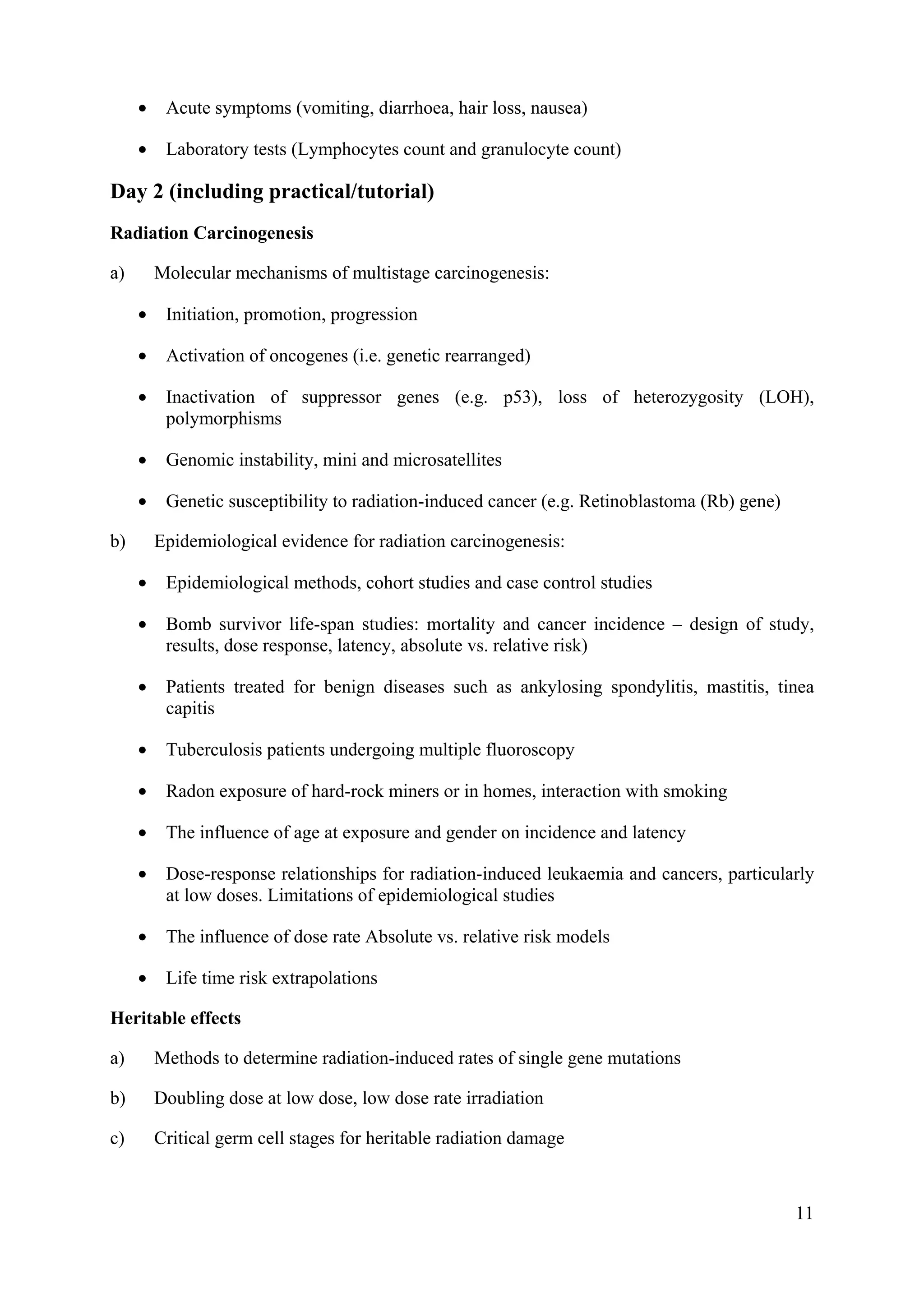
e-1
or 37 %. The linear relationship is consistent with data from some bacteria but it does not
apply in eukaryotic cells (except at high LET), which show shouldered survival curves that
can be accommodated by a single-hit multitarget model described by:
S = 1- [1 - e- (D/Do)
]n
.
This is reliable at high dose but not at low dose, because it does not describe accurately the
‘shoulder’ region at low doses, even if another single-hit term is added.
For practical purposes, there are merits of using survival at 2 Gy (SF2), because this is a dose
fraction using commonly in radiotherapy.
2.3.7. Cell cycle effects
Renewing cells in a growing population (e.g. skin, gut, bone marrow, tumour cells or cells in
culture), but not when resting in Go phase, participate in the cell cycle. Replication of the
genome occurs in S-phase and mitotic propagation to daughter generations occurs in G2/M
phases. Typical cell generation times are 10 – 40 hours with the G1 phase taking about 30 %,
S-phase 50 %, G2 phase 15 % and M-phase 5 % of the cell cycle time, although G1 phase
time may vary and be much longer in slowing proliferating populations. In interphase the
majority of cells are in G1 or Go. There are checkpoints at the G1/S and G2/M boundaries
that monitor the fidelity of genomic processing. Binding of cyclins to cyclin dependent
kinases activates the kinase complex to negotiate the checkpoints: cyclin B1/ p34 CDC-2 for
G2/M transition, cyclin D1/cdk-4 for M/G1 transition, cyclin E/cdk-2 for G1/S and
cyclinA/cdc-2 for S/G2 transition. Drugs that abrogate cell cycle blocks e.g. caffeine and
pentoxifylline, are radiosensitizing by rapidly re-establishing the B1/p34 CDC-2 pair,
promoting early mitotic progression before complete recovery and directly inhibiting HR
repair in G2. In p53 mutants (i.e. in most tumours) and in cells of p53 null status arising from
p53 destruction after viral infection (by the HPV E6 protein), p21 induction is abolished and
p21 controlled inhibition of G1/S transition cannot occur. In the absence of the G1 block,
cells enter a block at G2/M. Most tumour cells being p53 mutant hence would display altered
checkpoint expression and limited repair routes with opportunities for therapeutic
intervention. Tumour cell heterogeneity and multiple ploidy are complicating factors.
Radiosensitivity differs throughout the cell cycle with, in general, late S-phase being most
radioresistant, G2/M being most radiosensitive and G1 phase taking an intermediate position.
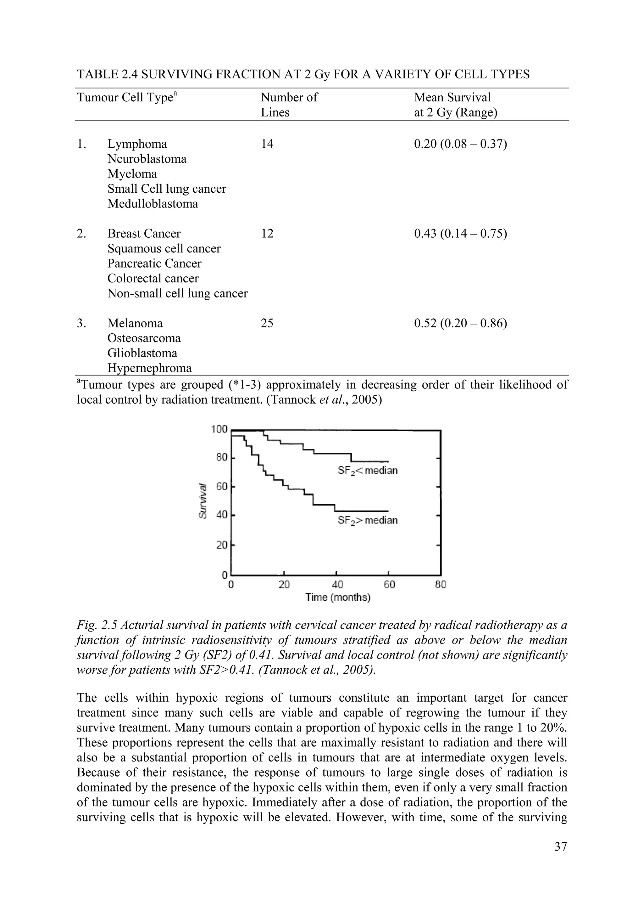
The greater proportion of repair by HR than by NHEJ in late S phase may explain the
resistance of late S phase cells. The open structure of DNA helps explain radioresistance in
G1. Chromatin compaction and poor repair competence (reduced enzyme access) could
explain the high radiosensitivity in G2/M. Attempts at cell synchronization in tumours by
irradiation to increase overall sensitivity and to harness this scenario clinically have not been
successful.
2.3.8. Relative biological effectiveness (RBE)
When effects of equal doses of different types of radiation are compared, they produce
unequal biological effects. Comparison of effects of different types of radiation is expressed
as relative biological effectiveness (RBE). Historically the effect of 250 kV X rays was taken
as the standard, but more usually now it is > 1 MeV photons (from Co-60). RBE is defined as
the ratio of doses of γ rays (Dγ-ray) and the test radiation (Dr) is required to produce an equal
29](https://image.slidesharecdn.com/ejcibduusi6m96yeuwei-140626002229-phpapp02/75/Tcs-42-web-40-2048.jpg)

![There are many other molecules that have been found to increase the radiosensitivity of cells
using clonogenic assays, including molecules that enhance DNA damage, such as halogenated
pyrimidines, inhibitors of DNA repair, modifiers of cell cycle checkpoints, such as caffeine
and modifiers of mitogen-activated protein (MAP) kinase signalling pathways, such as
inhibitors of RAS, epidermal growth factor receptor (EGFR), or protein kinase B (AKT). The
study of such molecules can illuminate our understanding of cellular response to irradiation
but their application in the clinic requires some expectation of specificity for tumour cells vs
normal cells. Previous studies have focused on biological or pathophysiological differences
between tumours and critical normal tissues such as hypoxia or proliferation. Recent studies
have been focusing on molecular differences such as levels of gene expression or mutations in
critical genes such as protein 53 (p53). Differential uptake of halogenated pyrimidines into
DNA in place of thymidine in proliferating cells provides one rationale and both
bromodeoxyuridine (BrdU) and iododeoxyuridine (IrdU) have been studied clinically. These
molecules are slightly larger than thymidine and partially disrupt the structure of the DNA
making it more susceptible to damage by X rays (or UV light), thereby radiosensitizing the
cells when a significant fraction of the DNA has incorporated the molecule (usually requires
several cell generations in the context of normal background thymidine levels). To date these
molecules have not shown great gains in clinical application, because of the difficulty of
obtaining sufficient differential uptake between tumour and exposed normal tissue. Inhibitors
of EGFR have recently been tested in the clinic with some success. EGFR is highly expressed
on some tumour types e.g. Head and Neck Squamous Cell Carcinoma (HNSCC) and Non
Small Cell Lung Cancer (NSCLC), and inhibition of the signaling pathway is believed to
reduce proliferation of tumour cells and block stimulation of this pathway by the radiation
treatment but the exact mechanisms of the effect remain uncertain.
Other approaches being investigated experimentally include antisense oligonucleotides to
inhibit the expression of anti-apoptotic factors such as Bcl-2; gene directed enzyme prodrug
therapy (GDEPT) targeting DNA synthesis; radiation-activated molecular switches to drive
specific promoters in tumours to increase expression of toxic molecules such as tumor
necrosis factor alpha (TNF-α): inhibitors of checkpoint kinases (Chk1 and Chk2) required for
expression of cell cycle blocks conceivably because blocking abrogation would inhibit repair.
2.3.13. Radiation protectors
In whole body irradiated mice, addition of cysteine or cysteamine is protective with a dose
reduction factor (DRF) of 1.8. This means that the dose becomes less effective i.e. the LD
50/30 (the dose of radiation required to kill [LD=Lethal Dose] 50% of the test cohort within
30 days) increases by this factor. These factors can also be demonstrated in vitro. Other
molecules giving similar levels of protection include: mercaptoethylamine, and Amifostine
(or WR 2721), which is a phosphorothioate that can be activated in vivo by alkaline
phosphatase to its thiol metabolite. This drug is currently used in the clinic as a normal tissue
protector based on data which suggests that the drug permeates normal tissue but not much in
the tumour because of hypoxia and chaotic vasculature. There is good evidence that it does
provide some normal tissue protection in HNSCC and NSCLC patients receiving radiotherapy
but there remains controversy about whether or not it it has been shown also to cause some
tumour protection. Amifostine is also claimed to protect against mutation and carcinogenesis,
as well as against nephrotoxicity from cisplatin. Sodium selenite, pentoxifylline, and vitamin
E all show clinical benefits in reducing morbidity e.g. less xerostomia, mucositis, proctitis,
enteritis, and fibrosis.
32](https://image.slidesharecdn.com/ejcibduusi6m96yeuwei-140626002229-phpapp02/75/Tcs-42-web-43-2048.jpg)


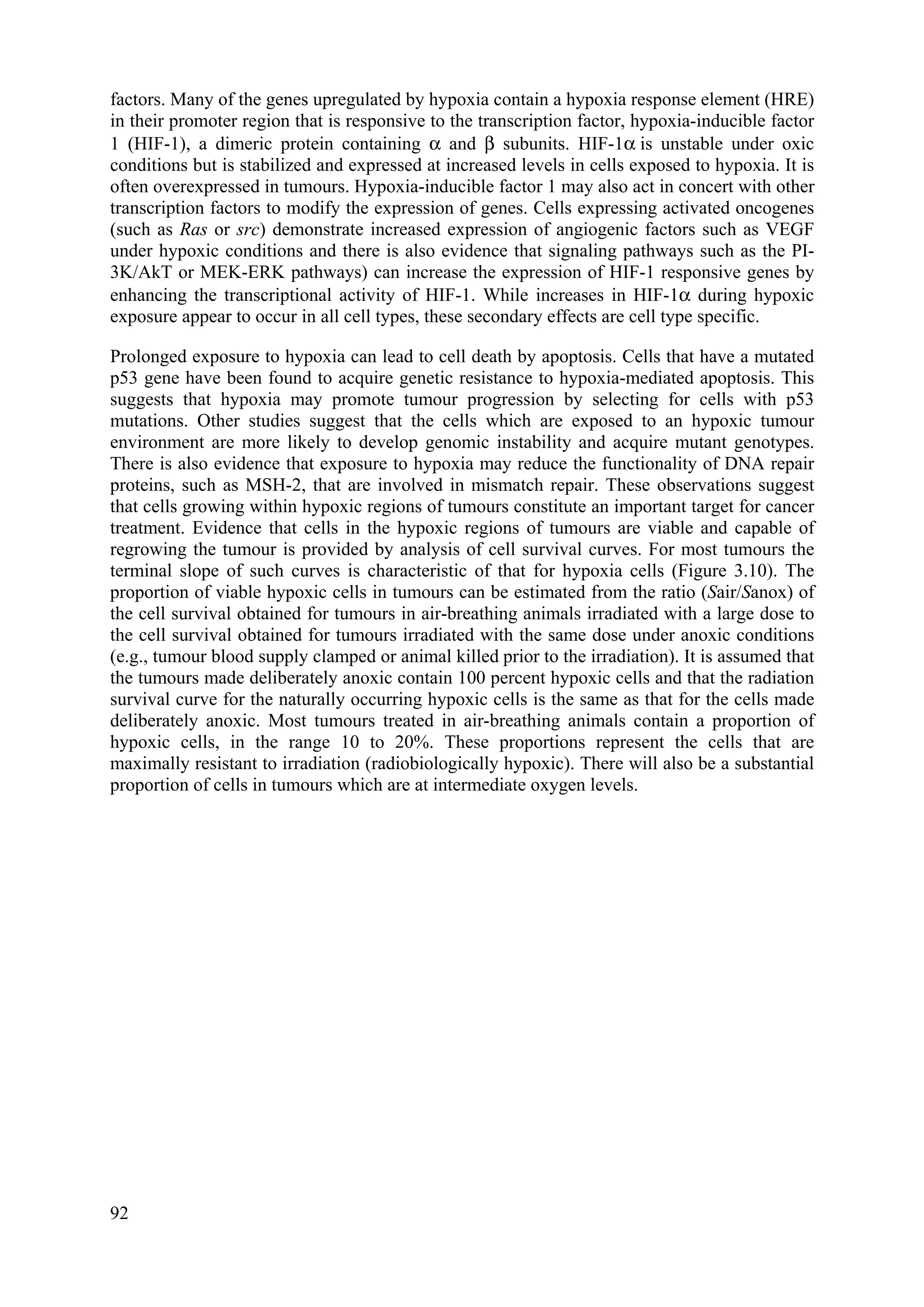
![These techniques often produce non-uniform dose distribution within the normal organs
which makes it difficult to determination a precise relationship between normal tissue
response and dose. Currently this is address by generating a dose-volume histogram (DVH)
for each exposed organ in a patient as part of a modern radiotherapy plan. These DVHs can
then be reduced to create one of more parameters (e.g. either an effective volume [Veff]
irradiated to a reference dose or an effective dose [equivalent uniform dose; EUD] uniformly
applied to the whole volume), which are used in a model for predicting normal tissue
complication probability (NTCP). Several models have been proposed; however, currently the
quality of clinical data available for such predictions is rarely sufficient to alter radiotherapy
practice. One important complexity with IMRT plans is that increased volumes of normal
tissue are exposed to lower doses within the entire body and this raises concerns as to the
possibility of increased radiation-induced second malignancies.
3.2.1. Brachytherapy, radionuclides, and radioimmunotherapy
Low-dose rate radiation sources implanted into or beside the tumour (known as
brachytherapy) can be used either alone or in combination with external beam radiotherapy
for accessible tumours such as those of the cervix, prostate, head and neck, breast, bladder,
lung, esophagus, and some sarcomas. Tissues close to the implanted source will receive a
high dose and tumour cell killing will be high. Further from the source, normal cell killing
will be less due to lower dose rates and a decreased total dose over the duration of treatment.
Recently computer controlled brachytherapy systems have been designed to deliver short
pulses of radiation (pulsed-dose brachytherapy) using a high-dose source traveling along a
catheter track within the tumour. Radiobiological modelling suggests that the acute and late
reactions are similar to traditional (continuous) brachytherapy as long as the gaps between
pulses are less than 1 hour. Injected radionuclides can also be used if there is selective uptake
by the tumour so that local irradiation may lead to death of the tumour cells. Iodine-131 (131
I)
is used to treat well-differentiated thyroid cancer, radiolabeled (e.g., indium-111) somatostatin
analogues for the treatment of neuroendocrine tumours, and strontium-89 (89
Sr) to treat bone
metastases mainly in prostate cancer. The conjugation of radionuclides to specific antibodies
allows targeted radiotherapy to tumours containing cells expressing the relevant antigens or
receptors and is termed radioimmunotherapy. Radionuclides emitting α-particles or short-
range beta particles or electrons (e.g. Auger electrons) that can kill cells within a radius of 1 to
3 cell diameters of the bound isotope are optimal for localized treatment. Currently, in
patients, this approach is limited by the lack of specific uptake in tumour cells and the
difficulties of accurate dosimetry and treatment planning.
3.2.2. Charged particles and high LET radiotherapy
Charged particles (protons, heavy ions) have a physical advantage because they can give
improved depth-dose distributions for deep-seated tumours. Much of their energy is deposited
in tissue at the end of particle tracks (i.e., in the region of the Bragg peak). Uncharged neutron
beams do not demonstrate a Bragg peak and their depth-dose distributions are similar to those
for low-LET radiation. There is also a potential biological advantage in that the oxygen
enhancement ratio is reduced with high LET ions, so hypoxic cells are protected to a lesser
degree. There is also reduced capacity for repair following high-LET radiation relative to that
following low-LET radiation, a property partially responsible for the increased relative
biological effectiveness (RBE) for high-LET radiations. The expected gains with protons are
largely confined to improved dose distribution, while for neutrons any gains are likely to be
related to the biological factors. One potential difficulty in using high-LET radiation is that
because late-responding tissues demonstrate greater repair capacity than early-responding
58](https://image.slidesharecdn.com/ejcibduusi6m96yeuwei-140626002229-phpapp02/75/Tcs-42-web-69-2048.jpg)


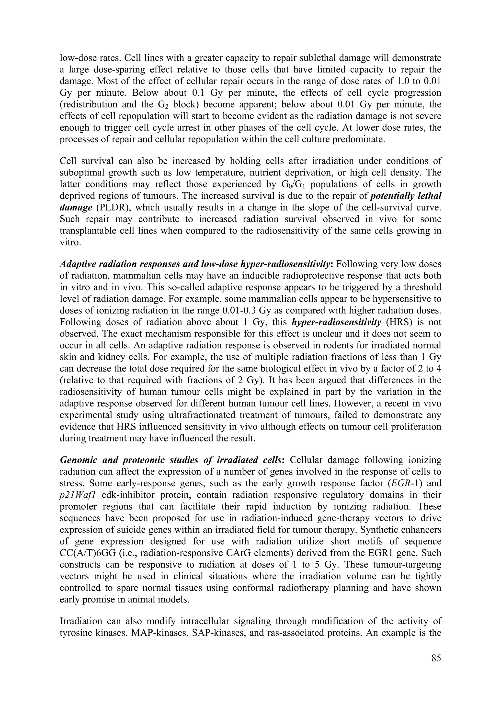
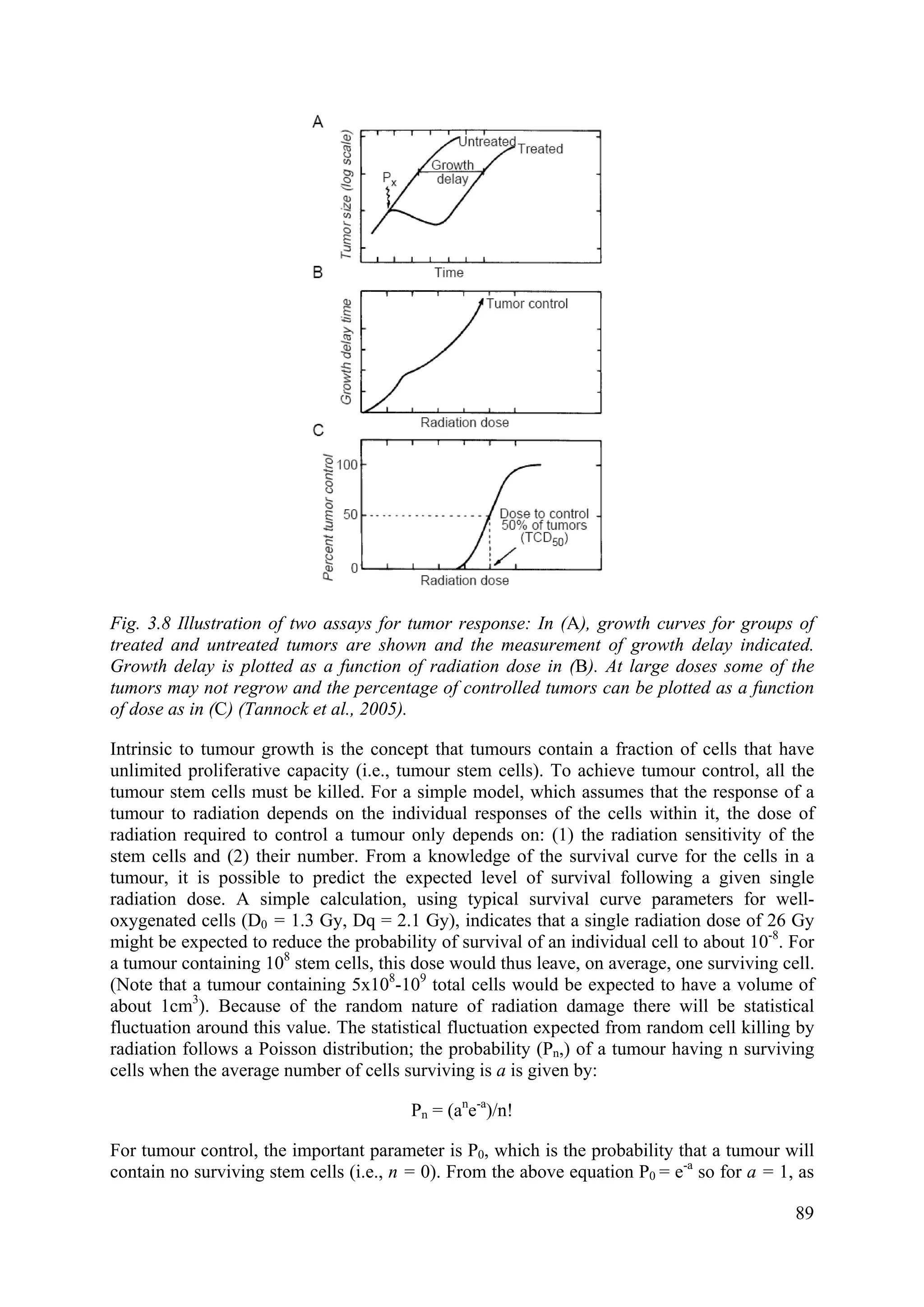
![comparing the rate of cell production (from assessment of the labeling index or fraction of S
phase cells by flow cytometry) with the rate of tumour growth. The overall rate of cell
production may be characterized by the potential doubling time of the tumour (Tpot), which is
the expected doubling time of the tumour in the absence of cell loss.
Flow cytometry: Flow cytometry is a method that allows the separation and sorting of cells
based on cellular fluorescence. Cells can be stained with a fluorescent dye whose binding (to
DNA) is proportional to DNA content, and flow cytometry then allows enumeration of cells
containing different amounts of DNA. Several fluorescent dyes are available which stain
DNA, including ethidium bromide, propidium iodide, acridine orange, and Hoechst 33342.
Most dyes require fixation of the cells to allow access of dye to the DNA, although selected
DNA specific dyes (e.g., Hoechst 33342) can enter viable cells; the Hoechst dye allows
isolation of viable cells according to DNA content. Often, fluorescent reagents are applied
concurrently or sequentially to allow for analysis or separation of cells on the basis of two or
more criteria (such as the expression of a specific protein and DNA content). Computer
analysis of a fluorescent DNA distribution provides estimates of the proportion of cells with
2N DNA content (i.e., G1 and most nonproliferating cells), with 4N DNA content (G2 and
mitotic cells), and with intermediate DNA content (S phase cells). In tumours, the presence of
aneuploidy (i.e. a G1-phase DNA content different from that of normal cells) and of variable
DNA content among G1 cells complicates analysis of DNA distributions and the estimation of
cell cycle parameters. The proportion of S phase cells obtained from a DNA distribution is
analogous to the thymidine labeling index and gives a broad indication of the proliferative
rate. Flow cytometry can be used to estimate cell cycle phase distribution, growth fraction,
and kinetic properties of cell populations. Precursors such as 5-bromodeoxyuridine (BrdUrd),
can be incorporated into newly synthesized DNA (like tritiated thymidine) and can be
recognized by flow cytometry using commercially-available fluorescently-tagged monoclonal
antibodies. Analysis of BrdUrd staining and DNA content at different times later allows
analysis of the tagged cells as they move through the cell cycle. Several methods allow
proliferating and nonproliferating cells to be distinguished by flow cytometry. A variety of
cellular antigens [e.g., that are recognized by the monoclonal antibody, Ki-67 and
proliferating cell nuclear antigen (PCNA)] appear to be expressed uniquely in cycling cells
and can be recognized by fluorescently-labelled antibodies. The Ki-67 antigen has been used
most often as a marker for proliferating cells although its function remains poorly understood.
A number of estimates of the duration of S phase (Ts) and of potential doubling time (Tpot) in
human tumours have been derived from PLM studies or BrdUrd (or IUrd) labeling and flow
cytometry. Mean values for Ts tend to be in the range of 12 to 24 hours and values of the
mean cell cycle time are in the range of 2 to 3 days, but the distribution of cell cycle times is
broad, and both the PLM and BrdUrd techniques tend to give information about the faster
proliferating cells in the population. Tpot values (range of 4.5 to 20 days) are much longer than
estimates of mean cycle time Tc, implying that many human tumours have a low growth
fraction (usually in the range 5-30% for solid adult tumours but may be much higher for
childhood tumours and adult tumours such a lymphoma or cancer of the testis). The mean
values of Tpot are also much lower than estimates of volume doubling time for common
human tumours (typically 2 to 3 months) because the rate of cell loss in many human tumours
is in the range of 75 to 90 percent of the rate of cell production. Not surprisingly, well-
nourished cells close to blood vessels have a more rapid rate of cell proliferation than poorly
nourished cells close to a region of necrosis. Slowly proliferating cells at a distance from
functional blood vessels may be resistant to radiation because of hypoxia and to cytotoxic
chemotherapeutic drugs because of their low proliferative rate and limited drug access.
77](https://image.slidesharecdn.com/ejcibduusi6m96yeuwei-140626002229-phpapp02/75/Tcs-42-web-88-2048.jpg)


![activation of the c-abl pathway which phosphorylates Rad51, a DNA repair protein, at sites of
DNA damage. Other genes induced by radiation include those encoding cell cycle related
proteins [e.g., growth arrest after DNA damage (GADD) genes, p34cdc2, cyclin B, p53],
growth factors, and cytokines [e.g., platelet-derived growth factor (PDGF), transforming
growth factor beta (TGF-β), basic fibroblast growth factor (bFGF)], and enzymes (e.g.,
plasminogen activator]. Liberation of inflammatory cytokines such as TGF-β, tumour
necrosis factor (TNF-α) and interleukin-1 (IL-1) by cells following radiation damage may
lead to a continuing cascade of cytokine production, and may be responsible for the acute
inflammation and late onset fibrosis observed in some irradiated tissues. Studies using cDNA
microarrays have found that radiation-induced gene expression can be cell-type specific.
Cell cycle sensitivity and DNA damage checkpoints: Mammalian cells have evolved
complex interrelated responses to DNA damage including cell cycle checkpoints, DNA repair,
and apoptosis. Cell cycle checkpoints are sites of cell cycle arrest in the G1, S, and G2 phases
that ensure successful and accurate DNA replication and repair prior to mitosis. Two general
types of cell cycle checkpoints exist. The mitotic spindle assembly checkpoint is responsible
for ensuring that the mitotic spindle is correctly formed prior to division. Additionally, there
are DNA integrity checkpoints that delay the cell cycle in response to DNA damage or to
defects in DNA replication (i.e., G1 to S, intra-S, and G2 to M checkpoints). Delaying cell
cycle progression could allow for the repair of DNA damage in cells prior to undergoing
DNA replication or mitosis and is thought to prevent genetic instability. Early kinetic studies
reported a rapid decrease in the mitotic index in an irradiated cell population, as both lethally
damaged and surviving cells ceased to enter mitosis, while cells already in mitosis continued
their cell cycle progression. After a period of time, which depends on both the cell type and
the radiation dose, surviving cells re-enter mitosis; this time is known as the mitotic delay.
Mitotic delay appears to be due largely to a block of cell cycle progression in G2 phase,
although cells in G1 and S phases are also delayed in their progression, albeit to a lesser
extent. There is approximately 3–4 hours of G2 delay per 1 Gy in a diploid cell. Cells may
continue to experience delays in their progression through the next and subsequent cell cycles.
As a result of radiation induced delays in the cell cycle and the fact that cells in different
phases of the cell cycle have different radiosensitivities, cell populations can be partially
synchronized by irradiation. If a single radiation dose is given to cells in different phases, then
a pattern of cell survival as a function of cell cycle position is obtained. Many cell lines
appear to have a resistant period in S phase and a sensitive period in G2 phase following
irradiation in vitro, however, some cell lines have different patterns of sensitivity throughout
the cell cycle. Some oncogene-transfected cells (e.g., overexpressing the ras oncogene) show
increased resistance in the G2 phase, whereas other cells, including DNA repair-deficient
cells, show similar sensitivity throughout all phases of the cell cycle. The pattern of
radiosensitivity throughout the cell cycle can be different for the same tumour cells growing
in vivo or in vitro, indicating the influence of cell-cell interactions on cell survival.
The ATM (ataxia telangiectasia mutated) protein plays a role in initiating checkpoint
pathways in all three cell cycle phases. G1 cell cycle arrest following irradiation depends on
an intact ATM-p53/Cdc25A-Rb pathway and decreased activity of cyclin D and E complexes.
This leads to continued hypophosphorylation of the Rb protein at the G1/S interface and
blocking of the initiation of DNA replication. Radiation-induced G1 arrest is abrogated in
cells that lack functional p53, ATM, or Rb proteins. Most data suggest that cells having
altered p53 protein function (and an abrogated G1 checkpoint) acquire relative radioresistance
in comparison with those cells having normal p53 protein function. The radioresistant
phenotype has been correlated with the level of expression of mutant p53 protein in
86](https://image.slidesharecdn.com/ejcibduusi6m96yeuwei-140626002229-phpapp02/75/Tcs-42-web-97-2048.jpg)


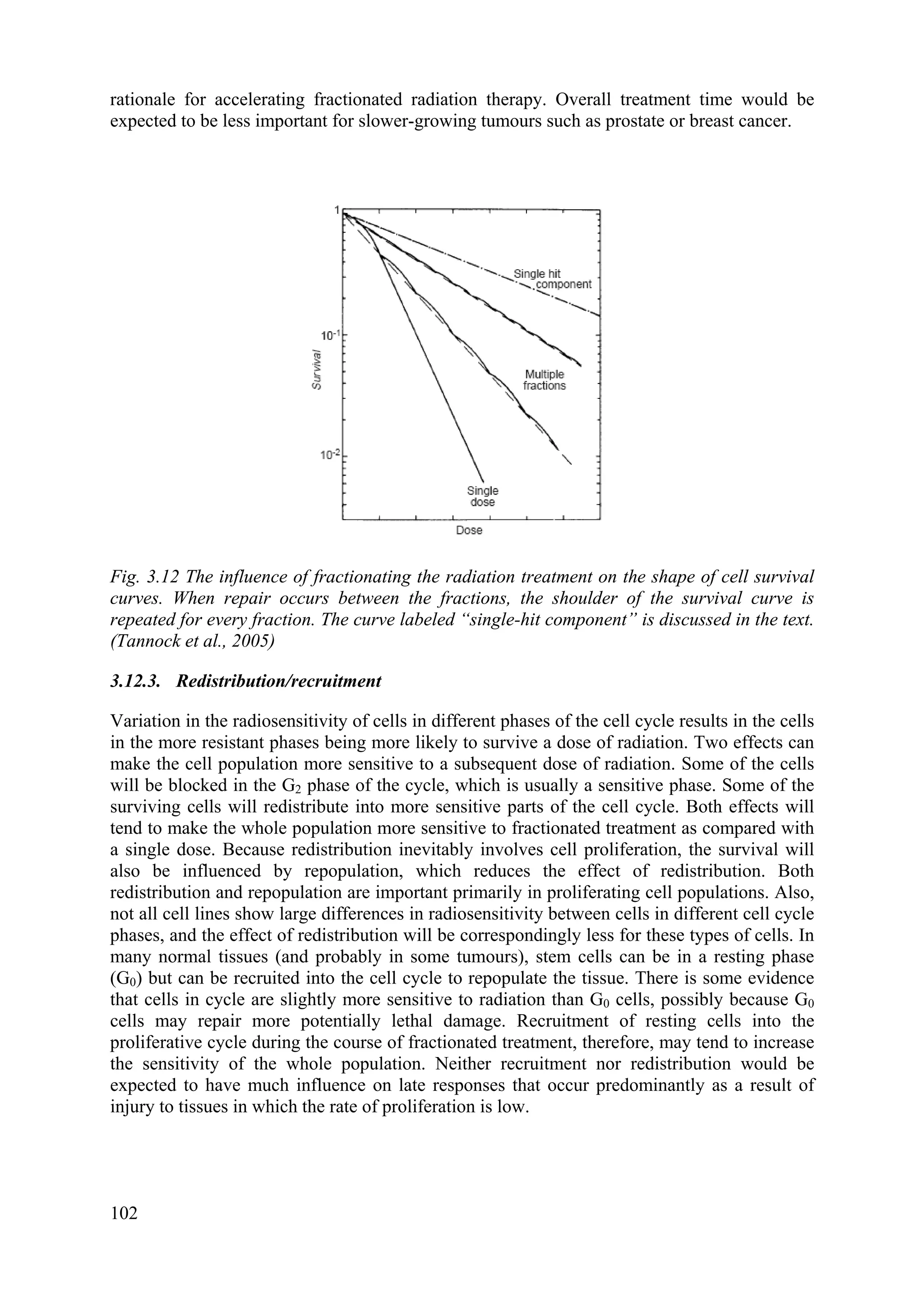

![3.12.7. The linear quadratic equation and models for isoeffect
Most isoeffect relationship used clinically are based on the linear-quadratic (LQ) equation.
SF = N/N0 = exp -(αD+βD2
)
In using the LQ model, it is assumed that each fraction has an equal effect, thus for a
fractionated regime (n fractions of size d):
SF = [exp -(αd+βd2
)]n
or -ln SF = n(αd+βd2
)
It is further assumed that if different fractionation regimes (e.g., n1 fractions of size d1 and n2
fractions of size d2) are isoeffective for a given tissue, they lead to the same surviving fraction
(SF). Thus we have:
Isoeffect (E) = -ln SF = n1(αd1+βd1
2
) = n2(αd2+βd2
2
)
which can be simplified to give:
n1d1/n2d2 = (α+βd2)/( α+βd1) = (α/β +d2)/(α/β+d1)
From this relationship and knowing the values of n1, d1, n2, and d2, the constant α/β can be
determined for the particular tissue and used in the equation to predict other isoeffective
treatment schedules. The parameter α/β has the units of dose (Gy) and is a measure of the
shape of the survival curve. The parameter α defines the initial slope of the survival curve; the
larger the value of α, the steeper the initial part of the curve. The parameter β defines the
curvature of the survival curve and a large value of β implies more curvature. Thus, a large
value of α/β implies a steep curve with little curvature (i.e., a small shoulder to the survival
curve) and a small value of α/β implies a shallow curve with greater curvature (i.e., a large
shoulder to the survival curve). Because the size of the shoulder of the survival curve is a
measure of the repair capacity of the cells, a small value of α/β is consistent with greater
repair capacity and a steep isoeffect curve, whereas a large value of α/β is consistent with
lesser repair capacity and a shallow isoeffect curve.
Derived α/β values for different normal tissues in rodents suggest that late-responding tissues
have values in the range 2 to 4 Gy (i.e., consistent with a steep isoeffect curve), while early-
responding tissues have values in the range 8 to 12 Gy (i.e., consistent with a shallow
isoeffect curve). The limited data available for human tissues suggest values in the same
ranges. Most tumours appear to have α/β values similar to or greater than those for early-
responding tissues, although recent data suggest that certain slow growing tumours (e.g.
prostate cancer) may have lower α/β values between 1 and 3 Gy. There is no consideration of
the effect of treatment time in the LQ model. In practice, this is a limitation that applies to
early normal tissue responses, which occur in proliferative tissues (and tumours), rather than
to late normal tissue responses, which generally occur in tissues that have slowly proliferating
parenchymal cell populations, and for which the response to radiation is less influenced by the
duration of fractionated treatment. In the LQ model, it is also assumed that there is complete
repair between the fractions, and predictions from the model may lead to serious overdosing
when the interfraction interval is too short or where repair of sublethal damage is slow (e.g. as
in spinal cord).
105](https://image.slidesharecdn.com/ejcibduusi6m96yeuwei-140626002229-phpapp02/75/Tcs-42-web-116-2048.jpg)


![shown in testis cancer patients treated with past radiation treatments to chest, abdominal, and
pelvic fields, with resultant 11-fold risks of leukaemia. Low-dose total body irradiation [e.g.
as used for the treatment of non–Hodgkin lymphoma in past years] has also been associated
with high risks of leukaemia. Radiation has been associated with increased risks of acute
myeloid leukemia (AML), chronic myelogenous leukaemia (CML), and acute lymphoblastic
leukaemia (ALL). Only chronic lymphocytic leukemia (CLL) has not been linked with either
prior radiotherapy or chemotherapy.
Breast cancer has emerged as the most common solid tumour among female survivors of
Hodgkin lymphoma after treatment. Excess breast cancers, which are largely due to high-
dose, large-field chest irradiation for Hodgkin lymphoma, are inversely correlated with age at
treatment. The highest risks are observed among women treated for Hodgkin lymphoma at
age less than 30 years, a finding that parallels the known sensitivity of the breast to ionizing
radiation in the young. One large analytic, international investigation of Hodgkin lymphoma
patients estimated long term risk according to radiation dose to the area in the breast where
cancer was later diagnosed and that took into account chemotherapy- or radiotherapy-related
ovarian damage. Statistical analyses were conducted to estimate the relative risk of breast
cancer in terms of radiation dose to site of breast cancer and to the ovaries, cumulative dose of
alkylating agent chemotherapy, and other risk factors. A radiation dose to the breast of more
than 4 Gy was followed by a significantly increased 3.2-fold risk of breast cancer compared
with women who received lower doses to the breast without alkylating agents. Risk of breast
cancer increased with increasing radiation dose to reach 8-fold at >40 Gy, and excess
radiotherapy-related breast cancers occurred for >25 years after exposure. The smaller
radiotherapy fields and lower doses now used to treat Hodgkin lymphoma should eventually
result in lower risks of breast cancer.
The interaction of chemotherapy with radiation or other risk factors in the development of
solid tumours also needs consideration. For example, smoking multiplies the risk of either
alkylating agent-associated or radiotherapy-associated lung cancer. In contrast, the effect of
chemotherapy and radiation on lung cancer risk after Hodgkin lymphoma seems additive, as
does the effect of cyclophosphamide and radiation on excess bladder cancers after non–
Hodgkin lymphoma. Other relevant questions include the effect of the sequence and timing of
exposures and interactions with other risk factors. Further, it will be important to understand
whether relations between cytotoxic drugs, radiation, and solid tumour risk represent either an
independent carcinogenic effect or radiosensitization by the chemotherapeutic agent, possibly
by drug interference with the repair of radiation-induced DNA damage.
For cervix cancer, the data come from series of patients analysed from Scandinavia, USA, and
Japan. In the regions of the body receiving high doses, the relative risk (RR) averaged over all
3 series for induced bladder cancer in cervix cancer patients surviving radiotherapy was about
1.6 (i.e. 60% greater) compared to the incidence in cervix cancer patients treated using non-
radiation methods, and about 3.3 compared to the incidence of primary cancers in the general
population. For induced rectal cancer the respective RR values were about 1.2 and about 1.5,
and for induced colon cancer were about 1.0 and about 1.1. This indicates that in these cervix
cancer patients there was a higher incidence of a second primary cancer in bladder and rectum
than of a first primary cancer in these sites in the general population, and the use of
radiotherapy increased the risk of a primary cancer in these sites.
For one large series of prostate cancer patients who received either radiotherapy it could be
concluded that if a prostate cancer patient is to be treated with radiotherapy, the risk of
developing a radiation-induced second cancer is approximately only 0.3%. Half of the 0.3%
115](https://image.slidesharecdn.com/ejcibduusi6m96yeuwei-140626002229-phpapp02/75/Tcs-42-web-126-2048.jpg)


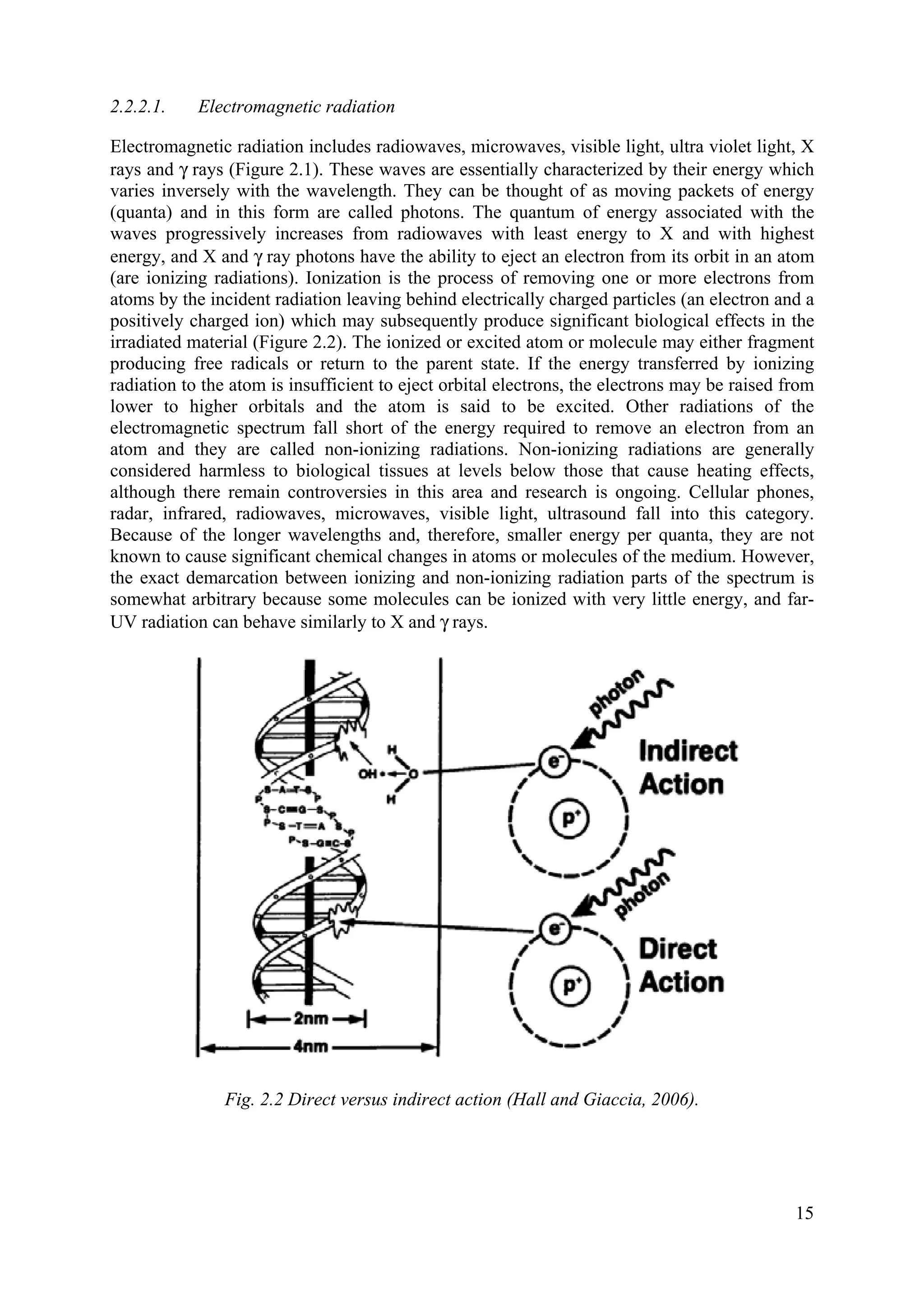
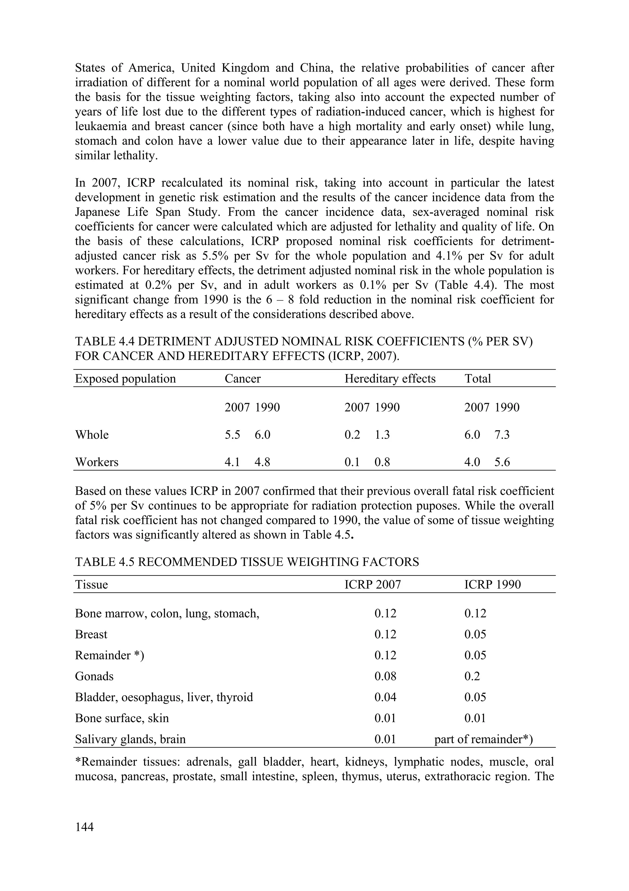
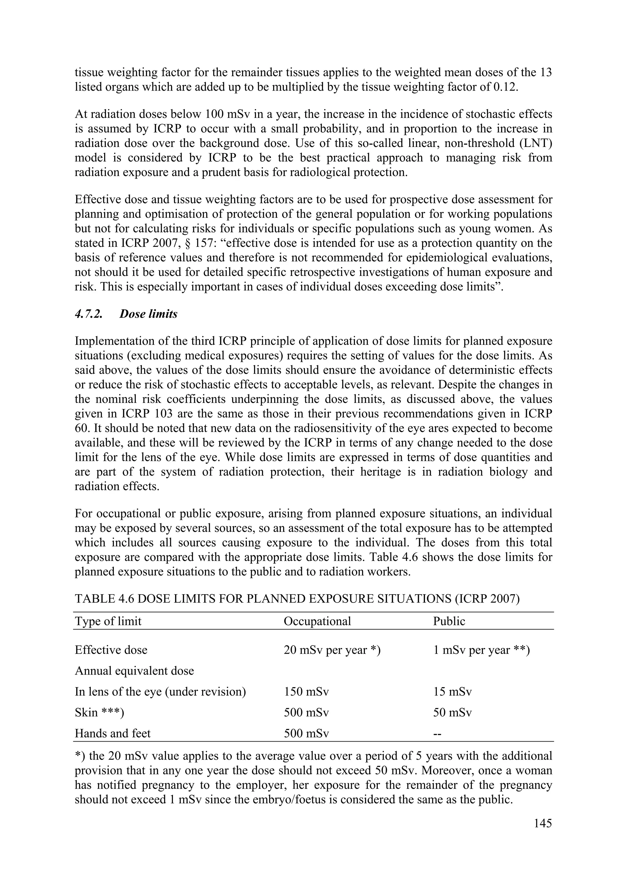
![the absorbed dose but also on the type and energy of the radiation causing the dose, in
particular the LET of the radiation. This is taken into account by weighting the absorbed dose
by a factor related to the quality of radiation. The radiation weighting factor is selected for the
type and energy of the radiation incident on the body of, in case of sources within the body,
emitted by the source. The weighted absorbed dose is the product of the absorbed dose
averaged over the respective tissue or organ and is called “equivalent dose”. The “effective
dose” is derived by weighting the equivalent doses of the different tissues and organs by a
factor which represents the sensitivity of the respective tissues and organs to stochastic
effects, primarily radiation-induced cancer. Thus, the detriment for all radiations is expressed
as effective dose E which is measured in sieverts (Sv). The values of the radiation weighting
factors for a specified type and energy of radiation has been selected to be representative for
values of the relative biological effectiveness (RBE) in inducing stochastic effects at low
radiation doses. The radiation weighting factors recommended by ICRP in 2007 are shown in
Table 4.3.
TABLE 4.3 RADIATION WEIGHTING FACTORS (ICRP 2007)
Radiation type Radiation weighting factor
Photons and electrons, all energies 1
Protons and charged pions 2
Alpha particles, fission fragments, heavy ions 20
Neutrons* En < 1 MeV 2.5 + 18.2 e[-ln En]2/6
1 MeV ≤ En ≤ 50 MeV 5.0 + 17.0 e[-ln 2En]2/6
En > 50 MeV 2.5 + 3.25 e[-ln 0.04En]2/6
*) these values have replaced the following step values that were recommended in 1990: < 10
keV, 5; 10 keV to 100 keV, 10; 100 keV to 2 MeV, 20; 2 MeV to 20 MeV, 10; >20 MeV, 5.
The tissue weighting factors were intended to ensure that a weighted tissue equivalent dose
would produce the same degree of detriment irrespective of the tissue or organ involved. They
are representative values averaged over age at exposure and sex and, besides the risk for fatal
cancer in the respective organs, also make allowance for different losses of life span, for the
morbidity resulting from the induction of non-fatal cancers and for the risk of serious
hereditary diseases in the first two generations of descendants of the irradiated individual. On
the other hand, any possible health damage arising from developmental damage in utero such
as severe mental retardation is not included in the tissue weighting factors.
The period of observation of exposed populations does not extend to a full life time in any of
the major epidemiological studies. Therefore, it is necessary to project the probability of
cancer induction or mortality from the period of observation to the full lifetime of the exposed
population. This is done using two alternative projection models: (1) the absolute or additive
risk model predicts a constant excess of induced cancers throughout a life-time unrelated to
the spontaneous cancer rate, (2) the relative or multiplicative model predicts that the excess of
induced cancers increases as a constant multiple of the age-dependent spontaneous rate. By
averaging the results of the dependence of risk on age, sex, projection model and their
influence on the base-line cancer risks in different populations e.g. those of Japan, United
143](https://image.slidesharecdn.com/ejcibduusi6m96yeuwei-140626002229-phpapp02/75/Tcs-42-web-154-2048.jpg)


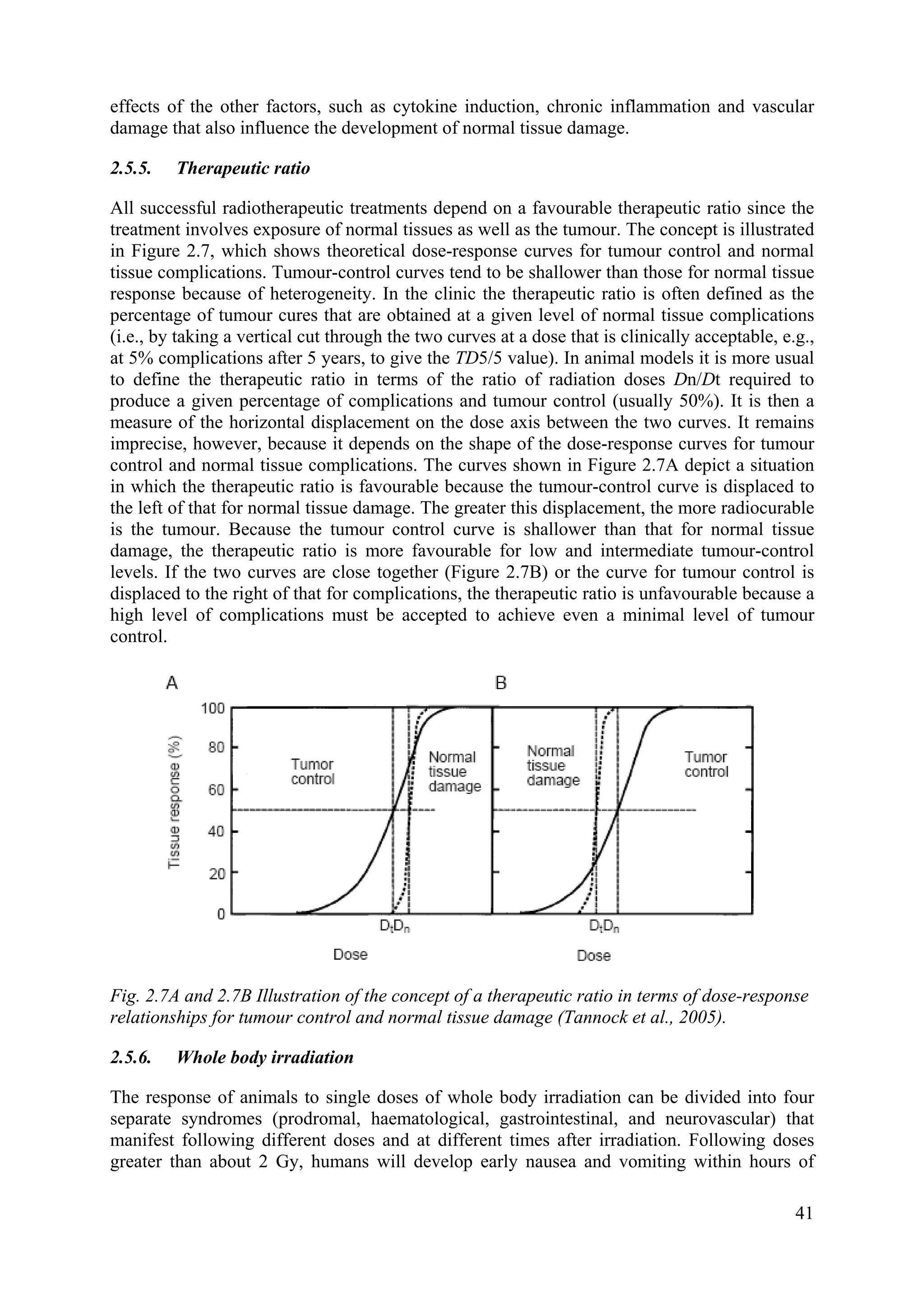
This document provides a teaching handbook for radiation biology. It includes: 1) A teaching programme with a basic 1-week course in radiation biology for various disciplines and additional advanced weeks for radiation oncologists and radiation protection personnel. 2) A minimum essential syllabus covering topics in physics and chemistry of radiation interactions, molecular and cellular radiobiology, tumour and normal tissue response, and the radiobiological basis of radiation protection. 3) Extra modules with more advanced content for radiation oncologists and radiation protection personnel respectively. The handbook is intended to provide a standardized curriculum to teach radiation biology, especially in low and middle income countries where access to training resources is limited. It compiles


