Download as PDF, PPTX
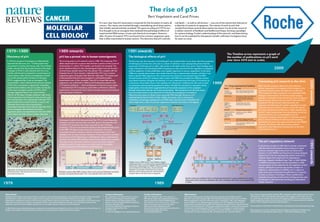

The document summarizes key developments in p53 research from its discovery in 1979 to current understanding and clinical translation efforts. It describes the initial view of p53 as an oncogene, the 1989 discovery that it is actually a tumor suppressor gene often mutated in cancer. Subsequent work revealed that p53 has a central role in stress response pathways, regulating genes that control cell cycle arrest, apoptosis, and other functions. Ongoing research aims to better understand p53 pathways and develop drugs to reactivate mutant p53 or inhibit its regulation by MDM2/MDM4 for cancer therapy.