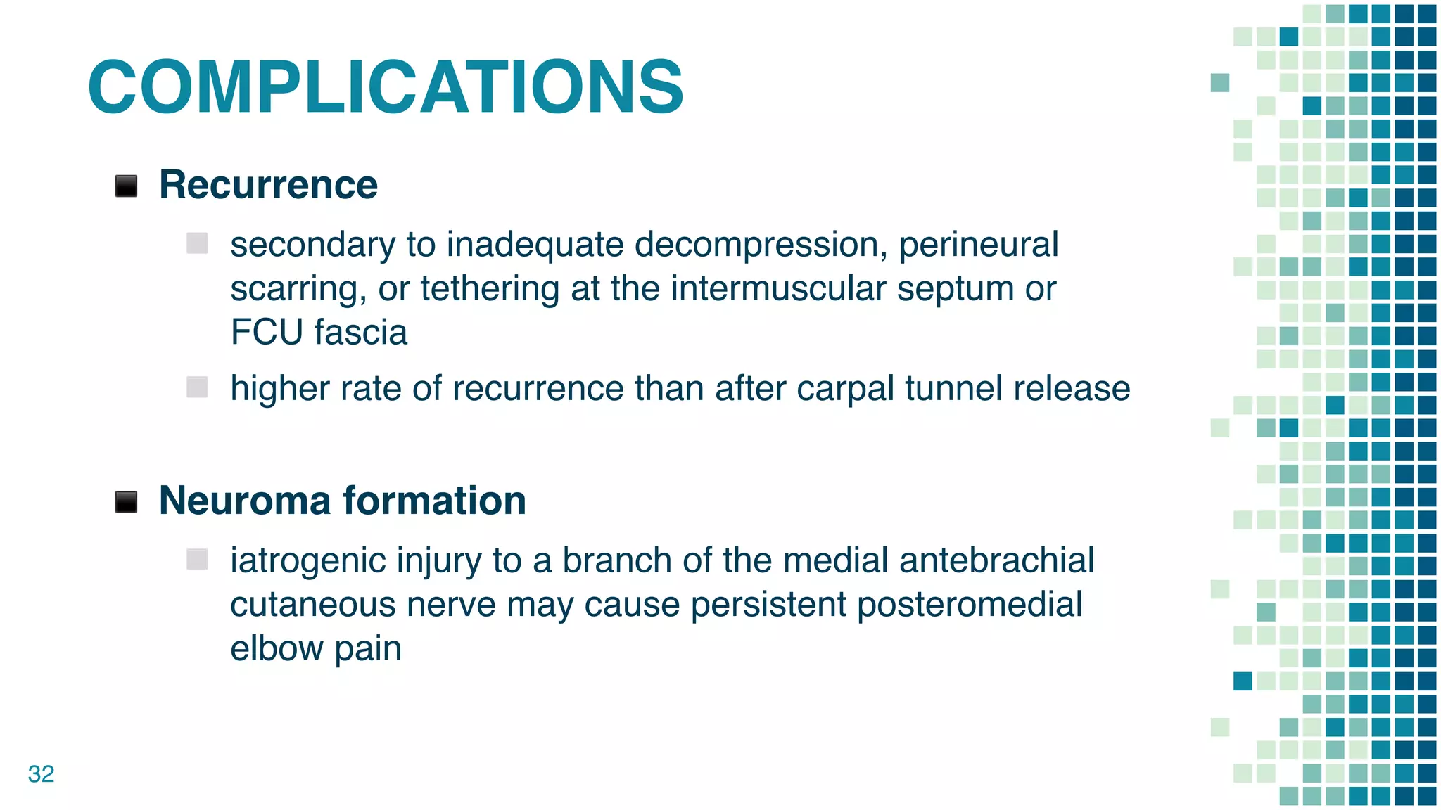
EXTERN CONFERENCE
EXT. RUNGSIMA SILPRACHAWONG
Patient Profile
▪ผู้ป่วยชายไทย อายุ 40 ปี
▪อาชีพ พระภิกษุ
▪ภูมิลำเนา อ.ด่านขุนทด จ.นครราชสีมา
2
Chief complaint
▪ชานิ้วนางและนิ้วก้อยมือขวา 1 เดือน PTA
Present Illness
3
▪1 เดือน PTA ข้อศอกขวากระแทกขอบปูน ไม่มีบาดแผล
ภายนอก จากนั้นเริ่มมีอาการชานิ้วนางและนิ้วก้อยมือขวา
เหมือนมีเข็มมาจิ้ม รู้สึกร้อนๆบริเวณเดียวกัน อาการค่อยๆ
เป็นมากขึ้น
▪3 week PTA มือขวาอ่อนแรง จับขันน้ำไม่อยู่
Physical Examination
▪Heart: normal S1S2, no
murmur
▪Lungs: equal breath
sound, no adventitious
sound
▪Abdomen: soft, no
tender
▪Neuro: E4V5M6, pupil 2
mm RTLBE,
▪ Affected part : Rt. Arm
▫ Ulnar Claw hand
▫ Hypothenar and 1st DI atrophy
▫ Slight wasting of IOM
▫ Tinel’s sign positive at medial
epicondyle and negative at
wrist
▫ Loss of sensation over the
palmar and dorsal surfaces of
medial 11/ 2 fingers
▫ Froment’s sign positive
4
Physical Examination
5
6
Investigation
7
▪Film Rt. elbow AP, Lateral
Diagnosis
8
▪Cubital Tunnel Syndrome at right elbow
Management
9
▪Set OR for Decompression with anterior
transposition right ulnar nerve
CUBITAL TUNNEL SYNDROME
10
ULNAR NERVE
11
INJURIES OF THE ULNAR NERVE
Injury of the ulnar nerve at
elbow:
▪ Fracture, dislocation of the
medial epicondyle.
▪ Thickening of the fibrous roof
of the cubital tunnel (cubital
tunnel syndrome).
▪ Compression between the 2
heads of flexor carpi ulnaris
(FCU) muscle.
▪ Valgus defect of elbow (tardy
or late ulnar nerve palsy).
▪ Injury of the ulnar
nerve at wrist: 
▪ Superficial position of
ulnar nerve at this site
makes it susceptible to
cuts and injuries.
▪ Compression in
the Guyon’s canal/Piso
hamate tunnel.
12
CUBITAL TUNNEL
SYNDROME
▪ A compressive neuropathy of
the ulnar nerve
▪ 2nd most common compression
neuropathy of the upper
extremity
13
CUBITAL TUNNEL SYNDROME
Cubital tunnel
▪ Roof
▫ formed by FCU
fascia and Osborne's
ligament 
▪ Floor
▫ formed by posterior and
transverse bands of MCL and
elbow joint capsule
▪ Walls
▫ formed by medial epicondyle
and olecranon
14
SYMPTOMS
▪ Paresthesia of small finger, ulnar half of ring finger, and
ulnar dorsal hand
▪ Pain - medial elbow area, ulnar hand and 4th and 5th fingers
▪ Exacerbating activities include
▫ cell phone use (excessive flexion)
▫ occupational or athletic activities requiring repetitive
elbow flexion and valgus stress
▪ Night symptoms
▪ Hand Weakness
▪ Painful clicking at the olecranon groove (ulnar nerve
subluxation)
15
ULNAR NERVE EXAMINATION
Inspection and palpation
▪ interosseous and first
web space atrophy
▪ ring and small finger
clawing
▪ observe ulnar nerve
subluxation over the
medial epicondyle as the
elbow moves through a
flexion-extension arc
16
ULNAR NERVE EXAMINATION
17
Sensory
▪ decreased sensation in
ulnar 1-1/2 digits
Motor
▪ paralysis of intrinsic muscles
(adductor pollicis, deep head
FPB, interossei, and lumbricals
4 and 5)
o weakened grasp
o weak pinch
ULNAR NERVE EXAMINATION
Advance signs
▪ Froment’s sign
▪ Wartenberg’s sign
▪ Jeanne’s sign
▪ Masse’s sign
▪ Cross Finger Test
▪ Intrinsic Atrophy
18
ULNAR NERVE EXAMINATION
Froment’s sign
▪ compensatory thumb IP
flexion by FPL (AIN) during
key pinch
▪ compensates for the loss of
MCP flexion by adductor
pollicis (ulnar n.)
▫ adductor pollicis muscle
normally acts as a MCP
flexor, first metacarpal
adductor, and IP extensor
19
ULNAR NERVE EXAMINATION
Wartenberg’s sign
▪ persistent small finger
abduction and extension
during attempted
adduction
▪ weak 3rd palmar
interosseous and small
finger lumbrical
20
Jeanne’s sign 
▪ compensatory thumb MCP
hyperextension and thumb
adduction by EPL (radial n.) with
key pinch
▪ compensates for loss of IP
extension and thumb adduction by
adductor pollicis (ulnar n.)
ULNAR NERVE EXAMINATION
Masse’s sign
▪ Loss of hypothenar and
flattened palmar
metacarpal arch.
21
Pollock's test
▪ Inability to flex DIPJ of
little and ring fingers.
ULNAR NERVE EXAMINATION
Duchenne's sign (Ulnar
clawing)
▪ Hyperextension MCP
joints and Flexion at IP
joints of two ulnar fingers
22
Cross Finger test
▪ Inability to cross index
and middle finger over
each other.
ULNAR NERVE EXAMINATION
Pitres Testut sign / Egawa’s sign
▪ Inability to abduct middle finger to either side.
Bouvier manoeuvre
▪ Correct the hyperextension of MCPJ and ask the patient to
extend IPJ. If IPJ extension is improved then Bouvier test
is positive and claw and is termed simple claw hand. If IPJ
extension doesn’t improve then test is negative and
clawing is called complex claw hand.
23
ULNAR NERVE EXAMINATION
Provocative tests
24
▪ Tinel sign positive over
cubital tunnel
▪Elbow flexion test
▫ positive when flexion of the
elbow for >60 sec
reproduces symptoms
ULNAR NERVE EXAMINATION
Provocative tests
Elbow flexion compression test
▪ Direct cubital tunnel compression exacerbates symptoms
25
INVESTIGATIONS
EMG : Electromyography
▪ Diagnosis
▫ conduction velocity <50 m/sec
across elbow
▫ low amplitudes of sensory
nerve action potentials and
compound muscle action
potentials
26
INVESTIGATIONS
MRI MRI may be helpful if a space-occupying lesion is
suspected
27
MCGOWAN CLASSIFICATION
Classification
▪ Grade 1: Purely subjective symptoms and mild
hypaesthesia
▪ Grade 2: Sensory loss and weakness of intrinsic hand
muscles, with or without slight wasting
▪ Grade 3: Severe sensorimotor deficit
28
NON-OPERATIVE TREATMENT
Indications
▫ first line of treatment with mild symptoms
Treatment
▪ NSAIDs
▪ Activity modification
▫ Avoid maximum elbow flexion
▫ Avoid resting elbow on hard surfaces
▫ Avoid repetitive hyperflexion elbow exercises
▪ Night-time elbow extension splinting
▫ night bracing in 45 degree extension with forearm in
neutral rotation
29
OPERATIVE TREATMENT
▪ in situ ulnar nerve decompression without transposition
▪ ulnar nerve decompression and anterior transposition
▪ medial epicondylectomy
30
TRANSPOSITION
31
COMPLICATIONS
▪ Recurrence
▫ secondary to inadequate decompression, perineural
scarring, or tethering at the intermuscular septum or
FCU fascia
▫ higher rate of recurrence than after carpal tunnel release
▪ Neuroma formation
▫ iatrogenic injury to a branch of the medial antebrachial
cutaneous nerve may cause persistent posteromedial
elbow pain
32
THANK YOU
33
“
Reference
1. https://www.handsurgeryresource.com/
2. Cubital Tunnel Syndrome, Manuel F. Dasilva M.D.
and Katia A. Dasilva B.A., Ferri&#39;s Clinical
Advisor 2019, 396.e2-396.e4
3. Entrapment Neuropathy of the Ulnar Nerve, Jane
Aitken OTR/L, CHT, Journal of Hand Therapy,
2008-07- 01, Volume 21, Issue 3, Pages 300-301,
Copyright © 2008 Hanley &amp; Belfus
4. https://www.orthobullets.com/hand/6021/cubital-
tunnel-syndrome
5. McGowan AJ. The results of transposition of the
ulnar nerve for traumatic ulnar neuritis. J Bone Joint
Surg Br. Aug 1950;32-B(3):293-301.
34
GUYON’S CANAL SYNDROME
35

Cubital tunnel syndrome 1

  • 1.
  • 2.
    Patient Profile ▪ผู้ป่วยชายไทย อายุ40 ปี ▪อาชีพ พระภิกษุ ▪ภูมิลำเนา อ.ด่านขุนทด จ.นครราชสีมา 2 Chief complaint ▪ชานิ้วนางและนิ้วก้อยมือขวา 1 เดือน PTA
  • 3.
    Present Illness 3 ▪1 เดือนPTA ข้อศอกขวากระแทกขอบปูน ไม่มีบาดแผล ภายนอก จากนั้นเริ่มมีอาการชานิ้วนางและนิ้วก้อยมือขวา เหมือนมีเข็มมาจิ้ม รู้สึกร้อนๆบริเวณเดียวกัน อาการค่อยๆ เป็นมากขึ้น ▪3 week PTA มือขวาอ่อนแรง จับขันน้ำไม่อยู่
  • 4.
    Physical Examination ▪Heart: normalS1S2, no murmur ▪Lungs: equal breath sound, no adventitious sound ▪Abdomen: soft, no tender ▪Neuro: E4V5M6, pupil 2 mm RTLBE, ▪ Affected part : Rt. Arm ▫ Ulnar Claw hand ▫ Hypothenar and 1st DI atrophy ▫ Slight wasting of IOM ▫ Tinel’s sign positive at medial epicondyle and negative at wrist ▫ Loss of sensation over the palmar and dorsal surfaces of medial 11/ 2 fingers ▫ Froment’s sign positive 4
  • 5.
  • 6.
  • 7.
  • 8.
  • 9.
    Management 9 ▪Set OR forDecompression with anterior transposition right ulnar nerve
  • 10.
  • 11.
  • 12.
    INJURIES OF THEULNAR NERVE Injury of the ulnar nerve at elbow: ▪ Fracture, dislocation of the medial epicondyle. ▪ Thickening of the fibrous roof of the cubital tunnel (cubital tunnel syndrome). ▪ Compression between the 2 heads of flexor carpi ulnaris (FCU) muscle. ▪ Valgus defect of elbow (tardy or late ulnar nerve palsy). ▪ Injury of the ulnar nerve at wrist:  ▪ Superficial position of ulnar nerve at this site makes it susceptible to cuts and injuries. ▪ Compression in the Guyon’s canal/Piso hamate tunnel. 12
  • 13.
    CUBITAL TUNNEL SYNDROME ▪ Acompressive neuropathy of the ulnar nerve ▪ 2nd most common compression neuropathy of the upper extremity 13
  • 14.
    CUBITAL TUNNEL SYNDROME Cubitaltunnel ▪ Roof ▫ formed by FCU fascia and Osborne's ligament  ▪ Floor ▫ formed by posterior and transverse bands of MCL and elbow joint capsule ▪ Walls ▫ formed by medial epicondyle and olecranon 14
  • 15.
    SYMPTOMS ▪ Paresthesia ofsmall finger, ulnar half of ring finger, and ulnar dorsal hand ▪ Pain - medial elbow area, ulnar hand and 4th and 5th fingers ▪ Exacerbating activities include ▫ cell phone use (excessive flexion) ▫ occupational or athletic activities requiring repetitive elbow flexion and valgus stress ▪ Night symptoms ▪ Hand Weakness ▪ Painful clicking at the olecranon groove (ulnar nerve subluxation) 15
  • 16.
    ULNAR NERVE EXAMINATION Inspectionand palpation ▪ interosseous and first web space atrophy ▪ ring and small finger clawing ▪ observe ulnar nerve subluxation over the medial epicondyle as the elbow moves through a flexion-extension arc 16
  • 17.
    ULNAR NERVE EXAMINATION 17 Sensory ▪decreased sensation in ulnar 1-1/2 digits Motor ▪ paralysis of intrinsic muscles (adductor pollicis, deep head FPB, interossei, and lumbricals 4 and 5) o weakened grasp o weak pinch
  • 18.
    ULNAR NERVE EXAMINATION Advancesigns ▪ Froment’s sign ▪ Wartenberg’s sign ▪ Jeanne’s sign ▪ Masse’s sign ▪ Cross Finger Test ▪ Intrinsic Atrophy 18
  • 19.
    ULNAR NERVE EXAMINATION Froment’ssign ▪ compensatory thumb IP flexion by FPL (AIN) during key pinch ▪ compensates for the loss of MCP flexion by adductor pollicis (ulnar n.) ▫ adductor pollicis muscle normally acts as a MCP flexor, first metacarpal adductor, and IP extensor 19
  • 20.
    ULNAR NERVE EXAMINATION Wartenberg’ssign ▪ persistent small finger abduction and extension during attempted adduction ▪ weak 3rd palmar interosseous and small finger lumbrical 20 Jeanne’s sign  ▪ compensatory thumb MCP hyperextension and thumb adduction by EPL (radial n.) with key pinch ▪ compensates for loss of IP extension and thumb adduction by adductor pollicis (ulnar n.)
  • 21.
    ULNAR NERVE EXAMINATION Masse’ssign ▪ Loss of hypothenar and flattened palmar metacarpal arch. 21 Pollock's test ▪ Inability to flex DIPJ of little and ring fingers.
  • 22.
    ULNAR NERVE EXAMINATION Duchenne'ssign (Ulnar clawing) ▪ Hyperextension MCP joints and Flexion at IP joints of two ulnar fingers 22 Cross Finger test ▪ Inability to cross index and middle finger over each other.
  • 23.
    ULNAR NERVE EXAMINATION PitresTestut sign / Egawa’s sign ▪ Inability to abduct middle finger to either side. Bouvier manoeuvre ▪ Correct the hyperextension of MCPJ and ask the patient to extend IPJ. If IPJ extension is improved then Bouvier test is positive and claw and is termed simple claw hand. If IPJ extension doesn’t improve then test is negative and clawing is called complex claw hand. 23
  • 24.
    ULNAR NERVE EXAMINATION Provocativetests 24 ▪ Tinel sign positive over cubital tunnel ▪Elbow flexion test ▫ positive when flexion of the elbow for >60 sec reproduces symptoms
  • 25.
    ULNAR NERVE EXAMINATION Provocativetests Elbow flexion compression test ▪ Direct cubital tunnel compression exacerbates symptoms 25
  • 26.
    INVESTIGATIONS EMG : Electromyography ▪Diagnosis ▫ conduction velocity <50 m/sec across elbow ▫ low amplitudes of sensory nerve action potentials and compound muscle action potentials 26
  • 27.
    INVESTIGATIONS MRI MRI maybe helpful if a space-occupying lesion is suspected 27
  • 28.
    MCGOWAN CLASSIFICATION Classification ▪ Grade1: Purely subjective symptoms and mild hypaesthesia ▪ Grade 2: Sensory loss and weakness of intrinsic hand muscles, with or without slight wasting ▪ Grade 3: Severe sensorimotor deficit 28
  • 29.
    NON-OPERATIVE TREATMENT Indications ▫ firstline of treatment with mild symptoms Treatment ▪ NSAIDs ▪ Activity modification ▫ Avoid maximum elbow flexion ▫ Avoid resting elbow on hard surfaces ▫ Avoid repetitive hyperflexion elbow exercises ▪ Night-time elbow extension splinting ▫ night bracing in 45 degree extension with forearm in neutral rotation 29
  • 30.
    OPERATIVE TREATMENT ▪ insitu ulnar nerve decompression without transposition ▪ ulnar nerve decompression and anterior transposition ▪ medial epicondylectomy 30
  • 31.
  • 32.
    COMPLICATIONS ▪ Recurrence ▫ secondaryto inadequate decompression, perineural scarring, or tethering at the intermuscular septum or FCU fascia ▫ higher rate of recurrence than after carpal tunnel release ▪ Neuroma formation ▫ iatrogenic injury to a branch of the medial antebrachial cutaneous nerve may cause persistent posteromedial elbow pain 32
  • 33.
  • 34.
    “ Reference 1. https://www.handsurgeryresource.com/ 2. CubitalTunnel Syndrome, Manuel F. Dasilva M.D. and Katia A. Dasilva B.A., Ferri&#39;s Clinical Advisor 2019, 396.e2-396.e4 3. Entrapment Neuropathy of the Ulnar Nerve, Jane Aitken OTR/L, CHT, Journal of Hand Therapy, 2008-07- 01, Volume 21, Issue 3, Pages 300-301, Copyright © 2008 Hanley &amp; Belfus 4. https://www.orthobullets.com/hand/6021/cubital- tunnel-syndrome 5. McGowan AJ. The results of transposition of the ulnar nerve for traumatic ulnar neuritis. J Bone Joint Surg Br. Aug 1950;32-B(3):293-301. 34
  • 35.