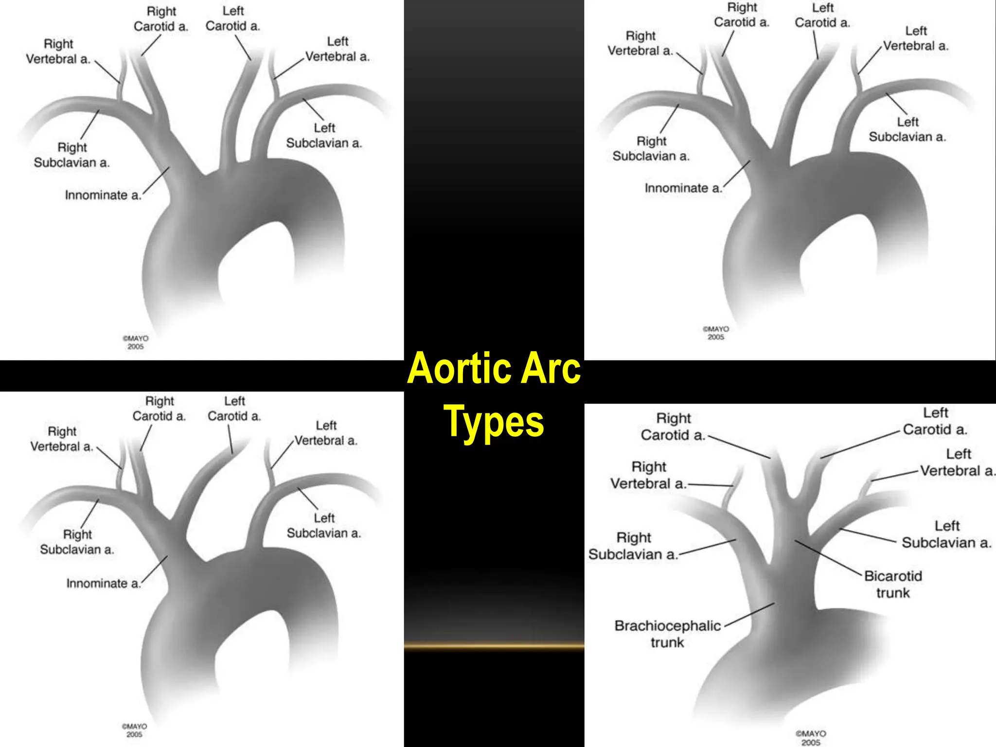
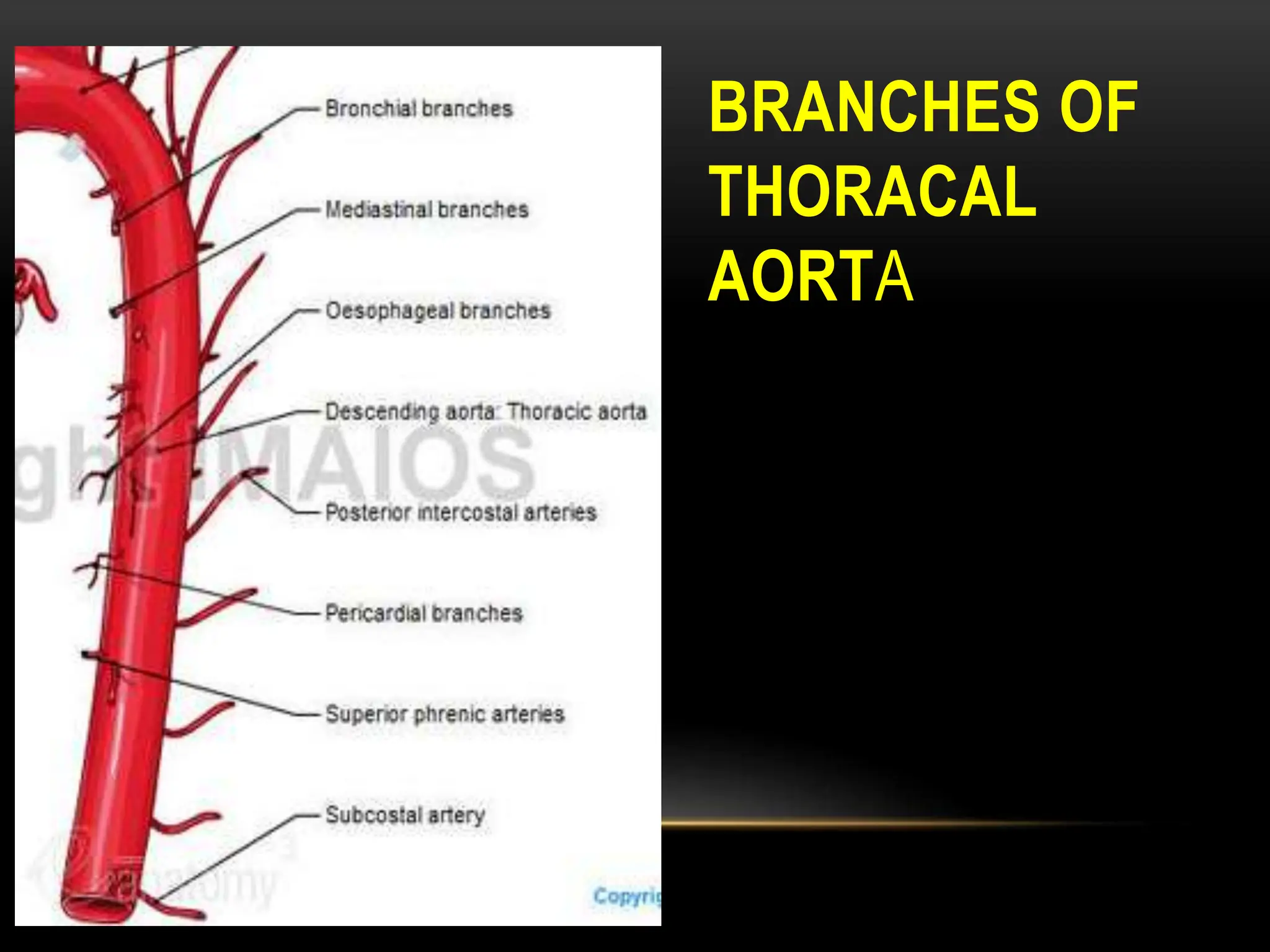
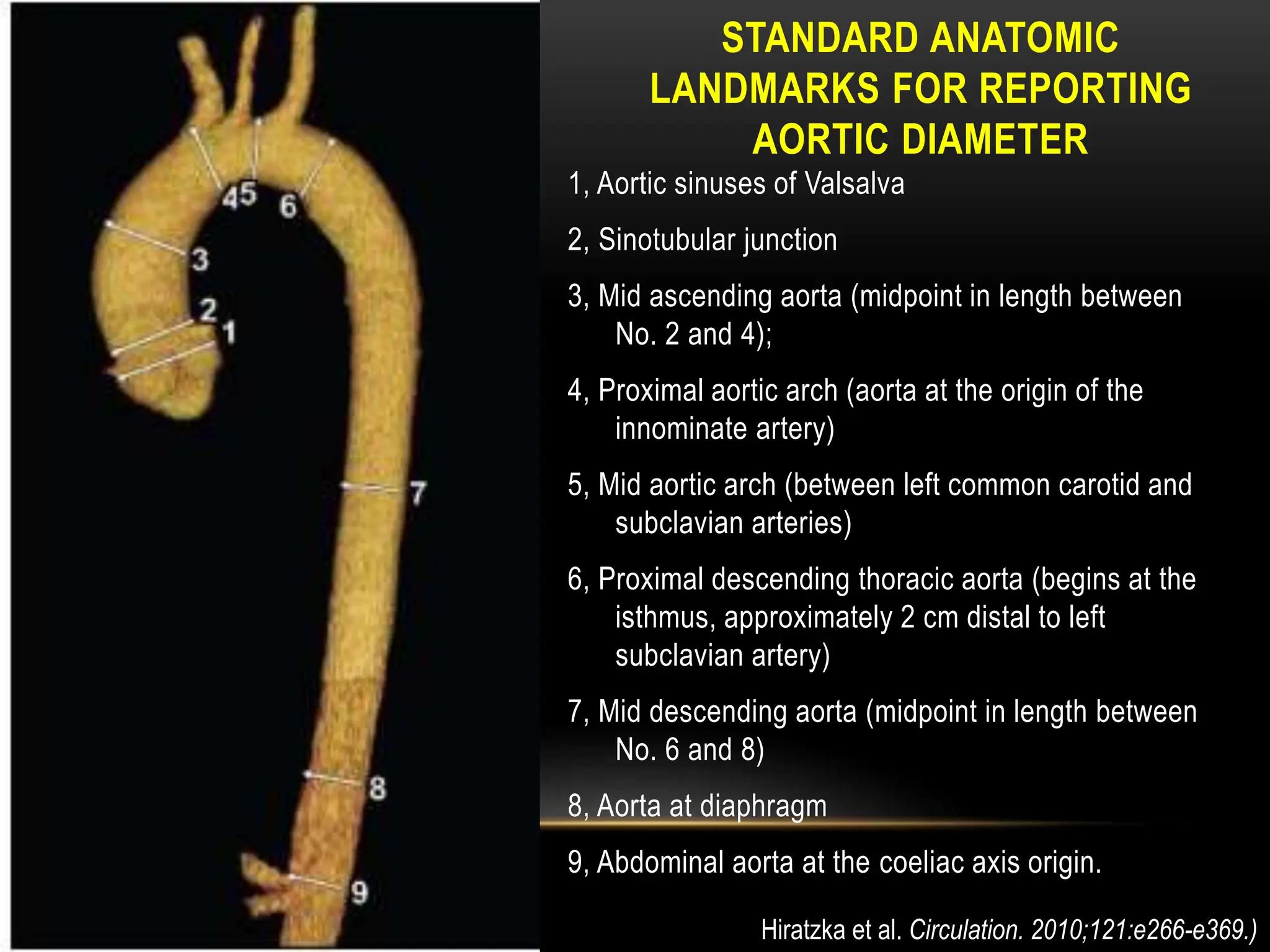
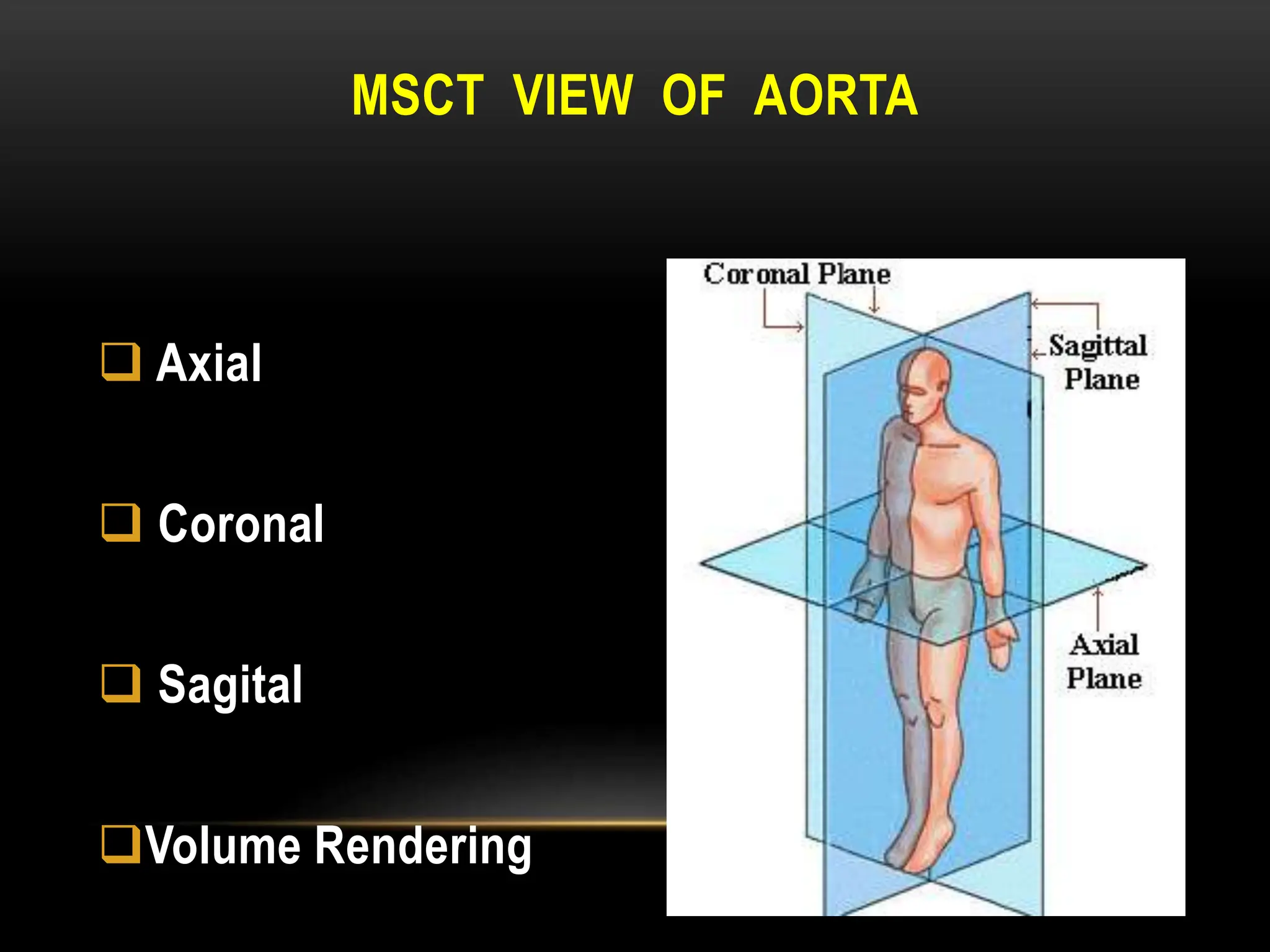
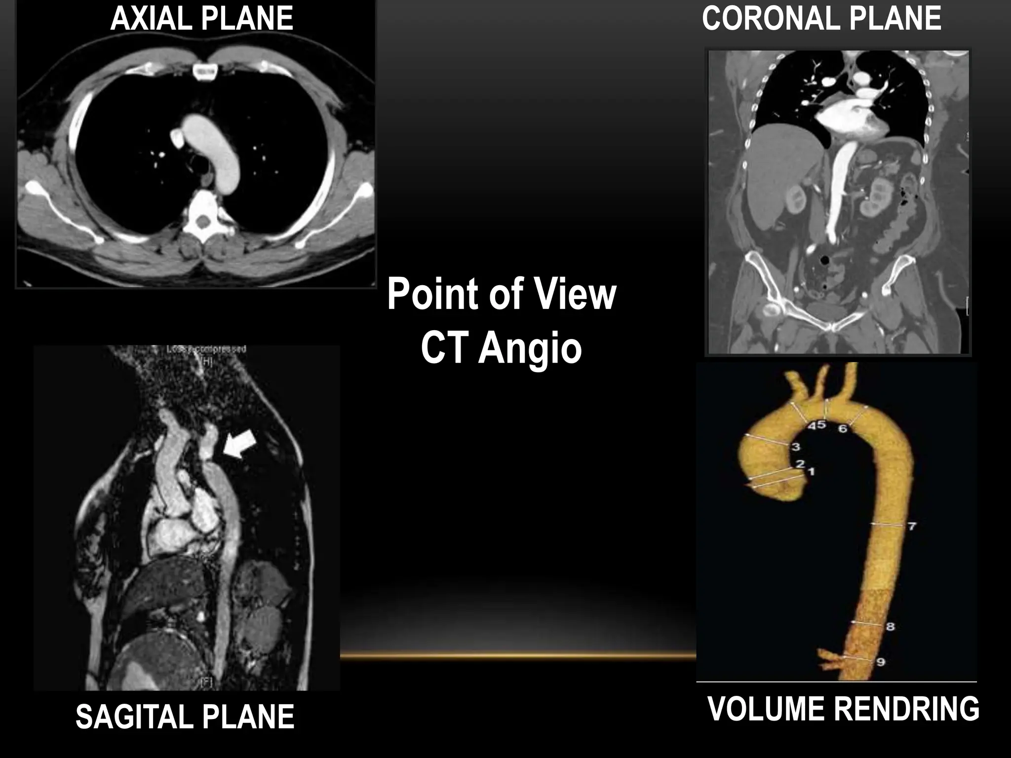
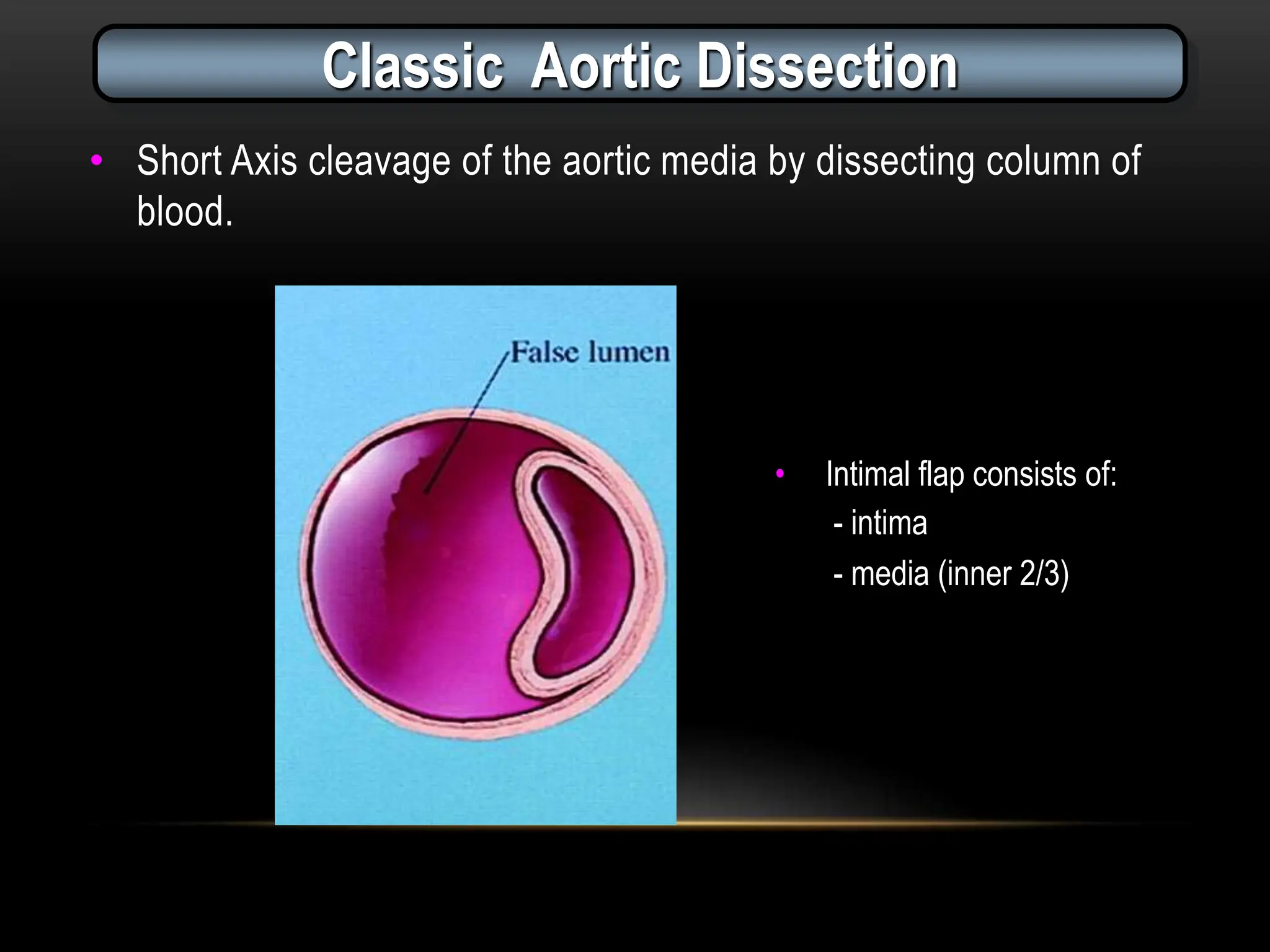
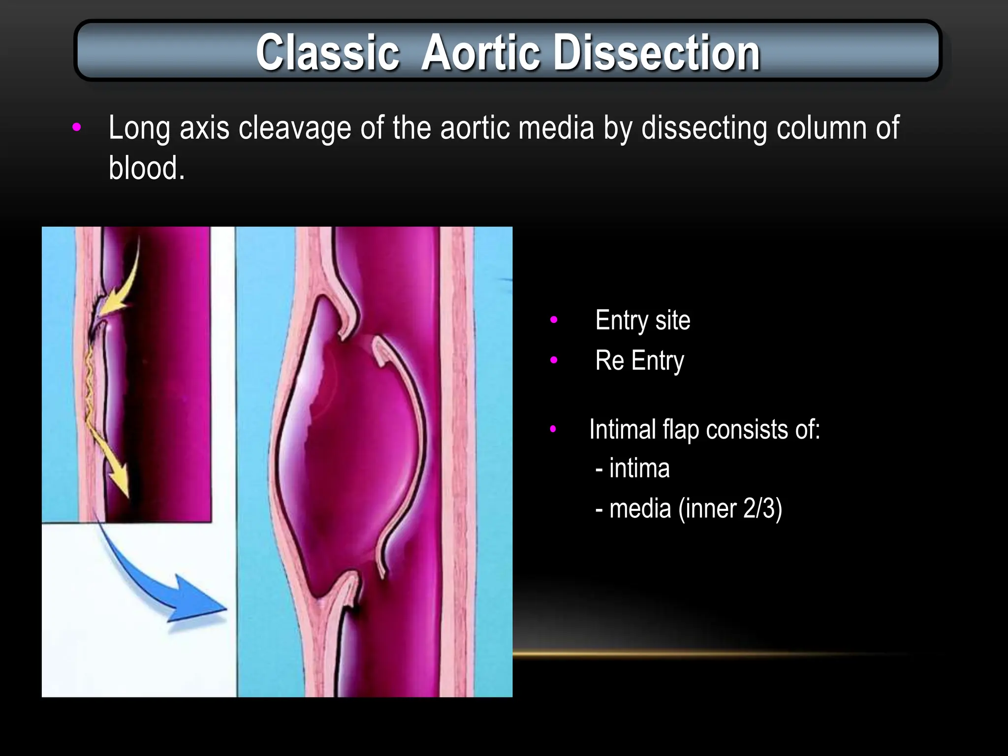
Anatomi, Topografi dan Modalitas Pencitraan
AORTA
ANATOMY
NORMAL ANATOMY OF THORACIC AORTA
BRANCHES
OF AORTIC
ARCH
Aortic Arc
Types
BRANCHES OF
THORACAL
AORTA
STANDARD ANATOMIC
LANDMARKS FOR REPORTING
AORTIC DIAMETER
1, Aortic sinuses of Valsalva
2, Sinotubular junction
3, Mid ascending aorta (midpoint in length between
No. 2 and 4);
4, Proximal aortic arch (aorta at the origin of the
innominate artery)
5, Mid aortic arch (between left common carotid and
subclavian arteries)
6, Proximal descending thoracic aorta (begins at the
isthmus, approximately 2 cm distal to left
subclavian artery)
7, Mid descending aorta (midpoint in length between
No. 6 and 8)
8, Aorta at diaphragm
9, Abdominal aorta at the coeliac axis origin.
Hiratzka et al. Circulation. 2010;121:e266-e369.)
NORMAL THORACIC AORTIC
DIAMETER
TOPOGRAPHY
BONE AND
ABDOMINAL
AORTIC
BRANCHES
MSCT
 Axial
 Coronal
 Sagital
Volume Rendering
MSCT VIEW OF AORTA
Point of View
CT Angio
AXIAL PLANE CORONAL PLANE
SAGITAL PLANE VOLUME RENDRING
AORTIC DISECTION
CLASIFICATION OF AORTIC DISECTION
Circulation. 1999;99:1331-1336
Characterized by :
1. True and false lumen
2. Intimal tear/ Entry site  communication between L.
3. Intimal Flap
CLASSIC DISSECTION
• Short Axis cleavage of the aortic media by dissecting column of
blood.
Classic Aortic Dissection
• Intimal flap consists of:
- intima
- media (inner 2/3)
• Long axis cleavage of the aortic media by dissecting column of
blood.
Classic Aortic Dissection
• Entry site
• Re Entry
• Intimal flap consists of:
- intima
- media (inner 2/3)
CTA MARKER – FALSE LUMEN
CLASSIC DISSECTION
AXIAL VIEW
• Beak Sign
• Cob Web Sign
• Wrap Sign usually in the Aortic Arch.
SAGITAL / CORONAL VIEW
• Must often is larger than true lumen .
• Less dencity than true lumen except entry site large
with expose reentry
IMH
IMH
PAU
PAU
Class 4 lesion : PAU
PAU
PAU
BRANCH-VESSEL COMPROMISE
Static Obstruction
Dynamic Obstruction
Williams DM et al. Radiology 1997;203:37-44
JVIR 1997;8:605-625
DYNAMIC OBSTRUCTION
STATIC OBSTRUCTION
AORTIC ANEURYM
I. Location
 TAA
 AAA
 TAAA
II Type
 Fusiform
 Saccular
II Diameter
IV NecK
V Iliac Arteries Tortuousity
FUSIFORM SACCULAR
SACCULAR ANEURYSM
• Still in controvesial
• Lobulated contour
• Rapid expansion or Development
and adjacent mass
• Stranding
• Fluid in unusual location
→ Highly suspicious for an infected
aneurysm
→ contraindication ? / stent-graft
placement with appropriate
antibiotic coverage
Macedo TA et al. Radiology 2004;231(1):250
AORTIC INFECTION
1,TB
2. Mycotic
3. Vasculitis
TUBERCULOUS MYCOTIC ANEURYSM
• Once symptomatic TBAA is identified,
treatment must not be delayed.
• The size of the aneurysm does not appear to
influence the need for treatment
• Aneurysms as small as 1.0-cm in diameter may
rupture
Long et al. Chest 1999;115(2):523
38/F
40-day 50-day
Tuberculous Aneurysm
MYCOTIC ANEURYSM
Vasculitis
Vasculitis
Aortic Disection
Aortic Aneurysm
TEVAR OR EVAR REQUISITE
NECK / LANDING ZONE
PROXIMAL TO THE ANEURYSM
Morphology suitable for endovascular repair, including:
1.with a length of at least 15 mm,
2.with a diameter measured outer wall to outer wall of no
greater than 28 mm and no less than18 mm,
3.with an angle less than 60 degrees relative to the long
axis of the aneurysm,
4.with an angle less than 45 degrees relative to the axis of
the suprarenal aorta.
NECK / LANDING ZONE MEASUREMENTS
TAA
• Untreated TAA likely to expand over time and
rupture
• Coady et al. rupture in descending thoracic
aortic aneurysm 7 cm or greater, and they
recommended surgical repair when aneurysms
reach a diameter of 6.5cm
Coady MA, Cardiol Clin 1999;17(4):827
DIAMETER
CTA AXIAL LEVEL- AORTA
RUPTURE AAA
Primary Sign
1. Periaortic Stranding
2. Retroperitoneal Haematoma
ex Perirenal Compartment
3. Extravasation of Contras
RUPTURE AAA
A
RUPTURE AORTA
RUPTURE AAA
Rupture AAA
IMPENDING RUPTURE AAA
Secondary Sign (Impending)
1. High-attenuating crescent
2. Focal discontinuity of intimal calcification
3. Tangential calcium
4. Draped aorta
Imp.Rupture AAA
Imp. Rupture AAA
Imp. Rupture AAA
Impending Rupture AAA
Rupture AAA
Impending Rupture AAA
Conclusion
MSCT of the Aorta is currently the imaging technique of choice
for evaluating patients with aortic diseases
THORACIC AORTIC
1
1
Rupture AAA
Primary Sign
1. Periaortic Stranding
2. Retroperitoneal Haematoma
ex Perirenal Compartment
3. Extravasation of Contras
Rupture AAA
Rupture AAA
Impending Rupture AAA
Secondary Sign (Impending)
1. High-attenuating crescent
2. Focal discontinuity of intimal calcification
3. Tangential calcium
4. Draped aorta
Rupture AAA
Rupture AAA
Rupture AAA
Rupture AAA
Rupture AAA
Rupture AAA
Rupture AAA
1. Caput Humeri Dex., 2.Oesophagus, 3.Trachea , 4.v. Subclavia Sin. 5.Spina
Scapula ,6.Columna Scapula .
1.Pulmo Dex, 2.Costa,3.Oesophagus ,4.Trachea,5.V.bracheocephalica
Sin.,6.a.Carotis Com. Sin,7.v.Axillaris sin.,8.Pulmo Sin., 9. Prosesus Transversus
,10.Scapula.
1.Trachea,2Costa, 3.a.Bracheocephalica dex,4.v.Bracheocephalica Sin.,5.m.Pectoralis
Major., 6.m.Pectoralis Minor, 7.a.Carotis Comm.Sin.,8 a.Subclavia Sin. 9,
Oesophagus,10.Scapula.
1.Pulmo dex,2.Costa ,3.Trachea,4.v.Bracheocephalica Sin,5.a. Bracheocephalica
,6.a.Carotis Comm. Sin.7.a. Subclavia Sin,8.Scapula,9.Oesophagus,10.Prosesus
Spinosus.
CT Imaging  examination about aorta and it's disease

CT Imaging examination about aorta and it's disease