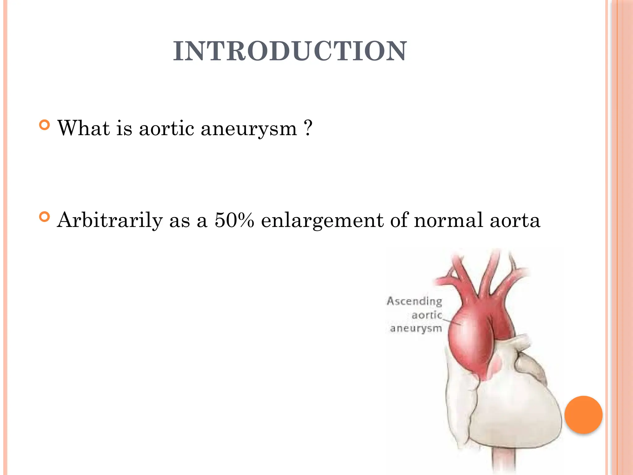
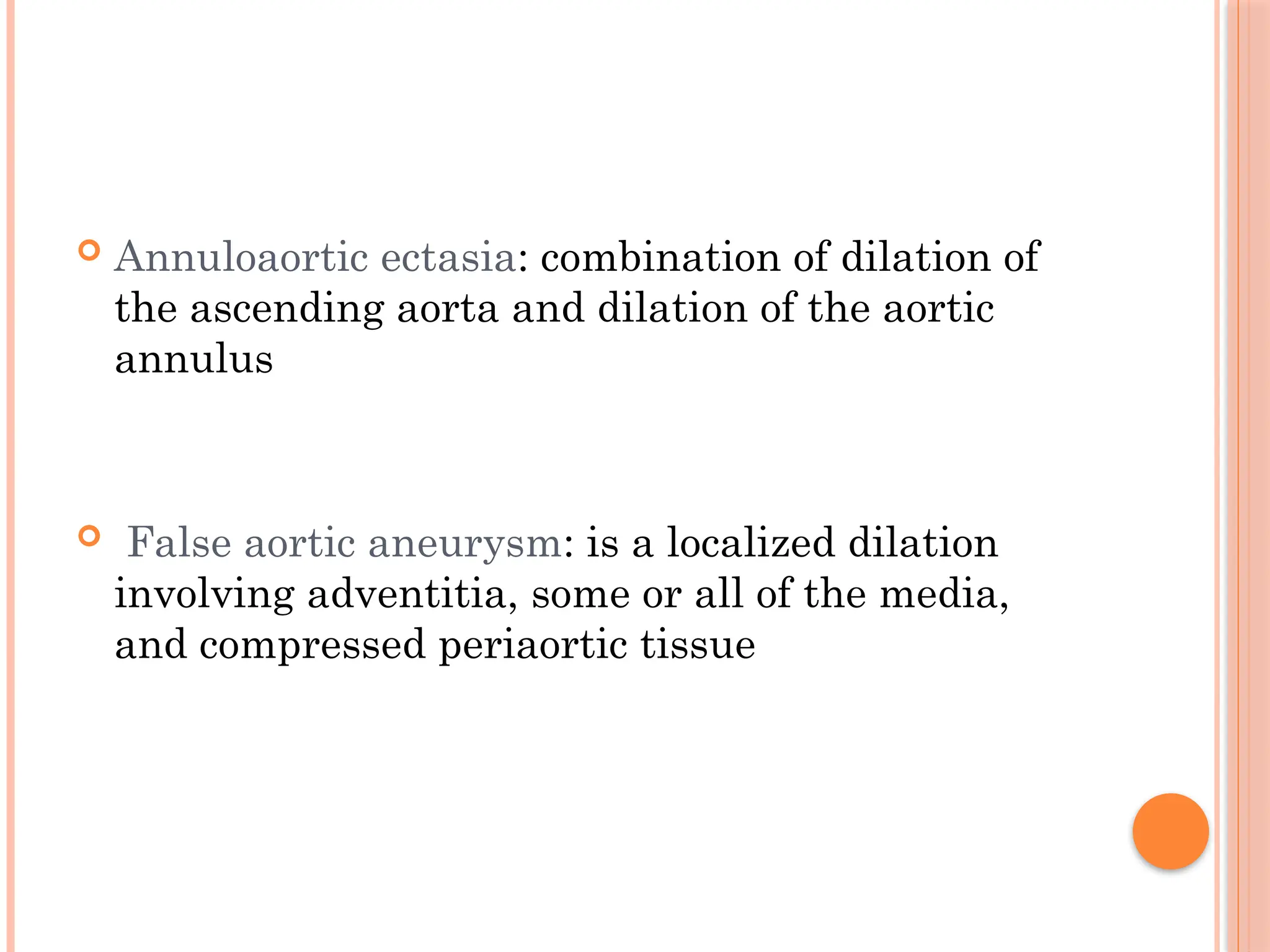
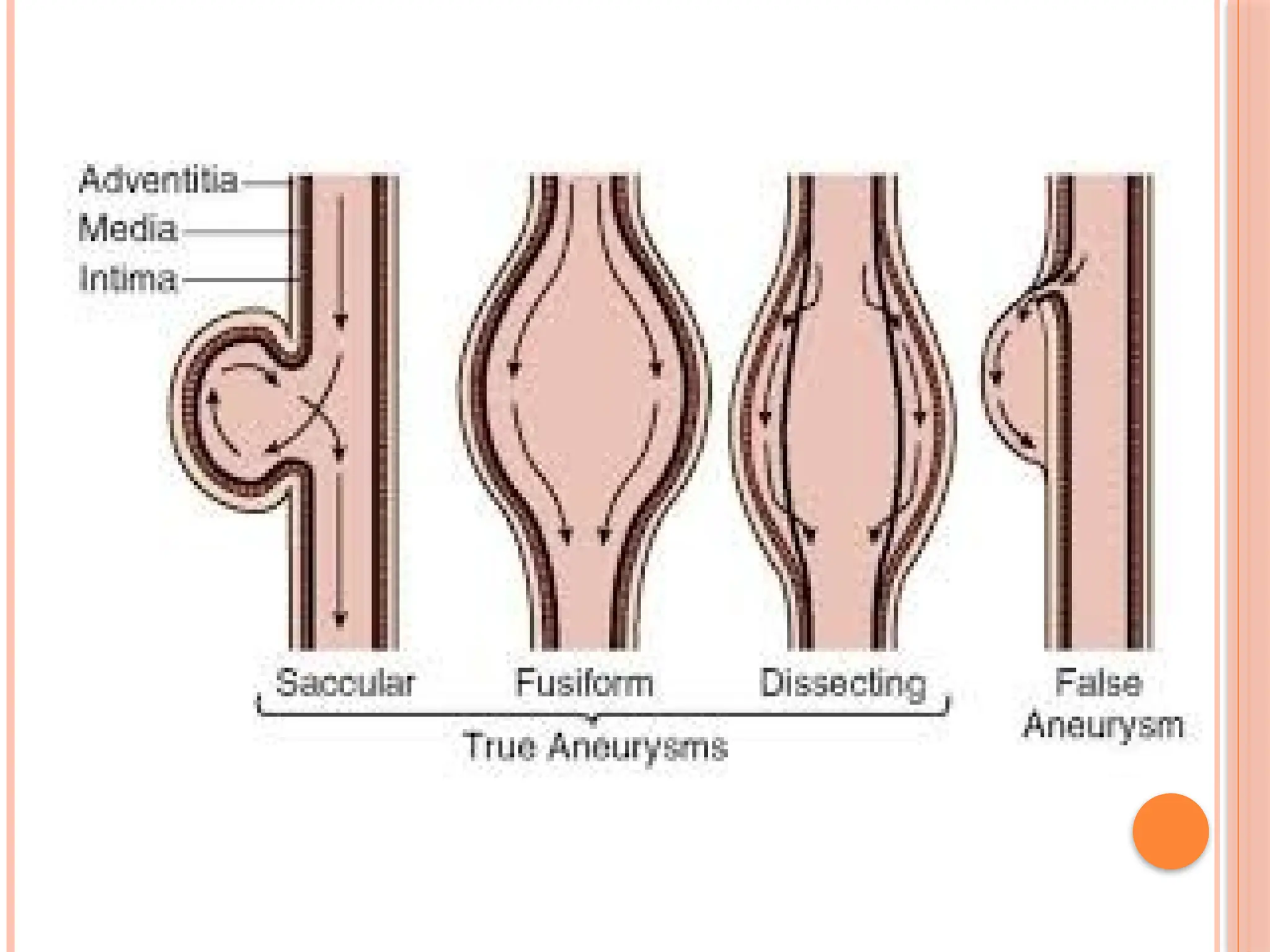
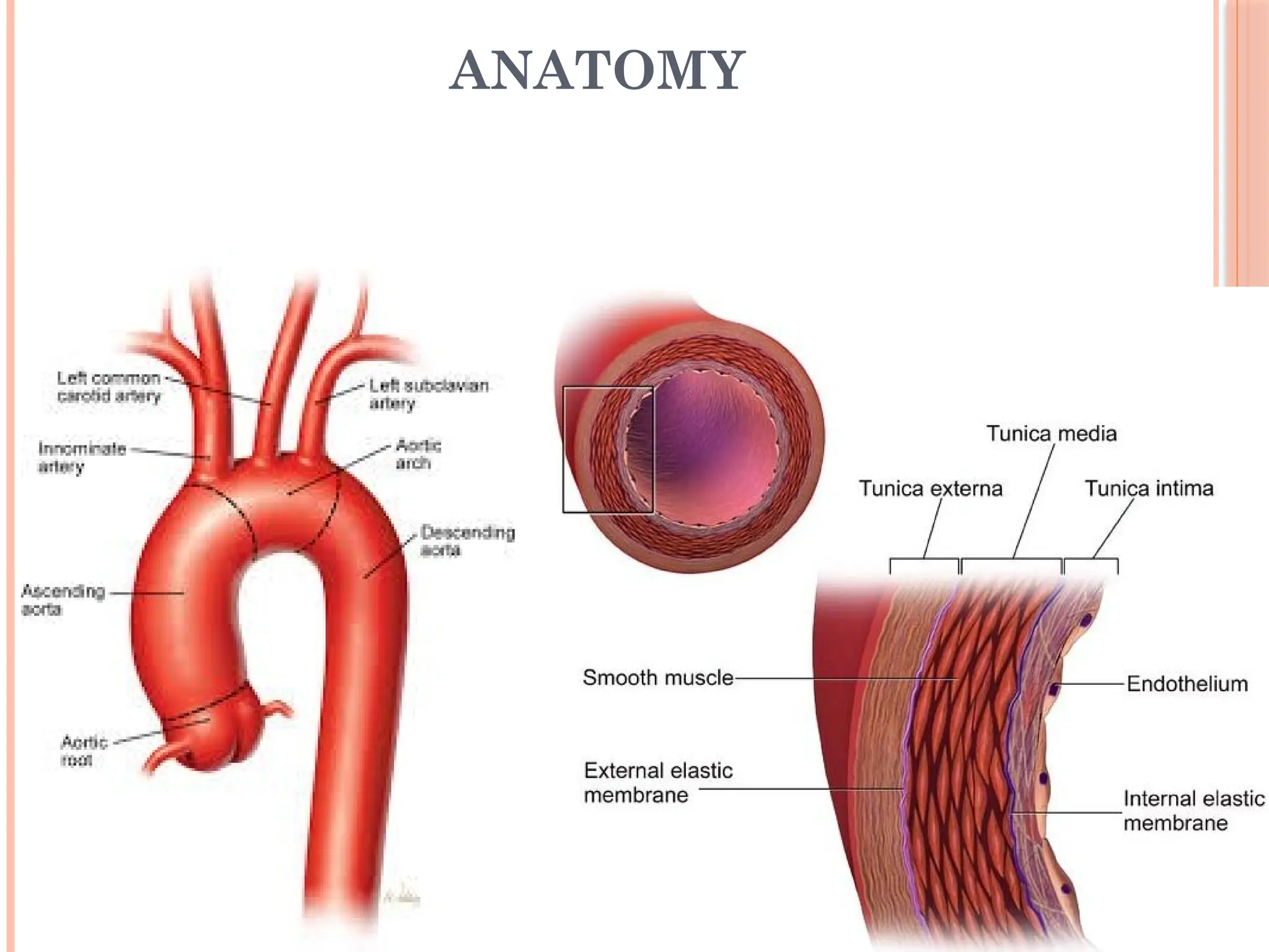
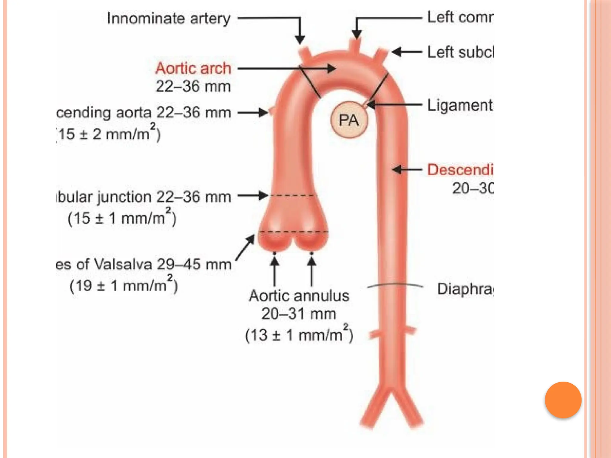
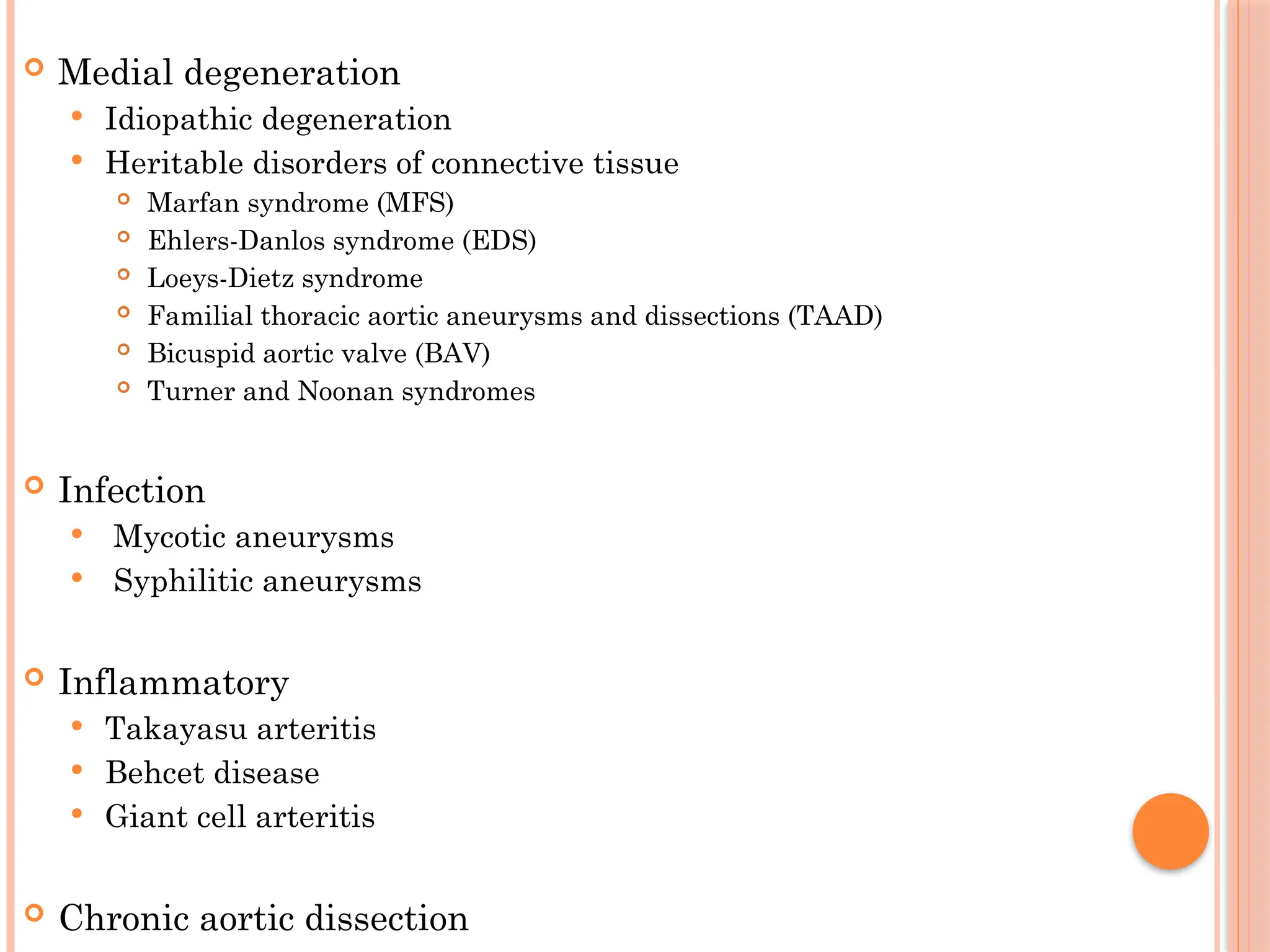
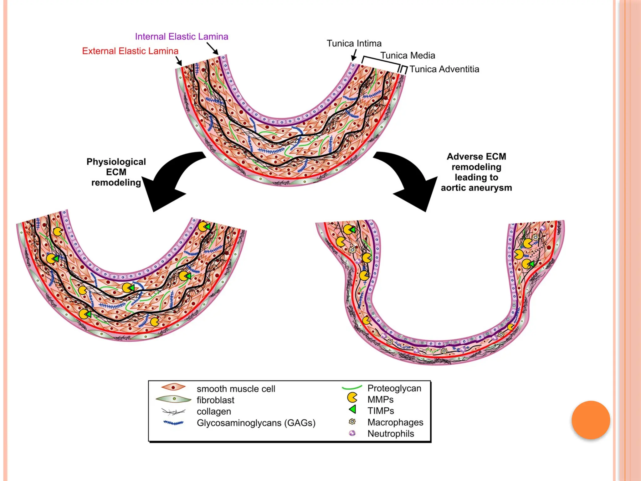
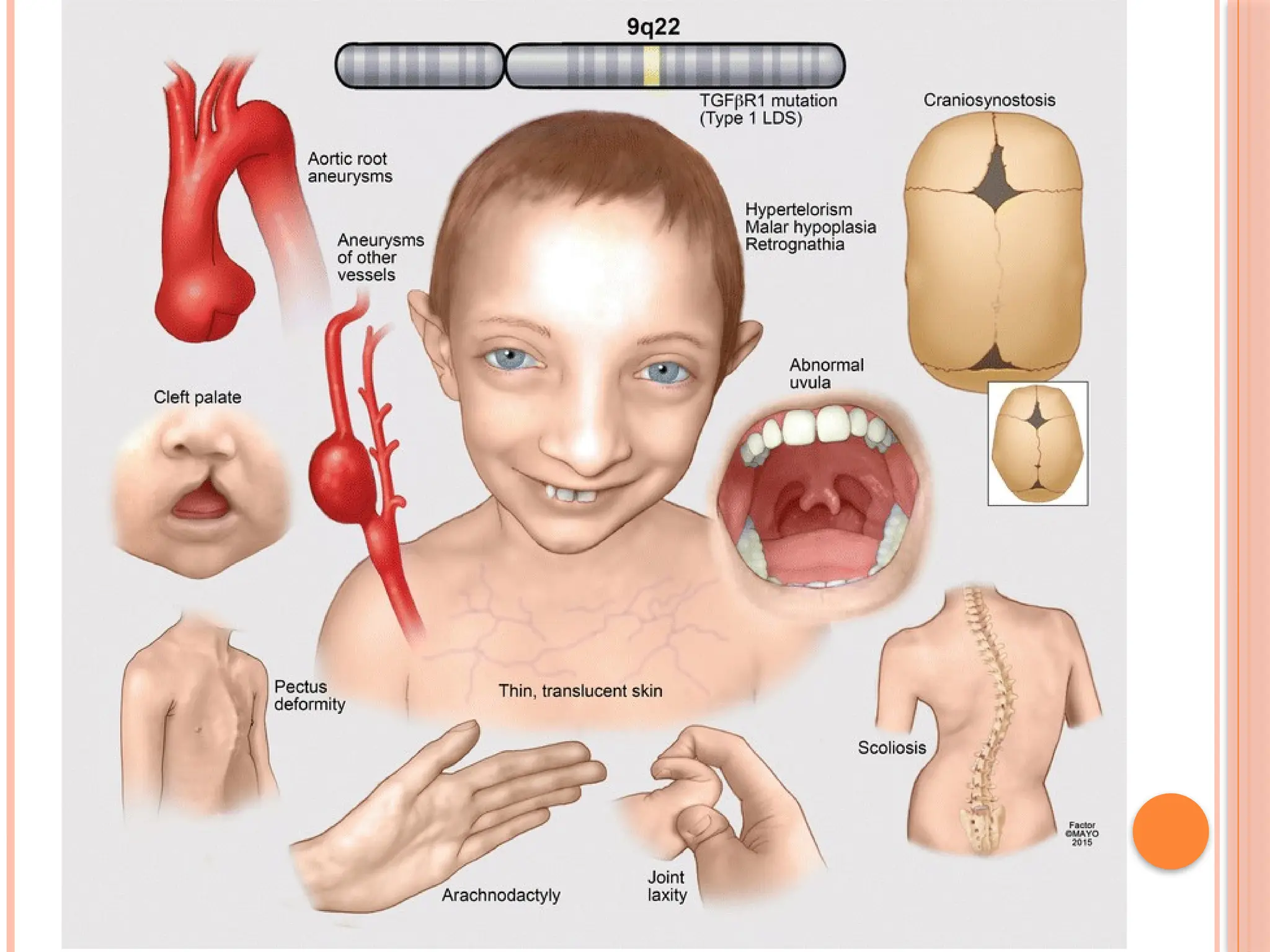
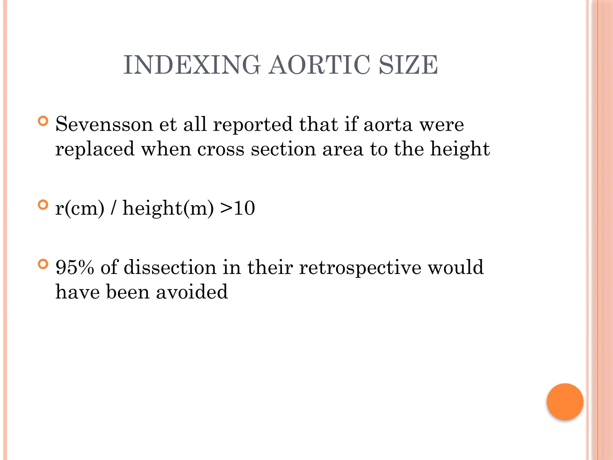
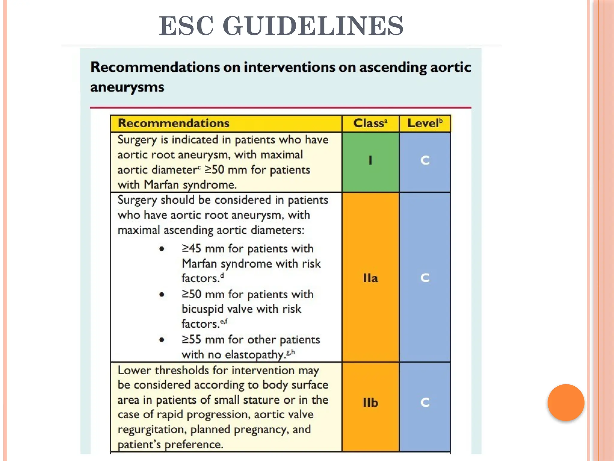
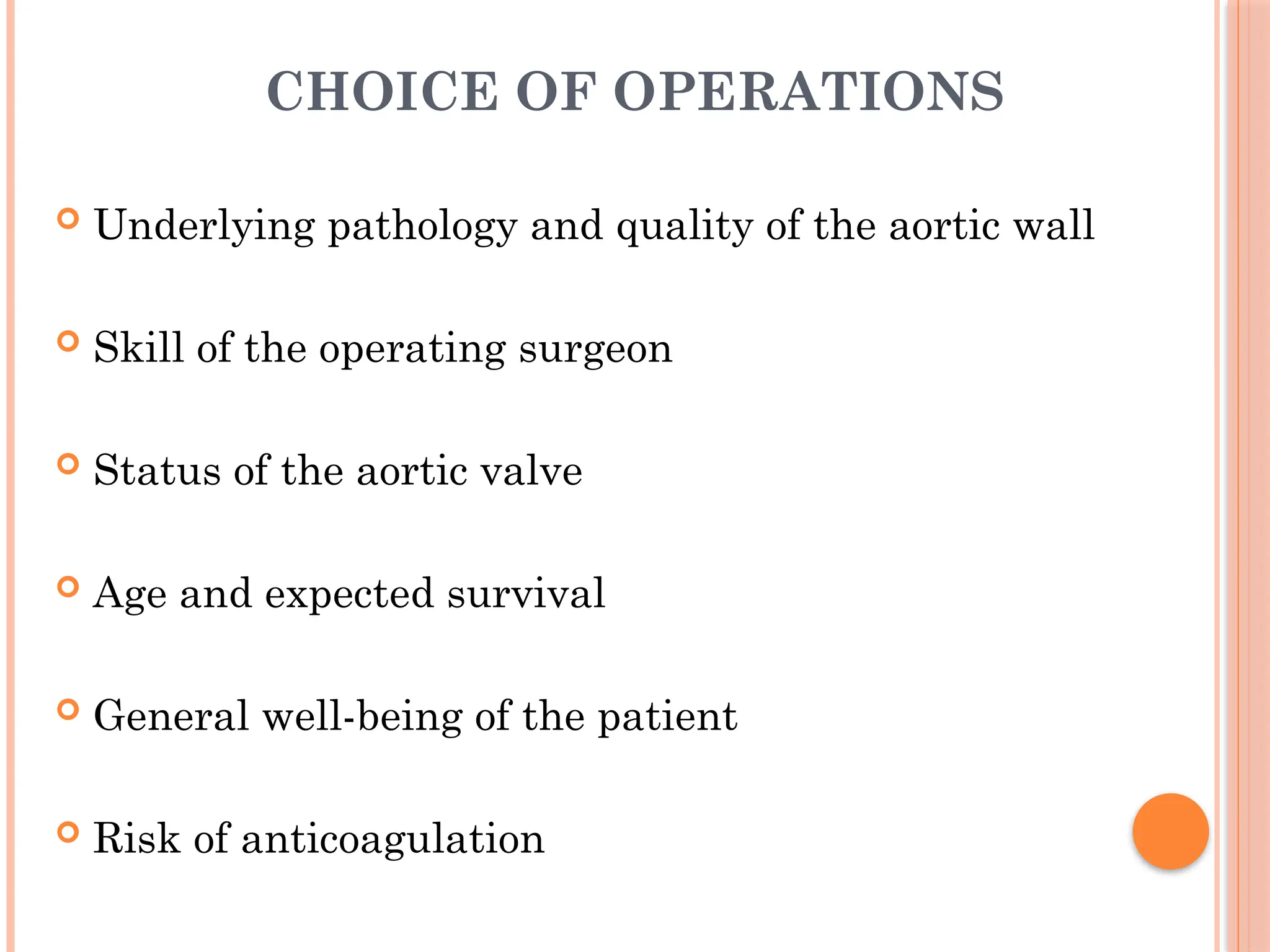
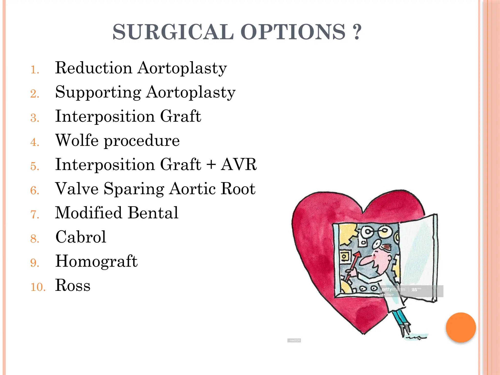
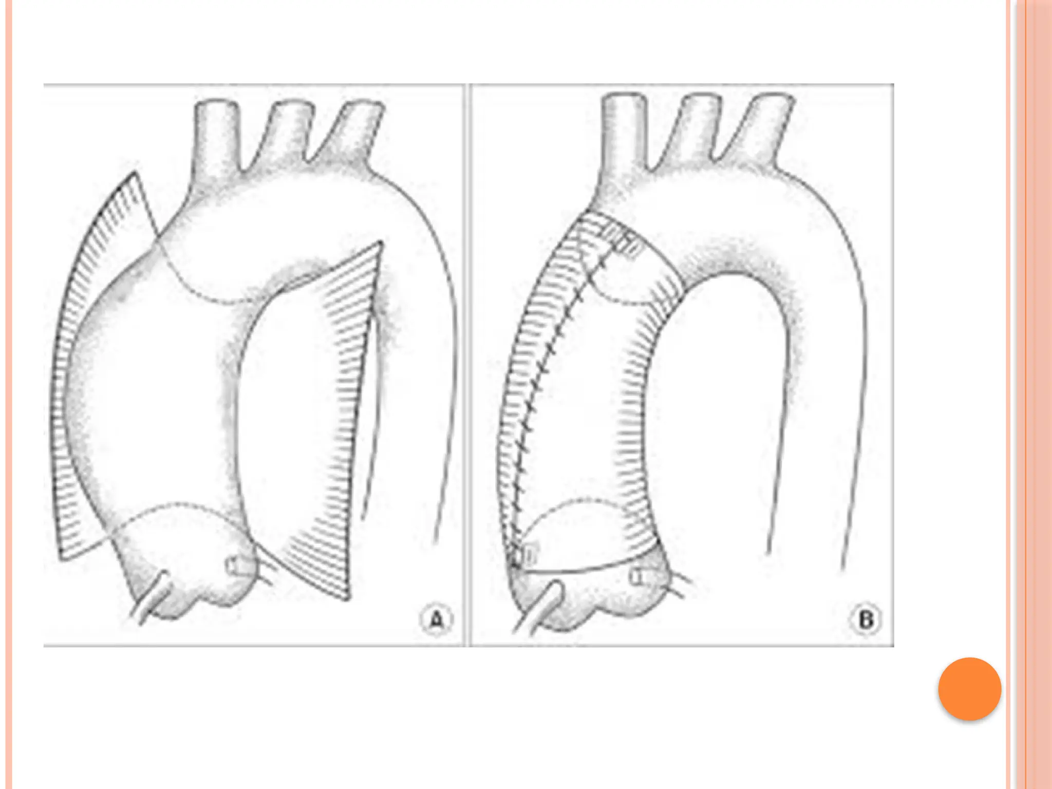
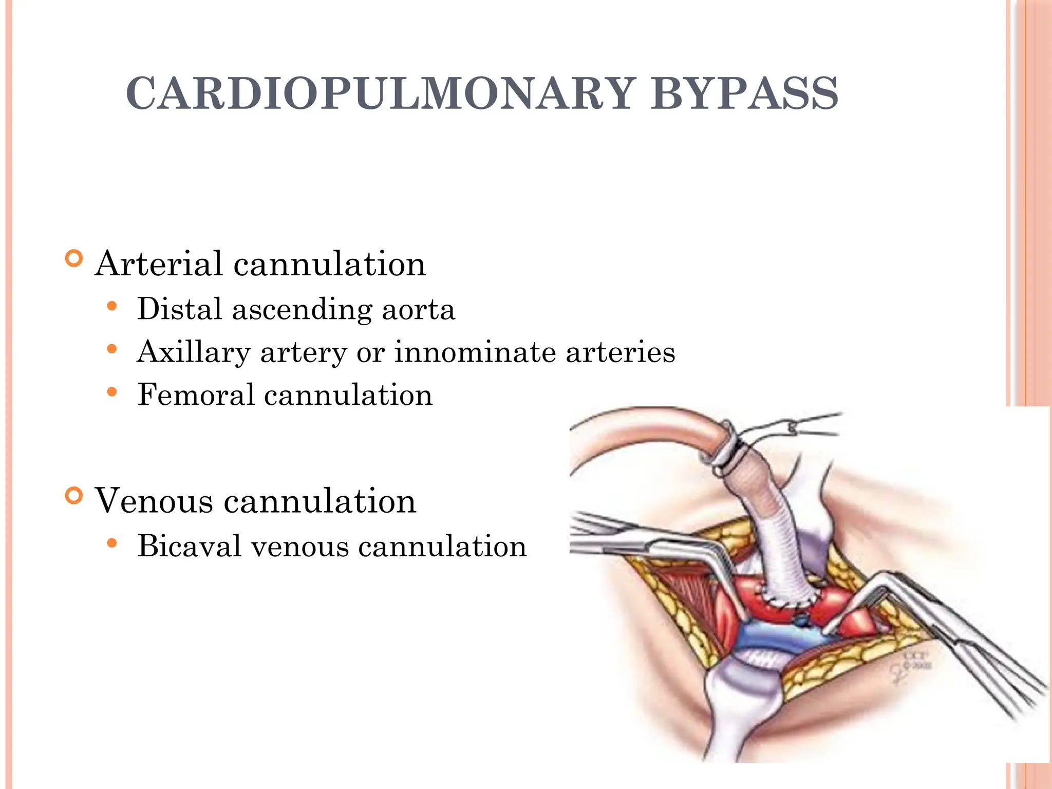
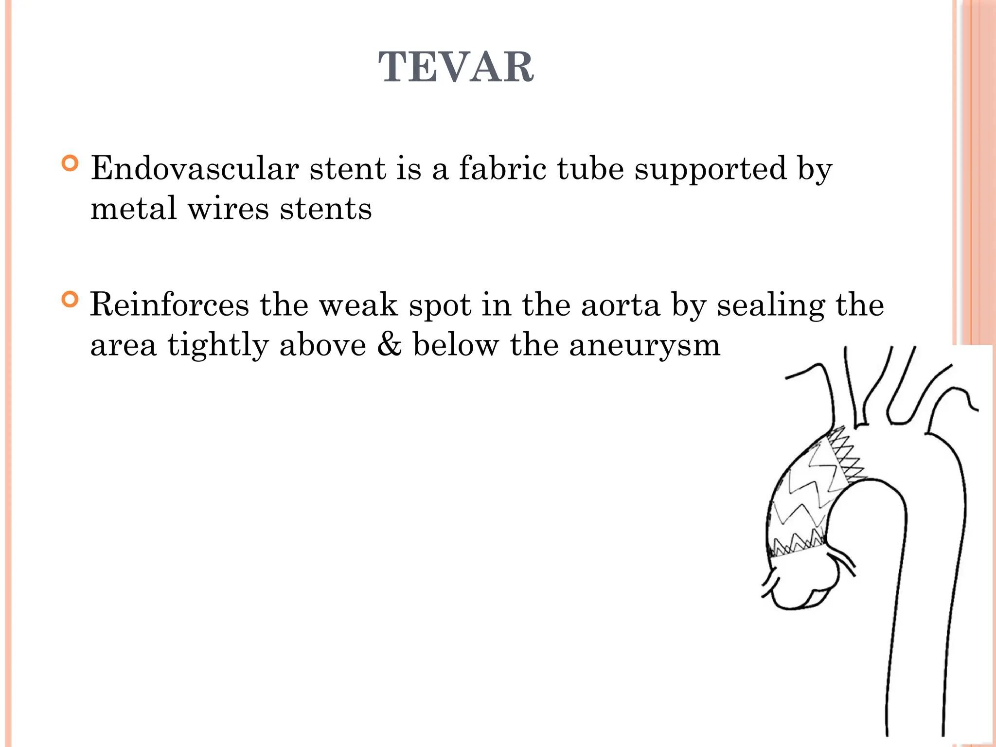
ASCENDING AORTIC
ANEURYSM
Dr. Rashid Aziz
Moderator
Dr. Zubair Brohi
Clinical Fellow
NICVD
INTRODUCTION
 What is aortic aneurysm ?
 Arbitrarily as a 50% enlargement of normal aorta
 Annuloaortic ectasia: combination of dilation of
the ascending aorta and dilation of the aortic
annulus
 False aortic aneurysm: is a localized dilation
involving adventitia, some or all of the media,
and compressed periaortic tissue
HISTORY
 Galen, who observed in gladiators injured during battle in
the second century.
 1543 Andeas, Vesalius described a thoracic aortic
aneurysm.
 1769 Morgagni seen postmortem findings of ruptured
thoracic aneurysms
 1773 Alexander Monro described three coats of the arterial
wall, and the destruction in the formation of true and false
aneurysms
 1952 Cooley and DeBakey first report of a descending
aortic repair . Performed on a saccular aneurysm
without cardiopulmonary bypass (CPB).
 1956, Cooley and DeBakey performed replacement of
the ascending aorta with a segment of homograft with
CPB
 1963 Bentall and De Bono first composite aortic root
replacement.
 1964 Wheat et al, resected the ascending aorta and
entire aortic root except for the aortic tissue
surrounding the coronary arteries, then performed a
mechanical valve insertion
 in 1981, Cabrol et al described the use of an 8 to 10
mm Dacron graft to attach to independently mobilized
coronary artery buttons
ANATOMY
CLASSIFICATION
 LOCATION
 Ascending aorta,
 Aortic Arch
 Descending aorta
 Morphology
 Fusiform
 Saccular
 Etiology
 Medial degeneration
 Infection
 Inflammation
 Chronic aortic dissection
 Medial degeneration
 Idiopathic degeneration
 Heritable disorders of connective tissue
 Marfan syndrome (MFS)
 Ehlers-Danlos syndrome (EDS)
 Loeys-Dietz syndrome
 Familial thoracic aortic aneurysms and dissections (TAAD)
 Bicuspid aortic valve (BAV)
 Turner and Noonan syndromes
 Infection
 Mycotic aneurysms
 Syphilitic aneurysms
 Inflammatory
 Takayasu arteritis
 Behcet disease
 Giant cell arteritis
 Chronic aortic dissection
INCIDENCE
 Estimated 5-10% per 100000 person/ year
 60% involve aortic root/ ascending aorta
 35% descending aorta
 <10% aortic arch
RISK FACTORS?
 Age
 Atherosclerosis
 Aortitis
 BAV
 Connective tissue disorders
 Dissection
 DM
 Hypertension
 Infections
 Pregnancy
 Smoking
 Trauma
INDEPENDENT RISK FACTOR FOR RUPTURE
 Age
 Aortic diameter
 Pain or symptoms of expansion
 COPD
 Smoking
PATHOPHYSIOLOGY
 The media of the aorta is composed of smooth muscle
cells within a matrix of elastin & collagen.
 Other structural proteins, including fibrillin, laminin,
proteoglycans, and fibronectin.
 Collagen provides the necessary structural strength.
 Elastin conveys the arterial recoil capabilities.
 Defects in either protein can cause aortic pathology.
IDIOPATHIC MEDIAL
DEGENERATION
 Triad of
1. Loss of non-inflammatory smooth muscle cells
2. Fragmentation of elastic fibers
3. Accumulation of basophilic ground substance in
depleted areas
 First weakens the aortic wall, thereby increasing the
wall tension, which can induce aortic dilatation
MARFAN SYNDROME
 Most prevalent inherited connective tissue disorder, 80%
develop Artic root aneurysm
 Mutations in the fibrillin- 1 gene & recently identified
mutations in the (TGF-B) gene
 Defective coding of fibrillin leading to elastin derangement &
excessive TGF-B activity negatively impact on smooth
muscle
 Additional cardiovascular complications
 Mitral valve prolapse and regurgitation
 Left ventricular dilation,
 Aortic root dilation is the most common cause of morbidity and
mortality
EHLERS-DANLOS
SYNDROME(EDS)
 Rare autosomal dominant inherited disorder of
connective tissue
 Site: carotid , descending and aortic arch
 Defective type III collagen
 Carries a substantial risk of rupture of the aorta.
LOEYS-DIETZ SYNDROME(LDS)
 Aortic aneurysm syndrome that has widespread systemic
involvement.
 Mutations in TGFBR1 and TGFBR2
 Loss of elastin conten in the aortic media.
 The phenotype overlaps with that of MFS (aortic aneurysm,
arachnodactyly, dural ectasia) ,
 Its distinctive features such as hypertelorism, bifid uvula and
generalized arterial tortuosity
 Average age at a first cardiovascularevent much lower than that for
patients with untreated MFS or vascular EDS
FAMILIAL THORACIC AORTIC
ANEURYSMS AND DISSECTIONS(TAAD)
 Patients without genetic syndromes also manifest familial
clustering of thoracic aortic aneurysms and dissections
 20% of aneurysm probands have a first-order relative with an
aortic aneurysm.
 TAAD 1 locus mapped to the long arm of chromosome 5, with
approximately half of the identified families.
 Early tendency to rupture in <5cm
 Therefore, TAAD is now a recognized heritable disorder of
connective tissue.
TURNER AND NOONAN
SYNDROMES
 Chromosome constitution is 45-XO; it has an
incidence of 1 in 5000 live births
 Cardiovascular problems include
 BAV (present in one-third of subjects)
 Coarctation of the aorta
 Hypertension
 Aortic root dilation in 40 %
BICUSPID AORTIC VALVE DISEASE
 Prevalence of 1 to 2 % with male predominance
 First -degree relative with BAV is 9%
 AS in three-quarters of patients & AR in 15%
 Independent risk factor for progressive aortic dilation
 Cause of this initially was attributed to "poststenotic dilation
 Aortic valvular cusps and ascending arterial media both a rise from
common neuralcrest cells.
 Increased elastic fragmentation & smooth muscle cell apoptosis
within the aortic media
CHRONIC AORTIC DISSECTION
 Chronic aortic dissections tend to dilate over time
 Debakey and colleagues found that aneurysms developed
 46% with uncontrolled hypertension
 17% with controlled hypertension
 Griepp and coworkers found that after repair of acute
type A dissections, the growth of the distal aorta
 0.85 mm a year for the aortic arch
 1.24 mm a year for the descending thoracic aorta.
MYCOTIC ANEURYSMS
 Rare but can be fatal if they are not diagnosed early.
 Staphylococcus aureus and salmonella predominant organisms
 Site: femoral , abdominal & visceral artery( coronary artery)
 Hematogenous spread to the intima or the vasa vasorum,
lymphatic spread, or direct extension from an adjacent infected
focus.
 Endothelial intimal lining of the aorta is generally highly
resistant to infection, but disruption of this barrier by
atherosclerosis reduces resistance
SYPHILITIC ANEURYSMS
 Once perhaps the most common cause of ascending aortic
aneurysms (Treponema pallidum)
 Females 20 - 30 years
 Stie: Arch of aorta
 Type: Saccular
 Inflamation of adventetia & vasavasorum endarteritis
reduce blood flow that weakens aortic wall
 Aneurysm develop 10 to 20 years after onset of disease
INFLAMMATORY ANEURYSMS
 Walker and colleagues were the first to defined the term
inflammatory aneurysm
 Takayasu arteritis
 Pulmonary artery, the subclavian artery, and abdominal aorta
 Behcet disease
 Cardiovascular involvement only 7 to 29 percent
 Arterial lesion develops in the aorta and the pulmonary artery
 Giant cell arteritis
 17.3 times more likely to develop a thoracic aortic aneurysm
than is the general population
NATURAL HISTORY
 TAA growth rate of 0.2 - 0.4cm/year and marked
individual variability
 5year survival of about 40%
 Mean rate of Rupture or Dissection
 2%/ year for < 5 cm
 5%/ year for 5 - 5.9 cm
 10%/ year for > 6 cm
CLINICAL PRESENTATION ?
 Mostly clinically silent in many cases
 Anatomic location and the space-occupying nature
 Hoarseness
 Stridor
 Dysphagia
 Dyspnea
 Plethora and edema
 Signs and symptoms of AR.
 Chest or back pain may indicate acute expansion or leakage
of the aneurysm
SIGNS
 Often normal in a patient without rupture aneurysm
 Large aneurysm can be palpated in the suprasternal notch
 Venous distention due to SVCl or innominate vein
obstruction
 Signs of AR
 Abdominal aortic aneurysms are present in 10 - 20 %
 EDS, LDS & Marfanoid features
DIAGNOSTIC INVESTIGATIONS
 BLOOD TESTS
 XRAY
 ECHO
 CT SCAN
 MRI
 ANGIOGRAPHY
XRAY
ECHO
 Can visualize the aortic root and ascending aorta
accurately
 Less ideal for full extent of the aortic arch and origins of
neck vessels.
 Assessment AR, LV function, and hemopericardium
 TEE is the modality of choice for the diagnosis and
exclusion of aortic dissection in a n unstable patient
CT-SCAN
 Currently the most common diagnostic imaging for the aorta
 Provides rapid and precise evaluation of the ascending aorta
in regards to size, extent, and location
 Excellent visualization of the aorta and its branch vessels
 Advantage of demonstrating aortic wall thickening,
calcification, and luminal thrombus
 Entire thoracic and abdominal aorta for evidence of
concomitant aneurysm disease in the arterial tree.
 64-bit scanners also are capable of imaging coronary arteries
MRI
 Emerging as the premier imaging method for the
diagnosis of diseases of the thoracic aorta in stable
patients
 ( CE-MRA) is the most widely used MRA method
 Assessment of the vessel wall and AR in the presence of
disease of the ascending aorta
MEDICAL MANAGEMENT
 The main aim of medical therapy is to reduce the shear
stress on the diseased segment of aorta by reducing BP
& cardiac contractility.
 Smoking cessation.
 Competitive sports should be avoided in pts with
enlarged aorta.
 In chronic conditions BP should be controlled below
140/90 mm Hg, with lifestyle changes &
antihypertensives.
 Prophylactic use of B-B, ACE, ARBs reduce
either progression/ occurrence of
complications.
OPERATIVE INDICATIONS ?
1. Aortic diameter 5 - 5.5 cm.
2. LDS > 4cm
3. MFS 4 - 4.5 cm
4. BAV 4.5 - 5 cm
5. EDS or TAAD > 5cm
6. < 5.0 cm
1. Rapid growth (more than 1 cm/year),
2. Family history of premature aortic dissection
3. Presence of moderate or severe AR
4. Symptoms suggest impending to rupture
INDEXING AORTIC SIZE
 Sevensson et all reported that if aorta were
replaced when cross section area to the height
 r(cm) / height(m) >10
 95% of dissection in their retrospective would
have been avoided
ESC GUIDELINES
CHOICE OF OPERATIONS
 Underlying pathology and quality of the aortic wall
 Skill of the operating surgeon
 Status of the aortic valve
 Age and expected survival
 General well-being of the patient
 Risk of anticoagulation
SURGICAL OPTIONS ?
1. Reduction Aortoplasty
2. Supporting Aortoplasty
3. Interposition Graft
4. Wolfe procedure
5. Interposition Graft + AVR
6. Valve Sparing Aortic Root
7. Modified Bental
8. Cabrol
9. Homograft
10. Ross
ANESTHESIA CONSIDERATIONS
 Induction reduces sympathetically mediated vasoconstriction
and tachycardia due to acute thoracic aortic disruption
 Femoral cannulation under local anesthesia with simultaneous
institution of GA and CPB in the setting of aneurysmal rupture
 Left and right radial artery pressures
 Pulmonary artery catheter.
 Intraoperative TEE
 Thermistor probe in bladder and in tympanic
CARDIOPULMONARY BYPASS
 Arterial cannulation
 Distal ascending aorta
 Axillary artery or innominate arteries
 Femoral cannulation
 Venous cannulation
 Bicaval venous cannulation
CEREBRAL PROTECTION
 Permanent and Transient neurologic dysfunction (TND)
 TND was observed commonly with ischemic periods longer than
40 to 60 min
 Hypothermia
 Cerebral monitoring
 Ante grade and retrograde perfusion
 Metabolic monitoring
 Pharmacologic
DHCA
 This technique allows the open distal anastomosis in
better visualization
 Disadvantages: coagulopathy, increased CPB time, renal
& neurologic dysfunction
 As low as 18°C the safe arrest time is 30 to 40 min
 Precise temperature of the brain, monitoring sites
esophageal, rectal, bladder, nasopharyngeal, tympanic &
pulmonary arterial
CEREBRAL MONITORING
 EEG- isoelectric between 20°C - 18°C,
 BIS
 SjVo2 >95%
 Core temprature
 DHCA time and the age of the patients are the
most important risk factors for mortality and
postoperative neurologic deficit
ANTIGRADE CEREBRAL PERFUSION
 Using the axillary artery or selective carotid cannulation
 Cerebral auto regulation is significantly better preserved
 Flow rate 10ml/kg/m( 700-900l/m)
 Perfusion pressure 30-70mmhg
 Recent trend away from low temperatures toward
moderate hypothermia up to 25°c with selective cold
( 15°C)
RETROGRADE CEREBRAL PERFUSION
 Mills and Ochsner in 1980 as a treatment for massive air
embolism
 Via a cannula placed in the SVC
 Maintenance of cerebral hypothermia, washout of
embolic air or debris, cerebral perfusion, and metabolic
support
 Flow rate 250-400ml/m and pressure 25-40mmhg
 Compared with ASCP, less effective but still provides
some what more brain preservation than does DHCA
VALVE-SPARING AORTIC SURGICAL
REPAIR
 Yacoub and subsequently david pioneered
 Yacoub procedure is the remodeling technique
 David procedure is the reimplantation technique
 Aortic root aneurysms if the aortic valve is structurally
normal
 If AR is present because of STJ dilation or cusp prolapse, can
be corrected
 Operative mortality rate for either procedure is low
 Need to return to the operating room for bleeding was six
fold higher after a Yacoub than after David
 Yacoub group 22% of patients had moderate AR at follow-
up (median follow-up of 3 years)
 Prevalence of late AR was lower in David's series, but 25%
of patients at 10 years
 In David's series, it is remarkable that no patient required
reoperation, only 9 patients remained at risk at 8 years.
 Yacoub's experience, in which 17% patients required
reoperation by 10 years
MODIFIED BENTAL PROCEDURE
 After the aorta is cross clamped proximal to the origin of
the innominate artery
 Antegrade and retro cardioplegia
 Aorta is completely transected proximal to the aortic
clamp and just above the level of the main coronary
artery ostia
 Aortic valve then is excised, and a CVG repair
commences
 Mechanical valve, a dacron valved conduit is sutured
into the annulus with 2-0 pledgeted horizontal mattress
sutures.
 Biological valve, a porcine root replacement is instituted
with interrupted 3-0 horizontal mattress sutures.
 Left and right coronary artery orifices are excised from
the aorta, leaving a 4- to 5-mm rim of aortic wall.
 1 to 2 cm of the coronary arteries are mobilized carefully
to avoid tension;
 the left coronary button is anastomosed to the valved
conduit, using a running 5-0 polypropylene suture.
 To avoid traction and subsequent distortion of the right
coronary artery, the distal graft anastomosis is completed
 valve conduit is measured transected, and sutured end to
end to the distal aorta with a running 4-0 polypropylene
suture and allowed to fill retrograde,
 followed by the right coronary button anastomosis
 before the completion of this suture line, the heart is filled
with blood and standard de-airing maneuvers
REPLACEMENT WITH HOMOGRAFT OR
XENOGRAFT
 In infective aneurysm is a good option
 After radical debridement of all infected or devitalized tissue.
 The proximal end of t he graft is sutured to the native aortic annulus
with interrupted or continuous 4-0 polypropylene sutures
 Occasionally suture line is reinforced with a strip of pericardium to
ensure hemostasis
 Homograft anterior mitral valve leaflet can be utilized to patch
erosions into the septum
 Native left and r ight coronary artery ostia and the corresponding
coronary ostia of the aortic homograft
RE ESTABLISHING CORONARY FLOW
 Large aneurysms the coronary ostia are laterally displaced from
the new aortic lumen
 Cabrol and coworkers described connecting the two coronary
ostia end-to-end with a separate Dacron interposition graft t hat
then was anastomosed side-to-side to the aortic conduit
 modified Cabrol technique involves resecting the entire aortic
wall and forming coronary ostial buttons, which are mobilized,
sutured end-to-end with a smaller Dacron tube graft, and
anastomosed side-to-side with the aortic conduit.
 Kay-Zubiate technique' relies solely on autologous tissue;
saphenous vein as an interposition graft for the displaced
coronary ostia
COMPLICATIONS
 Bleeding (2.4 – 11%)
 Myocardial infarction (0.5 – 1%)
 Temporary Heart block (3 – 6%)
 Neurologic injury (1.9 – 5%)
OUTCOMES/PROGNOSIS
 Operative mortality rates of 2 to 5%
 Operative mortality varies with
 Acuity of the operation,
 Patient age,
 LV function, and
 Extent of operation.
 The late survival rate after operation is
approximately
 65% at 5 years
 55% at 7 years
FOLLOW UP
 Primarily on medical therapy
 Surveillance should be perform after 6month then
yearly
 Under going repair
 First month to exclude early complication
 Surveillance should be at 6,12month then yearly
TEVAR
 Endovascular stent is a fabric tube supported by
metal wires stents
 Reinforces the weak spot in the aorta by sealing the
area tightly above & below the aneurysm
Aortic aneurysm basic, types and management

Aortic aneurysm basic, types and management

  • 2.
    ASCENDING AORTIC ANEURYSM Dr. RashidAziz Moderator Dr. Zubair Brohi Clinical Fellow NICVD
  • 3.
    INTRODUCTION  What isaortic aneurysm ?  Arbitrarily as a 50% enlargement of normal aorta
  • 4.
     Annuloaortic ectasia:combination of dilation of the ascending aorta and dilation of the aortic annulus  False aortic aneurysm: is a localized dilation involving adventitia, some or all of the media, and compressed periaortic tissue
  • 6.
    HISTORY  Galen, whoobserved in gladiators injured during battle in the second century.  1543 Andeas, Vesalius described a thoracic aortic aneurysm.  1769 Morgagni seen postmortem findings of ruptured thoracic aneurysms  1773 Alexander Monro described three coats of the arterial wall, and the destruction in the formation of true and false aneurysms
  • 7.
     1952 Cooleyand DeBakey first report of a descending aortic repair . Performed on a saccular aneurysm without cardiopulmonary bypass (CPB).  1956, Cooley and DeBakey performed replacement of the ascending aorta with a segment of homograft with CPB  1963 Bentall and De Bono first composite aortic root replacement.
  • 8.
     1964 Wheatet al, resected the ascending aorta and entire aortic root except for the aortic tissue surrounding the coronary arteries, then performed a mechanical valve insertion  in 1981, Cabrol et al described the use of an 8 to 10 mm Dacron graft to attach to independently mobilized coronary artery buttons
  • 10.
  • 12.
    CLASSIFICATION  LOCATION  Ascendingaorta,  Aortic Arch  Descending aorta  Morphology  Fusiform  Saccular  Etiology  Medial degeneration  Infection  Inflammation  Chronic aortic dissection
  • 13.
     Medial degeneration Idiopathic degeneration  Heritable disorders of connective tissue  Marfan syndrome (MFS)  Ehlers-Danlos syndrome (EDS)  Loeys-Dietz syndrome  Familial thoracic aortic aneurysms and dissections (TAAD)  Bicuspid aortic valve (BAV)  Turner and Noonan syndromes  Infection  Mycotic aneurysms  Syphilitic aneurysms  Inflammatory  Takayasu arteritis  Behcet disease  Giant cell arteritis  Chronic aortic dissection
  • 14.
    INCIDENCE  Estimated 5-10%per 100000 person/ year  60% involve aortic root/ ascending aorta  35% descending aorta  <10% aortic arch
  • 15.
    RISK FACTORS?  Age Atherosclerosis  Aortitis  BAV  Connective tissue disorders  Dissection  DM  Hypertension  Infections  Pregnancy  Smoking  Trauma
  • 16.
    INDEPENDENT RISK FACTORFOR RUPTURE  Age  Aortic diameter  Pain or symptoms of expansion  COPD  Smoking
  • 17.
    PATHOPHYSIOLOGY  The mediaof the aorta is composed of smooth muscle cells within a matrix of elastin & collagen.  Other structural proteins, including fibrillin, laminin, proteoglycans, and fibronectin.  Collagen provides the necessary structural strength.  Elastin conveys the arterial recoil capabilities.  Defects in either protein can cause aortic pathology.
  • 19.
    IDIOPATHIC MEDIAL DEGENERATION  Triadof 1. Loss of non-inflammatory smooth muscle cells 2. Fragmentation of elastic fibers 3. Accumulation of basophilic ground substance in depleted areas  First weakens the aortic wall, thereby increasing the wall tension, which can induce aortic dilatation
  • 20.
    MARFAN SYNDROME  Mostprevalent inherited connective tissue disorder, 80% develop Artic root aneurysm  Mutations in the fibrillin- 1 gene & recently identified mutations in the (TGF-B) gene  Defective coding of fibrillin leading to elastin derangement & excessive TGF-B activity negatively impact on smooth muscle  Additional cardiovascular complications  Mitral valve prolapse and regurgitation  Left ventricular dilation,  Aortic root dilation is the most common cause of morbidity and mortality
  • 23.
    EHLERS-DANLOS SYNDROME(EDS)  Rare autosomaldominant inherited disorder of connective tissue  Site: carotid , descending and aortic arch  Defective type III collagen  Carries a substantial risk of rupture of the aorta.
  • 25.
    LOEYS-DIETZ SYNDROME(LDS)  Aorticaneurysm syndrome that has widespread systemic involvement.  Mutations in TGFBR1 and TGFBR2  Loss of elastin conten in the aortic media.  The phenotype overlaps with that of MFS (aortic aneurysm, arachnodactyly, dural ectasia) ,  Its distinctive features such as hypertelorism, bifid uvula and generalized arterial tortuosity  Average age at a first cardiovascularevent much lower than that for patients with untreated MFS or vascular EDS
  • 27.
    FAMILIAL THORACIC AORTIC ANEURYSMSAND DISSECTIONS(TAAD)  Patients without genetic syndromes also manifest familial clustering of thoracic aortic aneurysms and dissections  20% of aneurysm probands have a first-order relative with an aortic aneurysm.  TAAD 1 locus mapped to the long arm of chromosome 5, with approximately half of the identified families.  Early tendency to rupture in <5cm  Therefore, TAAD is now a recognized heritable disorder of connective tissue.
  • 28.
    TURNER AND NOONAN SYNDROMES Chromosome constitution is 45-XO; it has an incidence of 1 in 5000 live births  Cardiovascular problems include  BAV (present in one-third of subjects)  Coarctation of the aorta  Hypertension  Aortic root dilation in 40 %
  • 29.
    BICUSPID AORTIC VALVEDISEASE  Prevalence of 1 to 2 % with male predominance  First -degree relative with BAV is 9%  AS in three-quarters of patients & AR in 15%  Independent risk factor for progressive aortic dilation  Cause of this initially was attributed to "poststenotic dilation  Aortic valvular cusps and ascending arterial media both a rise from common neuralcrest cells.  Increased elastic fragmentation & smooth muscle cell apoptosis within the aortic media
  • 31.
    CHRONIC AORTIC DISSECTION Chronic aortic dissections tend to dilate over time  Debakey and colleagues found that aneurysms developed  46% with uncontrolled hypertension  17% with controlled hypertension  Griepp and coworkers found that after repair of acute type A dissections, the growth of the distal aorta  0.85 mm a year for the aortic arch  1.24 mm a year for the descending thoracic aorta.
  • 32.
    MYCOTIC ANEURYSMS  Rarebut can be fatal if they are not diagnosed early.  Staphylococcus aureus and salmonella predominant organisms  Site: femoral , abdominal & visceral artery( coronary artery)  Hematogenous spread to the intima or the vasa vasorum, lymphatic spread, or direct extension from an adjacent infected focus.  Endothelial intimal lining of the aorta is generally highly resistant to infection, but disruption of this barrier by atherosclerosis reduces resistance
  • 33.
    SYPHILITIC ANEURYSMS  Onceperhaps the most common cause of ascending aortic aneurysms (Treponema pallidum)  Females 20 - 30 years  Stie: Arch of aorta  Type: Saccular  Inflamation of adventetia & vasavasorum endarteritis reduce blood flow that weakens aortic wall  Aneurysm develop 10 to 20 years after onset of disease
  • 35.
    INFLAMMATORY ANEURYSMS  Walkerand colleagues were the first to defined the term inflammatory aneurysm  Takayasu arteritis  Pulmonary artery, the subclavian artery, and abdominal aorta  Behcet disease  Cardiovascular involvement only 7 to 29 percent  Arterial lesion develops in the aorta and the pulmonary artery  Giant cell arteritis  17.3 times more likely to develop a thoracic aortic aneurysm than is the general population
  • 36.
    NATURAL HISTORY  TAAgrowth rate of 0.2 - 0.4cm/year and marked individual variability  5year survival of about 40%  Mean rate of Rupture or Dissection  2%/ year for < 5 cm  5%/ year for 5 - 5.9 cm  10%/ year for > 6 cm
  • 37.
    CLINICAL PRESENTATION ? Mostly clinically silent in many cases  Anatomic location and the space-occupying nature  Hoarseness  Stridor  Dysphagia  Dyspnea  Plethora and edema  Signs and symptoms of AR.  Chest or back pain may indicate acute expansion or leakage of the aneurysm
  • 38.
    SIGNS  Often normalin a patient without rupture aneurysm  Large aneurysm can be palpated in the suprasternal notch  Venous distention due to SVCl or innominate vein obstruction  Signs of AR  Abdominal aortic aneurysms are present in 10 - 20 %  EDS, LDS & Marfanoid features
  • 39.
    DIAGNOSTIC INVESTIGATIONS  BLOODTESTS  XRAY  ECHO  CT SCAN  MRI  ANGIOGRAPHY
  • 40.
  • 41.
    ECHO  Can visualizethe aortic root and ascending aorta accurately  Less ideal for full extent of the aortic arch and origins of neck vessels.  Assessment AR, LV function, and hemopericardium  TEE is the modality of choice for the diagnosis and exclusion of aortic dissection in a n unstable patient
  • 42.
    CT-SCAN  Currently themost common diagnostic imaging for the aorta  Provides rapid and precise evaluation of the ascending aorta in regards to size, extent, and location  Excellent visualization of the aorta and its branch vessels  Advantage of demonstrating aortic wall thickening, calcification, and luminal thrombus  Entire thoracic and abdominal aorta for evidence of concomitant aneurysm disease in the arterial tree.  64-bit scanners also are capable of imaging coronary arteries
  • 44.
    MRI  Emerging asthe premier imaging method for the diagnosis of diseases of the thoracic aorta in stable patients  ( CE-MRA) is the most widely used MRA method  Assessment of the vessel wall and AR in the presence of disease of the ascending aorta
  • 45.
    MEDICAL MANAGEMENT  Themain aim of medical therapy is to reduce the shear stress on the diseased segment of aorta by reducing BP & cardiac contractility.  Smoking cessation.  Competitive sports should be avoided in pts with enlarged aorta.  In chronic conditions BP should be controlled below 140/90 mm Hg, with lifestyle changes & antihypertensives.
  • 46.
     Prophylactic useof B-B, ACE, ARBs reduce either progression/ occurrence of complications.
  • 47.
    OPERATIVE INDICATIONS ? 1.Aortic diameter 5 - 5.5 cm. 2. LDS > 4cm 3. MFS 4 - 4.5 cm 4. BAV 4.5 - 5 cm 5. EDS or TAAD > 5cm 6. < 5.0 cm 1. Rapid growth (more than 1 cm/year), 2. Family history of premature aortic dissection 3. Presence of moderate or severe AR 4. Symptoms suggest impending to rupture
  • 48.
    INDEXING AORTIC SIZE Sevensson et all reported that if aorta were replaced when cross section area to the height  r(cm) / height(m) >10  95% of dissection in their retrospective would have been avoided
  • 49.
  • 51.
    CHOICE OF OPERATIONS Underlying pathology and quality of the aortic wall  Skill of the operating surgeon  Status of the aortic valve  Age and expected survival  General well-being of the patient  Risk of anticoagulation
  • 52.
    SURGICAL OPTIONS ? 1.Reduction Aortoplasty 2. Supporting Aortoplasty 3. Interposition Graft 4. Wolfe procedure 5. Interposition Graft + AVR 6. Valve Sparing Aortic Root 7. Modified Bental 8. Cabrol 9. Homograft 10. Ross
  • 59.
    ANESTHESIA CONSIDERATIONS  Inductionreduces sympathetically mediated vasoconstriction and tachycardia due to acute thoracic aortic disruption  Femoral cannulation under local anesthesia with simultaneous institution of GA and CPB in the setting of aneurysmal rupture  Left and right radial artery pressures  Pulmonary artery catheter.  Intraoperative TEE  Thermistor probe in bladder and in tympanic
  • 60.
    CARDIOPULMONARY BYPASS  Arterialcannulation  Distal ascending aorta  Axillary artery or innominate arteries  Femoral cannulation  Venous cannulation  Bicaval venous cannulation
  • 61.
    CEREBRAL PROTECTION  Permanentand Transient neurologic dysfunction (TND)  TND was observed commonly with ischemic periods longer than 40 to 60 min  Hypothermia  Cerebral monitoring  Ante grade and retrograde perfusion  Metabolic monitoring  Pharmacologic
  • 62.
    DHCA  This techniqueallows the open distal anastomosis in better visualization  Disadvantages: coagulopathy, increased CPB time, renal & neurologic dysfunction  As low as 18°C the safe arrest time is 30 to 40 min  Precise temperature of the brain, monitoring sites esophageal, rectal, bladder, nasopharyngeal, tympanic & pulmonary arterial
  • 63.
    CEREBRAL MONITORING  EEG-isoelectric between 20°C - 18°C,  BIS  SjVo2 >95%  Core temprature  DHCA time and the age of the patients are the most important risk factors for mortality and postoperative neurologic deficit
  • 64.
    ANTIGRADE CEREBRAL PERFUSION Using the axillary artery or selective carotid cannulation  Cerebral auto regulation is significantly better preserved  Flow rate 10ml/kg/m( 700-900l/m)  Perfusion pressure 30-70mmhg  Recent trend away from low temperatures toward moderate hypothermia up to 25°c with selective cold ( 15°C)
  • 66.
    RETROGRADE CEREBRAL PERFUSION Mills and Ochsner in 1980 as a treatment for massive air embolism  Via a cannula placed in the SVC  Maintenance of cerebral hypothermia, washout of embolic air or debris, cerebral perfusion, and metabolic support  Flow rate 250-400ml/m and pressure 25-40mmhg  Compared with ASCP, less effective but still provides some what more brain preservation than does DHCA
  • 67.
    VALVE-SPARING AORTIC SURGICAL REPAIR Yacoub and subsequently david pioneered  Yacoub procedure is the remodeling technique  David procedure is the reimplantation technique  Aortic root aneurysms if the aortic valve is structurally normal  If AR is present because of STJ dilation or cusp prolapse, can be corrected  Operative mortality rate for either procedure is low
  • 68.
     Need toreturn to the operating room for bleeding was six fold higher after a Yacoub than after David  Yacoub group 22% of patients had moderate AR at follow- up (median follow-up of 3 years)  Prevalence of late AR was lower in David's series, but 25% of patients at 10 years  In David's series, it is remarkable that no patient required reoperation, only 9 patients remained at risk at 8 years.  Yacoub's experience, in which 17% patients required reoperation by 10 years
  • 70.
    MODIFIED BENTAL PROCEDURE After the aorta is cross clamped proximal to the origin of the innominate artery  Antegrade and retro cardioplegia  Aorta is completely transected proximal to the aortic clamp and just above the level of the main coronary artery ostia  Aortic valve then is excised, and a CVG repair commences
  • 71.
     Mechanical valve,a dacron valved conduit is sutured into the annulus with 2-0 pledgeted horizontal mattress sutures.  Biological valve, a porcine root replacement is instituted with interrupted 3-0 horizontal mattress sutures.  Left and right coronary artery orifices are excised from the aorta, leaving a 4- to 5-mm rim of aortic wall.  1 to 2 cm of the coronary arteries are mobilized carefully to avoid tension;
  • 72.
     the leftcoronary button is anastomosed to the valved conduit, using a running 5-0 polypropylene suture.  To avoid traction and subsequent distortion of the right coronary artery, the distal graft anastomosis is completed  valve conduit is measured transected, and sutured end to end to the distal aorta with a running 4-0 polypropylene suture and allowed to fill retrograde,  followed by the right coronary button anastomosis  before the completion of this suture line, the heart is filled with blood and standard de-airing maneuvers
  • 75.
    REPLACEMENT WITH HOMOGRAFTOR XENOGRAFT  In infective aneurysm is a good option  After radical debridement of all infected or devitalized tissue.  The proximal end of t he graft is sutured to the native aortic annulus with interrupted or continuous 4-0 polypropylene sutures  Occasionally suture line is reinforced with a strip of pericardium to ensure hemostasis  Homograft anterior mitral valve leaflet can be utilized to patch erosions into the septum  Native left and r ight coronary artery ostia and the corresponding coronary ostia of the aortic homograft
  • 76.
    RE ESTABLISHING CORONARYFLOW  Large aneurysms the coronary ostia are laterally displaced from the new aortic lumen  Cabrol and coworkers described connecting the two coronary ostia end-to-end with a separate Dacron interposition graft t hat then was anastomosed side-to-side to the aortic conduit  modified Cabrol technique involves resecting the entire aortic wall and forming coronary ostial buttons, which are mobilized, sutured end-to-end with a smaller Dacron tube graft, and anastomosed side-to-side with the aortic conduit.  Kay-Zubiate technique' relies solely on autologous tissue; saphenous vein as an interposition graft for the displaced coronary ostia
  • 78.
    COMPLICATIONS  Bleeding (2.4– 11%)  Myocardial infarction (0.5 – 1%)  Temporary Heart block (3 – 6%)  Neurologic injury (1.9 – 5%)
  • 80.
    OUTCOMES/PROGNOSIS  Operative mortalityrates of 2 to 5%  Operative mortality varies with  Acuity of the operation,  Patient age,  LV function, and  Extent of operation.  The late survival rate after operation is approximately  65% at 5 years  55% at 7 years
  • 81.
    FOLLOW UP  Primarilyon medical therapy  Surveillance should be perform after 6month then yearly  Under going repair  First month to exclude early complication  Surveillance should be at 6,12month then yearly
  • 82.
    TEVAR  Endovascular stentis a fabric tube supported by metal wires stents  Reinforces the weak spot in the aorta by sealing the area tightly above & below the aneurysm