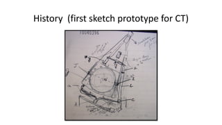
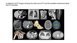
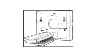
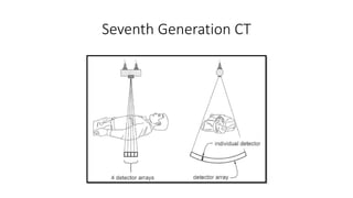
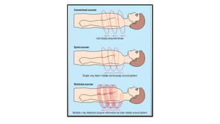
CT Computed Tomography
History
• First lab CT took 9 days to produce a single
image
• 1971 (first commercial CT) by Sir Godfrey
Hounsfield
• 1974 (3rd generation CT)
• 1979 Nobel Prize in physiology, Medicine "for
the development of computer assisted
tomography.“ with Allen Cormack
(theoretical calculations)
• 1994 Spiral CT
• 2007 320-Row Spiral CT
Sir Godfrey Hounsfield
1919- 2004
History (first sketch prototype for CT)
A collection of CT images showing the wide use of CT and the excellent anatomical detail
that CT provides.
Introduction
• Computed Tomography (Tomos means (slice) graphy means (write) in
Greek.
• (CT) scan is a medical device that uses a series of X-ray images taken
from different angles around the patient's body and uses computer
processing to create cross-sectional images (slices) of the internal
body organs.
The Device
• Generators: High frequency generators are small in size and used to
produce electrical energy that sent to x ray tube
• Gantry: The gantry is the donut like or ring shaped part of the CT
scanner. It houses many of the components necessary to produce and
detect x-rays. These components are mounted on a rotating scan
frame.
• X-ray tube: Modern CT scanners make use of slip ring technology in
which high voltage is supplied to the tube through contact rings in
the gantry. X-ray generation is related to rotation angle. CT x-ray
tubes are very expensive, with the price of some tubes exceeding
$200,000.
The Device
• Filtration:
• x-ray tube is positioned perpendicular to the imaging plane to reduce the
heel effect
• typical filtration on a CT x-ray tube is ~6 mm aluminum filter, though we need
heavy filtration in some scans.
• Bow tie filters attenuate little in the center, but attenuation increases with
increasing distance from the central ray, (they reduce scatter and patient
dose)
• Collimators:
• located at the x-ray tube as well as at the x-ray detectors.
• defines the section thickness on a single-slice scanner.
• also help reduce the amount of scatter radiation reaching the CT detectors.
The Device
• Radiation detectors:
• Energy integrating detectors: Most recent commercial CT detectors consist of a
scintillator crystal in combination with a photodiode. Scintillator material converts X-rays into
visible light, which then hits the photodiode, causing it to produce an electric current. The
multichannel readout electronics or data acquisition system (DAS) connects to the photodiode.
DAS converts the electric charge signal from diode to voltage using a transimpedance amplifier
and perform analog to digital conversion.
• Photon counting detectors: direct conversion material (such as cadmium telluride or
cadmium-zinc-telluride) converts an X-ray photon into a certain electronic charge proportional
to its energy. The charge produced in direct conversion is about ten times that produced by the
scintillator/ photodiode combination and the electronic noise no longer dominates the signal
from individual X-rays. Remaining challenges for commercial introduction of direct conversion
detectors for CT applications include stability and the count rate limits, and therefore it will be
several years before scintillator based detectors will be replaced on commercial CT scanners.
The Device
The Device
• Image reconstruction:
• Preprocessing
• Back-projection: logarithm
conversion based on tissue attenuation
• Filtered Back-projection
• Fourier-Based Reconstruction
First Generation CT
• Single detector, single x-ray tube,
rotate/translate pencil beam system,
rotation angle/step 1°
• About 6 Min to complete a single
scan.
Second Generation CT
• Linear array of about 30 detectors,
single x-ray tube, rotate/translate
motion, narrow angle (10 ° ) fan beam
• rotation angle/step 10°
• Shortest scan time was about 18 s per
slice
Third Generation CT
• Linear array of about 800 detectors,
single x-ray tube, rotate/rotate
motion only, wide fan beam to cover
the entire patient
• Scan time of newer scanners is about
½ s per slice
• Can produce ring artifacts
Fourth Generation CT
• Complete circular array of about 4800
stationary detectors
• Single x-ray tube rotates with in the
circular array of detectors
• Wide fan beam to cover the entire
patient
• Scan time of newer scanners is about ½ s
per slice
• Designed to address ring artifacts
Fifth Generation CT
• Scan time is about 50 ms per slice
• Developed for cardiac tomographic imaging
Sixth Generation CT
• Design: x-ray tube rotates as patient is moved smoothly into x-ray
scan field
• Simultaneous source rotation, table translation and data acquisition
• Produces one continuous volume set of data for entire region
• Data for multiple slices from patient acquired at 1sec/slice
• In some instances the entire scan be done within a single breath-hold
of the patient
Sixth Generation CT
Seventh Generation CT
• Cone Beam & multiple parallel rows of detectors
• Widened (z-direction) x ray beam & detector array to acquire multiple
(4-64-320 slices simultaneously)
• Very short scan time
Seventh Generation CT
Radiation Dose
Radiation Dose
Future Expectations
• CT will remain an important modality for the visualization of the skeleton,
calcifications, the lungs and the gastrointestinal tract.
• It will be the only alternative for patients with implants who are not allowed to
enter the MR room.
• Until recently all manufacturers were competing to have the largest number of
detector rows, also referred to as the slice wars.
• From a technical viewpoint the tendency is toward dose reduction, increased
volume coverage, higher contrast-to-noise ratio and improved spatial and
temporal resolution.
Thank you

Ct computed tomography

  • 1.
  • 2.
    History • First labCT took 9 days to produce a single image • 1971 (first commercial CT) by Sir Godfrey Hounsfield • 1974 (3rd generation CT) • 1979 Nobel Prize in physiology, Medicine "for the development of computer assisted tomography.“ with Allen Cormack (theoretical calculations) • 1994 Spiral CT • 2007 320-Row Spiral CT Sir Godfrey Hounsfield 1919- 2004
  • 3.
    History (first sketchprototype for CT)
  • 4.
    A collection ofCT images showing the wide use of CT and the excellent anatomical detail that CT provides.
  • 5.
    Introduction • Computed Tomography(Tomos means (slice) graphy means (write) in Greek. • (CT) scan is a medical device that uses a series of X-ray images taken from different angles around the patient's body and uses computer processing to create cross-sectional images (slices) of the internal body organs.
  • 6.
    The Device • Generators:High frequency generators are small in size and used to produce electrical energy that sent to x ray tube • Gantry: The gantry is the donut like or ring shaped part of the CT scanner. It houses many of the components necessary to produce and detect x-rays. These components are mounted on a rotating scan frame. • X-ray tube: Modern CT scanners make use of slip ring technology in which high voltage is supplied to the tube through contact rings in the gantry. X-ray generation is related to rotation angle. CT x-ray tubes are very expensive, with the price of some tubes exceeding $200,000.
  • 7.
    The Device • Filtration: •x-ray tube is positioned perpendicular to the imaging plane to reduce the heel effect • typical filtration on a CT x-ray tube is ~6 mm aluminum filter, though we need heavy filtration in some scans. • Bow tie filters attenuate little in the center, but attenuation increases with increasing distance from the central ray, (they reduce scatter and patient dose) • Collimators: • located at the x-ray tube as well as at the x-ray detectors. • defines the section thickness on a single-slice scanner. • also help reduce the amount of scatter radiation reaching the CT detectors.
  • 8.
    The Device • Radiationdetectors: • Energy integrating detectors: Most recent commercial CT detectors consist of a scintillator crystal in combination with a photodiode. Scintillator material converts X-rays into visible light, which then hits the photodiode, causing it to produce an electric current. The multichannel readout electronics or data acquisition system (DAS) connects to the photodiode. DAS converts the electric charge signal from diode to voltage using a transimpedance amplifier and perform analog to digital conversion. • Photon counting detectors: direct conversion material (such as cadmium telluride or cadmium-zinc-telluride) converts an X-ray photon into a certain electronic charge proportional to its energy. The charge produced in direct conversion is about ten times that produced by the scintillator/ photodiode combination and the electronic noise no longer dominates the signal from individual X-rays. Remaining challenges for commercial introduction of direct conversion detectors for CT applications include stability and the count rate limits, and therefore it will be several years before scintillator based detectors will be replaced on commercial CT scanners.
  • 9.
  • 10.
    The Device • Imagereconstruction: • Preprocessing • Back-projection: logarithm conversion based on tissue attenuation • Filtered Back-projection • Fourier-Based Reconstruction
  • 11.
    First Generation CT •Single detector, single x-ray tube, rotate/translate pencil beam system, rotation angle/step 1° • About 6 Min to complete a single scan.
  • 12.
    Second Generation CT •Linear array of about 30 detectors, single x-ray tube, rotate/translate motion, narrow angle (10 ° ) fan beam • rotation angle/step 10° • Shortest scan time was about 18 s per slice
  • 13.
    Third Generation CT •Linear array of about 800 detectors, single x-ray tube, rotate/rotate motion only, wide fan beam to cover the entire patient • Scan time of newer scanners is about ½ s per slice • Can produce ring artifacts
  • 14.
    Fourth Generation CT •Complete circular array of about 4800 stationary detectors • Single x-ray tube rotates with in the circular array of detectors • Wide fan beam to cover the entire patient • Scan time of newer scanners is about ½ s per slice • Designed to address ring artifacts
  • 15.
    Fifth Generation CT •Scan time is about 50 ms per slice • Developed for cardiac tomographic imaging
  • 16.
    Sixth Generation CT •Design: x-ray tube rotates as patient is moved smoothly into x-ray scan field • Simultaneous source rotation, table translation and data acquisition • Produces one continuous volume set of data for entire region • Data for multiple slices from patient acquired at 1sec/slice • In some instances the entire scan be done within a single breath-hold of the patient
  • 17.
  • 19.
    Seventh Generation CT •Cone Beam & multiple parallel rows of detectors • Widened (z-direction) x ray beam & detector array to acquire multiple (4-64-320 slices simultaneously) • Very short scan time
  • 20.
  • 22.
  • 23.
  • 24.
    Future Expectations • CTwill remain an important modality for the visualization of the skeleton, calcifications, the lungs and the gastrointestinal tract. • It will be the only alternative for patients with implants who are not allowed to enter the MR room. • Until recently all manufacturers were competing to have the largest number of detector rows, also referred to as the slice wars. • From a technical viewpoint the tendency is toward dose reduction, increased volume coverage, higher contrast-to-noise ratio and improved spatial and temporal resolution.
  • 25.