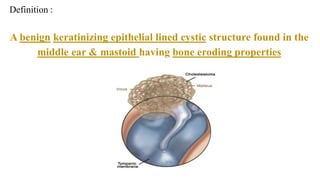
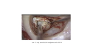
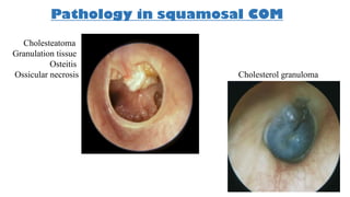
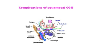
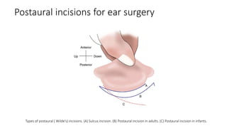
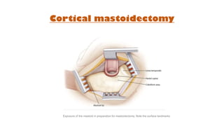
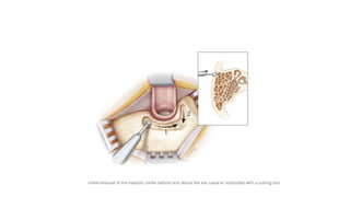
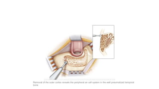
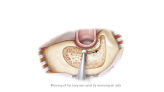
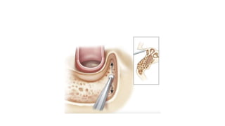
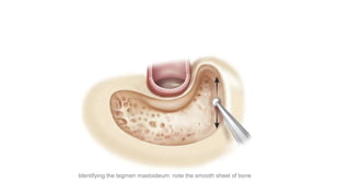
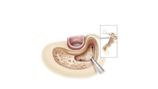
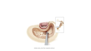
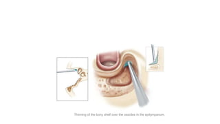
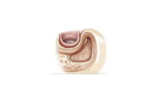
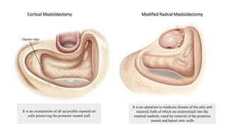
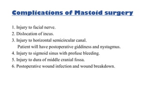
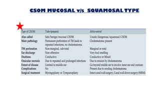
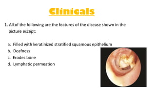
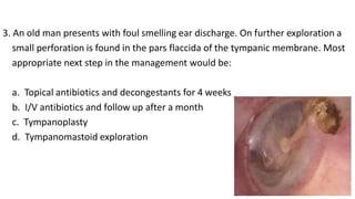
- Dr. Alka Kapil
( Assistant Professor,
Dept. of ENT & Head Neck Surgery )
CHR NIC TITIS MEDIA
Cholesteatoma
 It is a growth of keratinizing squamous epithelium originating from the external layer of the
tympanic membrane or ear canal that invades the middle ear cleft
 Cholesteatoma has two components—
i. the acellular keratin debris, which forms the contents of the sac,
ii. the matrix, which forms the sac itself
 Misnomer : neither cholesterol crystals nor a tumour
 BONE ERODING PROPERTY
 enzymes like collagenases , acid proteases
& acid phosphatases
Definition :
A benign keratinizing epithelial lined cystic structure found in the
middle ear & mastoid having bone eroding properties
Right ear large cholesteatoma filling the mastoid antrum
Theories of Cholesteatoma Pathogenesis
Wittmaack's
Sade's
Ruedi's
Habermann's
Types of Cholesteatoma
Congenital Acquired
Primary Secondary
- Develops Secondary to
perforation
- Hebermann’s theory
- Develops from Congenital
cell rest
Levenson’s Criteria for Congenital cholesteatoma
GENESIS OF PRIMARY AND SECONDARY CHOLESTEATOMAS
Tos Classification of Pars Flaccida Retraction
partial erosion of scutum definite erosion of scutum
PF retraction
not adherent to the malleus
PF retraction
adherent to the malleus
i. In stage 1, the pars flaccida is dimpled and more retracted than
normal but not adherent to the malleus
ii. In stage 2, the retraction is adherent to the neck of the malleus
and the full extent of the retraction can be seen
iii. In stage 3, part of the retraction is out of view and there may be
partial erosion of the bony attic wall
iv. In stage 4, there is definite erosion of the attic wall with the full
extent of the retraction being uncertain because it is out of view
Sadé classification of Pars Tensa retraction
I. Stage I: Retracted tympanic membrane
II. Stage II: Retraction with contact onto the incus
III. Stage III: Middle ear atelectasis
IV. Stage IV: Adhesive otitis media
Pathology in squamosal COM
Cholesteatoma
Granulation tissue
Osteitis
Ossicular necrosis Cholesterol granuloma
Cinical features of Squamosal COM
1. Otorrhea
- A persistent ,scanty ,foul-smelling painless otorrhea is the hallmark of cholesteatoma
- patients not responsive to systemic antibiotics, topical antibiotics may help temporarily
2. Hearing Loss
- A conductive hearing loss is a common finding in cholesteatoma, as ossicular chain erosion is common (70%) ; however,
a relatively good hearing could be present even the ossicular chain is eroded, this is the result of the conductive mass
effect of the cholesteatoma itself ( Cholesteatoma hearers )
- Evidence of sensorineural hearing loss may indicate an involvement of the labyrinth.
3. Vertigo/Imbalance
- A destruction of the bone which overlies the otic capsule, especially the lateral semicircular canal, can trigger vertigo
or a balance dysfunction.
4 . Facial Nerve Palsy
5. Otalgia, headache, vomiting, and fever are not typical presentations of cholesteatoma; however, their occurrence indicates
the possibility of impending intratemporal or intracranial complications.
Complications of squamosal COM
Investigations
and
Assessment
1. Examination under microscope
2.Audiogram - PTA
3. Culture & sensitivity of ear discharge
4. Routine haematological workup
5. Radiology – X ray b/l mastoids Schuller's view
Investigations
and
Assessment
6. High resolution CT scan B/L temporal bone
Investigations
and
Assessment
Treatment
1. Conservative : little role only where surgery is not possible
2. Surgical : Mastoid exploration
- The main aim of the surgery is to give the patient a safe, dry and hearing ear by eradicating
the disease and reconstructing the hearing mechanism
-Types of mastoid surgeries ( Intact canal wall ; Canal wall down )
Postaural incisions for ear surgery
Types of postaural ( Wilde’s) incisions. (A) Sulcus incision. (B) Postaural incision in adults. (C) Postaural incision in infants.
Cortical mastoidectomy
Exposure of the mastoid in preparation for mastoidectomy. Note the surface landmarks
Initial removal of the mastoid cortex behind and above the ear canal is conducted with a cutting burr
Removal of the outer cortex reveals the peripheral air cell system in the well pneumatized temporal
bone
Thinning of the bony ear canal by removing air cells
Identifying the tegmen mastoideum: note the smooth sheet of bone
Initial entry into the mastoid antrum
Thinning of the bony shelf over the ossicles in the epitympanum.
Thinning of air cells over the sigmoid sinus and the sinodural angle (also known as Citelli’s
angle)
Tip cells are opened as necessitated by the extent of pneumatization
Steps of Cortical Mastoidectomy
1. Incision
2. Exposure of lateral surface of mastoid and Macewen’s triangle
3. Removal of mastoid cortex and exposure of antrum
4. Removal of mastoid air cells
5. Removal of mastoid tip and finishing the cavity
6. Closure of wound
Sinodural angle
Posterior wall of EAC
Sinus Plate
Dural
plate
Cortical Mastoidectomy Modified Radical Mastoidectomy
It is an exenteration of all accessible mastoid air
cells preserving the posterior meatal wall
It is an operation to eradicate disease of the attic and
mastoid, both of which are exteriorized into the
external auditory canal by removal of the posterior
meatal and lateral attic walls
Steps of Modified Radical Mastoidectomy
1. Incision
2. Exposure of lateral surface of mastoid and Macewen’s triangle
3. Removal of mastoid cortex and exposure of antrum
4. Removal of diseased tissue & mastoid air cells
5. Posterior wall of EAC is sacrificed : Facial ridge is lowered & bridge is broken ;
anterior & posterior buttress removed
6. Removal of mastoid tip and finishing the cavity
7. Meatoplasty
8. Closure of wound
Complications of Mastoid surgery
1. Injury to facial nerve.
2. Dislocation of incus.
3. Injury to horizontal semicircular canal.
Patient will have postoperative giddiness and nystagmus.
4. Injury to sigmoid sinus with profuse bleeding.
5. Injury to dura of middle cranial fossa.
6. Postoperative wound infection and wound breakdown.
CSOM MUCOSAL v/s SQUAMOSAL TYPE
Management of chronic otitis media (COM)
Clinicals
1. All of the following are the features of the disease shown in the
picture except:
a. Filled with keratinized stratified squamous epithelium
b. Deafness
c. Erodes bone
d. Lymphatic permeation
2. Cholesteatoma is usually present at:
a. Anteroinferior quadrant of TM
b. Posteroinferior quadrant of TM
c. Attic region
d. Central part
3. Scanty, foul smelling , painless discharge from the ear is characteristic
feature of which of the following lesions:
a. ASOM
b. Cholesteatoma
c. Central perforation
d. Otitis externa
3. An old man presents with foul smelling ear discharge. On further exploration a
small perforation is found in the pars flaccida of the tympanic membrane. Most
appropriate next step in the management would be:
a. Topical antibiotics and decongestants for 4 weeks
b. I/V antibiotics and follow up after a month
c. Tympanoplasty
d. Tympanomastoid exploration
4. A 5-year-old boy has been diagnosed to have posterior superior retraction
pocket. All would constitute part of the management except:
a. Audiometry
b. Mastoid exploration
c. Tympanoplasty
d. Myringoplasty
Thank You

Chronic Otitis Media - Squamosal type ( UG)

  • 1.
    - Dr. AlkaKapil ( Assistant Professor, Dept. of ENT & Head Neck Surgery ) CHR NIC TITIS MEDIA
  • 2.
    Cholesteatoma  It isa growth of keratinizing squamous epithelium originating from the external layer of the tympanic membrane or ear canal that invades the middle ear cleft  Cholesteatoma has two components— i. the acellular keratin debris, which forms the contents of the sac, ii. the matrix, which forms the sac itself  Misnomer : neither cholesterol crystals nor a tumour  BONE ERODING PROPERTY  enzymes like collagenases , acid proteases & acid phosphatases
  • 3.
    Definition : A benignkeratinizing epithelial lined cystic structure found in the middle ear & mastoid having bone eroding properties
  • 4.
    Right ear largecholesteatoma filling the mastoid antrum
  • 5.
    Theories of CholesteatomaPathogenesis Wittmaack's Sade's Ruedi's Habermann's
  • 6.
    Types of Cholesteatoma CongenitalAcquired Primary Secondary - Develops Secondary to perforation - Hebermann’s theory - Develops from Congenital cell rest
  • 7.
    Levenson’s Criteria forCongenital cholesteatoma
  • 8.
    GENESIS OF PRIMARYAND SECONDARY CHOLESTEATOMAS
  • 9.
    Tos Classification ofPars Flaccida Retraction partial erosion of scutum definite erosion of scutum PF retraction not adherent to the malleus PF retraction adherent to the malleus
  • 10.
    i. In stage1, the pars flaccida is dimpled and more retracted than normal but not adherent to the malleus ii. In stage 2, the retraction is adherent to the neck of the malleus and the full extent of the retraction can be seen iii. In stage 3, part of the retraction is out of view and there may be partial erosion of the bony attic wall iv. In stage 4, there is definite erosion of the attic wall with the full extent of the retraction being uncertain because it is out of view
  • 11.
    Sadé classification ofPars Tensa retraction I. Stage I: Retracted tympanic membrane II. Stage II: Retraction with contact onto the incus III. Stage III: Middle ear atelectasis IV. Stage IV: Adhesive otitis media
  • 12.
    Pathology in squamosalCOM Cholesteatoma Granulation tissue Osteitis Ossicular necrosis Cholesterol granuloma
  • 13.
    Cinical features ofSquamosal COM 1. Otorrhea - A persistent ,scanty ,foul-smelling painless otorrhea is the hallmark of cholesteatoma - patients not responsive to systemic antibiotics, topical antibiotics may help temporarily 2. Hearing Loss - A conductive hearing loss is a common finding in cholesteatoma, as ossicular chain erosion is common (70%) ; however, a relatively good hearing could be present even the ossicular chain is eroded, this is the result of the conductive mass effect of the cholesteatoma itself ( Cholesteatoma hearers ) - Evidence of sensorineural hearing loss may indicate an involvement of the labyrinth. 3. Vertigo/Imbalance - A destruction of the bone which overlies the otic capsule, especially the lateral semicircular canal, can trigger vertigo or a balance dysfunction. 4 . Facial Nerve Palsy 5. Otalgia, headache, vomiting, and fever are not typical presentations of cholesteatoma; however, their occurrence indicates the possibility of impending intratemporal or intracranial complications.
  • 14.
  • 15.
  • 16.
    3. Culture &sensitivity of ear discharge 4. Routine haematological workup 5. Radiology – X ray b/l mastoids Schuller's view Investigations and Assessment
  • 17.
    6. High resolutionCT scan B/L temporal bone Investigations and Assessment
  • 18.
    Treatment 1. Conservative :little role only where surgery is not possible 2. Surgical : Mastoid exploration - The main aim of the surgery is to give the patient a safe, dry and hearing ear by eradicating the disease and reconstructing the hearing mechanism -Types of mastoid surgeries ( Intact canal wall ; Canal wall down )
  • 19.
    Postaural incisions forear surgery Types of postaural ( Wilde’s) incisions. (A) Sulcus incision. (B) Postaural incision in adults. (C) Postaural incision in infants.
  • 20.
    Cortical mastoidectomy Exposure ofthe mastoid in preparation for mastoidectomy. Note the surface landmarks
  • 21.
    Initial removal ofthe mastoid cortex behind and above the ear canal is conducted with a cutting burr
  • 22.
    Removal of theouter cortex reveals the peripheral air cell system in the well pneumatized temporal bone
  • 23.
    Thinning of thebony ear canal by removing air cells
  • 25.
    Identifying the tegmenmastoideum: note the smooth sheet of bone
  • 27.
    Initial entry intothe mastoid antrum
  • 28.
    Thinning of thebony shelf over the ossicles in the epitympanum.
  • 30.
    Thinning of aircells over the sigmoid sinus and the sinodural angle (also known as Citelli’s angle)
  • 31.
    Tip cells areopened as necessitated by the extent of pneumatization
  • 33.
    Steps of CorticalMastoidectomy 1. Incision 2. Exposure of lateral surface of mastoid and Macewen’s triangle 3. Removal of mastoid cortex and exposure of antrum 4. Removal of mastoid air cells 5. Removal of mastoid tip and finishing the cavity 6. Closure of wound Sinodural angle Posterior wall of EAC Sinus Plate Dural plate
  • 34.
    Cortical Mastoidectomy ModifiedRadical Mastoidectomy It is an exenteration of all accessible mastoid air cells preserving the posterior meatal wall It is an operation to eradicate disease of the attic and mastoid, both of which are exteriorized into the external auditory canal by removal of the posterior meatal and lateral attic walls
  • 35.
    Steps of ModifiedRadical Mastoidectomy 1. Incision 2. Exposure of lateral surface of mastoid and Macewen’s triangle 3. Removal of mastoid cortex and exposure of antrum 4. Removal of diseased tissue & mastoid air cells 5. Posterior wall of EAC is sacrificed : Facial ridge is lowered & bridge is broken ; anterior & posterior buttress removed 6. Removal of mastoid tip and finishing the cavity 7. Meatoplasty 8. Closure of wound
  • 37.
    Complications of Mastoidsurgery 1. Injury to facial nerve. 2. Dislocation of incus. 3. Injury to horizontal semicircular canal. Patient will have postoperative giddiness and nystagmus. 4. Injury to sigmoid sinus with profuse bleeding. 5. Injury to dura of middle cranial fossa. 6. Postoperative wound infection and wound breakdown.
  • 38.
    CSOM MUCOSAL v/sSQUAMOSAL TYPE
  • 39.
    Management of chronicotitis media (COM)
  • 40.
    Clinicals 1. All ofthe following are the features of the disease shown in the picture except: a. Filled with keratinized stratified squamous epithelium b. Deafness c. Erodes bone d. Lymphatic permeation
  • 41.
    2. Cholesteatoma isusually present at: a. Anteroinferior quadrant of TM b. Posteroinferior quadrant of TM c. Attic region d. Central part
  • 42.
    3. Scanty, foulsmelling , painless discharge from the ear is characteristic feature of which of the following lesions: a. ASOM b. Cholesteatoma c. Central perforation d. Otitis externa
  • 43.
    3. An oldman presents with foul smelling ear discharge. On further exploration a small perforation is found in the pars flaccida of the tympanic membrane. Most appropriate next step in the management would be: a. Topical antibiotics and decongestants for 4 weeks b. I/V antibiotics and follow up after a month c. Tympanoplasty d. Tympanomastoid exploration
  • 44.
    4. A 5-year-oldboy has been diagnosed to have posterior superior retraction pocket. All would constitute part of the management except: a. Audiometry b. Mastoid exploration c. Tympanoplasty d. Myringoplasty
  • 45.