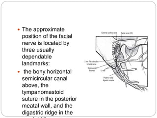
Dr surbhi
Patna medical college
Open Cavity Mastoid Operations
 Open cavity procedures can be broadly defined
as those requiring the removal of the posterior
wall of the external auditory canal.
 These procedures are identified by many
names—canal wall down mastoidectomy,
modified radical mastoidectomy, radical
mastoidectomy, and the Bondy mastoidectomy—
depending on how the middle ear and the
disease are managed
 The purpose of open cavity procedures is to
exteriorize the mastoid cavity for future monitoring
of recurrent cholesteatoma, provide drainage for
unresectable temporal bone infection, and,
occasionally, provide exposure for difficult-to-
access areas of the temporal bone.
HISTORICAL NOTES
 In 1873, Von Tröltsch was the first surgeon to
suggest that Schwartze’s simple mastoidectomy
technique needed to be modified to reduce
persistent otorrhea after initial surgery.
 He had observed that remnants of cholesteatoma
in the attic, antrum, or mastoid process would
invariably result in chronic drainage.
 Von Bergmann applied the term “radical” to any
case in which the posterior and superior bony
canal walls were removed to develop an open
cavity.
 . In 1890, Zaufal described in detail the technique
of the radical mastoidectomy to eradicate disease
in the middle ear and mastoid. This operation
converted the attic, antrum, mastoid process,
tympanum, and external auditory canal into a
common “radical cavity” that could be inspected
and cleaned for the rest of the patient’s life.
 Hearing, however, was at times quite good in
patients who presented with cholesteatoma
confined to the attic in which the pars tensa of the
tympanic membrane was intact. Körner
recognized this situation in 1899 and suggested
that the tympanic membrane and ossicles could
be left in place during radical operations in certain
cases of chronic otitis.
 In 1910, Bondy
described the
indications and
technique for a
modification of the
radical operation in
cases involving a pars
flaccida perforation with
an intact pars tensa.
 In this technique, the
superior osseous
meatal wall and a
portion of the posterior
osseous meatal wall
were removed without
disturbing the intact
tympanic membrane
(except for the attic
 This technique thus
exteriorized the attic
and antral
cholesteatoma into a
permanently open
“modified radical”
cavity that could be
cleaned through the
external meatus
without further
destroying hearing.
INDICATIONS FOR THE CLASSIC RADICAL
MASTOID OPERATION
 1. Unresectable cholesteatoma extending down
the eustachian tube or into the petrous apex
 2. Promontory cochlear fistula caused by
cholesteatoma
 3. Chronic perilabyrinthine osteitis or
cholesteatoma that cannot be removed and must
be cleaned or inspected periodically
 4. Resection of temporal bone neoplasms with
periodic monitoring
INDICATIONS FOR MODIFIED RADICAL
MASTOIDECTOMY
 Modified radical mastoidectomy is an effective
method to manage cholesteatoma in a single-
stage approach
Absolute indications :
 unresectable disease,
 an unreconstructable posterior canal wall,
 failure of a first-stage canal wall up procedure
because of poor eustachian tube function,
 and inadequate patient follow-up.
The relative indications
 disease in an only hearing ear or in a dead ear,
medical illness, severe otologic or central nervous
system complications, and neoplasms
 is poor eustachian tube function.
Conservative management of
cholesteatoma
 Conservative management of cholesteatoma can
be attempted when the attic defect is large and
the cholesteatoma sac shallow, allowing the
accumulated desquamated debris to be removed
by microdébridement and suction.
Conservative management is
contraindicated
 1. Radiographic evidence of an enlarged,
smoothwalled antrum indicates a large
cholesteatoma cavity.
 2. Otorrhea persists after several cleanings.
 3. A very small attic perforation makes cleaning
painful, difficult, and unsatisfactory.
 4. Cholesteatoma is observed behind the pars
tensa.
 5. There are symptoms or signs of erosion of
vital structures such as the fallopian canal,
semicircular canals, cochlea, or dura.
 6. Hearing loss, either conductive or
sensorineural, indicating progression of
cholesteatoma.
 7. The patient is uncooperative or is
geographically unable to return for necessary
management.
CONTRAINDICATIONS FOR THE
OPEN CAVITY MASTOID
OPERATION
 cases of chronic otitis media without
cholesteatoma
 cases of acute otitis media with coalescent
mastoiditis, persistent secretory otitis media, or
chronic allergic otitis media.
 . Relative contraindications for open cavity
procedures include wide exposure of the sigmoid
sinus, dura, and the facial nerve caused by
aggressive disease.
RADICAL MASTOIDECTOMY AND BONDY
MODIFIED RADICAL MASTOIDECTOMY
 The techniques of the radical mastoidectomy and
the Bondy radical mastoidectomy are presented
for historical interest and perspective.
 The objective of these procedures was to remove
safely all bone-invading disease; create an
accessible, exteriorized cavity for lifelong cleaning
and care; and promote epithelialization of the
cavity with healthy skin.
 Hearing improvement was of secondary
importance.
 The radical and Bondy operations began with
exposure of the attic and antrum, followed by
removal of the superior and posterior canal walls.
By performing the “inside-out” mastoidectomy, the
resultant cavity was smaller than if a complete
mastoidectomy with tympanoplasty were
performed.
 However, as a result of this approach, peripheral
air cells were isolated from the eustachian tube.
 If the mucosa continued to produce mucus, it
discharged into the mastoid cavity.
All matrix is removed, with the
following exceptions:
 1. Matrix firmly adherent to exposed dura or
sigmoid sinus may be left rather than risk injury to
these structures.
 2. Matrix over a fistula of a semicircular canal
may be left to avoid postoperative serous
labyrinthitis.
 3. Matrix firmly attached to exposed facial nerve
may be left
 4. Matrix extending into the mesotympanum and
covering the stapes footplate may be left
Bone Removal beyond
Cholesteatoma
 Remembering that chronic otorrhea is the result
of infected epidermal debris in the cholesteatoma
sac, in most cases, evacuation of the sac,
removal of matrix (epithelial lining), and curettage
of softened osteitic bone adjacent to the matrix
suffice to control the disease. The surgeon needs
to exercise prudent judgment with regard to
mastoid cells outside the cholesteatoma sac.
 These cells may be infected and osteitic
(softened), with granulations requiring removal,
but in many cases, mastoid cells are intact and
need not be removed.
Taking Down the Bridge and the Facial Ridge
 The remaining superior osseous meatal wall
bridging the notch of Rivinus is removed in small
bites
 With a small (000) curet, always working outward
away from the fallopian canal and facial nerve,
the anterior and posterior spines of the notch of
Rivinus, composing the anterior and posterior
buttresses of the bridge, are taken down.
 Wherever cholesteatoma envelops or extends
onto the medial surface of the malleus head or
incus, these ossicles must be removed.
 When cholesteatoma matrix lies against and
lateral to these ossicles, the matrix may be left or
carefully removed and the ossicles left
undisturbed.
 When the long process of the incus is absent and
matrix lies against the mobile stapes head, with
excellent hearing producing nature’s
myringostapediopexy, this portion of the matrix is
left undisturbed.
 The step in the radical
or Bondy operations
most often
accomplished poorly
is taking down the
posterior osseous
meatal wall, which,
deeper in, houses the
posterior bend and
vertical facial nerve
and thus is called the
facial ridge.
 The approximate
position of the facial
nerve is located by
three usually
dependable
landmarks:
 the bony horizontal
semicircular canal
above, the
tympanomastoid
suture in the posterior
meatal wall, and the
digastric ridge in the
 The bony facial ridge is taken down slowly and
carefully with a drill or curet, working under the
operating microscope, always parallel to and
never across the direction of the facial nerve.
 A pinkish color and bleeding are encountered
when the facial nerve is approached. It is better
not to expose the nerve unnecessarily
MODIFIED RADICAL
MASTOIDECTOMY
 Modified radical mastoidectomy, also known as
complete mastoidectomy and tympanoplasty,
complete removal of the posterior canal wall with
sealing of the middle ear space to avoid chronic
drainage from exposed mucous membrane
 The modification of the radical procedure (ie,
adding the technique of tympanoplasty)
potentially eliminates the expected intermittent
discharge from the middle ear mucosa. Hearing, it
should be noted, is a secondary consideration of
the modified radical procedure.
Preoperative Assessment
 The decision to perform a modified radical
mastoidectomy rather than a staged intact canal
wall approach depends on the extent and location
of the disease, previous surgery, eustachian tube
function and patient age, medical condition, and
aftercare preference.
 Extensive destruction of the posterior canal wall
with obvious cholesteatoma invading the mastoid
indicates the need for modified radical
mastoidectomy.
 Active suppuration should be controlled prior to
surgery whenever possible.
 Acetic acid (1.5% solution) irrigations followed by
antibiotic otic drops should be instituted for
several weeks prior to surgery
Meatoplasty
 One percent lidocaine with 1:100,000 epinephrine
is infiltrated into the conchal bowl.
 The entire posterior aspect of the conchal bowl is
exposed using sharp dissection with an iris
scissors through the fibrous periosteum and soft
tissue.
 With a finger in the conchal bowl, a semilunar
incision is made into the cartilage posteriorly until
the knife tip is felt through the anterior skin.
 This crescent-shaped cartilage measures about
1.5 2cm
 A Körner flap is now
developed by making
incisions through the
external auditory canal
skin.
 An inferior incision is
begun in the inferior
canal at 6 o’clock,
carried into the conchal
bowl, and curved
around the inferior
margin of the bowl.
 A superior incision is
made at 12 o’clock and
carried between the
tragus and the anterior
helix.
 These incisions create
a long (vascular strip)
flap that is based in
the posterosuperior
aspect of the conchal
bowl and will
constitute the back
wall of the mastoid
cavity
Postoperative Care
 At the first postoperative visit, any area that has
not been grafted is covered by a layer of
granulation tissue. Exuberant granulation tissue
should be débrided and treated with silver nitrate.
 The granulation tissue should then be painted
with 2% gentian violet and the patient instructed
to use antibiotic otic drops two or three times per
day until the next (at 2 to 3 weeks) visit.
 Drainage decreases with ensuing visits as re-
epithelialization occurs. As epithelialization
progresses, acetic acid irrigations can replace the
use of antibiotic otic drops.
 Once the cavity is healed, the patient should
return for a yearly visit and is given full water
sport privileges.
COMPLICATIONS OF OPEN CAVITY
PROCEDURES
 deafness or further hearing loss, facial paralysis,
vestibular symptoms, cerebrospinal fluid leak,
infection, and recurrent cholesteatoma or
drainage.
 Facial nerve paralysis is the most common major
complication associated with open cavity
procedures.
 The second most common complication of open
cavity procedures is wound infection.
 This infection usually results in perichondritis of
the auricle, manifested by a painful, swollen
auricle with copious discharge. Pseudomonas
aeruginosa is the causative organism.
 A “chocolate” or mucous retention cyst can occur
in a healed mastoid cavity as a result of a
collection of serum within a mucous membrane–
lined pocket.
 Cholesteatoma recurrence in open cavity
procedures occurs is usually caused by
inaccessible disease or a remnant of matrix that
was amputated at the time of surgery
 Recurrent aural drainage from a previously
healed and dry cavity is usually the result of poor
aural toilet.
conclusion
 Open cavity mastoid procedures are indicated
when canal wall up procedures are inadequate to
control disease.
 The vast majority of these procedures will result
in a modified radical mastoidectomy.
 Identification of the facial nerve is critical in this
procedure.
 Lowering of the facial ridge to the level of the
facial nerve and development of a large external
auditory meatus are mandatory for successful
outcome.
 Long-term postoperative care is minimal, with
Thank you

Open cavity mastoid operations

  • 1.
    Dr surbhi Patna medicalcollege Open Cavity Mastoid Operations
  • 2.
     Open cavityprocedures can be broadly defined as those requiring the removal of the posterior wall of the external auditory canal.  These procedures are identified by many names—canal wall down mastoidectomy, modified radical mastoidectomy, radical mastoidectomy, and the Bondy mastoidectomy— depending on how the middle ear and the disease are managed
  • 3.
     The purposeof open cavity procedures is to exteriorize the mastoid cavity for future monitoring of recurrent cholesteatoma, provide drainage for unresectable temporal bone infection, and, occasionally, provide exposure for difficult-to- access areas of the temporal bone.
  • 4.
    HISTORICAL NOTES  In1873, Von Tröltsch was the first surgeon to suggest that Schwartze’s simple mastoidectomy technique needed to be modified to reduce persistent otorrhea after initial surgery.  He had observed that remnants of cholesteatoma in the attic, antrum, or mastoid process would invariably result in chronic drainage.
  • 5.
     Von Bergmannapplied the term “radical” to any case in which the posterior and superior bony canal walls were removed to develop an open cavity.
  • 6.
     . In1890, Zaufal described in detail the technique of the radical mastoidectomy to eradicate disease in the middle ear and mastoid. This operation converted the attic, antrum, mastoid process, tympanum, and external auditory canal into a common “radical cavity” that could be inspected and cleaned for the rest of the patient’s life.
  • 7.
     Hearing, however,was at times quite good in patients who presented with cholesteatoma confined to the attic in which the pars tensa of the tympanic membrane was intact. Körner recognized this situation in 1899 and suggested that the tympanic membrane and ossicles could be left in place during radical operations in certain cases of chronic otitis.
  • 8.
     In 1910,Bondy described the indications and technique for a modification of the radical operation in cases involving a pars flaccida perforation with an intact pars tensa.  In this technique, the superior osseous meatal wall and a portion of the posterior osseous meatal wall were removed without disturbing the intact tympanic membrane (except for the attic
  • 9.
     This techniquethus exteriorized the attic and antral cholesteatoma into a permanently open “modified radical” cavity that could be cleaned through the external meatus without further destroying hearing.
  • 10.
    INDICATIONS FOR THECLASSIC RADICAL MASTOID OPERATION  1. Unresectable cholesteatoma extending down the eustachian tube or into the petrous apex  2. Promontory cochlear fistula caused by cholesteatoma  3. Chronic perilabyrinthine osteitis or cholesteatoma that cannot be removed and must be cleaned or inspected periodically  4. Resection of temporal bone neoplasms with periodic monitoring
  • 11.
    INDICATIONS FOR MODIFIEDRADICAL MASTOIDECTOMY  Modified radical mastoidectomy is an effective method to manage cholesteatoma in a single- stage approach Absolute indications :  unresectable disease,  an unreconstructable posterior canal wall,  failure of a first-stage canal wall up procedure because of poor eustachian tube function,  and inadequate patient follow-up.
  • 12.
    The relative indications disease in an only hearing ear or in a dead ear, medical illness, severe otologic or central nervous system complications, and neoplasms  is poor eustachian tube function.
  • 13.
    Conservative management of cholesteatoma Conservative management of cholesteatoma can be attempted when the attic defect is large and the cholesteatoma sac shallow, allowing the accumulated desquamated debris to be removed by microdébridement and suction.
  • 14.
    Conservative management is contraindicated 1. Radiographic evidence of an enlarged, smoothwalled antrum indicates a large cholesteatoma cavity.  2. Otorrhea persists after several cleanings.  3. A very small attic perforation makes cleaning painful, difficult, and unsatisfactory.  4. Cholesteatoma is observed behind the pars tensa.
  • 15.
     5. Thereare symptoms or signs of erosion of vital structures such as the fallopian canal, semicircular canals, cochlea, or dura.  6. Hearing loss, either conductive or sensorineural, indicating progression of cholesteatoma.  7. The patient is uncooperative or is geographically unable to return for necessary management.
  • 16.
    CONTRAINDICATIONS FOR THE OPENCAVITY MASTOID OPERATION  cases of chronic otitis media without cholesteatoma  cases of acute otitis media with coalescent mastoiditis, persistent secretory otitis media, or chronic allergic otitis media.  . Relative contraindications for open cavity procedures include wide exposure of the sigmoid sinus, dura, and the facial nerve caused by aggressive disease.
  • 17.
    RADICAL MASTOIDECTOMY ANDBONDY MODIFIED RADICAL MASTOIDECTOMY  The techniques of the radical mastoidectomy and the Bondy radical mastoidectomy are presented for historical interest and perspective.  The objective of these procedures was to remove safely all bone-invading disease; create an accessible, exteriorized cavity for lifelong cleaning and care; and promote epithelialization of the cavity with healthy skin.  Hearing improvement was of secondary importance.
  • 18.
     The radicaland Bondy operations began with exposure of the attic and antrum, followed by removal of the superior and posterior canal walls. By performing the “inside-out” mastoidectomy, the resultant cavity was smaller than if a complete mastoidectomy with tympanoplasty were performed.  However, as a result of this approach, peripheral air cells were isolated from the eustachian tube.  If the mucosa continued to produce mucus, it discharged into the mastoid cavity.
  • 19.
    All matrix isremoved, with the following exceptions:  1. Matrix firmly adherent to exposed dura or sigmoid sinus may be left rather than risk injury to these structures.  2. Matrix over a fistula of a semicircular canal may be left to avoid postoperative serous labyrinthitis.  3. Matrix firmly attached to exposed facial nerve may be left  4. Matrix extending into the mesotympanum and covering the stapes footplate may be left
  • 20.
    Bone Removal beyond Cholesteatoma Remembering that chronic otorrhea is the result of infected epidermal debris in the cholesteatoma sac, in most cases, evacuation of the sac, removal of matrix (epithelial lining), and curettage of softened osteitic bone adjacent to the matrix suffice to control the disease. The surgeon needs to exercise prudent judgment with regard to mastoid cells outside the cholesteatoma sac.  These cells may be infected and osteitic (softened), with granulations requiring removal, but in many cases, mastoid cells are intact and need not be removed.
  • 21.
    Taking Down theBridge and the Facial Ridge  The remaining superior osseous meatal wall bridging the notch of Rivinus is removed in small bites  With a small (000) curet, always working outward away from the fallopian canal and facial nerve, the anterior and posterior spines of the notch of Rivinus, composing the anterior and posterior buttresses of the bridge, are taken down.
  • 26.
     Wherever cholesteatomaenvelops or extends onto the medial surface of the malleus head or incus, these ossicles must be removed.  When cholesteatoma matrix lies against and lateral to these ossicles, the matrix may be left or carefully removed and the ossicles left undisturbed.  When the long process of the incus is absent and matrix lies against the mobile stapes head, with excellent hearing producing nature’s myringostapediopexy, this portion of the matrix is left undisturbed.
  • 27.
     The stepin the radical or Bondy operations most often accomplished poorly is taking down the posterior osseous meatal wall, which, deeper in, houses the posterior bend and vertical facial nerve and thus is called the facial ridge.
  • 28.
     The approximate positionof the facial nerve is located by three usually dependable landmarks:  the bony horizontal semicircular canal above, the tympanomastoid suture in the posterior meatal wall, and the digastric ridge in the
  • 29.
     The bonyfacial ridge is taken down slowly and carefully with a drill or curet, working under the operating microscope, always parallel to and never across the direction of the facial nerve.  A pinkish color and bleeding are encountered when the facial nerve is approached. It is better not to expose the nerve unnecessarily
  • 30.
    MODIFIED RADICAL MASTOIDECTOMY  Modifiedradical mastoidectomy, also known as complete mastoidectomy and tympanoplasty, complete removal of the posterior canal wall with sealing of the middle ear space to avoid chronic drainage from exposed mucous membrane
  • 31.
     The modificationof the radical procedure (ie, adding the technique of tympanoplasty) potentially eliminates the expected intermittent discharge from the middle ear mucosa. Hearing, it should be noted, is a secondary consideration of the modified radical procedure.
  • 32.
    Preoperative Assessment  Thedecision to perform a modified radical mastoidectomy rather than a staged intact canal wall approach depends on the extent and location of the disease, previous surgery, eustachian tube function and patient age, medical condition, and aftercare preference.
  • 33.
     Extensive destructionof the posterior canal wall with obvious cholesteatoma invading the mastoid indicates the need for modified radical mastoidectomy.  Active suppuration should be controlled prior to surgery whenever possible.  Acetic acid (1.5% solution) irrigations followed by antibiotic otic drops should be instituted for several weeks prior to surgery
  • 34.
    Meatoplasty  One percentlidocaine with 1:100,000 epinephrine is infiltrated into the conchal bowl.  The entire posterior aspect of the conchal bowl is exposed using sharp dissection with an iris scissors through the fibrous periosteum and soft tissue.  With a finger in the conchal bowl, a semilunar incision is made into the cartilage posteriorly until the knife tip is felt through the anterior skin.  This crescent-shaped cartilage measures about 1.5 2cm
  • 35.
     A Körnerflap is now developed by making incisions through the external auditory canal skin.  An inferior incision is begun in the inferior canal at 6 o’clock, carried into the conchal bowl, and curved around the inferior margin of the bowl.  A superior incision is made at 12 o’clock and carried between the tragus and the anterior helix.
  • 36.
     These incisionscreate a long (vascular strip) flap that is based in the posterosuperior aspect of the conchal bowl and will constitute the back wall of the mastoid cavity
  • 37.
    Postoperative Care  Atthe first postoperative visit, any area that has not been grafted is covered by a layer of granulation tissue. Exuberant granulation tissue should be débrided and treated with silver nitrate.  The granulation tissue should then be painted with 2% gentian violet and the patient instructed to use antibiotic otic drops two or three times per day until the next (at 2 to 3 weeks) visit.
  • 38.
     Drainage decreaseswith ensuing visits as re- epithelialization occurs. As epithelialization progresses, acetic acid irrigations can replace the use of antibiotic otic drops.  Once the cavity is healed, the patient should return for a yearly visit and is given full water sport privileges.
  • 39.
    COMPLICATIONS OF OPENCAVITY PROCEDURES  deafness or further hearing loss, facial paralysis, vestibular symptoms, cerebrospinal fluid leak, infection, and recurrent cholesteatoma or drainage.
  • 40.
     Facial nerveparalysis is the most common major complication associated with open cavity procedures.  The second most common complication of open cavity procedures is wound infection.  This infection usually results in perichondritis of the auricle, manifested by a painful, swollen auricle with copious discharge. Pseudomonas aeruginosa is the causative organism.
  • 41.
     A “chocolate”or mucous retention cyst can occur in a healed mastoid cavity as a result of a collection of serum within a mucous membrane– lined pocket.
  • 42.
     Cholesteatoma recurrencein open cavity procedures occurs is usually caused by inaccessible disease or a remnant of matrix that was amputated at the time of surgery
  • 43.
     Recurrent auraldrainage from a previously healed and dry cavity is usually the result of poor aural toilet.
  • 44.
    conclusion  Open cavitymastoid procedures are indicated when canal wall up procedures are inadequate to control disease.  The vast majority of these procedures will result in a modified radical mastoidectomy.  Identification of the facial nerve is critical in this procedure.  Lowering of the facial ridge to the level of the facial nerve and development of a large external auditory meatus are mandatory for successful outcome.  Long-term postoperative care is minimal, with
  • 45.