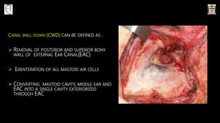
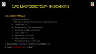
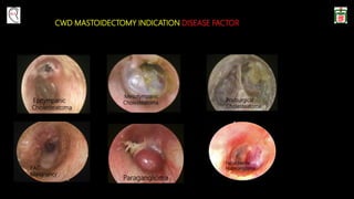
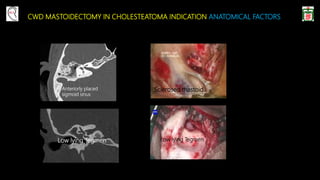
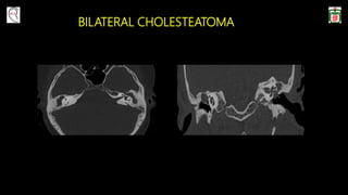
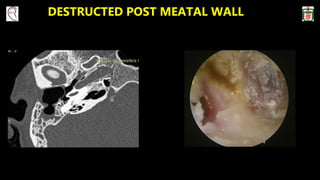
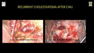
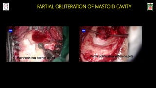
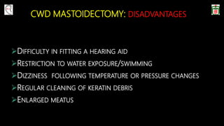
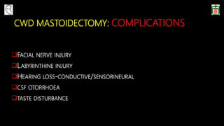
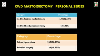
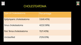
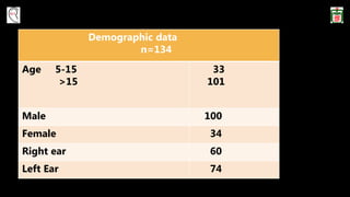
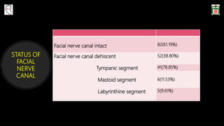
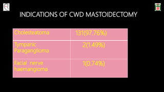
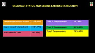
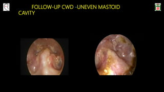
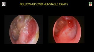
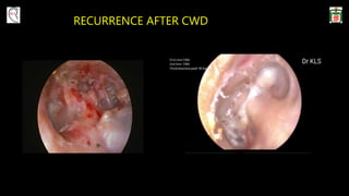
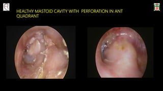
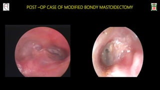
SlideShare a Scribd company logo
CANAL WALL DOWN MASTOIDECTOMY
DR KANU LAL SAHA
ASSOCIATE PROFESSOR
OTOLOGY DIVISION
DEPT. OF OTOLARYNGOLOGY-HEAD AND NECK
SURGERY
BANGABANDHU SHEIKH MUJIB MEDICAL UNIVERSITY
SHAHBAG,DHAKA
CANAL WALL DOWN (CWD) CAN BE DEFINED AS :
 REMOVAL OF POSTERIOR AND SUPERIOR BONY
WALL OF EXTERNAL EAR CANAL(EAC)
 EXENTERATION OF ALL MASTOID AIR CELLS
 CONVERTING MASTOID CAVITY, MIDDLE EAR AND
EAC INTO A SINGLE CAVITY EXTERIORIZED
THROUGH EAC
CWD :SYNONYMOUS OR SUBCLASSIFICATION
 RADICAL MASTOIDECTOMY
 MODIFIED RADICAL MASTOIDECTOMY
 BONDY MASTOIDECTOMY
 MODIFIED BONDY MASTOIDECTOMY(SANNA MODIFICATION)
 ATTICOTOMY
 ATTICOANTROTOMY
OPEN CAVITY MASTOIDECTOMY
CWD MASTOIDECTOMY- INDICATIONS
CHOLESTEATOMA
 CONTRACTED MASTOID
 VERY LOW LYING TEGMEN AND ANTERIORLY PLACED SIGMOID SINUS
 ERODED BONY EAC
 RECURRENCE AFTER CWU MASTOIDECTOMY
 CLEFT PALATE AND DOWN’S SYNDROME
 ONLY HEARING EAR
 BILATERAL CHOLESTEATOMA
 LARGE LABYRINTHINE FISTULA
 SEVERE SENSORINEURAL HEARING LOSS
 SOME BENIGN TUMOURS INVOLVING THE MIDDLE EAR
 SOME MALIGNANCY IN THE EAC
CWD MASTOIDECTOMY INDICATION DISEASE FACTOR
Epitympanic
Cholesteatoma
Mesotympanic
Cholesteatoma
Postsurgical
Cholesteatoma
EAC
Malignancy
Paraganglioma
Facial Nerve
Haemangioma
CWD MASTOIDECTOMY IN CHOLESTEATOMA INDICATION ANATOMICAL FACTORS
Low lying Tegmen
Anteriorly placed
sigmoid sinus
Low lying Tegmen
Sclerosed mastoid
BILATERAL CHOLESTEATOMA
DESTRUCTED POST MEATAL WALL
RECURRENT CHOLESTEATOMA AFTER CWU
LABYRINTHINE FISTULA
CWD IN CHOLESTEATOMA INDICATION PATIENT FACTORS
INVESTIGATION FACILITIES
UNRELIABLE FOLLOW UP
UNWILLING TO UNDERGO A SECOND STAGE PROCEDURE
HIGH RISK FOR GENERAL ANESTHESIA
LACK OF RELIABLE IMAGING FACILITIES
CWD IN OTHER CONDITIONS: NOW REPLACED BY STP
PARAGANGLIOMA
FACIAL NERVE TUMOR
EAC MALIGNANCY
TYMPANIC PARAGANGLIOMA
FACIAL NERVE HAEMANGIOMA
TEMPORAL BONE MALIGNANCY
MODIFIED BONDY MASTOIDECTOMY
THE MODIFIED BONDY MASTOIDECTOMY
 A CWD MASTOIDECTOMY PROCEDURE
 REMOVAL OF POSTERIOR CANAL WALL
 INVOLVES EXTERIORIZING THE LATERAL EPITYMPANIC SPACE AND MASTOID CAVITY
 REMOVAL OF CHOLESTEATOMA
 PRESERVATION OF THE OSSICULAR CHAIN
 PLACING A FASCIA GRAFT ONTO THE MEDIAL ASPECT OF THE OSSICULAR CHAIN
BASIC SURGICAL STEPS
APPROACH:
RETROAURICULAR
ENDAURAL
ROUTES:
TRANSCORTICAL OR OUTSIDE-IN :
DRILLING STARTS ON THE SURFACE OF THE CORTICAL BONE
CORTICAL MASTOIDECTOMY-ANTROTOMY-REMOVAL OF BONY
EAR CANAL.
TRANSMEATAL OR INSIDE-OUT :
DRILLING STARTS IN THE EAR CANAL
ATTICOTOMY-ATTICOANTROSTOMY-MASTOIDECTOMY
RULES OF INITIAL DRILLING
• BONE COVERING THE MIDDLE FOSSA DURA
AND SIGMOID SINUS IS THINNED USING LARGE
BURRS.
• BURRS ARE MOVED PARALLEL TO THE
STRUCTURES.
• CAVITY MUST ALWAYS BE WELL SAUCERIZED AND
GRADUALLY DEEPENED.
• BONY OVERHANGS SHOULD BE REMOVED FROM
THE EDGES.
LOWERING FACIAL RIDGE
• THE FACIAL RIDGE IS LOWERED EVENLY USING A LARGE
CUTTING BURR WITH CONTINUOUS SUCTION IRRIGATION.
• DRILLING PARALLEL TO THE COURSE OF THE FACIAL NERVE.
• DIAMOND BURRS - FINAL DRILLING TO THIN THE BONY
COVERAGE OF THE NERVE.
• THE NERVE IS SKELETONIZED BUT NEVER EXPOSED
• INFERIOR CANAL WALL SHOULD BE CONTOURED
SMOOTHLY IN ITS JUNCTION WITH THE CAVITY.
REMOVAL OF ANTERIOR BUTTRESS AND CANALPLASTY
If prominent protrusion of anterior and inferior wall,
canalplasty should be done.
Meatal skin is cut laterally,detached from the bone
towards annulus and protected with a sheet.
Removal of anterior buttress produces a continuous
plane between middle fossa plate and anterior canal.
MASTOID CAVITY SAUCERIZATION
BY BEVELING THE EDGES OF THE CAVITY (BLUE),
THE LATERAL SOFT TISSUES (RED) ARE INVITED TO
COLLAPSE INWARD, WHICH ACTS TO REDUCE CAVITY
VOLUME.
CONTOURING THE TYMPANIC BONE JUNCTION WITH MASTOID CAVITY
DRILLING OF THE TYMPANIC BONE INFERIOR TO THE
TYMPANIC RING AND ANTERIOR TO THE FACIAL NERVE IS
NECESSARY TO ELIMINATE THE “KIDNEY SHAPE” OF THE
RESULTANT CAVITY AND TO PROVIDE A SMOOTH JUNCTION
BETWEEN THE NATIVE EXTERNAL AUDITORY CANAL AND
MASTOID CAVITY
PARTIAL OBLITERATION OF MASTOID CAVITY
RECONSTRUCTION
 BLEEDING SHOULD BE STOPPED COMPLETELY BEFORE RECONSTRUCTION OF MIDDLE EAR
 USUALLY THE TEMPORALIS FASCIA IS GRAFTED IN UNDERLAY TECHNIQUE
 ANTERIOR END OF GRAFT IS PLACED UNDER THE ANNULUS
 THE GRAFT MUST HAVE ABUNDANT POSTERIOR EXTENSION TO COVER THE WHOLE MIDDLE EAR AND OBLITERATED
CELLS
 IF NECESSARY, ANOTHER PIECE OF GRAFT MAY BE PLACED TO COVER EXPOSED BONE IN MASTOID CAVITY
 EPITHELIALIZATION IS FACILITATED IF THE EXPOSED BONE IS COVERED WITH THE FASCIA
 OSSICULOPLASTY MAY BE DONE IN FIRST-STAGE OR SECOND-STAGE
TYPE 2 TYMPANOPLASTY
TYPE 3 TYMPANOPLASTY
TYPE 3 USING AUTO MALLEUS HEAD TYPE 3 USING TORP
TYPE 3 TYMPANOPLASTY
TYPE-3 USING CARTILAGE TYPE 3 USING MALLEUS SHOE
MEATOPLASTY
 USING NASAL SPECULUM A CONCHAL INCISION IS MADE TOWARD THE ANTIHELIX, PARALLEL TO
CRUS OF THE HELIX
 CONCHAL CARTILAGE IS DISSECTED FROM SKIN AND UNDERLYING CONNECTIVE TISSUE
 A TRIANGULAR PIECE OF CARTILAGE IS REMOVED
 THE AMOUNT OF CARTILAGE TO BE REMOVED DEPENDS ON THE SIZE AND CONTOUR OF THE CAVITY
 IMPORTANT TO PRESERVE THE CARTILAGE IN THE CRUS OF THE HELIX TO PREVENT COSMETIC
DEFORMITY
SERIAL FOLLOW-UP AFTER CWD MASTOIDECTOMY
PROPERLY PERFORMED CANAL WALL DOWN MASTOIDECTOMY
A. SAUCERIZED EDGES
B. MASTOID TIP AMPUTATED FROM DIGASTRIC MUSCLE
C. CELLS DRILLED AWAY UP TO SMOOTH DURAL PLATE
D. FACIAL RIDGE LOWERED
E. TYMPANIC BONE SMOOTHLY FRACTURED WITH
JUNCTION OF MASTOID
F. INTACT TYMPANIC MEMBRANE
G. MIDDLE EAR SEALED FROM EPI
F
E
UNFAVOURABLE MASTOID CAVITY
A. THE STEEP NON-SAUCERIZED EDGES
B. THE POTHOLES FROM RESIDUAL AIR CELLS
C. THE INTACT OPEN MASTOID TIP WITH DEBRIS
D. THE HIGH FACIAL RIDGE
E. THE UNCONTOURED TYMPANIC BONE FORMING A
KIDNEY-SHAPED CAVITY
F. TYMPANIC MEMBRANE PERFORATION
G. UNSEALED COMMUNICATION WITH EPITYMPANUM
A
B
C
D
E
F
G
CAUSES OF FAILURE IN CANAL WALL DOWN MASTOIDECTOMY
POOR EXECUTION OF SURGICAL TECHNIQUE
HIGH FACIAL RIDGE
BONY OVERHANGS
NARROW MEATUS
Anatomical irregularity Percentage
High Facial ridge 67%
Stenotic meatus 64%
Bony overhang 29%
Canal wall down mastoidectomy: causes of failure,
pitfalls and their management. Mario Sanna et al
Journal of laryngology and otology,1995,vol-105
FAILURE IN CANAL WALL DOWN MASTOIDECTOMY :OTHER CAUSES
 PROMINENT OR INACCESSIBLE MASTOID TIP
 RECURRENT RETRACTION OR CHOLESTEATOMA
 REMUCOSALIZATION OF RESIDUAL MASTOID CELL
 VERY LARGE AND UNEVEN MASTOID CAVITY
 OPEN EUSTACHIAN TUBE OR PERFORATED
HIGH FACIAL RIDGE
Bony overhang
BONY OVERHANG
PROMINENT MASTOID TIP
CWD MASTOIDECTOMY: DISADVANTAGES
DIFFICULTY IN FITTING A HEARING AID
RESTRICTION TO WATER EXPOSURE/SWIMMING
DIZZINESS FOLLOWING TEMPERATURE OR PRESSURE CHANGES
REGULAR CLEANING OF KERATIN DEBRIS
ENLARGED MEATUS
CWD MASTOIDECTOMY: COMPLICATIONS
FACIAL NERVE INJURY
LABYRINTHINE INJURY
HEARING LOSS-CONDUCTIVE/SENSORINEURAL
CSF OTORRHOEA
TASTE DISTURBANCE
LONG TERM RESULTS OF CWD
MASTOIDECTOMY
Demographic and clinical data
n=259
Age 2-96(mean-35)
Male 39%
Female 61%
Type III tympanoplasty 175(67.4%)
Recurrent infection in cavity 17(6.5%)
Meatal stenosis 5(1.9%)
Residual cholesteatoma 5(1.9%)
Recurrent cholesteatoma 1(0.4%)
Sensorineural hearing loss 2(0.7%)
Facial paralysis 1(0.3%)
Persistent vertigo 4(1.5%)
Kos et al, Canal Wall-Down Mastoidectomy
Ann Otol Rhinal LaryngoI 113:2004
CWD MASTOIDECTOMY PERSONAL SERIES
Category Percentage
Modified radical mastoidectomy 124 (92.53%)
Modified bondy mastoidectomy 10(7.46%)
Category Percentage
Primary procedure 113(84.33%)
Revision surgery 21(15.67%)
CHOLESTEATOMA
Type of Cholesteatoma Percentage
Epitympanic cholesteatoma 53(40.45%)
Sinus cholesteatoma 42(32.06%)
Pars Tensa cholesteatoma 15(11.45%)
Unclassified 21(16.03%)
Demographic data
n=134
Age 5-15
>15
33
101
Male 100
Female 34
Right ear 60
Left Ear 74
STATUS OF
FACIAL
NERVE
CANAL
Facial nerve canal intact 82(61.19%)
Facial nerve canal dehiscent
Tympanic segment
Mastoid segment
Labyrinthine segment
52(38.80%)
41(78.85%)
6(11.53%)
5(9.61%)
INDICATIONS OF CWD MASTOIDECTOMY
Cholesteatoma 131(97.76%)
Tympanic
Paraganglioma
2(1.49%)
Facial nerve
haemangioma
1(0.74%)
OSSICULAR STATUS AND MIDDLE EAR RECONSTRUCTION
Type 1 Tympanoplasty 10(7.46%)
Type 2 Tympanoplasty 51(38.47%)
Type 3 Tympanoplasty 73(54.47%)
Stapes suprastructure present 51(38.05%)
Stapes suprastructure absent 73(54.47%)
Intact ossicular chain 10(7.46%)
FOLLOW-UP CWD CASE - PRIMARY SURGERY
Pre-op
FOLLOW- UP CWD CASE – AFTER REVISION
Post-op
FOLLOW-UP CWD -UNEVEN MASTOID
CAVITY
FOLLOW-UP CWD –UNSTABLE CAVITY
RECURRENCE AFTER CWD
HEALTHY MASTOID CAVITY WITH PERFORATION IN ANT
QUADRANT
POST –OP CASE OF MODIFIED BONDY MASTOIDECTOMY
CWD MASTOIDECTOMY CHALLENGES
PROPER EXECUTION OF SURGICAL TECHNIQUE
KEEP THE PATIENT IN REGULAR FOLLOW UP
OSSICULOPLASTY AND OPTIMUM HEARING IMPROVEMENT IN ONLY MOBILE
FOOTPLATE
CONCLUSION
 IT IS NOT WHICH TECHNIQUE ONE USES BUT HOW WELL ONE USES THE TECHNIQUE –THAT IS, THE INDIVIDUAL
SURGEON’S JUDGEMENT AND ABILITY -SHEEHY
 CWD IS RATHER MORE CRITICIZED OR DEFAMED FOR FAILURE THAN IT IS PERFECTLY PERFORMED
 A PERFECTLY PERFORMED CWD MASTOIDECTOMY RESULTS IN A TROUBLE-FREE, DRY EAR
 IN DEVELOPING COUNTRY LIKE US WHERE REGULAR FOLLOW-UP AND TECHNICAL SUPPORTS ARE LIMITED
CWD IS THE SINGLE STAGE SAFE PROCEDURE
THANK YOU
Otology Dr Kanu BSMMU Bangladesh
drklsaha@gmail.com
www.drkanuotology.com

More Related Content

What's hot

Sinus tympani prof dr bikash
Sinus tympani prof dr bikashSinus tympani prof dr bikash
Sinus tympani prof dr bikash
Bikash Shrestha
 
surgical management of ototsclerosis
surgical management of ototsclerosissurgical management of ototsclerosis
surgical management of ototsclerosis
Priyanka Shastri
 
11 surgery for otosclerosis.ppt copy
11 surgery for otosclerosis.ppt   copy11 surgery for otosclerosis.ppt   copy
11 surgery for otosclerosis.ppt copy
social service
 
Ossiculoplasty
OssiculoplastyOssiculoplasty
Ossiculoplasty
Md Roohia
 
Cholesteatoma: General Considerations by dr rajendra singh lakhawat
Cholesteatoma: General Considerations by dr rajendra singh lakhawatCholesteatoma: General Considerations by dr rajendra singh lakhawat
Cholesteatoma: General Considerations by dr rajendra singh lakhawat
Dr. Rajendra Singh Lakhawat
 
Thyroplasty
ThyroplastyThyroplasty
Mastoidectomy; Types, Indications, Procedure
Mastoidectomy; Types, Indications, ProcedureMastoidectomy; Types, Indications, Procedure
Mastoidectomy; Types, Indications, Procedure
Prasanna Datta
 
Ossicluloplasty in detail
Ossicluloplasty in detailOssicluloplasty in detail
Ossicluloplasty in detail
Abhineet Jain
 
Inner ear malformations and Implantation
Inner ear malformations and ImplantationInner ear malformations and Implantation
Inner ear malformations and Implantation
Utkal Mishra
 
OME-Otitis Media with effusion in children
OME-Otitis Media with effusion in childrenOME-Otitis Media with effusion in children
OME-Otitis Media with effusion in children
Razal M
 
Fisch approaches Dr Zeeshan Ahmad
Fisch approaches Dr Zeeshan AhmadFisch approaches Dr Zeeshan Ahmad
Fisch approaches Dr Zeeshan Ahmad
Dr Zeeshan Ahmad
 
Canal wall up Mastoidectomy ( Intact Bridge Mastoidectomy) by Dr.Aditya Tiwari
Canal wall up Mastoidectomy ( Intact Bridge Mastoidectomy) by Dr.Aditya TiwariCanal wall up Mastoidectomy ( Intact Bridge Mastoidectomy) by Dr.Aditya Tiwari
Canal wall up Mastoidectomy ( Intact Bridge Mastoidectomy) by Dr.Aditya Tiwari
Aditya Tiwari
 
Stapes presentation
Stapes presentationStapes presentation
Stapes presentation
Yasha Gupta
 
Fess complications
Fess complicationsFess complications
Fess complications
Balasubramanian Thiagarajan
 
Infratemporal fossa approaches
Infratemporal fossa approachesInfratemporal fossa approaches
Infratemporal fossa approaches
Md Roohia
 
The pneumatic system of the temporal bone
The pneumatic system of the temporal boneThe pneumatic system of the temporal bone
The pneumatic system of the temporal bone
Surbhi narayan
 
Endoscope assisted middle ear surgery
Endoscope assisted middle ear surgeryEndoscope assisted middle ear surgery
Endoscope assisted middle ear surgery
Ram Raju
 
Mastoid surgeries KK .ppt
Mastoid surgeries KK .pptMastoid surgeries KK .ppt
Mastoid surgeries KK .ppt
DrKrishnaKoiralaENT
 
Hadad.bassagasteguy flap
Hadad.bassagasteguy flap Hadad.bassagasteguy flap
Hadad.bassagasteguy flap
Chandra Veer Suryavanshi
 

What's hot (20)

Sinus tympani prof dr bikash
Sinus tympani prof dr bikashSinus tympani prof dr bikash
Sinus tympani prof dr bikash
 
surgical management of ototsclerosis
surgical management of ototsclerosissurgical management of ototsclerosis
surgical management of ototsclerosis
 
11 surgery for otosclerosis.ppt copy
11 surgery for otosclerosis.ppt   copy11 surgery for otosclerosis.ppt   copy
11 surgery for otosclerosis.ppt copy
 
Ossiculoplasty
OssiculoplastyOssiculoplasty
Ossiculoplasty
 
Cholesteatoma: General Considerations by dr rajendra singh lakhawat
Cholesteatoma: General Considerations by dr rajendra singh lakhawatCholesteatoma: General Considerations by dr rajendra singh lakhawat
Cholesteatoma: General Considerations by dr rajendra singh lakhawat
 
Thyroplasty
ThyroplastyThyroplasty
Thyroplasty
 
Mastoidectomy; Types, Indications, Procedure
Mastoidectomy; Types, Indications, ProcedureMastoidectomy; Types, Indications, Procedure
Mastoidectomy; Types, Indications, Procedure
 
Ossicluloplasty in detail
Ossicluloplasty in detailOssicluloplasty in detail
Ossicluloplasty in detail
 
Atticotmy
AtticotmyAtticotmy
Atticotmy
 
Inner ear malformations and Implantation
Inner ear malformations and ImplantationInner ear malformations and Implantation
Inner ear malformations and Implantation
 
OME-Otitis Media with effusion in children
OME-Otitis Media with effusion in childrenOME-Otitis Media with effusion in children
OME-Otitis Media with effusion in children
 
Fisch approaches Dr Zeeshan Ahmad
Fisch approaches Dr Zeeshan AhmadFisch approaches Dr Zeeshan Ahmad
Fisch approaches Dr Zeeshan Ahmad
 
Canal wall up Mastoidectomy ( Intact Bridge Mastoidectomy) by Dr.Aditya Tiwari
Canal wall up Mastoidectomy ( Intact Bridge Mastoidectomy) by Dr.Aditya TiwariCanal wall up Mastoidectomy ( Intact Bridge Mastoidectomy) by Dr.Aditya Tiwari
Canal wall up Mastoidectomy ( Intact Bridge Mastoidectomy) by Dr.Aditya Tiwari
 
Stapes presentation
Stapes presentationStapes presentation
Stapes presentation
 
Fess complications
Fess complicationsFess complications
Fess complications
 
Infratemporal fossa approaches
Infratemporal fossa approachesInfratemporal fossa approaches
Infratemporal fossa approaches
 
The pneumatic system of the temporal bone
The pneumatic system of the temporal boneThe pneumatic system of the temporal bone
The pneumatic system of the temporal bone
 
Endoscope assisted middle ear surgery
Endoscope assisted middle ear surgeryEndoscope assisted middle ear surgery
Endoscope assisted middle ear surgery
 
Mastoid surgeries KK .ppt
Mastoid surgeries KK .pptMastoid surgeries KK .ppt
Mastoid surgeries KK .ppt
 
Hadad.bassagasteguy flap
Hadad.bassagasteguy flap Hadad.bassagasteguy flap
Hadad.bassagasteguy flap
 

Similar to Canal Wall Down Mastoidectomy(MRM)

Approach to a patient with cholesteatoma
Approach to a patient with cholesteatoma Approach to a patient with cholesteatoma
Approach to a patient with cholesteatoma
Dr Safika Zaman
 
Open cavity mastoid operations
Open cavity mastoid operationsOpen cavity mastoid operations
Open cavity mastoid operations
Surbhi narayan
 
Chronic Otitis Media - Squamosal type ( UG)
Chronic Otitis Media - Squamosal type ( UG)Chronic Otitis Media - Squamosal type ( UG)
Chronic Otitis Media - Squamosal type ( UG)
AlkaKapil
 
Mastoidectomy
MastoidectomyMastoidectomy
Mastoidectomy
Kartik Mittal
 
Mastoidectomy
MastoidectomyMastoidectomy
Mastoidectomy
Kartik Mittal
 
Temporal bone dissection (house)
Temporal bone dissection (house) Temporal bone dissection (house)
Temporal bone dissection (house)
Prasanna Datta
 
Surgical management of vestibular schwannoma by drdhiru456
Surgical management of vestibular schwannoma by drdhiru456Surgical management of vestibular schwannoma by drdhiru456
Surgical management of vestibular schwannoma by drdhiru456
Dr Dhirendra Patil
 
Sinus lift procedures.pptx
Sinus lift procedures.pptxSinus lift procedures.pptx
Sinus lift procedures.pptx
NAMITHA ANAND
 
Nasal septum and its diseases
Nasal septum and its diseasesNasal septum and its diseases
Nasal septum and its diseasesVinay Bhat
 
Mastoidectomy (by drdhiru456)
Mastoidectomy (by drdhiru456)Mastoidectomy (by drdhiru456)
Mastoidectomy (by drdhiru456)
Dr Dhirendra Patil
 
Maxillary reconstruction ih
Maxillary reconstruction  ihMaxillary reconstruction  ih
Maxillary reconstruction ih
itrat hussain
 
Mid facial fractures ( Mid facial trauma )
Mid facial fractures ( Mid facial trauma ) Mid facial fractures ( Mid facial trauma )
Mid facial fractures ( Mid facial trauma )
Ahmed Elgamal
 
caudal devation correction.pptx
caudal devation correction.pptxcaudal devation correction.pptx
caudal devation correction.pptx
EmanZayed17
 
Parotid Gland
Parotid GlandParotid Gland
Parotid Gland
VarshaEsakki
 
Surgical approaches to the facial skeleton
Surgical approaches to the facial skeletonSurgical approaches to the facial skeleton
Surgical approaches to the facial skeleton
Abhishek Roy
 
ATTICO-ANTRAL CSOM
ATTICO-ANTRAL CSOMATTICO-ANTRAL CSOM
ATTICO-ANTRAL CSOM
Dr Harjitpal Singh
 
Diseases of the nasal septum
Diseases of the nasal septumDiseases of the nasal septum
Diseases of the nasal septum
Dr Asmatullah Achakzai
 
Sinus Lift ppt about maxillary sinus lift
Sinus Lift ppt about maxillary sinus liftSinus Lift ppt about maxillary sinus lift
Sinus Lift ppt about maxillary sinus lift
senthilnathanhl
 
Surgical anatomy of Salivary glands - ORAL AND MAXILLOFACIAL SURGERY
Surgical anatomy of Salivary glands - ORAL AND MAXILLOFACIAL SURGERYSurgical anatomy of Salivary glands - ORAL AND MAXILLOFACIAL SURGERY
Surgical anatomy of Salivary glands - ORAL AND MAXILLOFACIAL SURGERY
ANIKET SARKAR
 

Similar to Canal Wall Down Mastoidectomy(MRM) (20)

Approach to a patient with cholesteatoma
Approach to a patient with cholesteatoma Approach to a patient with cholesteatoma
Approach to a patient with cholesteatoma
 
Open cavity mastoid operations
Open cavity mastoid operationsOpen cavity mastoid operations
Open cavity mastoid operations
 
Chronic Otitis Media - Squamosal type ( UG)
Chronic Otitis Media - Squamosal type ( UG)Chronic Otitis Media - Squamosal type ( UG)
Chronic Otitis Media - Squamosal type ( UG)
 
Mastoidectomy
MastoidectomyMastoidectomy
Mastoidectomy
 
Mastoidectomy
MastoidectomyMastoidectomy
Mastoidectomy
 
Temporal bone dissection (house)
Temporal bone dissection (house) Temporal bone dissection (house)
Temporal bone dissection (house)
 
Surgical management of vestibular schwannoma by drdhiru456
Surgical management of vestibular schwannoma by drdhiru456Surgical management of vestibular schwannoma by drdhiru456
Surgical management of vestibular schwannoma by drdhiru456
 
Sinus lift procedures.pptx
Sinus lift procedures.pptxSinus lift procedures.pptx
Sinus lift procedures.pptx
 
Nasal septum and its diseases
Nasal septum and its diseasesNasal septum and its diseases
Nasal septum and its diseases
 
Mastoidectomy (by drdhiru456)
Mastoidectomy (by drdhiru456)Mastoidectomy (by drdhiru456)
Mastoidectomy (by drdhiru456)
 
Maxillary reconstruction ih
Maxillary reconstruction  ihMaxillary reconstruction  ih
Maxillary reconstruction ih
 
Mid facial fractures ( Mid facial trauma )
Mid facial fractures ( Mid facial trauma ) Mid facial fractures ( Mid facial trauma )
Mid facial fractures ( Mid facial trauma )
 
caudal devation correction.pptx
caudal devation correction.pptxcaudal devation correction.pptx
caudal devation correction.pptx
 
Parotid Gland
Parotid GlandParotid Gland
Parotid Gland
 
Surgical approaches to the facial skeleton
Surgical approaches to the facial skeletonSurgical approaches to the facial skeleton
Surgical approaches to the facial skeleton
 
External rhinoplasty
External rhinoplastyExternal rhinoplasty
External rhinoplasty
 
ATTICO-ANTRAL CSOM
ATTICO-ANTRAL CSOMATTICO-ANTRAL CSOM
ATTICO-ANTRAL CSOM
 
Diseases of the nasal septum
Diseases of the nasal septumDiseases of the nasal septum
Diseases of the nasal septum
 
Sinus Lift ppt about maxillary sinus lift
Sinus Lift ppt about maxillary sinus liftSinus Lift ppt about maxillary sinus lift
Sinus Lift ppt about maxillary sinus lift
 
Surgical anatomy of Salivary glands - ORAL AND MAXILLOFACIAL SURGERY
Surgical anatomy of Salivary glands - ORAL AND MAXILLOFACIAL SURGERYSurgical anatomy of Salivary glands - ORAL AND MAXILLOFACIAL SURGERY
Surgical anatomy of Salivary glands - ORAL AND MAXILLOFACIAL SURGERY
 

Recently uploaded

Physiology of Chemical Sensation of smell.pdf
Physiology of Chemical Sensation of smell.pdfPhysiology of Chemical Sensation of smell.pdf
Physiology of Chemical Sensation of smell.pdf
MedicoseAcademics
 
Physiology of Special Chemical Sensation of Taste
Physiology of Special Chemical Sensation of TastePhysiology of Special Chemical Sensation of Taste
Physiology of Special Chemical Sensation of Taste
MedicoseAcademics
 
KDIGO 2024 guidelines for diabetologists
KDIGO 2024 guidelines for diabetologistsKDIGO 2024 guidelines for diabetologists
KDIGO 2024 guidelines for diabetologists
د.محمود نجيب
 
Pharynx and Clinical Correlations BY Dr.Rabia Inam Gandapore.pptx
Pharynx and Clinical Correlations BY Dr.Rabia Inam Gandapore.pptxPharynx and Clinical Correlations BY Dr.Rabia Inam Gandapore.pptx
Pharynx and Clinical Correlations BY Dr.Rabia Inam Gandapore.pptx
Dr. Rabia Inam Gandapore
 
Alcohol_Dr. Jeenal Mistry MD Pharmacology.pdf
Alcohol_Dr. Jeenal Mistry MD Pharmacology.pdfAlcohol_Dr. Jeenal Mistry MD Pharmacology.pdf
Alcohol_Dr. Jeenal Mistry MD Pharmacology.pdf
Dr Jeenal Mistry
 
Novas diretrizes da OMS para os cuidados perinatais de mais qualidade
Novas diretrizes da OMS para os cuidados perinatais de mais qualidadeNovas diretrizes da OMS para os cuidados perinatais de mais qualidade
Novas diretrizes da OMS para os cuidados perinatais de mais qualidade
Prof. Marcus Renato de Carvalho
 
Lung Cancer: Artificial Intelligence, Synergetics, Complex System Analysis, S...
Lung Cancer: Artificial Intelligence, Synergetics, Complex System Analysis, S...Lung Cancer: Artificial Intelligence, Synergetics, Complex System Analysis, S...
Lung Cancer: Artificial Intelligence, Synergetics, Complex System Analysis, S...
Oleg Kshivets
 
ANATOMY AND PHYSIOLOGY OF URINARY SYSTEM.pptx
ANATOMY AND PHYSIOLOGY OF URINARY SYSTEM.pptxANATOMY AND PHYSIOLOGY OF URINARY SYSTEM.pptx
ANATOMY AND PHYSIOLOGY OF URINARY SYSTEM.pptx
Swetaba Besh
 
Superficial & Deep Fascia of the NECK.pptx
Superficial & Deep Fascia of the NECK.pptxSuperficial & Deep Fascia of the NECK.pptx
Superficial & Deep Fascia of the NECK.pptx
Dr. Rabia Inam Gandapore
 
Ocular injury ppt Upendra pal optometrist upums saifai etawah
Ocular injury  ppt  Upendra pal  optometrist upums saifai etawahOcular injury  ppt  Upendra pal  optometrist upums saifai etawah
Ocular injury ppt Upendra pal optometrist upums saifai etawah
pal078100
 
263778731218 Abortion Clinic /Pills In Harare ,
263778731218 Abortion Clinic /Pills In Harare ,263778731218 Abortion Clinic /Pills In Harare ,
263778731218 Abortion Clinic /Pills In Harare ,
sisternakatoto
 
The Normal Electrocardiogram - Part I of II
The Normal Electrocardiogram - Part I of IIThe Normal Electrocardiogram - Part I of II
The Normal Electrocardiogram - Part I of II
MedicoseAcademics
 
HOT NEW PRODUCT! BIG SALES FAST SHIPPING NOW FROM CHINA!! EU KU DB BK substit...
HOT NEW PRODUCT! BIG SALES FAST SHIPPING NOW FROM CHINA!! EU KU DB BK substit...HOT NEW PRODUCT! BIG SALES FAST SHIPPING NOW FROM CHINA!! EU KU DB BK substit...
HOT NEW PRODUCT! BIG SALES FAST SHIPPING NOW FROM CHINA!! EU KU DB BK substit...
GL Anaacs
 
How STIs Influence the Development of Pelvic Inflammatory Disease.pptx
How STIs Influence the Development of Pelvic Inflammatory Disease.pptxHow STIs Influence the Development of Pelvic Inflammatory Disease.pptx
How STIs Influence the Development of Pelvic Inflammatory Disease.pptx
FFragrant
 
basicmodesofventilation2022-220313203758.pdf
basicmodesofventilation2022-220313203758.pdfbasicmodesofventilation2022-220313203758.pdf
basicmodesofventilation2022-220313203758.pdf
aljamhori teaching hospital
 
Cervical & Brachial Plexus By Dr. RIG.pptx
Cervical & Brachial Plexus By Dr. RIG.pptxCervical & Brachial Plexus By Dr. RIG.pptx
Cervical & Brachial Plexus By Dr. RIG.pptx
Dr. Rabia Inam Gandapore
 
How to Give Better Lectures: Some Tips for Doctors
How to Give Better Lectures: Some Tips for DoctorsHow to Give Better Lectures: Some Tips for Doctors
How to Give Better Lectures: Some Tips for Doctors
LanceCatedral
 
NVBDCP.pptx Nation vector borne disease control program
NVBDCP.pptx Nation vector borne disease control programNVBDCP.pptx Nation vector borne disease control program
NVBDCP.pptx Nation vector borne disease control program
Sapna Thakur
 
ACUTE SCROTUM.....pdf. ACUTE SCROTAL CONDITIOND
ACUTE SCROTUM.....pdf. ACUTE SCROTAL CONDITIONDACUTE SCROTUM.....pdf. ACUTE SCROTAL CONDITIOND
ACUTE SCROTUM.....pdf. ACUTE SCROTAL CONDITIOND
DR SETH JOTHAM
 
ARTHROLOGY PPT NCISM SYLLABUS AYURVEDA STUDENTS
ARTHROLOGY PPT NCISM SYLLABUS AYURVEDA STUDENTSARTHROLOGY PPT NCISM SYLLABUS AYURVEDA STUDENTS
ARTHROLOGY PPT NCISM SYLLABUS AYURVEDA STUDENTS
Dr. Vinay Pareek
 

Recently uploaded (20)

Physiology of Chemical Sensation of smell.pdf
Physiology of Chemical Sensation of smell.pdfPhysiology of Chemical Sensation of smell.pdf
Physiology of Chemical Sensation of smell.pdf
 
Physiology of Special Chemical Sensation of Taste
Physiology of Special Chemical Sensation of TastePhysiology of Special Chemical Sensation of Taste
Physiology of Special Chemical Sensation of Taste
 
KDIGO 2024 guidelines for diabetologists
KDIGO 2024 guidelines for diabetologistsKDIGO 2024 guidelines for diabetologists
KDIGO 2024 guidelines for diabetologists
 
Pharynx and Clinical Correlations BY Dr.Rabia Inam Gandapore.pptx
Pharynx and Clinical Correlations BY Dr.Rabia Inam Gandapore.pptxPharynx and Clinical Correlations BY Dr.Rabia Inam Gandapore.pptx
Pharynx and Clinical Correlations BY Dr.Rabia Inam Gandapore.pptx
 
Alcohol_Dr. Jeenal Mistry MD Pharmacology.pdf
Alcohol_Dr. Jeenal Mistry MD Pharmacology.pdfAlcohol_Dr. Jeenal Mistry MD Pharmacology.pdf
Alcohol_Dr. Jeenal Mistry MD Pharmacology.pdf
 
Novas diretrizes da OMS para os cuidados perinatais de mais qualidade
Novas diretrizes da OMS para os cuidados perinatais de mais qualidadeNovas diretrizes da OMS para os cuidados perinatais de mais qualidade
Novas diretrizes da OMS para os cuidados perinatais de mais qualidade
 
Lung Cancer: Artificial Intelligence, Synergetics, Complex System Analysis, S...
Lung Cancer: Artificial Intelligence, Synergetics, Complex System Analysis, S...Lung Cancer: Artificial Intelligence, Synergetics, Complex System Analysis, S...
Lung Cancer: Artificial Intelligence, Synergetics, Complex System Analysis, S...
 
ANATOMY AND PHYSIOLOGY OF URINARY SYSTEM.pptx
ANATOMY AND PHYSIOLOGY OF URINARY SYSTEM.pptxANATOMY AND PHYSIOLOGY OF URINARY SYSTEM.pptx
ANATOMY AND PHYSIOLOGY OF URINARY SYSTEM.pptx
 
Superficial & Deep Fascia of the NECK.pptx
Superficial & Deep Fascia of the NECK.pptxSuperficial & Deep Fascia of the NECK.pptx
Superficial & Deep Fascia of the NECK.pptx
 
Ocular injury ppt Upendra pal optometrist upums saifai etawah
Ocular injury  ppt  Upendra pal  optometrist upums saifai etawahOcular injury  ppt  Upendra pal  optometrist upums saifai etawah
Ocular injury ppt Upendra pal optometrist upums saifai etawah
 
263778731218 Abortion Clinic /Pills In Harare ,
263778731218 Abortion Clinic /Pills In Harare ,263778731218 Abortion Clinic /Pills In Harare ,
263778731218 Abortion Clinic /Pills In Harare ,
 
The Normal Electrocardiogram - Part I of II
The Normal Electrocardiogram - Part I of IIThe Normal Electrocardiogram - Part I of II
The Normal Electrocardiogram - Part I of II
 
HOT NEW PRODUCT! BIG SALES FAST SHIPPING NOW FROM CHINA!! EU KU DB BK substit...
HOT NEW PRODUCT! BIG SALES FAST SHIPPING NOW FROM CHINA!! EU KU DB BK substit...HOT NEW PRODUCT! BIG SALES FAST SHIPPING NOW FROM CHINA!! EU KU DB BK substit...
HOT NEW PRODUCT! BIG SALES FAST SHIPPING NOW FROM CHINA!! EU KU DB BK substit...
 
How STIs Influence the Development of Pelvic Inflammatory Disease.pptx
How STIs Influence the Development of Pelvic Inflammatory Disease.pptxHow STIs Influence the Development of Pelvic Inflammatory Disease.pptx
How STIs Influence the Development of Pelvic Inflammatory Disease.pptx
 
basicmodesofventilation2022-220313203758.pdf
basicmodesofventilation2022-220313203758.pdfbasicmodesofventilation2022-220313203758.pdf
basicmodesofventilation2022-220313203758.pdf
 
Cervical & Brachial Plexus By Dr. RIG.pptx
Cervical & Brachial Plexus By Dr. RIG.pptxCervical & Brachial Plexus By Dr. RIG.pptx
Cervical & Brachial Plexus By Dr. RIG.pptx
 
How to Give Better Lectures: Some Tips for Doctors
How to Give Better Lectures: Some Tips for DoctorsHow to Give Better Lectures: Some Tips for Doctors
How to Give Better Lectures: Some Tips for Doctors
 
NVBDCP.pptx Nation vector borne disease control program
NVBDCP.pptx Nation vector borne disease control programNVBDCP.pptx Nation vector borne disease control program
NVBDCP.pptx Nation vector borne disease control program
 
ACUTE SCROTUM.....pdf. ACUTE SCROTAL CONDITIOND
ACUTE SCROTUM.....pdf. ACUTE SCROTAL CONDITIONDACUTE SCROTUM.....pdf. ACUTE SCROTAL CONDITIOND
ACUTE SCROTUM.....pdf. ACUTE SCROTAL CONDITIOND
 
ARTHROLOGY PPT NCISM SYLLABUS AYURVEDA STUDENTS
ARTHROLOGY PPT NCISM SYLLABUS AYURVEDA STUDENTSARTHROLOGY PPT NCISM SYLLABUS AYURVEDA STUDENTS
ARTHROLOGY PPT NCISM SYLLABUS AYURVEDA STUDENTS
 

Canal Wall Down Mastoidectomy(MRM)

  • 1. CANAL WALL DOWN MASTOIDECTOMY DR KANU LAL SAHA ASSOCIATE PROFESSOR OTOLOGY DIVISION DEPT. OF OTOLARYNGOLOGY-HEAD AND NECK SURGERY BANGABANDHU SHEIKH MUJIB MEDICAL UNIVERSITY SHAHBAG,DHAKA
  • 2. CANAL WALL DOWN (CWD) CAN BE DEFINED AS :  REMOVAL OF POSTERIOR AND SUPERIOR BONY WALL OF EXTERNAL EAR CANAL(EAC)  EXENTERATION OF ALL MASTOID AIR CELLS  CONVERTING MASTOID CAVITY, MIDDLE EAR AND EAC INTO A SINGLE CAVITY EXTERIORIZED THROUGH EAC
  • 3. CWD :SYNONYMOUS OR SUBCLASSIFICATION  RADICAL MASTOIDECTOMY  MODIFIED RADICAL MASTOIDECTOMY  BONDY MASTOIDECTOMY  MODIFIED BONDY MASTOIDECTOMY(SANNA MODIFICATION)  ATTICOTOMY  ATTICOANTROTOMY OPEN CAVITY MASTOIDECTOMY
  • 4. CWD MASTOIDECTOMY- INDICATIONS CHOLESTEATOMA  CONTRACTED MASTOID  VERY LOW LYING TEGMEN AND ANTERIORLY PLACED SIGMOID SINUS  ERODED BONY EAC  RECURRENCE AFTER CWU MASTOIDECTOMY  CLEFT PALATE AND DOWN’S SYNDROME  ONLY HEARING EAR  BILATERAL CHOLESTEATOMA  LARGE LABYRINTHINE FISTULA  SEVERE SENSORINEURAL HEARING LOSS  SOME BENIGN TUMOURS INVOLVING THE MIDDLE EAR  SOME MALIGNANCY IN THE EAC
  • 5. CWD MASTOIDECTOMY INDICATION DISEASE FACTOR Epitympanic Cholesteatoma Mesotympanic Cholesteatoma Postsurgical Cholesteatoma EAC Malignancy Paraganglioma Facial Nerve Haemangioma
  • 6. CWD MASTOIDECTOMY IN CHOLESTEATOMA INDICATION ANATOMICAL FACTORS Low lying Tegmen Anteriorly placed sigmoid sinus Low lying Tegmen Sclerosed mastoid
  • 11. CWD IN CHOLESTEATOMA INDICATION PATIENT FACTORS INVESTIGATION FACILITIES UNRELIABLE FOLLOW UP UNWILLING TO UNDERGO A SECOND STAGE PROCEDURE HIGH RISK FOR GENERAL ANESTHESIA LACK OF RELIABLE IMAGING FACILITIES
  • 12. CWD IN OTHER CONDITIONS: NOW REPLACED BY STP PARAGANGLIOMA FACIAL NERVE TUMOR EAC MALIGNANCY
  • 16. MODIFIED BONDY MASTOIDECTOMY THE MODIFIED BONDY MASTOIDECTOMY  A CWD MASTOIDECTOMY PROCEDURE  REMOVAL OF POSTERIOR CANAL WALL  INVOLVES EXTERIORIZING THE LATERAL EPITYMPANIC SPACE AND MASTOID CAVITY  REMOVAL OF CHOLESTEATOMA  PRESERVATION OF THE OSSICULAR CHAIN  PLACING A FASCIA GRAFT ONTO THE MEDIAL ASPECT OF THE OSSICULAR CHAIN
  • 17. BASIC SURGICAL STEPS APPROACH: RETROAURICULAR ENDAURAL ROUTES: TRANSCORTICAL OR OUTSIDE-IN : DRILLING STARTS ON THE SURFACE OF THE CORTICAL BONE CORTICAL MASTOIDECTOMY-ANTROTOMY-REMOVAL OF BONY EAR CANAL. TRANSMEATAL OR INSIDE-OUT : DRILLING STARTS IN THE EAR CANAL ATTICOTOMY-ATTICOANTROSTOMY-MASTOIDECTOMY
  • 18. RULES OF INITIAL DRILLING • BONE COVERING THE MIDDLE FOSSA DURA AND SIGMOID SINUS IS THINNED USING LARGE BURRS. • BURRS ARE MOVED PARALLEL TO THE STRUCTURES. • CAVITY MUST ALWAYS BE WELL SAUCERIZED AND GRADUALLY DEEPENED. • BONY OVERHANGS SHOULD BE REMOVED FROM THE EDGES.
  • 19. LOWERING FACIAL RIDGE • THE FACIAL RIDGE IS LOWERED EVENLY USING A LARGE CUTTING BURR WITH CONTINUOUS SUCTION IRRIGATION. • DRILLING PARALLEL TO THE COURSE OF THE FACIAL NERVE. • DIAMOND BURRS - FINAL DRILLING TO THIN THE BONY COVERAGE OF THE NERVE. • THE NERVE IS SKELETONIZED BUT NEVER EXPOSED • INFERIOR CANAL WALL SHOULD BE CONTOURED SMOOTHLY IN ITS JUNCTION WITH THE CAVITY.
  • 20. REMOVAL OF ANTERIOR BUTTRESS AND CANALPLASTY If prominent protrusion of anterior and inferior wall, canalplasty should be done. Meatal skin is cut laterally,detached from the bone towards annulus and protected with a sheet. Removal of anterior buttress produces a continuous plane between middle fossa plate and anterior canal.
  • 21. MASTOID CAVITY SAUCERIZATION BY BEVELING THE EDGES OF THE CAVITY (BLUE), THE LATERAL SOFT TISSUES (RED) ARE INVITED TO COLLAPSE INWARD, WHICH ACTS TO REDUCE CAVITY VOLUME.
  • 22. CONTOURING THE TYMPANIC BONE JUNCTION WITH MASTOID CAVITY DRILLING OF THE TYMPANIC BONE INFERIOR TO THE TYMPANIC RING AND ANTERIOR TO THE FACIAL NERVE IS NECESSARY TO ELIMINATE THE “KIDNEY SHAPE” OF THE RESULTANT CAVITY AND TO PROVIDE A SMOOTH JUNCTION BETWEEN THE NATIVE EXTERNAL AUDITORY CANAL AND MASTOID CAVITY
  • 23. PARTIAL OBLITERATION OF MASTOID CAVITY
  • 24. RECONSTRUCTION  BLEEDING SHOULD BE STOPPED COMPLETELY BEFORE RECONSTRUCTION OF MIDDLE EAR  USUALLY THE TEMPORALIS FASCIA IS GRAFTED IN UNDERLAY TECHNIQUE  ANTERIOR END OF GRAFT IS PLACED UNDER THE ANNULUS  THE GRAFT MUST HAVE ABUNDANT POSTERIOR EXTENSION TO COVER THE WHOLE MIDDLE EAR AND OBLITERATED CELLS  IF NECESSARY, ANOTHER PIECE OF GRAFT MAY BE PLACED TO COVER EXPOSED BONE IN MASTOID CAVITY  EPITHELIALIZATION IS FACILITATED IF THE EXPOSED BONE IS COVERED WITH THE FASCIA  OSSICULOPLASTY MAY BE DONE IN FIRST-STAGE OR SECOND-STAGE
  • 26. TYPE 3 TYMPANOPLASTY TYPE 3 USING AUTO MALLEUS HEAD TYPE 3 USING TORP
  • 27. TYPE 3 TYMPANOPLASTY TYPE-3 USING CARTILAGE TYPE 3 USING MALLEUS SHOE
  • 28. MEATOPLASTY  USING NASAL SPECULUM A CONCHAL INCISION IS MADE TOWARD THE ANTIHELIX, PARALLEL TO CRUS OF THE HELIX  CONCHAL CARTILAGE IS DISSECTED FROM SKIN AND UNDERLYING CONNECTIVE TISSUE  A TRIANGULAR PIECE OF CARTILAGE IS REMOVED  THE AMOUNT OF CARTILAGE TO BE REMOVED DEPENDS ON THE SIZE AND CONTOUR OF THE CAVITY  IMPORTANT TO PRESERVE THE CARTILAGE IN THE CRUS OF THE HELIX TO PREVENT COSMETIC DEFORMITY
  • 29. SERIAL FOLLOW-UP AFTER CWD MASTOIDECTOMY
  • 30. PROPERLY PERFORMED CANAL WALL DOWN MASTOIDECTOMY A. SAUCERIZED EDGES B. MASTOID TIP AMPUTATED FROM DIGASTRIC MUSCLE C. CELLS DRILLED AWAY UP TO SMOOTH DURAL PLATE D. FACIAL RIDGE LOWERED E. TYMPANIC BONE SMOOTHLY FRACTURED WITH JUNCTION OF MASTOID F. INTACT TYMPANIC MEMBRANE G. MIDDLE EAR SEALED FROM EPI F E
  • 31. UNFAVOURABLE MASTOID CAVITY A. THE STEEP NON-SAUCERIZED EDGES B. THE POTHOLES FROM RESIDUAL AIR CELLS C. THE INTACT OPEN MASTOID TIP WITH DEBRIS D. THE HIGH FACIAL RIDGE E. THE UNCONTOURED TYMPANIC BONE FORMING A KIDNEY-SHAPED CAVITY F. TYMPANIC MEMBRANE PERFORATION G. UNSEALED COMMUNICATION WITH EPITYMPANUM A B C D E F G
  • 32. CAUSES OF FAILURE IN CANAL WALL DOWN MASTOIDECTOMY POOR EXECUTION OF SURGICAL TECHNIQUE HIGH FACIAL RIDGE BONY OVERHANGS NARROW MEATUS Anatomical irregularity Percentage High Facial ridge 67% Stenotic meatus 64% Bony overhang 29% Canal wall down mastoidectomy: causes of failure, pitfalls and their management. Mario Sanna et al Journal of laryngology and otology,1995,vol-105
  • 33. FAILURE IN CANAL WALL DOWN MASTOIDECTOMY :OTHER CAUSES  PROMINENT OR INACCESSIBLE MASTOID TIP  RECURRENT RETRACTION OR CHOLESTEATOMA  REMUCOSALIZATION OF RESIDUAL MASTOID CELL  VERY LARGE AND UNEVEN MASTOID CAVITY  OPEN EUSTACHIAN TUBE OR PERFORATED
  • 37. CWD MASTOIDECTOMY: DISADVANTAGES DIFFICULTY IN FITTING A HEARING AID RESTRICTION TO WATER EXPOSURE/SWIMMING DIZZINESS FOLLOWING TEMPERATURE OR PRESSURE CHANGES REGULAR CLEANING OF KERATIN DEBRIS ENLARGED MEATUS
  • 38. CWD MASTOIDECTOMY: COMPLICATIONS FACIAL NERVE INJURY LABYRINTHINE INJURY HEARING LOSS-CONDUCTIVE/SENSORINEURAL CSF OTORRHOEA TASTE DISTURBANCE
  • 39. LONG TERM RESULTS OF CWD MASTOIDECTOMY Demographic and clinical data n=259 Age 2-96(mean-35) Male 39% Female 61% Type III tympanoplasty 175(67.4%) Recurrent infection in cavity 17(6.5%) Meatal stenosis 5(1.9%) Residual cholesteatoma 5(1.9%) Recurrent cholesteatoma 1(0.4%) Sensorineural hearing loss 2(0.7%) Facial paralysis 1(0.3%) Persistent vertigo 4(1.5%) Kos et al, Canal Wall-Down Mastoidectomy Ann Otol Rhinal LaryngoI 113:2004
  • 40. CWD MASTOIDECTOMY PERSONAL SERIES Category Percentage Modified radical mastoidectomy 124 (92.53%) Modified bondy mastoidectomy 10(7.46%) Category Percentage Primary procedure 113(84.33%) Revision surgery 21(15.67%)
  • 41. CHOLESTEATOMA Type of Cholesteatoma Percentage Epitympanic cholesteatoma 53(40.45%) Sinus cholesteatoma 42(32.06%) Pars Tensa cholesteatoma 15(11.45%) Unclassified 21(16.03%)
  • 42. Demographic data n=134 Age 5-15 >15 33 101 Male 100 Female 34 Right ear 60 Left Ear 74
  • 43. STATUS OF FACIAL NERVE CANAL Facial nerve canal intact 82(61.19%) Facial nerve canal dehiscent Tympanic segment Mastoid segment Labyrinthine segment 52(38.80%) 41(78.85%) 6(11.53%) 5(9.61%)
  • 44. INDICATIONS OF CWD MASTOIDECTOMY Cholesteatoma 131(97.76%) Tympanic Paraganglioma 2(1.49%) Facial nerve haemangioma 1(0.74%)
  • 45. OSSICULAR STATUS AND MIDDLE EAR RECONSTRUCTION Type 1 Tympanoplasty 10(7.46%) Type 2 Tympanoplasty 51(38.47%) Type 3 Tympanoplasty 73(54.47%) Stapes suprastructure present 51(38.05%) Stapes suprastructure absent 73(54.47%) Intact ossicular chain 10(7.46%)
  • 46. FOLLOW-UP CWD CASE - PRIMARY SURGERY Pre-op
  • 47. FOLLOW- UP CWD CASE – AFTER REVISION Post-op
  • 48. FOLLOW-UP CWD -UNEVEN MASTOID CAVITY
  • 51. HEALTHY MASTOID CAVITY WITH PERFORATION IN ANT QUADRANT
  • 52. POST –OP CASE OF MODIFIED BONDY MASTOIDECTOMY
  • 53. CWD MASTOIDECTOMY CHALLENGES PROPER EXECUTION OF SURGICAL TECHNIQUE KEEP THE PATIENT IN REGULAR FOLLOW UP OSSICULOPLASTY AND OPTIMUM HEARING IMPROVEMENT IN ONLY MOBILE FOOTPLATE
  • 54. CONCLUSION  IT IS NOT WHICH TECHNIQUE ONE USES BUT HOW WELL ONE USES THE TECHNIQUE –THAT IS, THE INDIVIDUAL SURGEON’S JUDGEMENT AND ABILITY -SHEEHY  CWD IS RATHER MORE CRITICIZED OR DEFAMED FOR FAILURE THAN IT IS PERFECTLY PERFORMED  A PERFECTLY PERFORMED CWD MASTOIDECTOMY RESULTS IN A TROUBLE-FREE, DRY EAR  IN DEVELOPING COUNTRY LIKE US WHERE REGULAR FOLLOW-UP AND TECHNICAL SUPPORTS ARE LIMITED CWD IS THE SINGLE STAGE SAFE PROCEDURE
  • 55. THANK YOU Otology Dr Kanu BSMMU Bangladesh drklsaha@gmail.com www.drkanuotology.com

Editor's Notes

  1. Canal wall down mastoidectomy is one of the common procedures done in otolaryngological practice..
  2. Approach: Retroauricular Endaural Routes: Transcortical or Outside-in : Drilling starts on the surface of the cortical bone Cortical mastoidectomy-Antrotomy-Removal of bony ear canal. Transmeatal or Inside-out : Drilling starts in the ear canal Atticotomy-Atticoantrostomy-Mastoidectomy
  3. Bone covering the middle fossa dura and sigmoid sinus is thinned using large burrs. Burrs are moved parallel to the structures. Cavity must always be well saucerized and gradually deepened. Bony overhangs should be removed from the edges.
  4. The facial ridge is lowered evenly using a large cutting burr with continuous suction irrigation. Drilling parallel to the course of the facial nerve. Diamond burrs - final drilling to thin the bony coverage of the nerve. The nerve is skeletonized but never exposed Inferior canal wall should be contoured smoothly in its junction with the cavity.
  5. If prominent protrusion of anterior and inferior wall, canalplasty should be done. Meatal skin is cut laterally,detached from the bone towards annulus and protected with a sheet. Removal of anterior buttress produces a continuous plane between middle fossa plate and anterior canal.
  6. The edges of the mastoid cavity should be widely beveled superiorly over the lateral aspect of the middle fossa dura and posteriorly over the lateral edge of the sigmoid sinus, with removal of all retrosigmoid air cells.
  7. Using nasal speculum a conchal incision is made toward the antihelix, parallel to crus of the helix Conchal cartilage is dissected from skin and underlying connective tissue. A triangular piece of cartilage is removed The amount of cartilage to be removed depends on the size and contour of the cavity. Important to preserve the cartilage in the crus of the helix to prevent cosmetic deformity
  8. Operated in 6th January 2018.Serial follow up findings are recorded till March 2019