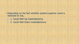
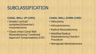
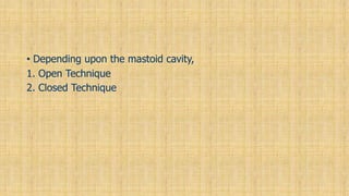
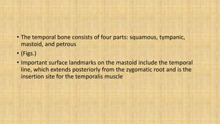
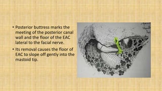
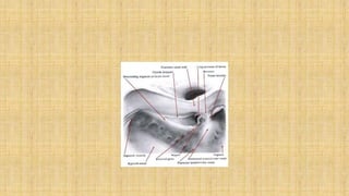
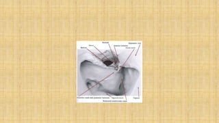
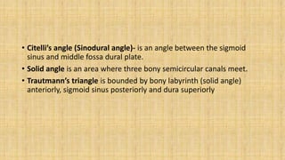
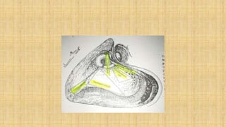
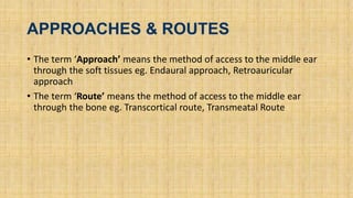
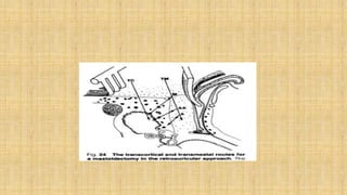
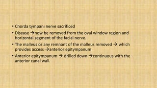
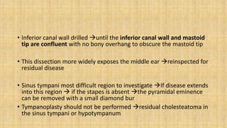
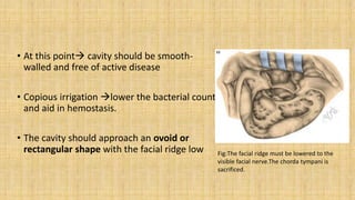
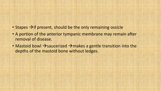
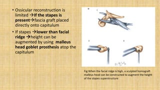
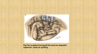
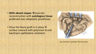
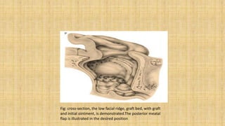
My MRM
HISTORY
• The first scholarly treatise on mastoid surgery for suppurative disease
was by ‘Schwartze’ in 1873.
• The procedure he described was a ‘cortical mastoidectomy’ with
limited exenteration of mastoid air cells.
• In1890, Zaufal described removing the superior and posterior canal
wall, tympanic membrane, and lateral ossicular chain, a procedure
now known as the ‘radical mastoidectomy
• This procedure was modified by Bondy, who recognized that disease
limited to the pars flaccida could simply be exteriorized, leaving the
uninvolved middle ear alone.
• His description of the “modified radical mastoidectomy” or “Bondy
procedure” in 1910 represented one of the first reports addressing
hearing function
• Interest in hearing preservation and restoration gained further
attention after Lempert introduced the fenestration operation in
1938, and Zollner and Wullstein described tympanoplasty techniques
in early1950s.
• Lempert popularized the use of a drill and loupe magnification in the
1920s
• During the next decade, Jansen, Sheehy, and others extended these
principles of restoring function and maintaining normal anatomy with
the introduction of the intact canal wall mastoidectomy with facial
recess approach.
• With the advent of CWU mastoidectomy, disease control as well as
preservation of anatomy and function became a reality
• The first postauricular incision was introduced in 1853 by Sir Willium
Wilde of Dublin.
INTRODUCTION
• Descriptions of chronic and suppurative infections of the mastoid
have been discovered dating back to ancient Greece.
• Prior to the advent of surgery and antibiotics, morbidity from acute
mastoiditis was considerable.
• Mastoid surgery has evolved from simple trephination for acute
infection, to the canalwall preserving mastoidectomy employed by
inost otologists today
• Chronic otitis media, with or without cholesteatoma, is one of the
more common indications for performing a mastoidectomy.
• Mastoidectomy permits access to remove cholesteatoma matrix or
diseased air cells in chronic otitis media.
CLASSIFICATIONS
• Traditionally, classified as :
• 1. Simple (cortical, complete) mastoidectomy
• 2. Modified radical mastoidectomy
• 3. Radical mastoidectomy
•  A fourth procedure, Tympanomastoidectomy , combines the
simple mastoidectomy with a middle-ear procedure, maintaining
the posterior and superior canal walls
• Depending on the fact whether postero-superior canal is
removed or not,
•  1. Canal Wall Up mastoidectomy
•  2. Canal Wall Down mastoidectomy
SUBCLASSIFICATION
CANAL WALL UP (CWU)
• Simple/ cortical/
complete/Schwartze’s
mastoidectomy
• Classic Intact Canal Wall
Mastoidectomy/ Combined
Approach Tympanoplasty (CAT)
CANAL WALL DOWN (CWD)
• Atticotomy
• Atticoantrotomy
• Radical Mastoidectomy
• Modified Radical
Mastoidectomy/ bondy’s
Procedure
• Retrograde Mastoidectomy
• Depending upon the mastoid cavity,
1. Open Technique
2. Closed Technique
•ANATOMICAL CONSIDERATIONS
• The temporal bone consists of four parts: squamous, tympanic,
mastoid, and petrous
• (Figs.)
• Important surface landmarks on the mastoid include the temporal
line, which extends posteriorly from the zygomatic root and is the
insertion site for the temporalis muscle
• The suprameatal spine of Henle is a small bony protuberance extending
superficially from the posterior and superior bony EAC.
• Posterior to the suprameatal spine, a group of small holes is seen,
described as the cribriform area.
• Small vessels pass through these foramina to the mucosa of the
underlying antrum in infants, and it’s here that a subperiosteal abscess
forms in cases of acute coalescent mastoiditis
• This cribriform area lies within Macewen’s triangle, an imaginary
triangle defined by three lines-
1. Temporal line
2. Line formed by the superior and posterior margins of the external
bony meatus (This line goes through the suprameatal spine)
3. Line drawn perpendiular to the first line and tangential to the second
• Mastoid antrum lies around 1.25 cm to 1.5 cm deep from the surface
of Macewen’s triangle.
• Cymba concha is the soft tissue anatomical landmark for the mastoid
antrum
• Facial Bridge is that portion of posterosuperior bony meatal wall that
bridges over the notch of Rivinus and overlies the ossicles.
• Facial Ridge is that part of the bony meatal wall which houses the
posterior bend and vertical segment of facial nerve.
• Anterior Buttress is the point at which the posterior bony canal wall
meets the tegmen
• Posterior buttress marks the
meeting of the posterior canal
wall and the floor of the EAC
lateral to the facial nerve.
• Its removal causes the floor of
EAC to slope off gently into the
mastoid tip.
• Citelli’s angle (Sinodural angle)- is an angle between the sigmoid
sinus and middle fossa dural plate.
• Solid angle is an area where three bony semicircular canals meet.
• Trautmann’s triangle is bounded by bony labyrinth (solid angle)
anteriorly, sigmoid sinus posteriorly and dura superiorly
• Donaldson’s line is a line passing through the horizontal semicircular
canal and bisects the posterior semicircular canal.
• This line is a landmark for the endolymphatic sac.
APPROACHES & ROUTES
• The term ‘Approach’ means the method of access to the middle ear
through the soft tissues eg. Endaural approach, Retroauricular
approach
• The term ‘Route’ means the method of access to the middle ear
through the bone eg. Transcortical route, Transmeatal Route
MODIFIED RADICAL
MASTOIDECTOMY
• Classically, modified radical mastoidectomy refers to the Bondy
procedure, in which disease limited to the epitympanum is simply
exteriorized by removing portions of the adjacent superior or
posterior canal wall.
• But, Frequently, the term modified radical mastoidectomy is used
interchangeably with canal wall down mastoidectomy
• A primary feature of the modified radical procedure is complete
removal of the posterior canal wall, the major reason for failure of the
Bondy procedure.
• MRM is an effective method to manage cholesteatoma in a ‘single-
stage’ approach (Unlike CAT).
• MRM is a surgical procedure where the disease process is eradicated
from the middle ear cleft; followed by converting the mastoid cavity,
middle ear and EAC into a single, smooth, selfcleansing cavity
exteriorised through EAC
INDICATIONS
Absolute Indications
1. Unresectable disease
2. Unreconstructable Posterior canal wall
3. Failure of first stage CWU procedure because of poor E T function.
4. Inadequate Patient Follow up.
Relative Indications
1. Disease in only hearing ear or in a dead ear.
2. Medical illness
3. Severe otologic or CNS complications
4. Neoplasms
5. Poor E T function
Contraindications
Chronic otitis media without cholesteatoma
2. Acute otitis media with coalescent mastoiditis,
3. persistent secretory otitis media, or
4. Chronic allergic otitis media.
5. Tuberculous otitis media.
CWU Vs CWD
• The choice for preserving or removing the posterior wall of the EAC,
ie, CWU versus CWD mastoidectomy, has been extensively debated.
• Preservation of the canal wall is preferred.
• The decision to remove the wall is most often made during surgery,
when the extent of the disease is fully appreciated
Intraoperative findings that may be indications for a CWD procedure
include
-labyrinthine fistula,
-unresectable disease on the facial nerve orstapes footplate,
-a low-lying tegmen that limits access to the attic,
-unresectable sinus tympani disease, and an unreconstructable
posterior canal wall defect.
• Removal of the canal wall does not improve access to the sinus
tympani.
• Rarely, our preoperative evaluation may result in the decision to take
down the canal wall.
1.Obvious posterior wall erosion,
2.larger labyrinthine fistula on CT scan,
3.elderly or infirmed patients in which second look is unadvisable.
4.Occasionally with disease in an only hearing ear, are preoperative
conditions that may warrant a CWD procedure.
Advantages and disadvantages of canal wall-
up and canal-down procedures
Advantages Disadvantages
Canal wall-up
Physiologic position of tympanic membrane Residual and recurrent cholesteatoma may occur
Enough middle ear space Incomplete exteriorization of facial recess
No mastoid cavity problem Second stage operation often required
Canal-down
Residual cholesteatoma easily found on follow-up
evaluation
Mastoid cavity problem often
Recurrent cholesteatoma is rare Middle ear is shallow and difficult to reconstruct
Total exteriorization of facial recess
Position of pinna may be altered; second stage operation
sometimes required
TECHNIQUE OF MODIFIED RADICAL
MASTOIDECTOMY(MRM)
• Also known as complete mastoidectomy and tympanoplasty
Major goal
→cholesteatoma surgery (i.e., exteriorization of disease) with sealing
of the middle car space
• A primary feature of the MRM is complete removal of the posterior
canal wall
Preoperative Assessment
• Decision to perform a MRM depends on:
the extent and location of the disease
previous surgery
 Eustachian tube function
 and patient age, medical condition, and aftercare preference
Careful microscopic inspection and cleaning of the ear aid in the decision
• Pus, mucus, and cholesteatomatous debris should be removed under
microscopic suction
• Active suppuration →controlled prior to surgery whenever possible.
(Acetic acid (1.5%solution) irrigations followed by antibiotic otic drops
should be instituted for several weeks prior to surgery)
• In cases of extensive mucosal Infection and cellulitis 10 - l4-day
course of oral fluoroquinolones with gram-positive coverage is
indicated prior to surgery
Surgical Procedure
• After induction of G.A
• Ear prepared by pouring povidone-
iodine solution into the ear canal and
scrubbing the auricle and postauricular
area with povidone-iodine
• 1 % lidocaine with 1:100,000
epinephrine injected postauricular
region & ear canal for hemostasis
• Incisions ear canal for the vascular strip
• A postauricular incision Plane
developed between the subcutaneous
tissue and the temporalis muscle and
periosteum of the mastoid
• Several large pieces of areolar tissue and
temporalis fascia are harvested and set
aside to dry
FIG: Standard tympanoplasty canal incisions
outline the vascular strip as well as the
superior and inferior canal wall flaps
• Horizontal incision superior to the
temporal line through the temporalis
muscle & vertical incision mastoid
tip perpendicular to and bisecting
the horizontal incision ( “T” shaped
Incision)
Fig: Loose areolar fascia is harvested from
temporal muscle, and a T-shaped incision
is made in soft tissue over mastoid.
• Mastoid bone is exposed using a
Lempert elevator
• Periosteum raised into the ear canal
vascular strip elevated & reflected
out of the ear canal anteriorly using
a self-retaining retractor
Fig:Exposure of mastoid in
crosssection showing vascular
strip held forward under anterior
blade of retractor.
• In revision casesElevation of the scarred
musculoperiosteum carefully to avoid
injury to exposed dura or sigmoid sinus
• Canal wall flaps elevated and rotated
anteriorly prior entering middle ear
• Disease in the mesotympanum is first
removed malleus handle and incus as
landmarks
Fig:The inferior flap is elevated to the fibrous
annulus
• Cholesteatoma, polyps, and granulation tissue are removed from all
areas except the posterosuperior quadrant(PSQ)
• Any atrophic tympanic membrane removed & middle ear
prepared for grafting
• Once all available landmarks have been identified  PSQ
inspected.
• If disease extends into the attic dissection of disease ceases and
Gelfoam with epinephrine  packed middle ear
Bone Work
• Simple mastoidectomy begunlarge
cutting bur.
• All mastoid air cellsremoved with
exposure of the middle fossa and
posterior fossa dural plates, the sigmoid
sinus, digastric ridge, and bony canal wall
• Cholesteatoma and granulations filling
the central mastoid tract removed at
this time.
FIG: With the posterior external auditory
canal (EAC) wall preserved, a complete,
simple mastoidectomy demonstrates
• As the labyrinth is approached lateral
capsule of the cholesteatoma opened
cholesteatoma should be removed
leaving the medial matrix of the
cholesteatoma on the bony labyrinth
• Under higher-power magnification the
matrix inspected labyrinthine fistula
• The vertical segment of the facial nerve
now identified opening of the facial
recess
• If the incus is involved with
cholesteatoma incudostapedial joint is
identified through the facial recess and cut
and the incus is removed
• The posterior canal wall now be safely
taken down rongeur
• Facial ridge lowered
Fig:The posterior EAC wall must be lowered
to the visible facial nerve. The chorda
tympani is sacrificed. Canal wall flaps are
preserved
• Chorda tympani nerve sacrificed
• Disease now be removed from the oval window region and
horizontal segment of the facial nerve.
• The malleus or any remnant of the malleus removed  which
provides access anterior epitympanum
• Anterior epitympanum  drilled down continuous with the
anterior canal wall.
• Inferior canal wall drilled until the inferior canal wall and mastoid
tip are confluent with no bony overhang to obscure the mastoid tip
• This dissection more widely exposes the middle ear reinspected for
residual disease
• Sinus tympani most difficult region to investigate If disease extends
into this region  if the stapes is absent the pyramidal eminence
can be removed with a small diamond bur
• Tympanoplasty should not be performed residual cholesteatoma in
the sinus tympani or hypotympanum
• At this point cavity should be smooth-
walled and free of active disease
• Copious irrigation lower the bacterial count
and aid in hemostasis.
• The cavity should approach an ovoid or
rectangular shape with the facial ridge low Fig:The facial ridge must be lowered to the
visible facial nerve.The chorda tympani is
sacrificed.
• Stapes if present, should be the only remaining ossicle
• A portion of the anterior tympanic membrane may remain after
removal of disease.
• Mastoid bowl saucerized makes a gentle transition into the
depths of the mastoid bone without ledges.
Meatoplasty
• 1% lidocaine with 1:100,000
epinephrine infiltrated into the
conchal bowl
• The entire posterior aspect of the
conchal bowl is exposed With
finger in conchal bowl  semilunar
incision cartilage posteriorly
• Crescent-shaped cartilage measures
about 1.5 x 2 cm
Fig: meatoplasty beginsby excising, from behind,
conchal cartilage
• Korner flap incisions through external
auditory canal skin
• Inferior incision inferior canal at 6
o'clock conchal bowl curved around
the inferior margin of the bowl.
• A superior incision 12 o'clock tragus
and anterior helix
• These incisions create a long (vascular
strip) flap posterosuperior aspect of
the conchal bowl constitute the back
wall of the mastoid cavity Fig: Superior and inferior meatal incisions create
a posterior Korner's flap, shown here as it will
be sutured in place
Grafting
• Auricle and flap retracted
anteriorly to expose the mastoid
and middle ear
• Epinephrine-soaked absorbable
gelatin sponge removed 
middle ear and Eustachian tube
packed with saline-moistened
absorbable gelatin sponge level
of the anterior annulus Fig:An absorable gelatin sponge (Gelfoam) bed is
prepared for the tympanic membrane graft. Note
the Korner's flap free in the meatus
• Fascia graft placed medial to the
anterior annulus and drum remnant
extending over the stapes to the
facial ridge into the mastoid bowl
Fig:The graft is placed medial to the tympanic
membrane remnant, superiorly over the labyrinth
and posteriorly over the facial ridge. The graft is
applied directly atop the stapes superstructure
• Ossicular reconstruction is
limited If the stapes is
presentfascia graft placed
directly onto capitulum
• If stapes lower than facial
ridge height can be
augmented by using malleus
head goblet prosthesis atop the
capitulum
Fig:When the facial ridge is high, a sculpted homograft
malleus head can be constructed to augment the height
of the stapes superstructure
Fig:The sculpted homograft fits atop the stapedial
capitulum, ready for grafting
• With absent stapes ossicular
reconstruction with autologous tissue
preferred over alloplastic prostheses
• Once the fascia graft is in place
surface covered with polymixin B and
bacitracin ophthalmic ointment
Fig: Ointment "packing" fills the cavity.
Fig: cross-section, the low facial ridge, graft bed, with graft
and initial ointment, is demonstrated.The posterior meatal
flap is illustrated in the desired position
• Korner flap secured
musculoperiosteum at edge
of the mastoid cavity
• Tension of these sutures
adjusted until meatus has
desired shape
Fig: The posterior flap is sewn to the
posterior soft tissue margins of the incision
• Postauricular incision closed subcuticular absorbable suture
• Mastoid bowl filled with ointment or packed with gauze
• Mastoid dressing applied
Postoperative Care
• Mastoid dressing removed on first postoperative day
• Large piece of cotton kept in meatus postauricular dressing placed
• Copious drainage occurs meatus  1 week requiring frequent
cotton changes
• Postauricular dressing removed second postoperative day 
antibiotic ointment applied to incision for 1 week
• Patient instructed keep ear dry and avoid nose blowing
• Pain medication prescribed
• Oral antibiotics not used routinely
• Patient returns in 2 to 3 weeks first postoperative visit
any area that has not been grafted is covered by layer of granulation
tissue
Exuberant granulation tissue debrided and treated with silver
nitrate
• Granulation tissue then painted 2% gentian violet instructed to
use antibiotic drops 2-3 times next visit
• Drainage decreases re-epithelialization occurs
• As epithelialization progresses acetic acid irrigations can replace
antibiotic otic drops.
• Once cavity is healed patient should return for yearly visit & given
full water sport privileges
COMPLICATIONS OF OPEN CAVITY PROCEDURES
Facial paralysis
Wound infection
Deafness or further hearing loss
Vestibular symptoms
Cerebrospinal fluid leak
Recurrent cholesteatoma or drainage.
• Facial nerve paralysis most common major complication
wound infection
2nd most common complication
perichondritis of auricle
 Pseudomonas aeruginosa causative organism
Treatment
High-dose fluoroquinolones and antibiotic-corticosteroid drops
“Chocolate" or mucous retention cyst
• Can occur in healed mastoid cavity
• Result of collection of serum within a mucous membrane-lined
pocket
Management
• Simple aspiration reduce the size of the cyst
• Recurrence common
Definitive management
• Exposure of the cyst & complete removal of mucoperiosteal pocket
Cholesteatoma recurrence
• In open cavity procedures  4 to 28% cases
Etiology
• Inaccessible disease or a remnant of matrix that was amputated at
the time of surgery
Management
• Routine follow-up these "pearls" of recurrent cholesteatoma
identified and removed in the office
Recurrent aural drainage
• Previously healed and dry cavity result of poor aural toilet
• Breakdown of epithelial lining & formation of granulation tissue
occurs when epidermal debris is allowed to accumulate and becomes
infected
Management
• Careful microscopic debridement of granulation tissue and
application of gentian violet followed by antibiotic corticosteroid
otic drops re-epithelialization & dry ear
Development of scar bands within mastoid defect
• Lead to keratin debris accumulation and subsequent infection
Management
• Transmeatal removal ofscar bands under local anesthesia
In extensive cases
• Transmeatal removal of scar bands under G.A.re-epithelialization of
mastoid bowl
• It is critical that the patient understand the need for
periodicusually annual examination to prevent such occurrence
Bibliography
Next presentation
Thank you

My MRM.pptx

  • 1.
  • 3.
    HISTORY • The firstscholarly treatise on mastoid surgery for suppurative disease was by ‘Schwartze’ in 1873. • The procedure he described was a ‘cortical mastoidectomy’ with limited exenteration of mastoid air cells. • In1890, Zaufal described removing the superior and posterior canal wall, tympanic membrane, and lateral ossicular chain, a procedure now known as the ‘radical mastoidectomy
  • 4.
    • This procedurewas modified by Bondy, who recognized that disease limited to the pars flaccida could simply be exteriorized, leaving the uninvolved middle ear alone. • His description of the “modified radical mastoidectomy” or “Bondy procedure” in 1910 represented one of the first reports addressing hearing function
  • 5.
    • Interest inhearing preservation and restoration gained further attention after Lempert introduced the fenestration operation in 1938, and Zollner and Wullstein described tympanoplasty techniques in early1950s. • Lempert popularized the use of a drill and loupe magnification in the 1920s
  • 6.
    • During thenext decade, Jansen, Sheehy, and others extended these principles of restoring function and maintaining normal anatomy with the introduction of the intact canal wall mastoidectomy with facial recess approach. • With the advent of CWU mastoidectomy, disease control as well as preservation of anatomy and function became a reality
  • 7.
    • The firstpostauricular incision was introduced in 1853 by Sir Willium Wilde of Dublin.
  • 8.
    INTRODUCTION • Descriptions ofchronic and suppurative infections of the mastoid have been discovered dating back to ancient Greece. • Prior to the advent of surgery and antibiotics, morbidity from acute mastoiditis was considerable. • Mastoid surgery has evolved from simple trephination for acute infection, to the canalwall preserving mastoidectomy employed by inost otologists today
  • 9.
    • Chronic otitismedia, with or without cholesteatoma, is one of the more common indications for performing a mastoidectomy. • Mastoidectomy permits access to remove cholesteatoma matrix or diseased air cells in chronic otitis media.
  • 10.
    CLASSIFICATIONS • Traditionally, classifiedas : • 1. Simple (cortical, complete) mastoidectomy • 2. Modified radical mastoidectomy • 3. Radical mastoidectomy •  A fourth procedure, Tympanomastoidectomy , combines the simple mastoidectomy with a middle-ear procedure, maintaining the posterior and superior canal walls
  • 11.
    • Depending onthe fact whether postero-superior canal is removed or not, •  1. Canal Wall Up mastoidectomy •  2. Canal Wall Down mastoidectomy
  • 12.
    SUBCLASSIFICATION CANAL WALL UP(CWU) • Simple/ cortical/ complete/Schwartze’s mastoidectomy • Classic Intact Canal Wall Mastoidectomy/ Combined Approach Tympanoplasty (CAT) CANAL WALL DOWN (CWD) • Atticotomy • Atticoantrotomy • Radical Mastoidectomy • Modified Radical Mastoidectomy/ bondy’s Procedure • Retrograde Mastoidectomy
  • 13.
    • Depending uponthe mastoid cavity, 1. Open Technique 2. Closed Technique
  • 14.
  • 15.
    • The temporalbone consists of four parts: squamous, tympanic, mastoid, and petrous • (Figs.) • Important surface landmarks on the mastoid include the temporal line, which extends posteriorly from the zygomatic root and is the insertion site for the temporalis muscle
  • 18.
    • The suprameatalspine of Henle is a small bony protuberance extending superficially from the posterior and superior bony EAC. • Posterior to the suprameatal spine, a group of small holes is seen, described as the cribriform area. • Small vessels pass through these foramina to the mucosa of the underlying antrum in infants, and it’s here that a subperiosteal abscess forms in cases of acute coalescent mastoiditis
  • 19.
    • This cribriformarea lies within Macewen’s triangle, an imaginary triangle defined by three lines- 1. Temporal line 2. Line formed by the superior and posterior margins of the external bony meatus (This line goes through the suprameatal spine) 3. Line drawn perpendiular to the first line and tangential to the second
  • 21.
    • Mastoid antrumlies around 1.25 cm to 1.5 cm deep from the surface of Macewen’s triangle. • Cymba concha is the soft tissue anatomical landmark for the mastoid antrum
  • 22.
    • Facial Bridgeis that portion of posterosuperior bony meatal wall that bridges over the notch of Rivinus and overlies the ossicles. • Facial Ridge is that part of the bony meatal wall which houses the posterior bend and vertical segment of facial nerve. • Anterior Buttress is the point at which the posterior bony canal wall meets the tegmen
  • 24.
    • Posterior buttressmarks the meeting of the posterior canal wall and the floor of the EAC lateral to the facial nerve. • Its removal causes the floor of EAC to slope off gently into the mastoid tip.
  • 27.
    • Citelli’s angle(Sinodural angle)- is an angle between the sigmoid sinus and middle fossa dural plate. • Solid angle is an area where three bony semicircular canals meet. • Trautmann’s triangle is bounded by bony labyrinth (solid angle) anteriorly, sigmoid sinus posteriorly and dura superiorly
  • 28.
    • Donaldson’s lineis a line passing through the horizontal semicircular canal and bisects the posterior semicircular canal. • This line is a landmark for the endolymphatic sac.
  • 30.
    APPROACHES & ROUTES •The term ‘Approach’ means the method of access to the middle ear through the soft tissues eg. Endaural approach, Retroauricular approach • The term ‘Route’ means the method of access to the middle ear through the bone eg. Transcortical route, Transmeatal Route
  • 32.
    MODIFIED RADICAL MASTOIDECTOMY • Classically,modified radical mastoidectomy refers to the Bondy procedure, in which disease limited to the epitympanum is simply exteriorized by removing portions of the adjacent superior or posterior canal wall. • But, Frequently, the term modified radical mastoidectomy is used interchangeably with canal wall down mastoidectomy • A primary feature of the modified radical procedure is complete removal of the posterior canal wall, the major reason for failure of the Bondy procedure. • MRM is an effective method to manage cholesteatoma in a ‘single- stage’ approach (Unlike CAT).
  • 33.
    • MRM isa surgical procedure where the disease process is eradicated from the middle ear cleft; followed by converting the mastoid cavity, middle ear and EAC into a single, smooth, selfcleansing cavity exteriorised through EAC
  • 35.
    INDICATIONS Absolute Indications 1. Unresectabledisease 2. Unreconstructable Posterior canal wall 3. Failure of first stage CWU procedure because of poor E T function. 4. Inadequate Patient Follow up.
  • 36.
    Relative Indications 1. Diseasein only hearing ear or in a dead ear. 2. Medical illness 3. Severe otologic or CNS complications 4. Neoplasms 5. Poor E T function
  • 37.
    Contraindications Chronic otitis mediawithout cholesteatoma 2. Acute otitis media with coalescent mastoiditis, 3. persistent secretory otitis media, or 4. Chronic allergic otitis media. 5. Tuberculous otitis media.
  • 38.
    CWU Vs CWD •The choice for preserving or removing the posterior wall of the EAC, ie, CWU versus CWD mastoidectomy, has been extensively debated. • Preservation of the canal wall is preferred. • The decision to remove the wall is most often made during surgery, when the extent of the disease is fully appreciated
  • 39.
    Intraoperative findings thatmay be indications for a CWD procedure include -labyrinthine fistula, -unresectable disease on the facial nerve orstapes footplate, -a low-lying tegmen that limits access to the attic, -unresectable sinus tympani disease, and an unreconstructable posterior canal wall defect.
  • 40.
    • Removal ofthe canal wall does not improve access to the sinus tympani. • Rarely, our preoperative evaluation may result in the decision to take down the canal wall. 1.Obvious posterior wall erosion, 2.larger labyrinthine fistula on CT scan, 3.elderly or infirmed patients in which second look is unadvisable. 4.Occasionally with disease in an only hearing ear, are preoperative conditions that may warrant a CWD procedure.
  • 41.
    Advantages and disadvantagesof canal wall- up and canal-down procedures Advantages Disadvantages Canal wall-up Physiologic position of tympanic membrane Residual and recurrent cholesteatoma may occur Enough middle ear space Incomplete exteriorization of facial recess No mastoid cavity problem Second stage operation often required Canal-down Residual cholesteatoma easily found on follow-up evaluation Mastoid cavity problem often Recurrent cholesteatoma is rare Middle ear is shallow and difficult to reconstruct Total exteriorization of facial recess Position of pinna may be altered; second stage operation sometimes required
  • 42.
    TECHNIQUE OF MODIFIEDRADICAL MASTOIDECTOMY(MRM) • Also known as complete mastoidectomy and tympanoplasty Major goal →cholesteatoma surgery (i.e., exteriorization of disease) with sealing of the middle car space • A primary feature of the MRM is complete removal of the posterior canal wall
  • 43.
    Preoperative Assessment • Decisionto perform a MRM depends on: the extent and location of the disease previous surgery  Eustachian tube function  and patient age, medical condition, and aftercare preference Careful microscopic inspection and cleaning of the ear aid in the decision • Pus, mucus, and cholesteatomatous debris should be removed under microscopic suction
  • 44.
    • Active suppuration→controlled prior to surgery whenever possible. (Acetic acid (1.5%solution) irrigations followed by antibiotic otic drops should be instituted for several weeks prior to surgery) • In cases of extensive mucosal Infection and cellulitis 10 - l4-day course of oral fluoroquinolones with gram-positive coverage is indicated prior to surgery
  • 45.
    Surgical Procedure • Afterinduction of G.A • Ear prepared by pouring povidone- iodine solution into the ear canal and scrubbing the auricle and postauricular area with povidone-iodine • 1 % lidocaine with 1:100,000 epinephrine injected postauricular region & ear canal for hemostasis
  • 46.
    • Incisions earcanal for the vascular strip • A postauricular incision Plane developed between the subcutaneous tissue and the temporalis muscle and periosteum of the mastoid • Several large pieces of areolar tissue and temporalis fascia are harvested and set aside to dry FIG: Standard tympanoplasty canal incisions outline the vascular strip as well as the superior and inferior canal wall flaps
  • 47.
    • Horizontal incisionsuperior to the temporal line through the temporalis muscle & vertical incision mastoid tip perpendicular to and bisecting the horizontal incision ( “T” shaped Incision) Fig: Loose areolar fascia is harvested from temporal muscle, and a T-shaped incision is made in soft tissue over mastoid.
  • 48.
    • Mastoid boneis exposed using a Lempert elevator • Periosteum raised into the ear canal vascular strip elevated & reflected out of the ear canal anteriorly using a self-retaining retractor Fig:Exposure of mastoid in crosssection showing vascular strip held forward under anterior blade of retractor.
  • 49.
    • In revisioncasesElevation of the scarred musculoperiosteum carefully to avoid injury to exposed dura or sigmoid sinus • Canal wall flaps elevated and rotated anteriorly prior entering middle ear • Disease in the mesotympanum is first removed malleus handle and incus as landmarks Fig:The inferior flap is elevated to the fibrous annulus
  • 50.
    • Cholesteatoma, polyps,and granulation tissue are removed from all areas except the posterosuperior quadrant(PSQ) • Any atrophic tympanic membrane removed & middle ear prepared for grafting • Once all available landmarks have been identified  PSQ inspected. • If disease extends into the attic dissection of disease ceases and Gelfoam with epinephrine  packed middle ear
  • 51.
    Bone Work • Simplemastoidectomy begunlarge cutting bur. • All mastoid air cellsremoved with exposure of the middle fossa and posterior fossa dural plates, the sigmoid sinus, digastric ridge, and bony canal wall • Cholesteatoma and granulations filling the central mastoid tract removed at this time. FIG: With the posterior external auditory canal (EAC) wall preserved, a complete, simple mastoidectomy demonstrates
  • 52.
    • As thelabyrinth is approached lateral capsule of the cholesteatoma opened cholesteatoma should be removed leaving the medial matrix of the cholesteatoma on the bony labyrinth • Under higher-power magnification the matrix inspected labyrinthine fistula • The vertical segment of the facial nerve now identified opening of the facial recess
  • 53.
    • If theincus is involved with cholesteatoma incudostapedial joint is identified through the facial recess and cut and the incus is removed • The posterior canal wall now be safely taken down rongeur • Facial ridge lowered Fig:The posterior EAC wall must be lowered to the visible facial nerve. The chorda tympani is sacrificed. Canal wall flaps are preserved
  • 54.
    • Chorda tympaninerve sacrificed • Disease now be removed from the oval window region and horizontal segment of the facial nerve. • The malleus or any remnant of the malleus removed  which provides access anterior epitympanum • Anterior epitympanum  drilled down continuous with the anterior canal wall.
  • 55.
    • Inferior canalwall drilled until the inferior canal wall and mastoid tip are confluent with no bony overhang to obscure the mastoid tip • This dissection more widely exposes the middle ear reinspected for residual disease • Sinus tympani most difficult region to investigate If disease extends into this region  if the stapes is absent the pyramidal eminence can be removed with a small diamond bur • Tympanoplasty should not be performed residual cholesteatoma in the sinus tympani or hypotympanum
  • 56.
    • At thispoint cavity should be smooth- walled and free of active disease • Copious irrigation lower the bacterial count and aid in hemostasis. • The cavity should approach an ovoid or rectangular shape with the facial ridge low Fig:The facial ridge must be lowered to the visible facial nerve.The chorda tympani is sacrificed.
  • 57.
    • Stapes ifpresent, should be the only remaining ossicle • A portion of the anterior tympanic membrane may remain after removal of disease. • Mastoid bowl saucerized makes a gentle transition into the depths of the mastoid bone without ledges.
  • 58.
    Meatoplasty • 1% lidocainewith 1:100,000 epinephrine infiltrated into the conchal bowl • The entire posterior aspect of the conchal bowl is exposed With finger in conchal bowl  semilunar incision cartilage posteriorly • Crescent-shaped cartilage measures about 1.5 x 2 cm Fig: meatoplasty beginsby excising, from behind, conchal cartilage
  • 59.
    • Korner flapincisions through external auditory canal skin • Inferior incision inferior canal at 6 o'clock conchal bowl curved around the inferior margin of the bowl. • A superior incision 12 o'clock tragus and anterior helix • These incisions create a long (vascular strip) flap posterosuperior aspect of the conchal bowl constitute the back wall of the mastoid cavity Fig: Superior and inferior meatal incisions create a posterior Korner's flap, shown here as it will be sutured in place
  • 60.
    Grafting • Auricle andflap retracted anteriorly to expose the mastoid and middle ear • Epinephrine-soaked absorbable gelatin sponge removed  middle ear and Eustachian tube packed with saline-moistened absorbable gelatin sponge level of the anterior annulus Fig:An absorable gelatin sponge (Gelfoam) bed is prepared for the tympanic membrane graft. Note the Korner's flap free in the meatus
  • 61.
    • Fascia graftplaced medial to the anterior annulus and drum remnant extending over the stapes to the facial ridge into the mastoid bowl Fig:The graft is placed medial to the tympanic membrane remnant, superiorly over the labyrinth and posteriorly over the facial ridge. The graft is applied directly atop the stapes superstructure
  • 62.
    • Ossicular reconstructionis limited If the stapes is presentfascia graft placed directly onto capitulum • If stapes lower than facial ridge height can be augmented by using malleus head goblet prosthesis atop the capitulum Fig:When the facial ridge is high, a sculpted homograft malleus head can be constructed to augment the height of the stapes superstructure
  • 63.
    Fig:The sculpted homograftfits atop the stapedial capitulum, ready for grafting
  • 64.
    • With absentstapes ossicular reconstruction with autologous tissue preferred over alloplastic prostheses • Once the fascia graft is in place surface covered with polymixin B and bacitracin ophthalmic ointment Fig: Ointment "packing" fills the cavity.
  • 65.
    Fig: cross-section, thelow facial ridge, graft bed, with graft and initial ointment, is demonstrated.The posterior meatal flap is illustrated in the desired position
  • 66.
    • Korner flapsecured musculoperiosteum at edge of the mastoid cavity • Tension of these sutures adjusted until meatus has desired shape Fig: The posterior flap is sewn to the posterior soft tissue margins of the incision
  • 67.
    • Postauricular incisionclosed subcuticular absorbable suture • Mastoid bowl filled with ointment or packed with gauze • Mastoid dressing applied
  • 68.
    Postoperative Care • Mastoiddressing removed on first postoperative day • Large piece of cotton kept in meatus postauricular dressing placed • Copious drainage occurs meatus  1 week requiring frequent cotton changes • Postauricular dressing removed second postoperative day  antibiotic ointment applied to incision for 1 week
  • 69.
    • Patient instructedkeep ear dry and avoid nose blowing • Pain medication prescribed • Oral antibiotics not used routinely • Patient returns in 2 to 3 weeks first postoperative visit any area that has not been grafted is covered by layer of granulation tissue Exuberant granulation tissue debrided and treated with silver nitrate
  • 70.
    • Granulation tissuethen painted 2% gentian violet instructed to use antibiotic drops 2-3 times next visit • Drainage decreases re-epithelialization occurs • As epithelialization progresses acetic acid irrigations can replace antibiotic otic drops. • Once cavity is healed patient should return for yearly visit & given full water sport privileges
  • 71.
    COMPLICATIONS OF OPENCAVITY PROCEDURES Facial paralysis Wound infection Deafness or further hearing loss Vestibular symptoms Cerebrospinal fluid leak Recurrent cholesteatoma or drainage.
  • 72.
    • Facial nerveparalysis most common major complication
  • 73.
    wound infection 2nd mostcommon complication perichondritis of auricle  Pseudomonas aeruginosa causative organism Treatment High-dose fluoroquinolones and antibiotic-corticosteroid drops
  • 74.
    “Chocolate" or mucousretention cyst • Can occur in healed mastoid cavity • Result of collection of serum within a mucous membrane-lined pocket Management • Simple aspiration reduce the size of the cyst • Recurrence common Definitive management • Exposure of the cyst & complete removal of mucoperiosteal pocket
  • 75.
    Cholesteatoma recurrence • Inopen cavity procedures  4 to 28% cases Etiology • Inaccessible disease or a remnant of matrix that was amputated at the time of surgery Management • Routine follow-up these "pearls" of recurrent cholesteatoma identified and removed in the office
  • 76.
    Recurrent aural drainage •Previously healed and dry cavity result of poor aural toilet • Breakdown of epithelial lining & formation of granulation tissue occurs when epidermal debris is allowed to accumulate and becomes infected Management • Careful microscopic debridement of granulation tissue and application of gentian violet followed by antibiotic corticosteroid otic drops re-epithelialization & dry ear
  • 77.
    Development of scarbands within mastoid defect • Lead to keratin debris accumulation and subsequent infection Management • Transmeatal removal ofscar bands under local anesthesia In extensive cases • Transmeatal removal of scar bands under G.A.re-epithelialization of mastoid bowl • It is critical that the patient understand the need for periodicusually annual examination to prevent such occurrence
  • 78.
  • 79.
  • 80.

Editor's Notes

  • #43 with sealing of the middle car space to avoid chronic drainage from exposed mucous membrane A primary feature of the modified radical procedure is complete removal of the posterior canal wall, the major reason for failure of the Bondy procedure The modification of the radical procedure (i.e., add ing the technique of tympanoplasty) potentially eliminates the expected intermittent discharge from the middle ear mucosa Hearing, it should be noted, is a secondary consideration of the modified radical procedure.
  • #44 Polyps can be removed with gentle traction with the suction or niicrocup forceps. Significant retraction should be avoided si nee the polyp 1nay be attached to the facial nerve, matrix of a labyrinthine fistula, or stapes superstructure or footplate
  • #45 Extensive destruction of the posterior canal wall with obvious cholesteatoma invading the mastoid indicates the need for modified radical mastoidectomy Acetic acid solution is made by 1nix.ing one part of white vinegar to two parts of boiling water. After cooling, the solution is instilled into the ear several tin1es using an infant nasal-bulb syringe to niechanically debride the area. Antibiotic eardrops are instilled after the irrigations, \.Yhich should be performed two to four tin1es daily
  • #47  A postauricular incision is made about 1 cm behind the postauricular crease and a plane is developed between the subcutaneous tissue and the temporalis muscle and periosteum of the mastoid
  • #49 And as the
  • #51 Cholesteatoma, polyps, and granulation tissue are removed from all areas except the posterosuperior quadrant
  • #52 The canal wall should be left up in all but the most contracted mastoid cavities
  • #53 Under higher-power magnification the matrix can be inspected for the telltale blue line or palpated for the presence of a labyrinthine fistula This is best accomplished by using the digastric ridge and the lateral semicircular canal as landmarks Through the facial recess, disease can be well managed in an intact canal wall (ICW) context. The malleus head and incus are shown for orientation only. They are customarily removed. Incudostapedial disarticulation is demonstrated.
  • #54 Facial ridge lowered until a thin layer of bone remains over the vertical segment of the facial nerve
  • #55 The malleus, or any remnant of the malleus, is removed by cutting the tensor tympani tendon at the cochleariform process, which provides access to the anterior epitympanum
  • #56 The sinus tympani is the most difficult region to investigate. If disease extends into this region, and if the stapes is absent, the pyramidal eminence can be removed with a small diamond bur. Right angle hooks, whirlybird dissectors, micromirrors, and surgical telescopes can aid in cholesteatoma removal from this region
  • #58 This attention to detail helps ensure soft tissue obliteration of much of the cavity space Rarely, the mastoid is so contracted that the posterior canal wall is taken down as the antrum is exposed. This approach is potentially more dangerous to the facial nerve and ossicular chain and should be avoided whenever possible Regardless of when the canal wall is removed, the remainder of the technique remains the same.
  • #59 The entire posterior aspect of the conchal bowl is exposed using sharp dissection with an iris scissors through the fibrous periosteum and soft tissue  semilunar incision cartilage posteriorly until the knife tip is felt through the anterior skin
  • #62 As much of the mastoid bone as possible should be covered with fascia grafts to reduce granulations and speed epithelialization. In particular, perilabyrinthine, retrofacial, zygomatic, and peritubal cell tracts should be covered
  • #67 A 3.0 polyglactin 910 (Vicryl®) suture is placed subdermally at both edges of the base of the Korner flap and affixed posteriorly to the soft tissue Overtightening of the sutures, especially the superior suture will result in a protruding auricle
  • #70 Silver nitrate should not be used near an exposed facial nerve to avoid facial palsy
  • #71 2-3 times until the next (at 2 to 3 weeks) visit.
  • #73 It is critical to identify facial nerve throughout its course in the mastoid as soon as possible, which is best accomplished after the lateral semicircular canal and any ossicles within the posterior epitympanum have been identified. Especially in revision cases, the most effective way to locate the vertical segment of the facial nerve is to follow the digastric ridge to the stylomastoid foramen Although facial nerve injury is at times unavoidable because of the extent of disease, most cases of postoperative facial paralysis are a result of unrecognized facial nerve trauma at the hands of an unskilled otologic surgeon Normal surgical landmarks are often distorted in the diseased mastoid, and positive identification of vital structures is mandatory to perform a successful open cavity procedure
  • #74 Second most common complication of open cavity procedures is wound infection perichondritis of auricle, manifested by a painful, swollen auricle with copious discharge
  • #75 Simple aspiration of the mucoid, brownish serum will reduce the size of the cyst
  • #76  Extensive recurrent disease, with its attendant complications, is more commonly found behind an intact canal wall rather than in an open cavity
  • #78 The development of scar bands within the mastoid defect can lead to keratin debris accumulation and subsequent infection. Transmeatal removal of the scar bands can often be accomplished under local anesthesia