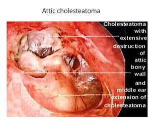
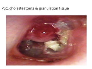
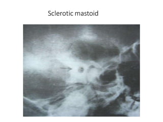
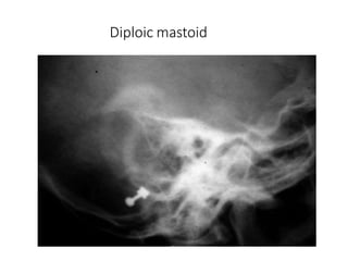
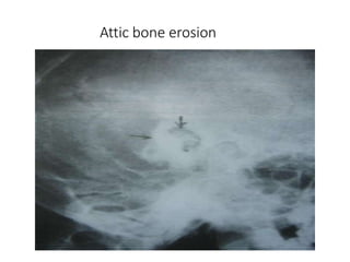
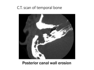
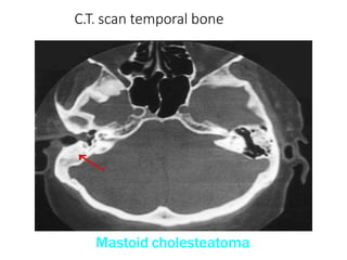
SlideShare a Scribd company logo
Chronic Suppurative Otitis
Media: Attico - antral disease
(CSOM-AAD)
(COM-Squamous)
By Dr Amit Jha
1st Year PG Resident , ENT-HNS
Definition:
• Chronic pyogenic infection of middle ear cleft lasting
for >3 months with cholesteatoma & granulation
tissue in attic or postero-superior quadrant of pars
tensa
• Unsafe/ Dangerous : Higher chances of complication
due to bone erosion
• Hallmark of Disease : Cholesteatoma/granulations
Cholesteatoma
• Johannes Müller ( 1858)
• Defined as a three dimensional sac lined by
matrix of keratinizing stratified squamous
epithelium that rests on a thin layer of fibrous
tissue and contains desquamated keratin debris
which grows at the expense of surrounding bone
• Not a tumor and has no cholesterol ( Ludman )
• Better term : Epidermosis ( Tumarkin )
Cholesteatoma :
•It is a three dimensional epidermal and connective
tissue structure , formed due to accumulation of
exfoliated Keratin in middle ear cleft . It has the
capacity for progressive independent growth at
the expense of underlying bone .
- Abramson , 1977
It has been accurately described as Keratoma
- by Schuknecht
Cholesteatoma
Histologically
• Cystic Content
o is composed of fully differentiated anucleate keratin squames.
• Matrix
o contains keratinizing squamous epithelium lining a cyst like
structure.
• Perimatrix
o known as lamina propria
o peripheral part of cholesteatoma consists of granulation tissue
and cholesterol granules.
o This layer is in contact with the bone. It is the granulation tissue
which releases enzymes that cause bone destruction.
Causes of bone destruction
• Hyperaemic decalcification
• Osteoclastic bone resorption
– Acid phosphatase ,collagenase, acid proteases
proteolytic enzymes, leukotrienes, cytokines
•Pressure necrosis
•Bacterial toxins
• Congenital
• Primary Acquired
– Retraction pocket (Wittmaack)
– Basal cell hyperplasia (Ruedi)
– Squamous metaplasia (Sade)
• Secondary Acquired
– Squamous metaplasia
– Epithelial migration (Habermann)
• Tertiary Acquired : Post-traumatic , post-tympanoplasty
Types of Cholesteatoma
Congenital Cholesteatoma
⦁ Definition (Levenson, 1989) – 5 points
White mass medial to normal tympanic membrane
Normal pars flaccida and pars tensa
No prior history of otorrhea or perforations
No prior otologic procedures
Prior bouts of otitis media not grounds for exclusion
Theories “Congenital cholesteatoma”
⦁ Epithelial cell rest theory
⦁ Squamous metaplasia theory
⦁ Epidermoid formation theory
⦁ Invagination theory
⦁ ESEI
Teed’s theory – Failure of involution of
embryonic cell rest
⦁ Proposed in 1936
⦁ The embryonic ectodermal epithelial cell rests
that is present during fetal development in
proximity to the geniculate ganglion fails to
involute.
⦁ Persistence of embryonic squamous cell rests in the
temporal bone led to the formation of congenital
cholesteatoma.
⦁ Presence of squamous cell rests in the temporal
bone – fairly common. Usually they involute at a
later date to become mature middle ear lining
Wendt’s Squamous Metaplastic theory
⦁ This was first proposed by Wendt in 1873
⦁ The attic area of the middle ear cavity is lined by
pavement epithelium.
⦁ According to Wendt, this pavement epithelium
undergoes squamous metaplasia in response to
infection thus forming a nidus for cholesteatoma
formation.
Michael’s epidermoid formation theory
⦁ Michaels in 1980s – fetal human temporal bones
⦁ Identified squamous cell tuft present from 10-33 wk
of gestation.
⦁ This “epidermoid formation” was noted in
anterosuperior wall of ME cleft.
⦁ Failure of involution could be basis of cholesteatoma
in anterosuperior mesotympanum
Reudi’s invagination theory
⦁ First proposed by Ruedi
⦁ Suggested in-utero infection of TM causing it to
invaginate into the middle ear cavity and produce
stratified squamous epithelium.
⦁ These invaginations predispose to cholesteatoma
formation.
Congenital Cholesteatoma
⦁ Origin remains uncertain
⦁ Usually starts from the antero superior quadrant
⦁ Spreads through the posterior superior quadrant,
attic and finally into the mastoid cavity
⦁ Mean age of presentation is 4.5 yo
⦁ M:F ratio is 3:1
⦁ Incidence is 0.12 per 100,000 people
⦁ Anterosuperior quadrant > Posterosuperior quadrant
Acquired Cholesteatoma
1. Invagination / Retraction pocket (Wittmack’s
theory)
–One of the primary mechanism of
cholesteatoma formation
–Develops in posterosuperior quadrant of Pars
tensa /Attic with adjacent canal wall erosion
Retraction pocket formation
Retraction pocket in pars flaccida or Postero-superior
quadrant of pars tensa due to E.T. dysfunction
cerebellopontine angle
Reasons for Retraction pocket in
posterosuperior quadrant :
• Poser- superior part of pars tensa is thinnest
• Rate of migration of epithelium is highest at posterosuperior
canal wall
• Poor ventilation of the postero-superioir mesotympanum
due to too much crowding due to presence of ossicles
• Presence of negative pressure in the posterosuperior
mesotympanum because of :
- Relatively large and deep posterior pouch
- Proximity of the aditus ( due to large volume of the mastoid
antrum )
2. Basal cell hyperplasia (Ruedi)
Hyperplasia of basal cells in epithelial layer of T.M. &
their invasion of sub-epithelial tissues .
The cause for hyperplasia is subclinical inflammation
in the attic .
3. Primary squamous metaplasia( Ruede ,
Tumarkin and McGuckin)
Transformation of middle ear mucosa into squamous
epithelium due to infection without TM perforation
4. Secondary squamous
metaplasia( Wendt and Sade )
Transformation of middle ear mucosa into squamous
epithelium due to infection via T.M. perforation
5. Epithelial migration
Migration of epithelium via T.M. perforation into
middle ear
6. Tertiary / Post-traumatic cholesteatoma
• Mechanisms:
1. Epithelial entrapment in fracture line
2. Ingrowth of epithelium through fracture line
3. Traumatic implantation of epithelium into
middle ear
4. Entrapment of epithelium medial to E.A.C.
stenosis
Sade classification of Pars Tensa
Retraction :
GradeIretraction
• Dull, lusterless T.M.
• Prominent annulus
• Cone of light absent
• Prominent lateral process
• Handle of malleus medialized
• Malleal folds sickleshaped
GradeIIretraction
TM touches theincus
Grade III retraction
• TMtouches thepromontory (atelectasis) butmobile
onValsalva maneuverorSiegelization
GradeIVretraction
TM firmly adherent to promontory & immobile onValsalva
maneuver or Siegelization
Tos classification – Pars Flaccida Retractions:
•Grade 1 – Simple Attic dimple
•Grade 2 – Pars flaccida retracted maximally and
drapped over neck of malleus
•Grade 3 – As grade 3 with erosion of outer attic wall
•Grade 4 – Deep Retraction with unreachable
accumulated keratin
Pathological Changes (Pathology)
1. T.M. perforation (marginal or attic)
2. T.M. retraction pocket (attic or P.S.Q.)
3. Cholesteatoma formation
4. Ossicles: destruction
5. Middle ear mucosa: edematous, red, polypoid
6. Aural polyp: red, fleshy
7. Osteitis & granulation tissue formation
8. Mastoid bone: erosion, sclerosis
Sites for congenital cholesteatoma :
•cerebellopontine angle
•Petrous apex
•Middle ear cleft
•Jugular fossa
Clinical Features
 Ear discharge: scanty, purulent, continuous, foul-
smelling, blood-stained
 Hearing Loss: conductive or sensori-neural
 T.M. perforation: marginal /attic /total
 T.M. retraction pocket: attic or P.S.Q.
 Cholesteatoma flakes
 Aural polyp, osteitis & granulation tissue
Features of Complications
• Severe otalgia, painful swelling around ear
• Vertigo, nausea, vomiting
• Headache + blurred vision + projectile vomiting
• Fever + neck rigidity + irritability / drowsiness
• Facial asymmetry
• Headache/retro-orbital pain (apex petrositis)
• Ataxia
Attic cholesteatoma
PSQ cholesteatoma & granulation tissue
Spread of cholesteatoma
- In the middle ear cleft, cholesteatoma follows
the path of least resistance and causes enzymatic
bone destruction.
- The growth of attic cholesteatoma is limited by
the mucosal folds and suspensory ligaments of
the ossicles.
- Attic cholesteatoma first invades Prussak’s
space (lateral most portion of epitympanum) and
then into the recesses of epitympanum
posteriorly, lateral to the body of incus.
•Inferiorly it goes into the middle ear via pouch of
von Troltsch. (anatomical spaces between
the malleolar folds & the tympanic membrane )
• Anteriorly cholesteatoma enters into the
protympanum.
•An attic cholesteatoma thus extends posteriorly
into the aditus, antrum and
•mastoid, inferiorly into the mesotympanum and
medially surrounds the incus and head of the
malleus
Prussak space :
• Prussak space is a
subcomponent of the
lateral epitympanic
• Boundaries :
• lateral: pars flaccida of
the tympanic membrane
• medial: neck of
the malleus
• superior: lateral malleal
ligament fold
• inferior: lateral (short)
process of the malleus
Destruction of bone :
• Cholesteatoma destroys the bones, which come in its way
such as ear ossicles, bony labyrinth, canal of facial nerve,
sinus plate and tegmen tympani.
• Formerly bone destruction was believed to be due to
pressure necrosis. Currently, bone destruction has been
attributed to enzymes.
• They are liberated by osteoclasts and mononuclear
inflammatory cells (associated with cholesteatoma) and
include as collagenase, acid phosphatase and proteolytic
enzymes.
Investigations
• Examination under microscope
• Ear discharge swab: for culture and sensitivity
• Pure tone audiometry
• X-ray mastoid : B/L 300 lateral oblique
(Schuller)
• CT scan: revision surgery, complications,
children
Advantages of E.U.M.
• Confirmation of otoscopic findings
• Epithelial migration from margin of perforation
• Cholesteatoma & granulations
• Adhesions & tympanosclerosis
• Assessment of ossicular chain integrity
• Collection of discharge for culture sensitivity
Uses of X-ray mastoid
1. Position of dural & sinus plates
2. Type of pneumatization : Cellular (80%), Diploic
(<1%), Sclerotic (20%)
3. Cholesteatoma (cotton wool appearance)
4. Bone destruction: presence & extent
5. Mastoid cavity
Dural & sinus plates
Cellular mastoid
Sclerotic mastoid
Diploic mastoid
Attic bone erosion
Causes of big mastoid cavity
• Cholesteatoma erosion
• Mastoidectomy cavity
• Tubercular mastoiditis
• Coalescent mastoiditis
• Malignancy
• Eosinophilic granuloma
• Mega-antrum
• Large emissary vein
C.T. scan of temporal bone
Posterior canal wall erosion
C.T. scan temporal bone
Mastoid cholesteatoma
Management
History:
1. Hearing loss
2. Otorrhea: malodorous
3. Otalgia
4. Tinnitus
5. Vertigo
Progressive unilateral hearing loss with a chronic foul smelling
otorrhea should raise suspicion.
⦁ Previous history of middle ear disease
1. Chronic otitis media
2. Tympanic membrane perforation: Pars flaccida
3. Prior surgery
Examination:
⦁ Otoscopy
⦁ Microscopy
⦁ Positive fistula (pneumatic otoscopy will result in
nystagmus and vertigo) response suggests erosion
of the semicircular canals or cochlea
⦁ 512Hz tuning fork exam
- Always relate with audiometry results
Hearing evaluation
⦁ PTA: Conductive hearing loss
⦁ Tympanometry
May suggest decreased compliance
Imaging
⦁ Preoperative imaging with computed tomographies (CTs
) of temporal bones (2mm ) section without contrast in
axial and coronal planes.
1. Allows for evaluation of anatomy
2. May reveal evidence of the extent
3. Screen for asymptomatic complications
Medical - Conservative
• Topical ear drops + frequent suction clearance
• Indications:
– Early disease with shallow retraction pocket
– Only hearing ear with cholesteatoma
– Elderly patients
– Pts who are not fit for surgery under G.A.
– Pts who can regularly come for follow up
Treatment Options
Management
Cholesteatoma is a surgical problem
Goals of surgery include:
1) To make the ear safe by eradicating the
cholesteatoma and infection
2) To conserve residual hearing Improvement of
hearing when possible
3) To provide acceptable cosmetic appearance
4) To reconstruct the ear in a manner that reduces
the chances of recurrence
Surgeries
• Grommet insertion (to manage early
retraction pockets)
• Canal wall down procedure
• Canal wall up procedure
Canal wall down procedure
• Most commonly used
• Good access
• Middle ear space reduced
• Large cavity (cavity problems)
⦁ Classic CWD operation is the modified
radical mastoidectomy in which middle ear
space is preserved
⦁ Radical mastoidectomy is CWD operation
in which: Middle ear space is eliminated ,
Eustachian tube is plugged
Canal wall down procedure
• Posterior canal wall is reduced up to the
level of vertical portion of facial nerve
• Middle ear, attic, antrum,and mastoid are
exteriorized and made into a single cavity
• Attic is obliterated
• Large meatoplasty is performed
Indications of canal wall down
• Cholesteatoma in the only hearing ear
• Erosion of posterior canal wall
• H/o vertigo suggesting labyrinthine fistula
• Recurrent cholesteatoma after canal wall
up surgery
• Poor Eustachean tube function
• Sclerosed mastoid with poor access to
attic region
Canal wall down procedure :
Advantages :
• Residual disease is easily
detected
• Recurrent disease
is rare
• Facial recess is
exteriorized
Disadvantages :
• Open cavity is created
• Takes longer time to heal
• Mastoid bowl maintenance
can be life long problem
• Shallow middle ear space
makes OCR (Ossicular Chain
Reconstruction) difficult
• Dry ear precautions are
essential
Bondy’s operation
⦁ A type of modified radical mastoidectomy in which
the mastoid cavity is exteriorized without disturbing
the intact ossicular chain and pars tensa.
⦁ Indication: epitympanic cholesteatoma with intact
ossicular chain, normal pars tensa, and good
hearing.
⦁ The advantages of the technique are one-stage
surgery with preservation of preoperative hearing
levels, which is not possible with any other
procedure.
Canal wall up surgery
• More conservative procedure
• Posterior canal wall is retained
• Middle ear space is maintained
• Normal physiology is maintained
• Usually is performed as a staged
procedure
Canal -Wall Up :
⦁ CWU procedure developed to avoid problems and
maintenance necessary with CWD procedures
⦁ CWU consists of preservation of posterior bony
external auditory canal wall during simple
mastoidectomy with or without a posterior with
tympanotomy
⦁ Staged procedure often necessary with a scheduled
second look operation at 6 to 18 months for:
- Removal of residual cholesteatoma
- Ossicular chain reconstruction if necessary
⦁ Procedure should be adapted to extent of disease as
well as skill of otologist
Indications of canal wall up procedure
• In patients with well pneumatized mastoid
• Patients with good Eustachean tube
function
• In patient’s with limited disease (confined
to the attic)
Contraindications of CWU
• Patients with poor Eustachean tube
function
• Patient with labyrinthine involvement
• Long standing ear disease
• Patients with intra cranial complications
Advantages of CWU
• Rapid healing time
• No cavity problems
• Hearing aids can be used without
problems
THANK YOU !!!

More Related Content

What's hot

Intractable aspiration
Intractable aspirationIntractable aspiration
Intractable aspiration
ENT Resident
 
Thyroplasty
ThyroplastyThyroplasty
Laryngeal tuberculosis
Laryngeal tuberculosisLaryngeal tuberculosis
Laryngeal tuberculosis
Dr Shrikant Phatak
 
The nasal valve & its management
The nasal valve & its managementThe nasal valve & its management
The nasal valve & its management
Shekhar Krishna Debnath
 
Septal perforation
Septal perforationSeptal perforation
Septal perforation
Junaid Ahmad
 
Nasal polypi
Nasal polypiNasal polypi
Canal wall up Mastoidectomy ( Intact Bridge Mastoidectomy) by Dr.Aditya Tiwari
Canal wall up Mastoidectomy ( Intact Bridge Mastoidectomy) by Dr.Aditya TiwariCanal wall up Mastoidectomy ( Intact Bridge Mastoidectomy) by Dr.Aditya Tiwari
Canal wall up Mastoidectomy ( Intact Bridge Mastoidectomy) by Dr.Aditya Tiwari
Aditya Tiwari
 
11 surgery for otosclerosis.ppt copy
11 surgery for otosclerosis.ppt   copy11 surgery for otosclerosis.ppt   copy
11 surgery for otosclerosis.ppt copy
social service
 
Fess
FessFess
Complications of stapes surgry
Complications of stapes surgryComplications of stapes surgry
Complications of stapes surgry
Mamoon Ameen
 
Theories of cholesteatoma
Theories of cholesteatomaTheories of cholesteatoma
Theories of cholesteatoma
Angus Shao
 
Open cavity mastoid operations
Open cavity mastoid operationsOpen cavity mastoid operations
Open cavity mastoid operations
Surbhi narayan
 
MASTOIDECTOMY PPT
MASTOIDECTOMY PPTMASTOIDECTOMY PPT
MASTOIDECTOMY PPT
RitchieShija
 
Csom mucosal
Csom mucosalCsom mucosal
Csom mucosal
amit jha
 
OME-Otitis Media with effusion in children
OME-Otitis Media with effusion in childrenOME-Otitis Media with effusion in children
OME-Otitis Media with effusion in children
Razal M
 
surgical approaches to frontal sinus ppt
surgical approaches to frontal sinus pptsurgical approaches to frontal sinus ppt
surgical approaches to frontal sinus ppt
Vaibhav Lahane
 
Granulomatous conditions of larynx
Granulomatous conditions of larynxGranulomatous conditions of larynx
Granulomatous conditions of larynx
Vinay Bhat
 
Laryngeal trauma
Laryngeal traumaLaryngeal trauma
Laryngeal trauma
Priyanka Shastri
 
8737 Coclia 84 Glottic Ans Subglottic Stenosis
8737 Coclia 84 Glottic Ans Subglottic Stenosis8737 Coclia 84 Glottic Ans Subglottic Stenosis
8737 Coclia 84 Glottic Ans Subglottic Stenosis
MedicineAndHealthResearch
 
Surgical approach to middle ear,mastoid mamoon
Surgical approach to middle ear,mastoid mamoonSurgical approach to middle ear,mastoid mamoon
Surgical approach to middle ear,mastoid mamoon
Mamoon Ameen
 

What's hot (20)

Intractable aspiration
Intractable aspirationIntractable aspiration
Intractable aspiration
 
Thyroplasty
ThyroplastyThyroplasty
Thyroplasty
 
Laryngeal tuberculosis
Laryngeal tuberculosisLaryngeal tuberculosis
Laryngeal tuberculosis
 
The nasal valve & its management
The nasal valve & its managementThe nasal valve & its management
The nasal valve & its management
 
Septal perforation
Septal perforationSeptal perforation
Septal perforation
 
Nasal polypi
Nasal polypiNasal polypi
Nasal polypi
 
Canal wall up Mastoidectomy ( Intact Bridge Mastoidectomy) by Dr.Aditya Tiwari
Canal wall up Mastoidectomy ( Intact Bridge Mastoidectomy) by Dr.Aditya TiwariCanal wall up Mastoidectomy ( Intact Bridge Mastoidectomy) by Dr.Aditya Tiwari
Canal wall up Mastoidectomy ( Intact Bridge Mastoidectomy) by Dr.Aditya Tiwari
 
11 surgery for otosclerosis.ppt copy
11 surgery for otosclerosis.ppt   copy11 surgery for otosclerosis.ppt   copy
11 surgery for otosclerosis.ppt copy
 
Fess
FessFess
Fess
 
Complications of stapes surgry
Complications of stapes surgryComplications of stapes surgry
Complications of stapes surgry
 
Theories of cholesteatoma
Theories of cholesteatomaTheories of cholesteatoma
Theories of cholesteatoma
 
Open cavity mastoid operations
Open cavity mastoid operationsOpen cavity mastoid operations
Open cavity mastoid operations
 
MASTOIDECTOMY PPT
MASTOIDECTOMY PPTMASTOIDECTOMY PPT
MASTOIDECTOMY PPT
 
Csom mucosal
Csom mucosalCsom mucosal
Csom mucosal
 
OME-Otitis Media with effusion in children
OME-Otitis Media with effusion in childrenOME-Otitis Media with effusion in children
OME-Otitis Media with effusion in children
 
surgical approaches to frontal sinus ppt
surgical approaches to frontal sinus pptsurgical approaches to frontal sinus ppt
surgical approaches to frontal sinus ppt
 
Granulomatous conditions of larynx
Granulomatous conditions of larynxGranulomatous conditions of larynx
Granulomatous conditions of larynx
 
Laryngeal trauma
Laryngeal traumaLaryngeal trauma
Laryngeal trauma
 
8737 Coclia 84 Glottic Ans Subglottic Stenosis
8737 Coclia 84 Glottic Ans Subglottic Stenosis8737 Coclia 84 Glottic Ans Subglottic Stenosis
8737 Coclia 84 Glottic Ans Subglottic Stenosis
 
Surgical approach to middle ear,mastoid mamoon
Surgical approach to middle ear,mastoid mamoonSurgical approach to middle ear,mastoid mamoon
Surgical approach to middle ear,mastoid mamoon
 

Similar to Com squamous

Cholesteatoma
CholesteatomaCholesteatoma
Cholesteatoma
Dr. Lavita Hazarika
 
CHOLESTEATOMA new.pptx
CHOLESTEATOMA new.pptxCHOLESTEATOMA new.pptx
CHOLESTEATOMA new.pptx
yashij1431
 
Cholesteatoma
Cholesteatoma Cholesteatoma
Cholesteatoma
Razal M
 
Cholesteatoma; Defination, type, etiology, management
Cholesteatoma; Defination, type, etiology, managementCholesteatoma; Defination, type, etiology, management
Cholesteatoma; Defination, type, etiology, management
Prasanna Datta
 
Cholesteatoma: General Considerations by dr rajendra singh lakhawat
Cholesteatoma: General Considerations by dr rajendra singh lakhawatCholesteatoma: General Considerations by dr rajendra singh lakhawat
Cholesteatoma: General Considerations by dr rajendra singh lakhawat
Dr. Rajendra Singh Lakhawat
 
anatomy and disease of ext ear.pptx
anatomy and disease of ext ear.pptxanatomy and disease of ext ear.pptx
anatomy and disease of ext ear.pptx
sricharitha7
 
Chronic suppurative otitis media
Chronic suppurative otitis media Chronic suppurative otitis media
Chronic suppurative otitis media
Ishta Thakur
 
CSOM atticoantral.pptx
CSOM atticoantral.pptxCSOM atticoantral.pptx
CSOM atticoantral.pptx
Awais irshad
 
Csom, cholesteatoma
Csom, cholesteatomaCsom, cholesteatoma
Csom, cholesteatoma
farranajwa
 
Chronic suppurative otitis media
Chronic suppurative otitis mediaChronic suppurative otitis media
Chronic suppurative otitis media
chandrabhan93
 
15-180318175059.pdf
15-180318175059.pdf15-180318175059.pdf
15-180318175059.pdf
AzeemHamdani
 
Chronic suppurative otitis media attico-antral disease (CSOM AA)
Chronic suppurative otitis media attico-antral disease (CSOM AA)Chronic suppurative otitis media attico-antral disease (CSOM AA)
Chronic suppurative otitis media attico-antral disease (CSOM AA)
Dr Krishna Koirala
 
14. chronic suppurative otitis media attico antral disease
14. chronic suppurative otitis media attico   antral disease14. chronic suppurative otitis media attico   antral disease
14. chronic suppurative otitis media attico antral disease
krishnakoirala4
 
Ear problems external problems ENT PG SPECIAL
Ear problems external problems ENT PG SPECIALEar problems external problems ENT PG SPECIAL
Ear problems external problems ENT PG SPECIAL
Suman Dash
 
Chronic Suppurative Otitis Media Attico - antral disease.ppt
Chronic Suppurative Otitis Media Attico - antral disease.pptChronic Suppurative Otitis Media Attico - antral disease.ppt
Chronic Suppurative Otitis Media Attico - antral disease.ppt
DrKrishnaKoiralaENT
 
Anatomy of middle ear
Anatomy of middle earAnatomy of middle ear
Anatomy of middle ear
Razal M
 
Anatomy of the external and middle ear
Anatomy of the external and middle  earAnatomy of the external and middle  ear
Anatomy of the external and middle ear
Dr Raja Preetham Betha
 
Csom aa, 10.04.17, s.s.bakshi
Csom aa, 10.04.17,  s.s.bakshiCsom aa, 10.04.17,  s.s.bakshi
Csom aa, 10.04.17, s.s.bakshi
ophthalmgmcri
 
Middle ear ventilatory pathway and Mucosal folds.pptx
Middle ear ventilatory pathway and Mucosal folds.pptxMiddle ear ventilatory pathway and Mucosal folds.pptx
Middle ear ventilatory pathway and Mucosal folds.pptx
SaneeshDamodaran
 
Anatomy of ear
Anatomy of ear Anatomy of ear
Anatomy of ear
amit jha
 

Similar to Com squamous (20)

Cholesteatoma
CholesteatomaCholesteatoma
Cholesteatoma
 
CHOLESTEATOMA new.pptx
CHOLESTEATOMA new.pptxCHOLESTEATOMA new.pptx
CHOLESTEATOMA new.pptx
 
Cholesteatoma
Cholesteatoma Cholesteatoma
Cholesteatoma
 
Cholesteatoma; Defination, type, etiology, management
Cholesteatoma; Defination, type, etiology, managementCholesteatoma; Defination, type, etiology, management
Cholesteatoma; Defination, type, etiology, management
 
Cholesteatoma: General Considerations by dr rajendra singh lakhawat
Cholesteatoma: General Considerations by dr rajendra singh lakhawatCholesteatoma: General Considerations by dr rajendra singh lakhawat
Cholesteatoma: General Considerations by dr rajendra singh lakhawat
 
anatomy and disease of ext ear.pptx
anatomy and disease of ext ear.pptxanatomy and disease of ext ear.pptx
anatomy and disease of ext ear.pptx
 
Chronic suppurative otitis media
Chronic suppurative otitis media Chronic suppurative otitis media
Chronic suppurative otitis media
 
CSOM atticoantral.pptx
CSOM atticoantral.pptxCSOM atticoantral.pptx
CSOM atticoantral.pptx
 
Csom, cholesteatoma
Csom, cholesteatomaCsom, cholesteatoma
Csom, cholesteatoma
 
Chronic suppurative otitis media
Chronic suppurative otitis mediaChronic suppurative otitis media
Chronic suppurative otitis media
 
15-180318175059.pdf
15-180318175059.pdf15-180318175059.pdf
15-180318175059.pdf
 
Chronic suppurative otitis media attico-antral disease (CSOM AA)
Chronic suppurative otitis media attico-antral disease (CSOM AA)Chronic suppurative otitis media attico-antral disease (CSOM AA)
Chronic suppurative otitis media attico-antral disease (CSOM AA)
 
14. chronic suppurative otitis media attico antral disease
14. chronic suppurative otitis media attico   antral disease14. chronic suppurative otitis media attico   antral disease
14. chronic suppurative otitis media attico antral disease
 
Ear problems external problems ENT PG SPECIAL
Ear problems external problems ENT PG SPECIALEar problems external problems ENT PG SPECIAL
Ear problems external problems ENT PG SPECIAL
 
Chronic Suppurative Otitis Media Attico - antral disease.ppt
Chronic Suppurative Otitis Media Attico - antral disease.pptChronic Suppurative Otitis Media Attico - antral disease.ppt
Chronic Suppurative Otitis Media Attico - antral disease.ppt
 
Anatomy of middle ear
Anatomy of middle earAnatomy of middle ear
Anatomy of middle ear
 
Anatomy of the external and middle ear
Anatomy of the external and middle  earAnatomy of the external and middle  ear
Anatomy of the external and middle ear
 
Csom aa, 10.04.17, s.s.bakshi
Csom aa, 10.04.17,  s.s.bakshiCsom aa, 10.04.17,  s.s.bakshi
Csom aa, 10.04.17, s.s.bakshi
 
Middle ear ventilatory pathway and Mucosal folds.pptx
Middle ear ventilatory pathway and Mucosal folds.pptxMiddle ear ventilatory pathway and Mucosal folds.pptx
Middle ear ventilatory pathway and Mucosal folds.pptx
 
Anatomy of ear
Anatomy of ear Anatomy of ear
Anatomy of ear
 

More from amit jha

Diseases of esophagus
Diseases of esophagusDiseases of esophagus
Diseases of esophagus
amit jha
 
Granulomatous nose
Granulomatous noseGranulomatous nose
Granulomatous nose
amit jha
 
Neoplasm of salivary glands
Neoplasm of salivary glandsNeoplasm of salivary glands
Neoplasm of salivary glands
amit jha
 
tracheostomy
 tracheostomy tracheostomy
tracheostomy
amit jha
 
Allergic and non allergic rhinitis
Allergic and non allergic rhinitisAllergic and non allergic rhinitis
Allergic and non allergic rhinitis
amit jha
 
Infectious Rhinosinusitis amit
Infectious Rhinosinusitis amitInfectious Rhinosinusitis amit
Infectious Rhinosinusitis amit
amit jha
 
Peritoneal Dialysis
Peritoneal Dialysis Peritoneal Dialysis
Peritoneal Dialysis
amit jha
 
CHEST XRAY
CHEST XRAY CHEST XRAY
CHEST XRAY
amit jha
 
Pregnancy Induced Hypertension
Pregnancy Induced Hypertension Pregnancy Induced Hypertension
Pregnancy Induced Hypertension
amit jha
 
Ectopic Pregnancy
Ectopic Pregnancy Ectopic Pregnancy
Ectopic Pregnancy
amit jha
 
Abdominal Aortic Aneurysms
Abdominal Aortic AneurysmsAbdominal Aortic Aneurysms
Abdominal Aortic Aneurysms
amit jha
 

More from amit jha (11)

Diseases of esophagus
Diseases of esophagusDiseases of esophagus
Diseases of esophagus
 
Granulomatous nose
Granulomatous noseGranulomatous nose
Granulomatous nose
 
Neoplasm of salivary glands
Neoplasm of salivary glandsNeoplasm of salivary glands
Neoplasm of salivary glands
 
tracheostomy
 tracheostomy tracheostomy
tracheostomy
 
Allergic and non allergic rhinitis
Allergic and non allergic rhinitisAllergic and non allergic rhinitis
Allergic and non allergic rhinitis
 
Infectious Rhinosinusitis amit
Infectious Rhinosinusitis amitInfectious Rhinosinusitis amit
Infectious Rhinosinusitis amit
 
Peritoneal Dialysis
Peritoneal Dialysis Peritoneal Dialysis
Peritoneal Dialysis
 
CHEST XRAY
CHEST XRAY CHEST XRAY
CHEST XRAY
 
Pregnancy Induced Hypertension
Pregnancy Induced Hypertension Pregnancy Induced Hypertension
Pregnancy Induced Hypertension
 
Ectopic Pregnancy
Ectopic Pregnancy Ectopic Pregnancy
Ectopic Pregnancy
 
Abdominal Aortic Aneurysms
Abdominal Aortic AneurysmsAbdominal Aortic Aneurysms
Abdominal Aortic Aneurysms
 

Recently uploaded

The Electrocardiogram - Physiologic Principles
The Electrocardiogram - Physiologic PrinciplesThe Electrocardiogram - Physiologic Principles
The Electrocardiogram - Physiologic Principles
MedicoseAcademics
 
TEST BANK For An Introduction to Brain and Behavior, 7th Edition by Bryan Kol...
TEST BANK For An Introduction to Brain and Behavior, 7th Edition by Bryan Kol...TEST BANK For An Introduction to Brain and Behavior, 7th Edition by Bryan Kol...
TEST BANK For An Introduction to Brain and Behavior, 7th Edition by Bryan Kol...
rightmanforbloodline
 
CHEMOTHERAPY_RDP_CHAPTER 1_ANTI TB DRUGS.pdf
CHEMOTHERAPY_RDP_CHAPTER 1_ANTI TB DRUGS.pdfCHEMOTHERAPY_RDP_CHAPTER 1_ANTI TB DRUGS.pdf
CHEMOTHERAPY_RDP_CHAPTER 1_ANTI TB DRUGS.pdf
rishi2789
 
CHEMOTHERAPY_RDP_CHAPTER 4_ANTI VIRAL DRUGS.pdf
CHEMOTHERAPY_RDP_CHAPTER 4_ANTI VIRAL DRUGS.pdfCHEMOTHERAPY_RDP_CHAPTER 4_ANTI VIRAL DRUGS.pdf
CHEMOTHERAPY_RDP_CHAPTER 4_ANTI VIRAL DRUGS.pdf
rishi2789
 
K CỔ TỬ CUNG.pdf tự ghi chép, chữ hơi xấu
K CỔ TỬ CUNG.pdf tự ghi chép, chữ hơi xấuK CỔ TỬ CUNG.pdf tự ghi chép, chữ hơi xấu
K CỔ TỬ CUNG.pdf tự ghi chép, chữ hơi xấu
HongBiThi1
 
CHEMOTHERAPY_RDP_CHAPTER 6_Anti Malarial Drugs.pdf
CHEMOTHERAPY_RDP_CHAPTER 6_Anti Malarial Drugs.pdfCHEMOTHERAPY_RDP_CHAPTER 6_Anti Malarial Drugs.pdf
CHEMOTHERAPY_RDP_CHAPTER 6_Anti Malarial Drugs.pdf
rishi2789
 
Top-Vitamin-Supplement-Brands-in-India List
Top-Vitamin-Supplement-Brands-in-India ListTop-Vitamin-Supplement-Brands-in-India List
Top-Vitamin-Supplement-Brands-in-India List
SwisschemDerma
 
Diabetic nephropathy diagnosis treatment
Diabetic nephropathy diagnosis treatmentDiabetic nephropathy diagnosis treatment
Diabetic nephropathy diagnosis treatment
arahmanzai5
 
Aortic Association CBL Pilot April 19 – 20 Bern
Aortic Association CBL Pilot April 19 – 20 BernAortic Association CBL Pilot April 19 – 20 Bern
Aortic Association CBL Pilot April 19 – 20 Bern
suvadeepdas911
 
Cardiac Assessment for B.sc Nursing Student.pdf
Cardiac Assessment for B.sc Nursing Student.pdfCardiac Assessment for B.sc Nursing Student.pdf
Cardiac Assessment for B.sc Nursing Student.pdf
shivalingatalekar1
 
8 Surprising Reasons To Meditate 40 Minutes A Day That Can Change Your Life.pptx
8 Surprising Reasons To Meditate 40 Minutes A Day That Can Change Your Life.pptx8 Surprising Reasons To Meditate 40 Minutes A Day That Can Change Your Life.pptx
8 Surprising Reasons To Meditate 40 Minutes A Day That Can Change Your Life.pptx
Holistified Wellness
 
A Classical Text Review on Basavarajeeyam
A Classical Text Review on BasavarajeeyamA Classical Text Review on Basavarajeeyam
A Classical Text Review on Basavarajeeyam
Dr. Jyothirmai Paindla
 
Osteoporosis - Definition , Evaluation and Management .pdf
Osteoporosis - Definition , Evaluation and Management .pdfOsteoporosis - Definition , Evaluation and Management .pdf
Osteoporosis - Definition , Evaluation and Management .pdf
Jim Jacob Roy
 
Best Ayurvedic medicine for Gas and Indigestion
Best Ayurvedic medicine for Gas and IndigestionBest Ayurvedic medicine for Gas and Indigestion
Best Ayurvedic medicine for Gas and Indigestion
Swastik Ayurveda
 
CHEMOTHERAPY_RDP_CHAPTER 2 _LEPROSY.pdf1
CHEMOTHERAPY_RDP_CHAPTER 2 _LEPROSY.pdf1CHEMOTHERAPY_RDP_CHAPTER 2 _LEPROSY.pdf1
CHEMOTHERAPY_RDP_CHAPTER 2 _LEPROSY.pdf1
rishi2789
 
Integrating Ayurveda into Parkinson’s Management: A Holistic Approach
Integrating Ayurveda into Parkinson’s Management: A Holistic ApproachIntegrating Ayurveda into Parkinson’s Management: A Holistic Approach
Integrating Ayurveda into Parkinson’s Management: A Holistic Approach
Ayurveda ForAll
 
Netter's Atlas of Human Anatomy 7.ed.pdf
Netter's Atlas of Human Anatomy 7.ed.pdfNetter's Atlas of Human Anatomy 7.ed.pdf
Netter's Atlas of Human Anatomy 7.ed.pdf
BrissaOrtiz3
 
Role of Mukta Pishti in the Management of Hyperthyroidism
Role of Mukta Pishti in the Management of HyperthyroidismRole of Mukta Pishti in the Management of Hyperthyroidism
Role of Mukta Pishti in the Management of Hyperthyroidism
Dr. Jyothirmai Paindla
 
Ketone bodies and metabolism-biochemistry
Ketone bodies and metabolism-biochemistryKetone bodies and metabolism-biochemistry
Ketone bodies and metabolism-biochemistry
Dhayanithi C
 
Post-Menstrual Smell- When to Suspect Vaginitis.pptx
Post-Menstrual Smell- When to Suspect Vaginitis.pptxPost-Menstrual Smell- When to Suspect Vaginitis.pptx
Post-Menstrual Smell- When to Suspect Vaginitis.pptx
FFragrant
 

Recently uploaded (20)

The Electrocardiogram - Physiologic Principles
The Electrocardiogram - Physiologic PrinciplesThe Electrocardiogram - Physiologic Principles
The Electrocardiogram - Physiologic Principles
 
TEST BANK For An Introduction to Brain and Behavior, 7th Edition by Bryan Kol...
TEST BANK For An Introduction to Brain and Behavior, 7th Edition by Bryan Kol...TEST BANK For An Introduction to Brain and Behavior, 7th Edition by Bryan Kol...
TEST BANK For An Introduction to Brain and Behavior, 7th Edition by Bryan Kol...
 
CHEMOTHERAPY_RDP_CHAPTER 1_ANTI TB DRUGS.pdf
CHEMOTHERAPY_RDP_CHAPTER 1_ANTI TB DRUGS.pdfCHEMOTHERAPY_RDP_CHAPTER 1_ANTI TB DRUGS.pdf
CHEMOTHERAPY_RDP_CHAPTER 1_ANTI TB DRUGS.pdf
 
CHEMOTHERAPY_RDP_CHAPTER 4_ANTI VIRAL DRUGS.pdf
CHEMOTHERAPY_RDP_CHAPTER 4_ANTI VIRAL DRUGS.pdfCHEMOTHERAPY_RDP_CHAPTER 4_ANTI VIRAL DRUGS.pdf
CHEMOTHERAPY_RDP_CHAPTER 4_ANTI VIRAL DRUGS.pdf
 
K CỔ TỬ CUNG.pdf tự ghi chép, chữ hơi xấu
K CỔ TỬ CUNG.pdf tự ghi chép, chữ hơi xấuK CỔ TỬ CUNG.pdf tự ghi chép, chữ hơi xấu
K CỔ TỬ CUNG.pdf tự ghi chép, chữ hơi xấu
 
CHEMOTHERAPY_RDP_CHAPTER 6_Anti Malarial Drugs.pdf
CHEMOTHERAPY_RDP_CHAPTER 6_Anti Malarial Drugs.pdfCHEMOTHERAPY_RDP_CHAPTER 6_Anti Malarial Drugs.pdf
CHEMOTHERAPY_RDP_CHAPTER 6_Anti Malarial Drugs.pdf
 
Top-Vitamin-Supplement-Brands-in-India List
Top-Vitamin-Supplement-Brands-in-India ListTop-Vitamin-Supplement-Brands-in-India List
Top-Vitamin-Supplement-Brands-in-India List
 
Diabetic nephropathy diagnosis treatment
Diabetic nephropathy diagnosis treatmentDiabetic nephropathy diagnosis treatment
Diabetic nephropathy diagnosis treatment
 
Aortic Association CBL Pilot April 19 – 20 Bern
Aortic Association CBL Pilot April 19 – 20 BernAortic Association CBL Pilot April 19 – 20 Bern
Aortic Association CBL Pilot April 19 – 20 Bern
 
Cardiac Assessment for B.sc Nursing Student.pdf
Cardiac Assessment for B.sc Nursing Student.pdfCardiac Assessment for B.sc Nursing Student.pdf
Cardiac Assessment for B.sc Nursing Student.pdf
 
8 Surprising Reasons To Meditate 40 Minutes A Day That Can Change Your Life.pptx
8 Surprising Reasons To Meditate 40 Minutes A Day That Can Change Your Life.pptx8 Surprising Reasons To Meditate 40 Minutes A Day That Can Change Your Life.pptx
8 Surprising Reasons To Meditate 40 Minutes A Day That Can Change Your Life.pptx
 
A Classical Text Review on Basavarajeeyam
A Classical Text Review on BasavarajeeyamA Classical Text Review on Basavarajeeyam
A Classical Text Review on Basavarajeeyam
 
Osteoporosis - Definition , Evaluation and Management .pdf
Osteoporosis - Definition , Evaluation and Management .pdfOsteoporosis - Definition , Evaluation and Management .pdf
Osteoporosis - Definition , Evaluation and Management .pdf
 
Best Ayurvedic medicine for Gas and Indigestion
Best Ayurvedic medicine for Gas and IndigestionBest Ayurvedic medicine for Gas and Indigestion
Best Ayurvedic medicine for Gas and Indigestion
 
CHEMOTHERAPY_RDP_CHAPTER 2 _LEPROSY.pdf1
CHEMOTHERAPY_RDP_CHAPTER 2 _LEPROSY.pdf1CHEMOTHERAPY_RDP_CHAPTER 2 _LEPROSY.pdf1
CHEMOTHERAPY_RDP_CHAPTER 2 _LEPROSY.pdf1
 
Integrating Ayurveda into Parkinson’s Management: A Holistic Approach
Integrating Ayurveda into Parkinson’s Management: A Holistic ApproachIntegrating Ayurveda into Parkinson’s Management: A Holistic Approach
Integrating Ayurveda into Parkinson’s Management: A Holistic Approach
 
Netter's Atlas of Human Anatomy 7.ed.pdf
Netter's Atlas of Human Anatomy 7.ed.pdfNetter's Atlas of Human Anatomy 7.ed.pdf
Netter's Atlas of Human Anatomy 7.ed.pdf
 
Role of Mukta Pishti in the Management of Hyperthyroidism
Role of Mukta Pishti in the Management of HyperthyroidismRole of Mukta Pishti in the Management of Hyperthyroidism
Role of Mukta Pishti in the Management of Hyperthyroidism
 
Ketone bodies and metabolism-biochemistry
Ketone bodies and metabolism-biochemistryKetone bodies and metabolism-biochemistry
Ketone bodies and metabolism-biochemistry
 
Post-Menstrual Smell- When to Suspect Vaginitis.pptx
Post-Menstrual Smell- When to Suspect Vaginitis.pptxPost-Menstrual Smell- When to Suspect Vaginitis.pptx
Post-Menstrual Smell- When to Suspect Vaginitis.pptx
 

Com squamous

  • 1. Chronic Suppurative Otitis Media: Attico - antral disease (CSOM-AAD) (COM-Squamous) By Dr Amit Jha 1st Year PG Resident , ENT-HNS
  • 2. Definition: • Chronic pyogenic infection of middle ear cleft lasting for >3 months with cholesteatoma & granulation tissue in attic or postero-superior quadrant of pars tensa • Unsafe/ Dangerous : Higher chances of complication due to bone erosion • Hallmark of Disease : Cholesteatoma/granulations
  • 3. Cholesteatoma • Johannes Müller ( 1858) • Defined as a three dimensional sac lined by matrix of keratinizing stratified squamous epithelium that rests on a thin layer of fibrous tissue and contains desquamated keratin debris which grows at the expense of surrounding bone • Not a tumor and has no cholesterol ( Ludman ) • Better term : Epidermosis ( Tumarkin )
  • 4. Cholesteatoma : •It is a three dimensional epidermal and connective tissue structure , formed due to accumulation of exfoliated Keratin in middle ear cleft . It has the capacity for progressive independent growth at the expense of underlying bone . - Abramson , 1977 It has been accurately described as Keratoma - by Schuknecht
  • 6. Histologically • Cystic Content o is composed of fully differentiated anucleate keratin squames. • Matrix o contains keratinizing squamous epithelium lining a cyst like structure. • Perimatrix o known as lamina propria o peripheral part of cholesteatoma consists of granulation tissue and cholesterol granules. o This layer is in contact with the bone. It is the granulation tissue which releases enzymes that cause bone destruction.
  • 7. Causes of bone destruction • Hyperaemic decalcification • Osteoclastic bone resorption – Acid phosphatase ,collagenase, acid proteases proteolytic enzymes, leukotrienes, cytokines •Pressure necrosis •Bacterial toxins
  • 8. • Congenital • Primary Acquired – Retraction pocket (Wittmaack) – Basal cell hyperplasia (Ruedi) – Squamous metaplasia (Sade) • Secondary Acquired – Squamous metaplasia – Epithelial migration (Habermann) • Tertiary Acquired : Post-traumatic , post-tympanoplasty Types of Cholesteatoma
  • 9. Congenital Cholesteatoma ⦁ Definition (Levenson, 1989) – 5 points White mass medial to normal tympanic membrane Normal pars flaccida and pars tensa No prior history of otorrhea or perforations No prior otologic procedures Prior bouts of otitis media not grounds for exclusion
  • 10. Theories “Congenital cholesteatoma” ⦁ Epithelial cell rest theory ⦁ Squamous metaplasia theory ⦁ Epidermoid formation theory ⦁ Invagination theory ⦁ ESEI
  • 11. Teed’s theory – Failure of involution of embryonic cell rest ⦁ Proposed in 1936 ⦁ The embryonic ectodermal epithelial cell rests that is present during fetal development in proximity to the geniculate ganglion fails to involute. ⦁ Persistence of embryonic squamous cell rests in the temporal bone led to the formation of congenital cholesteatoma. ⦁ Presence of squamous cell rests in the temporal bone – fairly common. Usually they involute at a later date to become mature middle ear lining
  • 12. Wendt’s Squamous Metaplastic theory ⦁ This was first proposed by Wendt in 1873 ⦁ The attic area of the middle ear cavity is lined by pavement epithelium. ⦁ According to Wendt, this pavement epithelium undergoes squamous metaplasia in response to infection thus forming a nidus for cholesteatoma formation.
  • 13. Michael’s epidermoid formation theory ⦁ Michaels in 1980s – fetal human temporal bones ⦁ Identified squamous cell tuft present from 10-33 wk of gestation. ⦁ This “epidermoid formation” was noted in anterosuperior wall of ME cleft. ⦁ Failure of involution could be basis of cholesteatoma in anterosuperior mesotympanum
  • 14. Reudi’s invagination theory ⦁ First proposed by Ruedi ⦁ Suggested in-utero infection of TM causing it to invaginate into the middle ear cavity and produce stratified squamous epithelium. ⦁ These invaginations predispose to cholesteatoma formation.
  • 15. Congenital Cholesteatoma ⦁ Origin remains uncertain ⦁ Usually starts from the antero superior quadrant ⦁ Spreads through the posterior superior quadrant, attic and finally into the mastoid cavity ⦁ Mean age of presentation is 4.5 yo ⦁ M:F ratio is 3:1 ⦁ Incidence is 0.12 per 100,000 people
  • 16. ⦁ Anterosuperior quadrant > Posterosuperior quadrant
  • 17. Acquired Cholesteatoma 1. Invagination / Retraction pocket (Wittmack’s theory) –One of the primary mechanism of cholesteatoma formation –Develops in posterosuperior quadrant of Pars tensa /Attic with adjacent canal wall erosion
  • 18. Retraction pocket formation Retraction pocket in pars flaccida or Postero-superior quadrant of pars tensa due to E.T. dysfunction cerebellopontine angle
  • 19. Reasons for Retraction pocket in posterosuperior quadrant : • Poser- superior part of pars tensa is thinnest • Rate of migration of epithelium is highest at posterosuperior canal wall • Poor ventilation of the postero-superioir mesotympanum due to too much crowding due to presence of ossicles • Presence of negative pressure in the posterosuperior mesotympanum because of : - Relatively large and deep posterior pouch - Proximity of the aditus ( due to large volume of the mastoid antrum )
  • 20. 2. Basal cell hyperplasia (Ruedi) Hyperplasia of basal cells in epithelial layer of T.M. & their invasion of sub-epithelial tissues . The cause for hyperplasia is subclinical inflammation in the attic .
  • 21. 3. Primary squamous metaplasia( Ruede , Tumarkin and McGuckin) Transformation of middle ear mucosa into squamous epithelium due to infection without TM perforation
  • 22. 4. Secondary squamous metaplasia( Wendt and Sade ) Transformation of middle ear mucosa into squamous epithelium due to infection via T.M. perforation
  • 23. 5. Epithelial migration Migration of epithelium via T.M. perforation into middle ear
  • 24. 6. Tertiary / Post-traumatic cholesteatoma • Mechanisms: 1. Epithelial entrapment in fracture line 2. Ingrowth of epithelium through fracture line 3. Traumatic implantation of epithelium into middle ear 4. Entrapment of epithelium medial to E.A.C. stenosis
  • 25. Sade classification of Pars Tensa Retraction :
  • 26. GradeIretraction • Dull, lusterless T.M. • Prominent annulus • Cone of light absent • Prominent lateral process • Handle of malleus medialized • Malleal folds sickleshaped
  • 28. Grade III retraction • TMtouches thepromontory (atelectasis) butmobile onValsalva maneuverorSiegelization
  • 29. GradeIVretraction TM firmly adherent to promontory & immobile onValsalva maneuver or Siegelization
  • 30. Tos classification – Pars Flaccida Retractions: •Grade 1 – Simple Attic dimple •Grade 2 – Pars flaccida retracted maximally and drapped over neck of malleus •Grade 3 – As grade 3 with erosion of outer attic wall •Grade 4 – Deep Retraction with unreachable accumulated keratin
  • 31.
  • 32. Pathological Changes (Pathology) 1. T.M. perforation (marginal or attic) 2. T.M. retraction pocket (attic or P.S.Q.) 3. Cholesteatoma formation 4. Ossicles: destruction 5. Middle ear mucosa: edematous, red, polypoid 6. Aural polyp: red, fleshy 7. Osteitis & granulation tissue formation 8. Mastoid bone: erosion, sclerosis
  • 33. Sites for congenital cholesteatoma : •cerebellopontine angle •Petrous apex •Middle ear cleft •Jugular fossa
  • 34. Clinical Features  Ear discharge: scanty, purulent, continuous, foul- smelling, blood-stained  Hearing Loss: conductive or sensori-neural  T.M. perforation: marginal /attic /total  T.M. retraction pocket: attic or P.S.Q.  Cholesteatoma flakes  Aural polyp, osteitis & granulation tissue
  • 35. Features of Complications • Severe otalgia, painful swelling around ear • Vertigo, nausea, vomiting • Headache + blurred vision + projectile vomiting • Fever + neck rigidity + irritability / drowsiness • Facial asymmetry • Headache/retro-orbital pain (apex petrositis) • Ataxia
  • 37. PSQ cholesteatoma & granulation tissue
  • 38. Spread of cholesteatoma - In the middle ear cleft, cholesteatoma follows the path of least resistance and causes enzymatic bone destruction. - The growth of attic cholesteatoma is limited by the mucosal folds and suspensory ligaments of the ossicles. - Attic cholesteatoma first invades Prussak’s space (lateral most portion of epitympanum) and then into the recesses of epitympanum posteriorly, lateral to the body of incus.
  • 39. •Inferiorly it goes into the middle ear via pouch of von Troltsch. (anatomical spaces between the malleolar folds & the tympanic membrane ) • Anteriorly cholesteatoma enters into the protympanum. •An attic cholesteatoma thus extends posteriorly into the aditus, antrum and •mastoid, inferiorly into the mesotympanum and medially surrounds the incus and head of the malleus
  • 40. Prussak space : • Prussak space is a subcomponent of the lateral epitympanic • Boundaries : • lateral: pars flaccida of the tympanic membrane • medial: neck of the malleus • superior: lateral malleal ligament fold • inferior: lateral (short) process of the malleus
  • 41. Destruction of bone : • Cholesteatoma destroys the bones, which come in its way such as ear ossicles, bony labyrinth, canal of facial nerve, sinus plate and tegmen tympani. • Formerly bone destruction was believed to be due to pressure necrosis. Currently, bone destruction has been attributed to enzymes. • They are liberated by osteoclasts and mononuclear inflammatory cells (associated with cholesteatoma) and include as collagenase, acid phosphatase and proteolytic enzymes.
  • 42. Investigations • Examination under microscope • Ear discharge swab: for culture and sensitivity • Pure tone audiometry • X-ray mastoid : B/L 300 lateral oblique (Schuller) • CT scan: revision surgery, complications, children
  • 43. Advantages of E.U.M. • Confirmation of otoscopic findings • Epithelial migration from margin of perforation • Cholesteatoma & granulations • Adhesions & tympanosclerosis • Assessment of ossicular chain integrity • Collection of discharge for culture sensitivity
  • 44. Uses of X-ray mastoid 1. Position of dural & sinus plates 2. Type of pneumatization : Cellular (80%), Diploic (<1%), Sclerotic (20%) 3. Cholesteatoma (cotton wool appearance) 4. Bone destruction: presence & extent 5. Mastoid cavity
  • 45. Dural & sinus plates
  • 50. Causes of big mastoid cavity • Cholesteatoma erosion • Mastoidectomy cavity • Tubercular mastoiditis • Coalescent mastoiditis • Malignancy • Eosinophilic granuloma • Mega-antrum • Large emissary vein
  • 51. C.T. scan of temporal bone Posterior canal wall erosion
  • 52. C.T. scan temporal bone Mastoid cholesteatoma
  • 53. Management History: 1. Hearing loss 2. Otorrhea: malodorous 3. Otalgia 4. Tinnitus 5. Vertigo Progressive unilateral hearing loss with a chronic foul smelling otorrhea should raise suspicion. ⦁ Previous history of middle ear disease 1. Chronic otitis media 2. Tympanic membrane perforation: Pars flaccida 3. Prior surgery
  • 54. Examination: ⦁ Otoscopy ⦁ Microscopy ⦁ Positive fistula (pneumatic otoscopy will result in nystagmus and vertigo) response suggests erosion of the semicircular canals or cochlea ⦁ 512Hz tuning fork exam - Always relate with audiometry results
  • 55. Hearing evaluation ⦁ PTA: Conductive hearing loss ⦁ Tympanometry May suggest decreased compliance
  • 56. Imaging ⦁ Preoperative imaging with computed tomographies (CTs ) of temporal bones (2mm ) section without contrast in axial and coronal planes. 1. Allows for evaluation of anatomy 2. May reveal evidence of the extent 3. Screen for asymptomatic complications
  • 57. Medical - Conservative • Topical ear drops + frequent suction clearance • Indications: – Early disease with shallow retraction pocket – Only hearing ear with cholesteatoma – Elderly patients – Pts who are not fit for surgery under G.A. – Pts who can regularly come for follow up Treatment Options
  • 58. Management Cholesteatoma is a surgical problem Goals of surgery include: 1) To make the ear safe by eradicating the cholesteatoma and infection 2) To conserve residual hearing Improvement of hearing when possible 3) To provide acceptable cosmetic appearance 4) To reconstruct the ear in a manner that reduces the chances of recurrence
  • 59. Surgeries • Grommet insertion (to manage early retraction pockets) • Canal wall down procedure • Canal wall up procedure
  • 60. Canal wall down procedure • Most commonly used • Good access • Middle ear space reduced • Large cavity (cavity problems)
  • 61. ⦁ Classic CWD operation is the modified radical mastoidectomy in which middle ear space is preserved ⦁ Radical mastoidectomy is CWD operation in which: Middle ear space is eliminated , Eustachian tube is plugged
  • 62. Canal wall down procedure • Posterior canal wall is reduced up to the level of vertical portion of facial nerve • Middle ear, attic, antrum,and mastoid are exteriorized and made into a single cavity • Attic is obliterated • Large meatoplasty is performed
  • 63. Indications of canal wall down • Cholesteatoma in the only hearing ear • Erosion of posterior canal wall • H/o vertigo suggesting labyrinthine fistula • Recurrent cholesteatoma after canal wall up surgery • Poor Eustachean tube function • Sclerosed mastoid with poor access to attic region
  • 64. Canal wall down procedure : Advantages : • Residual disease is easily detected • Recurrent disease is rare • Facial recess is exteriorized Disadvantages : • Open cavity is created • Takes longer time to heal • Mastoid bowl maintenance can be life long problem • Shallow middle ear space makes OCR (Ossicular Chain Reconstruction) difficult • Dry ear precautions are essential
  • 65. Bondy’s operation ⦁ A type of modified radical mastoidectomy in which the mastoid cavity is exteriorized without disturbing the intact ossicular chain and pars tensa. ⦁ Indication: epitympanic cholesteatoma with intact ossicular chain, normal pars tensa, and good hearing. ⦁ The advantages of the technique are one-stage surgery with preservation of preoperative hearing levels, which is not possible with any other procedure.
  • 66. Canal wall up surgery • More conservative procedure • Posterior canal wall is retained • Middle ear space is maintained • Normal physiology is maintained • Usually is performed as a staged procedure
  • 67. Canal -Wall Up : ⦁ CWU procedure developed to avoid problems and maintenance necessary with CWD procedures ⦁ CWU consists of preservation of posterior bony external auditory canal wall during simple mastoidectomy with or without a posterior with tympanotomy ⦁ Staged procedure often necessary with a scheduled second look operation at 6 to 18 months for: - Removal of residual cholesteatoma - Ossicular chain reconstruction if necessary ⦁ Procedure should be adapted to extent of disease as well as skill of otologist
  • 68. Indications of canal wall up procedure • In patients with well pneumatized mastoid • Patients with good Eustachean tube function • In patient’s with limited disease (confined to the attic)
  • 69. Contraindications of CWU • Patients with poor Eustachean tube function • Patient with labyrinthine involvement • Long standing ear disease • Patients with intra cranial complications
  • 70. Advantages of CWU • Rapid healing time • No cavity problems • Hearing aids can be used without problems