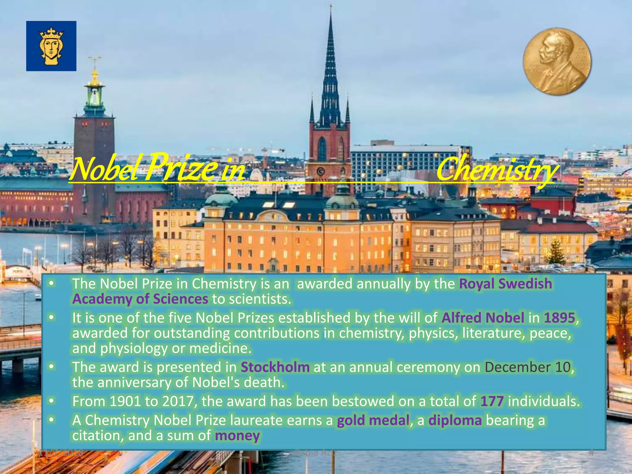
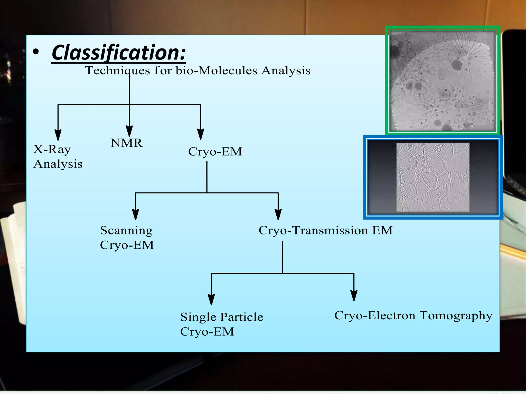
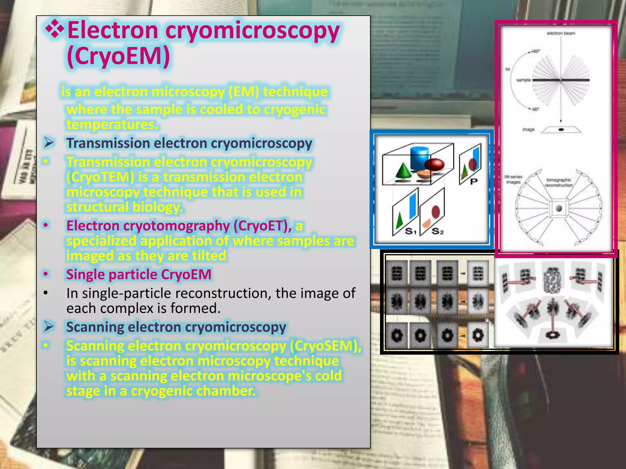
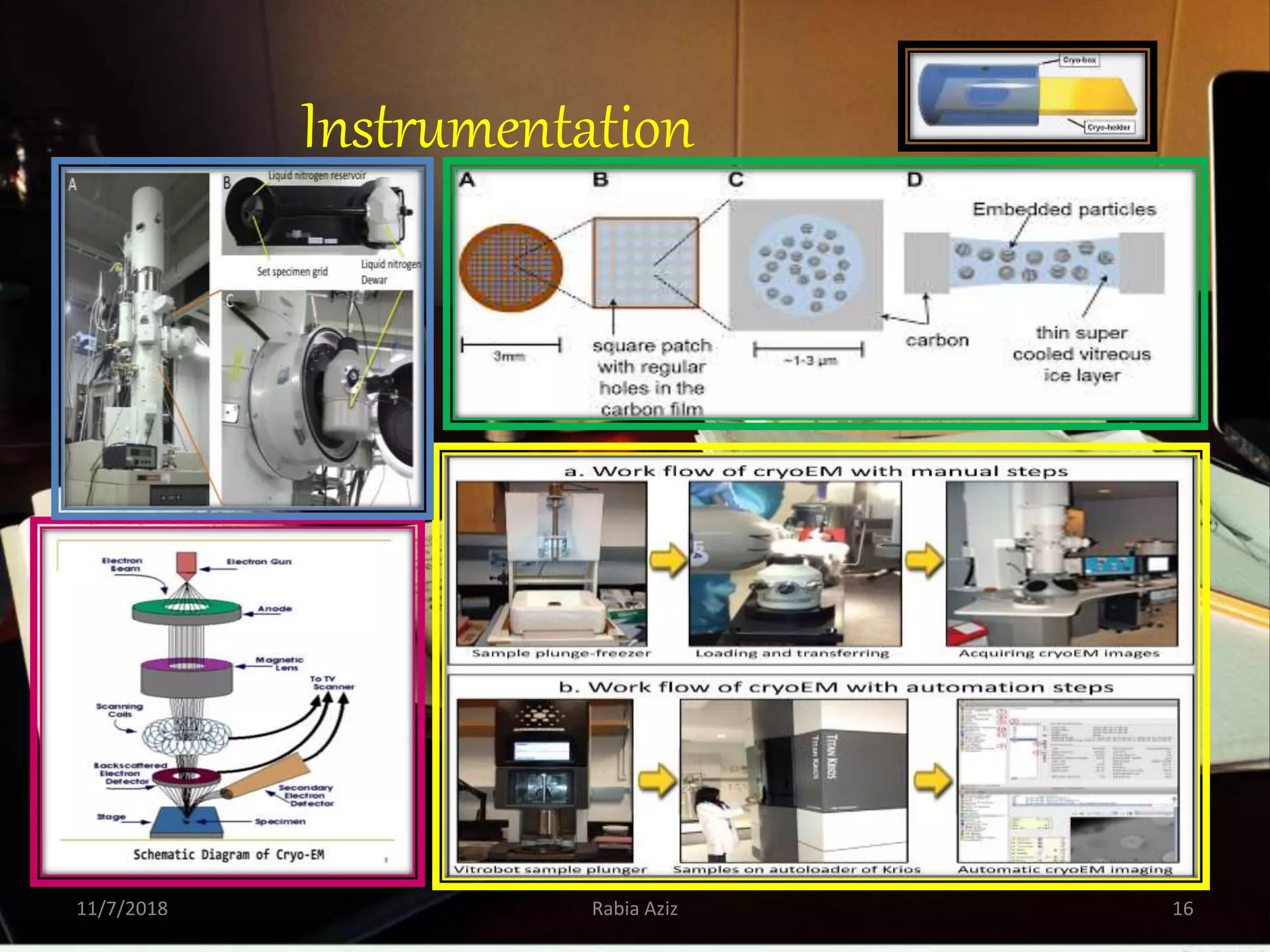
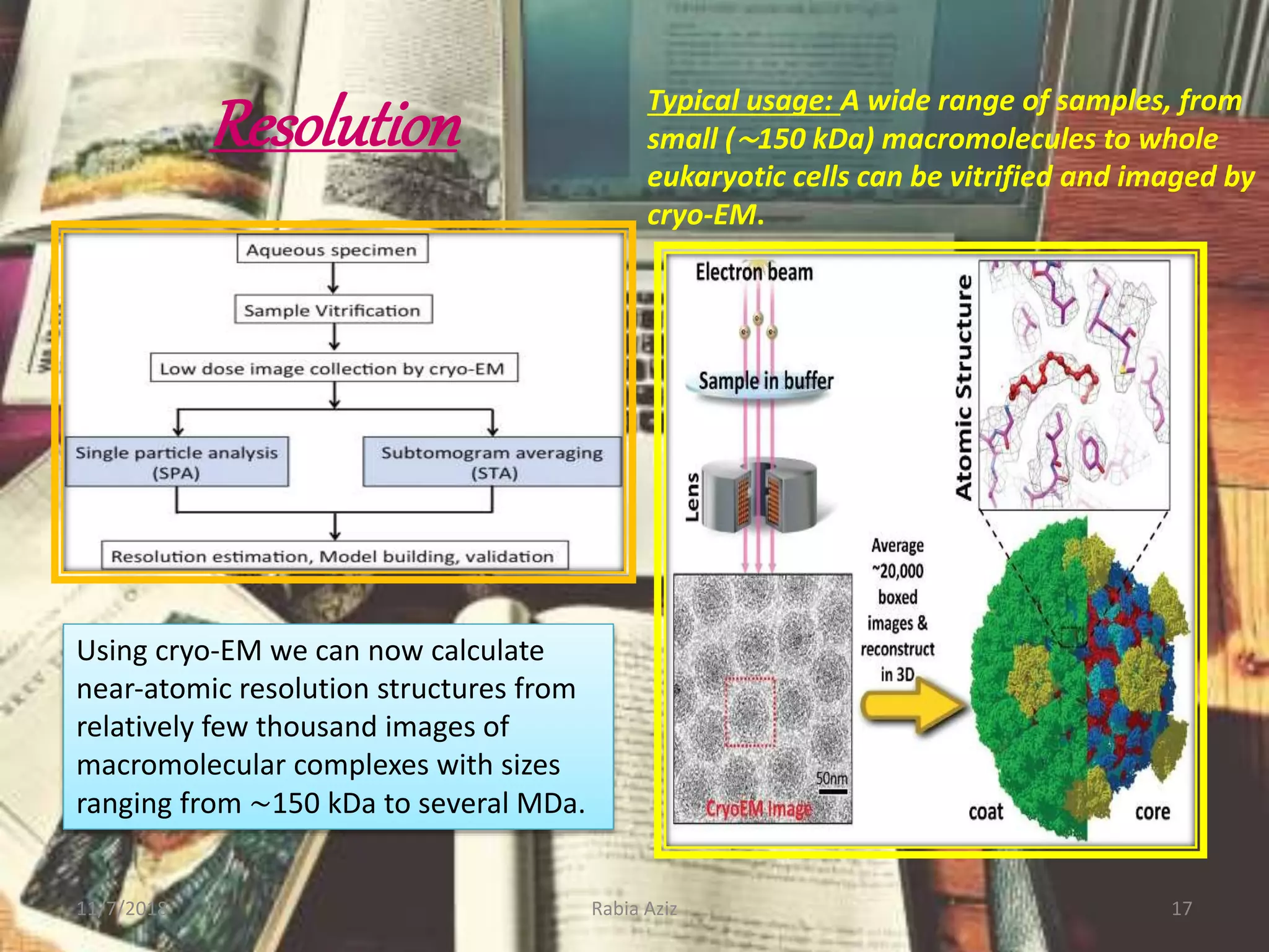
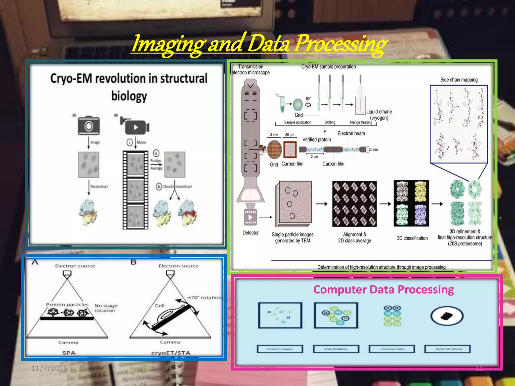
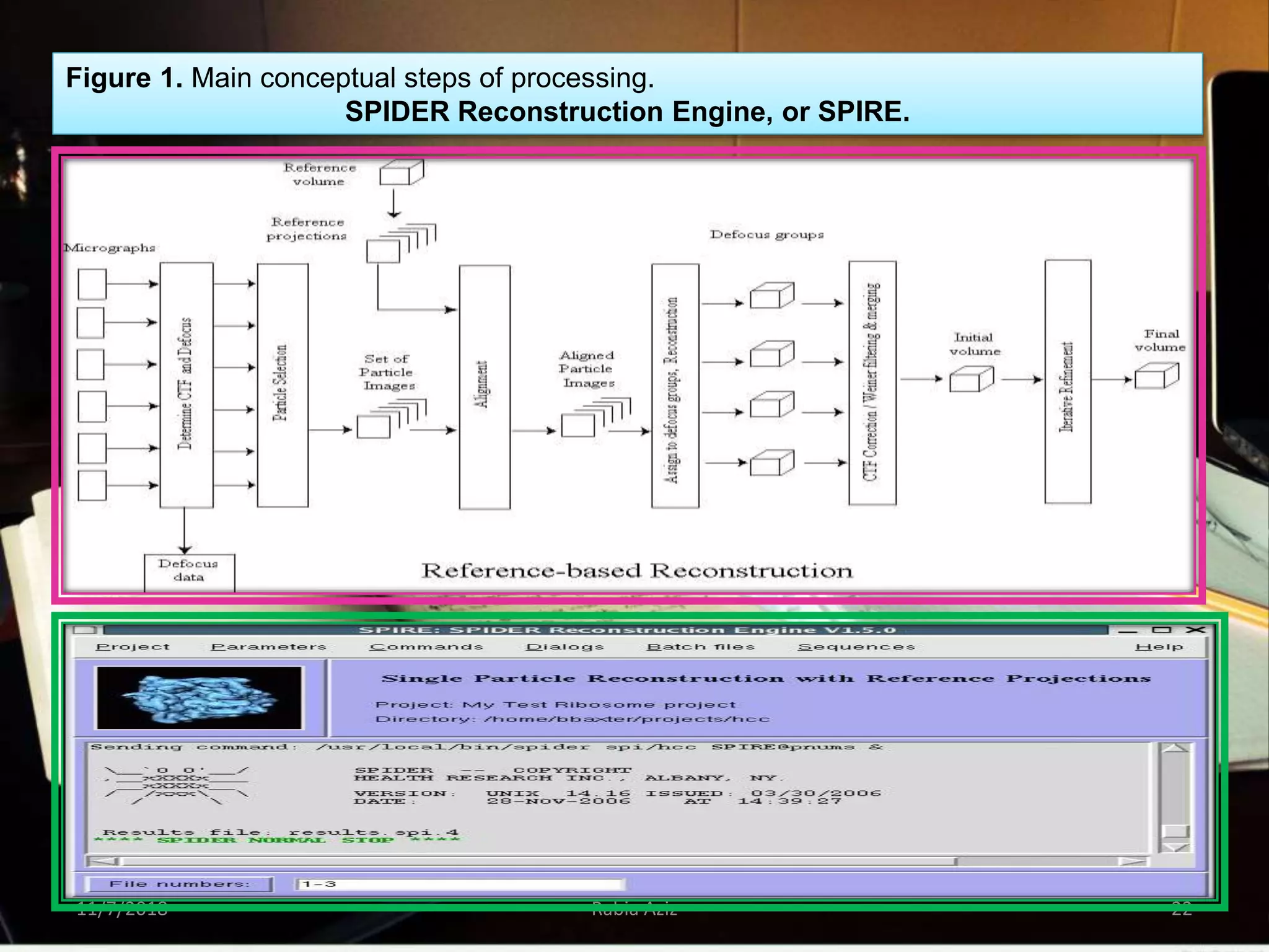
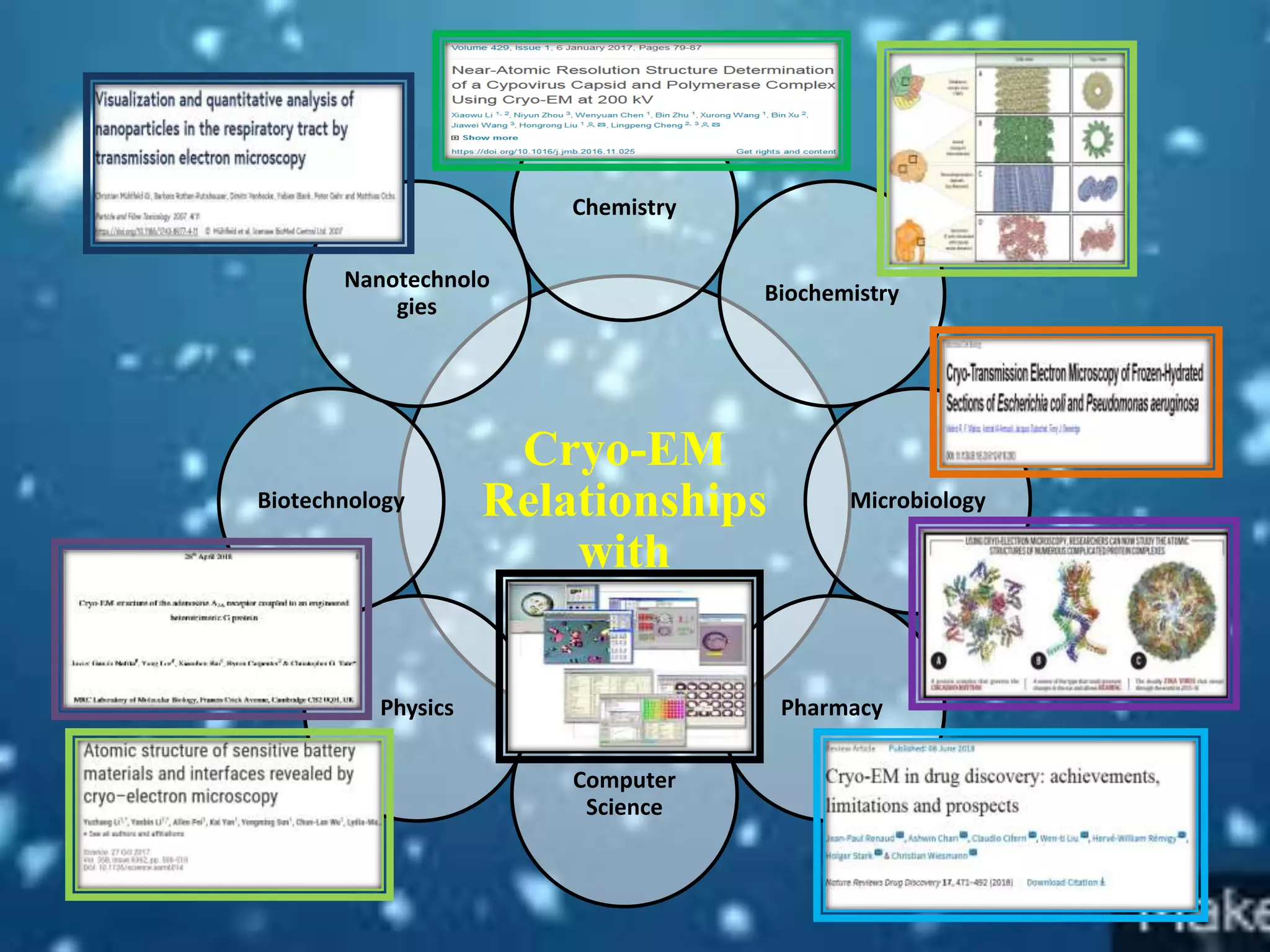
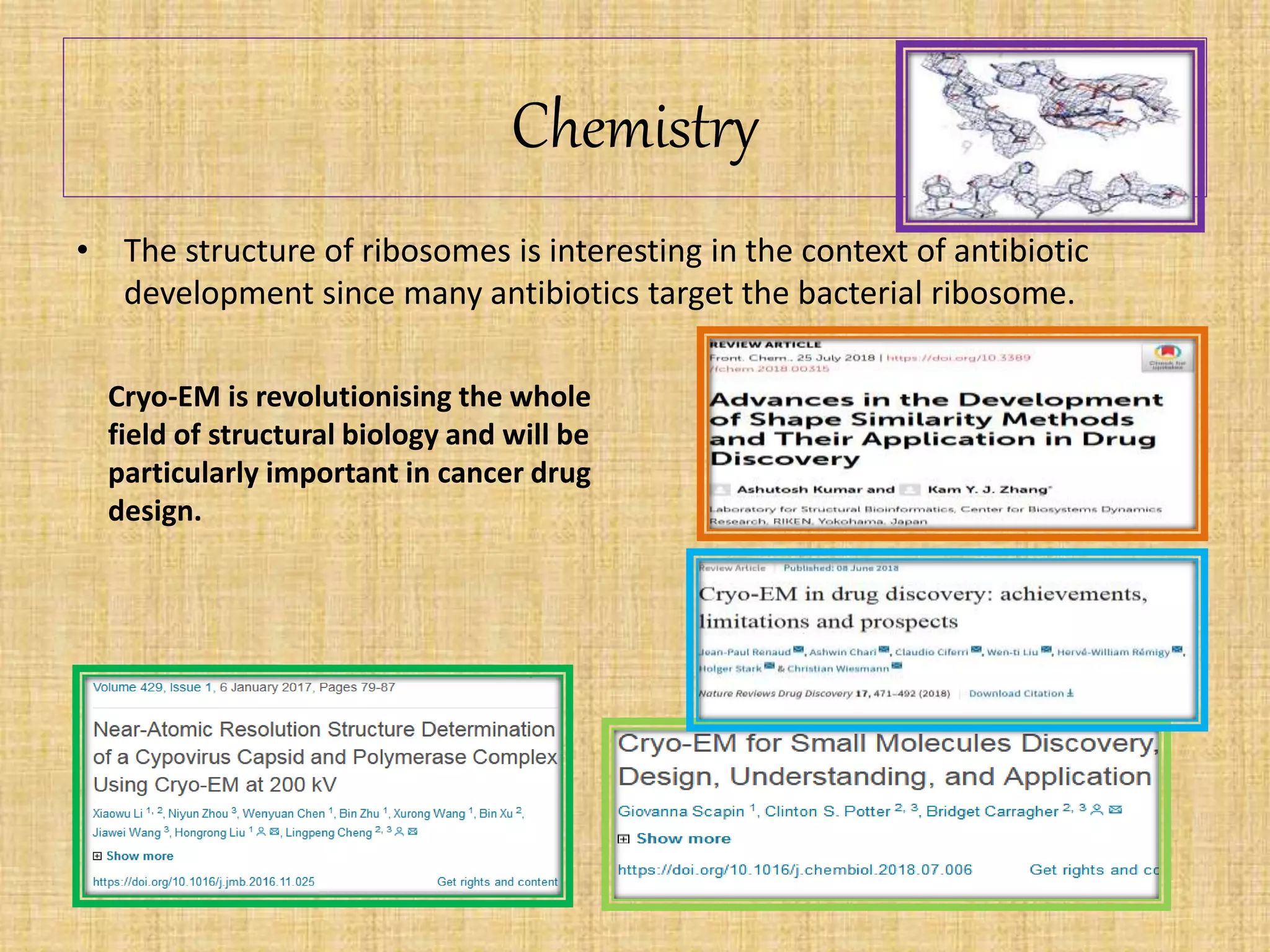
11/7/2018 Rabia Aziz 1
Nobel Prizein Chemistry2017
JoachimFrank
Rabia Aziz
BS-IV Organic Chemistry
Jinnah University for Women
Karachi
11/7/2018 2Rabia Aziz
Introduction
Nobel Prizein Chemistry
• The Nobel Prize in Chemistry is an awarded annually by the Royal Swedish
Academy of Sciences to scientists.
• It is one of the five Nobel Prizes established by the will of Alfred Nobel in 1895,
awarded for outstanding contributions in chemistry, physics, literature, peace,
and physiology or medicine.
• The award is presented in Stockholm at an annual ceremony on December 10,
the anniversary of Nobel's death.
• From 1901 to 2017, the award has been bestowed on a total of 177 individuals.
• A Chemistry Nobel Prize laureate earns a gold medal, a diploma bearing a
citation, and a sum of money
11/7/2018 4Rabia Aziz
11/7/2018 Rabia Aziz 5
1
2 3
4
5
Joachim Frank, Ph.D.
Profession:
Biography:
Fields of Specialization:
(1) Application of electron microscopy and image
analysis to the elucidation of three-dimensional
structure and function of macromolecular
assemblies and cell components.
(2) The mechanism of protein synthesis
2017NobelPrize Winner
Nobel Prize:
He was honoured for his
development of cryo-electron
microscopy, which improves the
imaging of biomolecules.11/7/2018 6Rabia Aziz
JoachimFrank’s
Thesis
1. Investigation of secondary electron
emission of gold at its melting point
2. Investigations of high-resolution
electron micrographs using image
difference and reconstruction methods
11/7/2018 7Rabia Aziz
1. Theory of partial coherence in electron
microscopy
2. Single-particle approaches in electron
microscopy
Working
1. Found in Translation –
Collection of Original
Articles on Single-Particle
Reconstruction and the
Structural Basis of Protein
Synthesis
2. Molecular Machines in
Biology: Workshop of the
Cell
3. Electron Crystallography of
Biological Macromolecules
4. Electron Tomography (2nd
ed.)
5. Three-Dimensional
Electron Microscopy of
Macromolecular
Assemblies (2nd ed.)
Books
Articles
1. "Generalized single-particle cryo-EM – a historical
perspective".
2. "Structure and dynamics of a processive Brownian
motor: the translating ribosome".
3. "The ribosome and the mechanism of protein
synthesis".
4. "Single-particle reconstruction of biological
macromolecules in electron microscopy -- 30 years"
Techniques for Bio-moleculesAnalysis
11/7/2018 Rabia Aziz 8
Cryo-ElectronMicroscopy
• Classification:
Electron cryomicroscopy
(CryoEM)
is an electron microscopy (EM) technique
where the sample is cooled to cryogenic
temperatures.
 Transmission electron cryomicroscopy
• Transmission electron cryomicroscopy
(CryoTEM) is a transmission electron
microscopy technique that is used in
structural biology.
• Electron cryotomography (CryoET), a
specialized application of where samples are
imaged as they are tilted
• Single particle CryoEM
• In single-particle reconstruction, the image of
each complex is formed.
 Scanning electron cryomicroscopy
• Scanning electron cryomicroscopy (CryoSEM),
is scanning electron microscopy technique
with a scanning electron microscope's cold
stage in a cryogenic chamber.
Cryo-ElectronmicroscopyCharacteristics
11/7/2018 12Rabia Aziz
1. Native state of specimen; not stained not
Fixed
2. Specimens are observed in vitreous ice
3. Vitreousbe maintained in microscope ice is
water frozen to -140° C in less than 10-4 sec
4, Low dose parameters are required so the
sample is not destroyed
5. Automated 3D image to get high resolution
images
Cryo-electron microscopy (cryo-EM) and cryo-electron
tomography are employed in structural biology to determine
the structure of vitrified biological samples, such as isolated
proteins and protein complexes, two-dimensional (2D)
protein/lipid crystals, or larger biological objects such as
bacteria and nanocrystals.
Cryo-ElectronMicroscopyProcess
Take a sample
Cool it so rapidly that it is
immobilized before it has
time to crystallize
Cut it into ultra-thin
sections
Observe, in a cryo-
electron microscope
Reconstruct the 3-D
distribution of the
material in the section by
means of computerized
electron tomography CET
The time span from the sample
to the image could be a as say
short as one hour rather than
day
11/7/2018 14Rabia Aziz
Preparation of the material
Vitrification
Cryo-sectioning
Observing in the cryo-electron microscope
Cryo-EM is a method that uncovers the structure of
macromolecular complexes. It involves purifying a
solution of a certain macromolecular complex,
placing the solution onto a grid, freezing it
(cryogenically), and then imaging the frozen film
using an electron microscope. This produces many
images of the same complex in different
orientations.
SampleAnalysis
Grid preparation method Typical samples Typical results
Cryogenic visualisation Macromolecular complexes
(>150 kDa, liposomes,
organelles, prokaryotic and
eukaryotic cells
Visualisation of specimen
with low contrast
Cryogenic reconstruction
(single particle)
Macromolecular complexes
(>150 kDa
Ribosomes, Icosahedral
Viruses
Depending on the specimen,
reconstructions of ∼3–20 Å
Cryogenic reconstruction
(tomography)
Organelles, prokaryotic cells,
thin edge or lamellar of
eukaryotic cells
Resolution of
tomograms < 10 nm,
subtomogram averaging can
produce reconstructions of
>10 Å
Instrumentation
11/7/2018 Rabia Aziz 16
Resolution
11/7/2018 Rabia Aziz 17
Using cryo‐EM we can now calculate
near‐atomic resolution structures from
relatively few thousand images of
macromolecular complexes with sizes
ranging from ∼150 kDa to several MDa.
Typical usage: A wide range of samples, from
small (∼150 kDa) macromolecules to whole
eukaryotic cells can be vitrified and imaged by
cryo-EM.
Imagingand DataProcessing
11/7/2018 Rabia Aziz 18
Computer Data Processing
Joachim Frank Image Analysis
FrankRefinesImageAnalysis:
• In 1975, Joachim Frank began work on the algorithms that would
analyze fuzzy 2D images and reconstruct them into sharp 3D
structures.
• He developed a mathematical method that allowed the computer
to identify different recurring patterns in the image.
• The computer then sorted similar patterns into the same group
and merged the information in these images to generate an
averaged, sharper image. In this way he obtained a number of
high-resolution, two-dimensional images that showed the same
protein but from different angles. The algorithms for the software
were complete in 1981.
• Joachim Frank develops Spider software for conversion of
2D images into 3D structures.
11/7/2018 Rabia Aziz 21
cryo‐sample preparation, data collection and processing workflows, and the
modeling and refinement of structures in cryo‐EM 3D maps
11/7/2018 Rabia Aziz 22
Figure 1. Main conceptual steps of processing.
SPIDER Reconstruction Engine, or SPIRE.
Cryo-EM
Relationships
with
Chemistry
Biochemistry
Microbiology
Pharmacy
Computer
Science
Physics
Biotechnology
Nanotechnolo
gies
Chemistry
• The structure of ribosomes is interesting in the context of antibiotic
development since many antibiotics target the bacterial ribosome.
Cryo-EM is revolutionising the whole
field of structural biology and will be
particularly important in cancer drug
design.
Advantagesand Disadvantages
Advantages:
• Sample is visualised in a native-like state. Processing of
data can yield high-resolution, 3D structural information.
• A great advantage with respect to pharmacological
application is the possibility to identify with high precision
the single amino acids residues and their ligands, as well
as hydrogen bonds and intermolecular interactions.
Disadvantages:
• Biological samples must be imaged with low electron
doses to prevent radiation damage resulting in poor
contrast.
• It is sometimes non-trivial to optimise sample distribution
and ice thickness.
Applications
1. Nanoparticle Research
2. Pharmaceutical Drug Research
3. 3D Structure Visualization of: Single Particles
such as Ribosome, tRNA, Viruses, Proteins,
Macromolecules; Lipid Vesicles
4. Structure and dynamics study
Cryo-EM has a huge potential for the
clarification of pathological mechanisms
and it could assist the early identification
and selection of drug candidates and the
validation of pathological targets.
Virology, neurosciences, tumors and
immunology are the most promising
areas of application,
FutureProspects:
• A number of improvements are expected in future
• High Voltage Electron Source
• Mathematical Correction of Lens Defects
• High Resolution
• Improved Scanners
• Better High-Pressure Freezing
• Improved CCD detectors will remove the need for
computer processing in future
Advancement: A hybrid approach involving the combination of structure
prediction, prospectrometry (CX‐MS), and identification and modeling of the
corresponding segment in the high‐resolution cryo‐EM map.
Conclusion:
• Electron cryomicroscopy (CryoEM) is an electron
microscopy (EM) technique where the sample is cooled to
cryogenic temperatures.
• The cryo-EM method can be used to determine the three-
dimensional structure of biomacromolecules in near native
condition at close to atomic resolution, and has the
potential to reveal conformations of dynamic molecular
complexes.
• Joachim Frank made the technology generally applicable.
Between 1975 and 1986 he developed an image processing
method in which the electron microscope’s fuzzy two
dimensional images are analysed and merged to reveal a
sharp three-dimensional structure.
11/7/2018 Rabia Aziz 29

Cryo-Electron Microscopy

  • 1.
  • 2.
    Nobel Prizein Chemistry2017 JoachimFrank RabiaAziz BS-IV Organic Chemistry Jinnah University for Women Karachi 11/7/2018 2Rabia Aziz
  • 3.
  • 4.
    Nobel Prizein Chemistry •The Nobel Prize in Chemistry is an awarded annually by the Royal Swedish Academy of Sciences to scientists. • It is one of the five Nobel Prizes established by the will of Alfred Nobel in 1895, awarded for outstanding contributions in chemistry, physics, literature, peace, and physiology or medicine. • The award is presented in Stockholm at an annual ceremony on December 10, the anniversary of Nobel's death. • From 1901 to 2017, the award has been bestowed on a total of 177 individuals. • A Chemistry Nobel Prize laureate earns a gold medal, a diploma bearing a citation, and a sum of money 11/7/2018 4Rabia Aziz
  • 5.
  • 6.
    Joachim Frank, Ph.D. Profession: Biography: Fieldsof Specialization: (1) Application of electron microscopy and image analysis to the elucidation of three-dimensional structure and function of macromolecular assemblies and cell components. (2) The mechanism of protein synthesis 2017NobelPrize Winner Nobel Prize: He was honoured for his development of cryo-electron microscopy, which improves the imaging of biomolecules.11/7/2018 6Rabia Aziz JoachimFrank’s
  • 7.
    Thesis 1. Investigation ofsecondary electron emission of gold at its melting point 2. Investigations of high-resolution electron micrographs using image difference and reconstruction methods 11/7/2018 7Rabia Aziz 1. Theory of partial coherence in electron microscopy 2. Single-particle approaches in electron microscopy Working 1. Found in Translation – Collection of Original Articles on Single-Particle Reconstruction and the Structural Basis of Protein Synthesis 2. Molecular Machines in Biology: Workshop of the Cell 3. Electron Crystallography of Biological Macromolecules 4. Electron Tomography (2nd ed.) 5. Three-Dimensional Electron Microscopy of Macromolecular Assemblies (2nd ed.) Books Articles 1. "Generalized single-particle cryo-EM – a historical perspective". 2. "Structure and dynamics of a processive Brownian motor: the translating ribosome". 3. "The ribosome and the mechanism of protein synthesis". 4. "Single-particle reconstruction of biological macromolecules in electron microscopy -- 30 years"
  • 8.
  • 9.
  • 10.
  • 11.
    Electron cryomicroscopy (CryoEM) is anelectron microscopy (EM) technique where the sample is cooled to cryogenic temperatures.  Transmission electron cryomicroscopy • Transmission electron cryomicroscopy (CryoTEM) is a transmission electron microscopy technique that is used in structural biology. • Electron cryotomography (CryoET), a specialized application of where samples are imaged as they are tilted • Single particle CryoEM • In single-particle reconstruction, the image of each complex is formed.  Scanning electron cryomicroscopy • Scanning electron cryomicroscopy (CryoSEM), is scanning electron microscopy technique with a scanning electron microscope's cold stage in a cryogenic chamber.
  • 12.
    Cryo-ElectronmicroscopyCharacteristics 11/7/2018 12Rabia Aziz 1.Native state of specimen; not stained not Fixed 2. Specimens are observed in vitreous ice 3. Vitreousbe maintained in microscope ice is water frozen to -140° C in less than 10-4 sec 4, Low dose parameters are required so the sample is not destroyed 5. Automated 3D image to get high resolution images Cryo-electron microscopy (cryo-EM) and cryo-electron tomography are employed in structural biology to determine the structure of vitrified biological samples, such as isolated proteins and protein complexes, two-dimensional (2D) protein/lipid crystals, or larger biological objects such as bacteria and nanocrystals.
  • 14.
    Cryo-ElectronMicroscopyProcess Take a sample Coolit so rapidly that it is immobilized before it has time to crystallize Cut it into ultra-thin sections Observe, in a cryo- electron microscope Reconstruct the 3-D distribution of the material in the section by means of computerized electron tomography CET The time span from the sample to the image could be a as say short as one hour rather than day 11/7/2018 14Rabia Aziz Preparation of the material Vitrification Cryo-sectioning Observing in the cryo-electron microscope Cryo-EM is a method that uncovers the structure of macromolecular complexes. It involves purifying a solution of a certain macromolecular complex, placing the solution onto a grid, freezing it (cryogenically), and then imaging the frozen film using an electron microscope. This produces many images of the same complex in different orientations.
  • 15.
    SampleAnalysis Grid preparation methodTypical samples Typical results Cryogenic visualisation Macromolecular complexes (>150 kDa, liposomes, organelles, prokaryotic and eukaryotic cells Visualisation of specimen with low contrast Cryogenic reconstruction (single particle) Macromolecular complexes (>150 kDa Ribosomes, Icosahedral Viruses Depending on the specimen, reconstructions of ∼3–20 Å Cryogenic reconstruction (tomography) Organelles, prokaryotic cells, thin edge or lamellar of eukaryotic cells Resolution of tomograms < 10 nm, subtomogram averaging can produce reconstructions of >10 Å
  • 16.
  • 17.
    Resolution 11/7/2018 Rabia Aziz17 Using cryo‐EM we can now calculate near‐atomic resolution structures from relatively few thousand images of macromolecular complexes with sizes ranging from ∼150 kDa to several MDa. Typical usage: A wide range of samples, from small (∼150 kDa) macromolecules to whole eukaryotic cells can be vitrified and imaged by cryo-EM.
  • 18.
    Imagingand DataProcessing 11/7/2018 RabiaAziz 18 Computer Data Processing
  • 19.
  • 20.
    FrankRefinesImageAnalysis: • In 1975,Joachim Frank began work on the algorithms that would analyze fuzzy 2D images and reconstruct them into sharp 3D structures. • He developed a mathematical method that allowed the computer to identify different recurring patterns in the image. • The computer then sorted similar patterns into the same group and merged the information in these images to generate an averaged, sharper image. In this way he obtained a number of high-resolution, two-dimensional images that showed the same protein but from different angles. The algorithms for the software were complete in 1981. • Joachim Frank develops Spider software for conversion of 2D images into 3D structures.
  • 21.
    11/7/2018 Rabia Aziz21 cryo‐sample preparation, data collection and processing workflows, and the modeling and refinement of structures in cryo‐EM 3D maps
  • 22.
    11/7/2018 Rabia Aziz22 Figure 1. Main conceptual steps of processing. SPIDER Reconstruction Engine, or SPIRE.
  • 23.
  • 24.
    Chemistry • The structureof ribosomes is interesting in the context of antibiotic development since many antibiotics target the bacterial ribosome. Cryo-EM is revolutionising the whole field of structural biology and will be particularly important in cancer drug design.
  • 25.
    Advantagesand Disadvantages Advantages: • Sampleis visualised in a native-like state. Processing of data can yield high-resolution, 3D structural information. • A great advantage with respect to pharmacological application is the possibility to identify with high precision the single amino acids residues and their ligands, as well as hydrogen bonds and intermolecular interactions. Disadvantages: • Biological samples must be imaged with low electron doses to prevent radiation damage resulting in poor contrast. • It is sometimes non-trivial to optimise sample distribution and ice thickness.
  • 26.
    Applications 1. Nanoparticle Research 2.Pharmaceutical Drug Research 3. 3D Structure Visualization of: Single Particles such as Ribosome, tRNA, Viruses, Proteins, Macromolecules; Lipid Vesicles 4. Structure and dynamics study Cryo-EM has a huge potential for the clarification of pathological mechanisms and it could assist the early identification and selection of drug candidates and the validation of pathological targets. Virology, neurosciences, tumors and immunology are the most promising areas of application,
  • 27.
    FutureProspects: • A numberof improvements are expected in future • High Voltage Electron Source • Mathematical Correction of Lens Defects • High Resolution • Improved Scanners • Better High-Pressure Freezing • Improved CCD detectors will remove the need for computer processing in future Advancement: A hybrid approach involving the combination of structure prediction, prospectrometry (CX‐MS), and identification and modeling of the corresponding segment in the high‐resolution cryo‐EM map.
  • 28.
    Conclusion: • Electron cryomicroscopy(CryoEM) is an electron microscopy (EM) technique where the sample is cooled to cryogenic temperatures. • The cryo-EM method can be used to determine the three- dimensional structure of biomacromolecules in near native condition at close to atomic resolution, and has the potential to reveal conformations of dynamic molecular complexes. • Joachim Frank made the technology generally applicable. Between 1975 and 1986 he developed an image processing method in which the electron microscope’s fuzzy two dimensional images are analysed and merged to reveal a sharp three-dimensional structure.
  • 29.