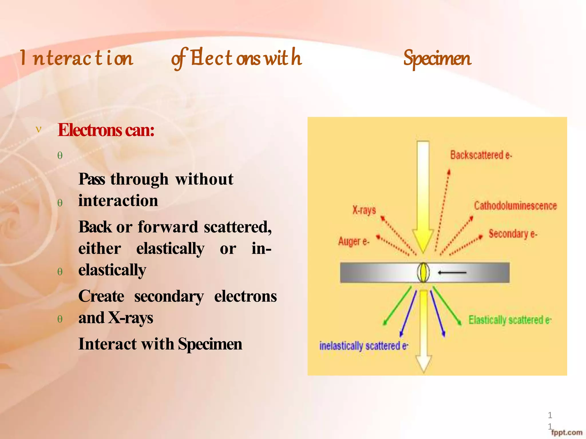
Khurshid anwar
BS BOTANY 8TH
Cryo- ELECTRON
Microscopy
4
Content
5
Introductionto ElectronMicroscopy
WhyUseCryo-EM?
ProceduresInvolvedin Cryo-EM
ProsandConsof theTechnique
Applications
FutureProspects
Conclusion
DEFINITIONS:
”Cryoelectron microscopy is a method for imaging
frozen-hydrated specimens at cryogenic
temperatures by electronmicroscopy”
MEANING
Cryo:
“A combining form meaning icy cold, frost”
Microscopy:
“Technical field of using microscopes to view objects
and areas of objects that cannot be seen with the
naked eye”
How Do We Study Cells?
8
Can study live cells
Colorimaging
Relatively fast andeasy
Relativelycheap
Resolution Limit 200nm
Light Microscopy Electron Miic roscopy
Can study ultra-structure
Needto kill and ‘fix’ cells
Difficult and time
Consuming Expensive
Resolution Limit 2nm
Light
Microscope
Electron
Microscope
9
Electron Microscopy
1
0
Twotypesof the ElectronMicroscope:
TransmissionElectronMicroscope(TEM):Abeam of
electrons interacts with the specimento form an
image
ScanningElectronMicroscope(SEM):
Abeamof electrons scansthe samplesurface to create
image
I nterac t ion of Elect onswith Specimen
 Electronscan:




Pass through without
interaction
Back or forward scattered,
either elastically or in-
elastically
Create secondary electrons
andX-rays
Interact with Specimen
1
1
Cryo-Electron Microscopy
1
2
A Formof EM;sampleisstudied at CryogenicTemperatures
Native state of specimen;not stainednot Fixed
Specimensare observedin vitreousice
Cryo-fixation;
Rapidfreezing of sample
Cont..
• Automated 3Dimageto get high resolutionimages
• Lowdoseparameters are required sothe sample isnot
destroyed
Origin and Development
1
4
1980s
Bruggellerand Mayer produced vitrified water
Dubochet and McDowall produced a thin layer of vitrified
waterusing liquid ethane
All these findings have been combined to produce a simple
but extremely powerful method for the production of high-
resolution images
WHy Use I T ?
1
6
Native State ofSample
Vitrified
Water
NoStaining
Automated 3DReconstruction
ValidatedStructures
10Schemat i c Di agr am of Cryo-EM
Procedures
Involv ed in Cryo-EM
18
The Microscope
ACryo-EM isaTEMwith an
additional specimenholder
which:
Enablethe viewing of the
frozen-hydrated specimen
Maintains Liquid Nitrogen
or Liquid Helium
temperatures
19
Specimen Preparation
Twomethods of specimenpreparationare:
ThinFilm:Specimenisplaced on EMgrid and is
rapidly frozen without crystallizingit
VitreousSections:Largersamplesare vitrified by
high pressurefreezing, cut thinly andplaced on
the EMgrid
20
Adrop of the sampleisplaced at the endof the plungerandrapidly
immersedinto the cryogentank
21
1) Vitrification
RapidCooling is required to
avoidthe formation of ice:
Rapidcooling traps the water ina
vitrified state in which it doesnot
crystallize
Vitrified state ismaintained by keeping
itat liquid nitrogentemperature
22
Cont..
• Vitrified state canbe maintained forlong periods
Sample is placed on carbon grid and dipped into a bath
of ethane held in acontainer of liquid nitrogen
2) Cryo-Sectioning
2
4
Wholecellsandtissuesaretoothickto bespreadintoathin layer
Firstvitrify sampleandthen cut into thin sectionsusingdiamond knives
Sectioningisadifficult task,distortions are madeinsample
Thesedistortions causealossin order of the structure and makesit
difficult for imagesto increasethe signal-to-noiseratio
Cryo-EM Gr i ds
The grid on which the sample is placed is made from
carbon
Highquality carbongrid isusedto get better results
Twotypes of Gridsare:
Continuous Films: Enable the sample to cover
the surface asaregular, thin layer
Holey Films: Have a network of holes of a
desired sizein which the sampleisspread
ACarbonGrid
25
A Holey Grid
Supporting
Carbon Film
Ice Holes
Metal Grid
26
Cryogens
27
Cryogensare usedfor Chilling and freezingpurpose
Typeof cryogenusedaffects the rate offreezing
Commoncryogensare LiquidNitrogen,Ethaneor Propane
Nitrogen isnot directly used; It canmakecrystalsdue to slow
cooling
Formation of i c e
At low temperature andpressure,
water freezes into threeforms:
Vitreous
Cubic
Hexagonal
Vitreous ice isobtained byrapid
cooling of liquidwater
An Ice Hole
Particles are randomly
positioned and
orientated
28
Observation of the Specimen
29
Thecontrastof the specimendependson:
Specimen itself
Defocusvalue of the objectivelens
Thicknessof theice
3D Reconstruction
30
3Dreconstructionprocessestimates the unknown
orientations and 3Dstructure at the sametime;
3Delectron density mapsare created from 2Dprojections
Anglesof projections relative toeachother are determined
Findcommon line projections todetermine relative angles
1) Re-projections
•3D density map can be used to generate
projections that can be used to realign the raw
images
•Processmay have to be repeated severaltimes
31
• Thereare three methodsof observingandrecording
images:
• FluorescentScreen
• PhotographicFilm
• COD Cameras
METHODS
2) Automated Particle Picking
Identify particles in micrograph and cut
out patchescontaining one particleeach
Thiscanbe doneautomatically
Manual processistedious anddifficult
34
3) Images Enhancement
Image Noise is the random variation of brightness or color
information in imagesproduced by thesensor
Cryo EMimagesare very noisy and havevery lowcontrast
Smooth the noise aswell asenhancethe contrast
35
Structure of Hepatitis B Virus Solved by
Cryo-EM
Example
36
Pros and Cons
37
Advantage:Structure remains native andno
dehydration isrequired
Limitation: It is not possible to look at the sample for a
long time because of beam damage and it causes poor
resolution
Applic a t ions
38
Nanoparticle Research
Pharmaceutical DrugResearch
CONT..
• 3DStructure Visualization of:
• SingleParticles suchasRibosome,
• Viruses
• Proteins
• Macromolecules; LipidVesicles
Future Prospects
40
Anumberof improvementsareexpectedinfuture
• HighVoltage ElectronSource
•Mathematical Correction of Lens
• DefectsHighResolution
Cont..
• Improved Scanners
• Better High-PressureFreezing
• Improved CODdetectors will remove the need for
computer
• processing in future
Conclusions
42
A. Cryo-EM is a form of Transmission Electron
Microscopy (TEM) where the sample is studied in its
native state at cryogenic temperatures
• Usedfor 3Dvisualization of biologicalmolecules
Cont..
• Resolution of Cryo-EM is not high enough but it is improving
using different computertechniques
• With the advancement of technology, this technique will
certainly improve
REFERENCES
44






• Cryo-electron microscopy, Methods in Molecular Biophysics,
Spring2009
• Cryo-electron microscopy: taking back the knight
Stephen Fullerpy, MICROBIOLOGYTODAY VOL
26/MAY 99
• http://www.ncbi.nlm.nih.gov/pmc/articles/PMC2678009/pd
f/nihms87373.pdf
• http://www.bbc.co.uk/dna/hub/A914302#back10
• http://www.physorg.com/news192189631.html
C0NT..
• http://en.wikipedia.org/wiki/Cryo-
electron_microscopy
• http://www.jic.ac.uk/microscopy/intro_EM.html
• http://en.wikibooks.org/wiki/Structural_Bi
ochemistry/Proteins/CryoElectron_Microscpy
• http://www.ncbi.nlm.nih.gov/pmc/articles/PMC2726
835/pdf/nihms100338.
cryo electron microscopy

cryo electron microscopy

  • 3.
  • 4.
  • 5.
  • 6.
    DEFINITIONS: ”Cryoelectron microscopy isa method for imaging frozen-hydrated specimens at cryogenic temperatures by electronmicroscopy”
  • 7.
    MEANING Cryo: “A combining formmeaning icy cold, frost” Microscopy: “Technical field of using microscopes to view objects and areas of objects that cannot be seen with the naked eye”
  • 8.
    How Do WeStudy Cells? 8 Can study live cells Colorimaging Relatively fast andeasy Relativelycheap Resolution Limit 200nm Light Microscopy Electron Miic roscopy Can study ultra-structure Needto kill and ‘fix’ cells Difficult and time Consuming Expensive Resolution Limit 2nm
  • 9.
  • 10.
    Electron Microscopy 1 0 Twotypesof theElectronMicroscope: TransmissionElectronMicroscope(TEM):Abeam of electrons interacts with the specimento form an image ScanningElectronMicroscope(SEM): Abeamof electrons scansthe samplesurface to create image
  • 11.
    I nterac tion of Elect onswith Specimen  Electronscan:     Pass through without interaction Back or forward scattered, either elastically or in- elastically Create secondary electrons andX-rays Interact with Specimen 1 1
  • 12.
    Cryo-Electron Microscopy 1 2 A FormofEM;sampleisstudied at CryogenicTemperatures Native state of specimen;not stainednot Fixed Specimensare observedin vitreousice Cryo-fixation; Rapidfreezing of sample
  • 13.
    Cont.. • Automated 3Dimagetoget high resolutionimages • Lowdoseparameters are required sothe sample isnot destroyed
  • 14.
    Origin and Development 1 4 1980s BruggellerandMayer produced vitrified water Dubochet and McDowall produced a thin layer of vitrified waterusing liquid ethane All these findings have been combined to produce a simple but extremely powerful method for the production of high- resolution images
  • 16.
    WHy Use IT ? 1 6 Native State ofSample Vitrified Water NoStaining Automated 3DReconstruction ValidatedStructures
  • 17.
    10Schemat i cDi agr am of Cryo-EM
  • 18.
  • 19.
    The Microscope ACryo-EM isaTEMwithan additional specimenholder which: Enablethe viewing of the frozen-hydrated specimen Maintains Liquid Nitrogen or Liquid Helium temperatures 19
  • 20.
    Specimen Preparation Twomethods ofspecimenpreparationare: ThinFilm:Specimenisplaced on EMgrid and is rapidly frozen without crystallizingit VitreousSections:Largersamplesare vitrified by high pressurefreezing, cut thinly andplaced on the EMgrid 20
  • 21.
    Adrop of thesampleisplaced at the endof the plungerandrapidly immersedinto the cryogentank 21
  • 22.
    1) Vitrification RapidCooling isrequired to avoidthe formation of ice: Rapidcooling traps the water ina vitrified state in which it doesnot crystallize Vitrified state ismaintained by keeping itat liquid nitrogentemperature 22
  • 23.
    Cont.. • Vitrified statecanbe maintained forlong periods Sample is placed on carbon grid and dipped into a bath of ethane held in acontainer of liquid nitrogen
  • 24.
    2) Cryo-Sectioning 2 4 Wholecellsandtissuesaretoothickto bespreadintoathinlayer Firstvitrify sampleandthen cut into thin sectionsusingdiamond knives Sectioningisadifficult task,distortions are madeinsample Thesedistortions causealossin order of the structure and makesit difficult for imagesto increasethe signal-to-noiseratio
  • 25.
    Cryo-EM Gr ids The grid on which the sample is placed is made from carbon Highquality carbongrid isusedto get better results Twotypes of Gridsare: Continuous Films: Enable the sample to cover the surface asaregular, thin layer Holey Films: Have a network of holes of a desired sizein which the sampleisspread ACarbonGrid 25
  • 26.
    A Holey Grid Supporting CarbonFilm Ice Holes Metal Grid 26
  • 27.
    Cryogens 27 Cryogensare usedfor Chillingand freezingpurpose Typeof cryogenusedaffects the rate offreezing Commoncryogensare LiquidNitrogen,Ethaneor Propane Nitrogen isnot directly used; It canmakecrystalsdue to slow cooling
  • 28.
    Formation of ic e At low temperature andpressure, water freezes into threeforms: Vitreous Cubic Hexagonal Vitreous ice isobtained byrapid cooling of liquidwater An Ice Hole Particles are randomly positioned and orientated 28
  • 29.
    Observation of theSpecimen 29 Thecontrastof the specimendependson: Specimen itself Defocusvalue of the objectivelens Thicknessof theice
  • 30.
    3D Reconstruction 30 3Dreconstructionprocessestimates theunknown orientations and 3Dstructure at the sametime; 3Delectron density mapsare created from 2Dprojections Anglesof projections relative toeachother are determined Findcommon line projections todetermine relative angles
  • 31.
    1) Re-projections •3D densitymap can be used to generate projections that can be used to realign the raw images •Processmay have to be repeated severaltimes 31
  • 32.
    • Thereare threemethodsof observingandrecording images: • FluorescentScreen • PhotographicFilm • COD Cameras METHODS
  • 34.
    2) Automated ParticlePicking Identify particles in micrograph and cut out patchescontaining one particleeach Thiscanbe doneautomatically Manual processistedious anddifficult 34
  • 35.
    3) Images Enhancement ImageNoise is the random variation of brightness or color information in imagesproduced by thesensor Cryo EMimagesare very noisy and havevery lowcontrast Smooth the noise aswell asenhancethe contrast 35
  • 36.
    Structure of HepatitisB Virus Solved by Cryo-EM Example 36
  • 37.
    Pros and Cons 37 Advantage:Structureremains native andno dehydration isrequired Limitation: It is not possible to look at the sample for a long time because of beam damage and it causes poor resolution
  • 38.
    Applic a tions 38 Nanoparticle Research Pharmaceutical DrugResearch
  • 39.
    CONT.. • 3DStructure Visualizationof: • SingleParticles suchasRibosome, • Viruses • Proteins • Macromolecules; LipidVesicles
  • 40.
    Future Prospects 40 Anumberof improvementsareexpectedinfuture •HighVoltage ElectronSource •Mathematical Correction of Lens • DefectsHighResolution
  • 41.
    Cont.. • Improved Scanners •Better High-PressureFreezing • Improved CODdetectors will remove the need for computer • processing in future
  • 42.
    Conclusions 42 A. Cryo-EM isa form of Transmission Electron Microscopy (TEM) where the sample is studied in its native state at cryogenic temperatures • Usedfor 3Dvisualization of biologicalmolecules
  • 43.
    Cont.. • Resolution ofCryo-EM is not high enough but it is improving using different computertechniques • With the advancement of technology, this technique will certainly improve
  • 44.
    REFERENCES 44       • Cryo-electron microscopy,Methods in Molecular Biophysics, Spring2009 • Cryo-electron microscopy: taking back the knight Stephen Fullerpy, MICROBIOLOGYTODAY VOL 26/MAY 99 • http://www.ncbi.nlm.nih.gov/pmc/articles/PMC2678009/pd f/nihms87373.pdf • http://www.bbc.co.uk/dna/hub/A914302#back10 • http://www.physorg.com/news192189631.html
  • 45.
    C0NT.. • http://en.wikipedia.org/wiki/Cryo- electron_microscopy • http://www.jic.ac.uk/microscopy/intro_EM.html •http://en.wikibooks.org/wiki/Structural_Bi ochemistry/Proteins/CryoElectron_Microscpy • http://www.ncbi.nlm.nih.gov/pmc/articles/PMC2726 835/pdf/nihms100338.