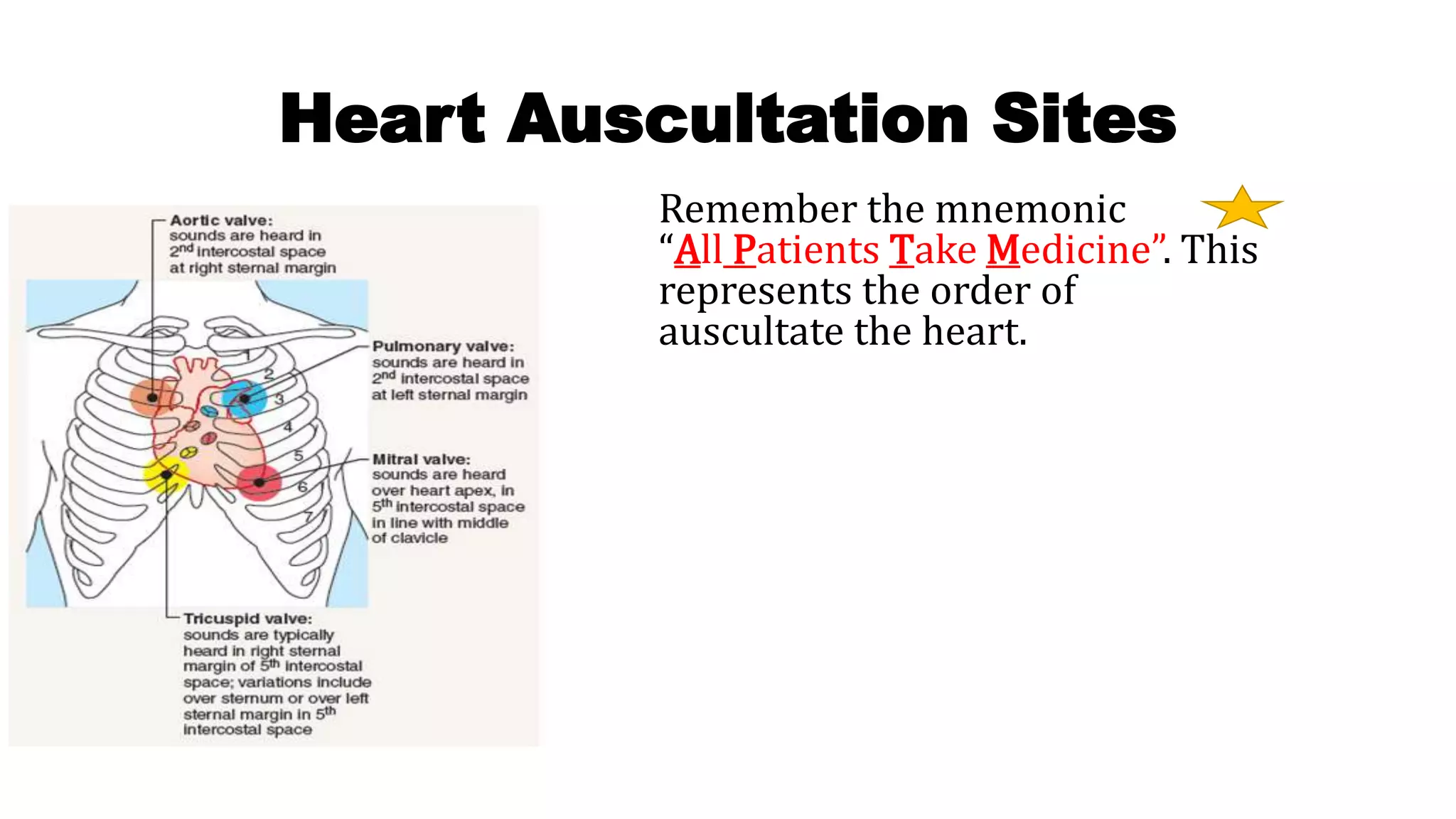
2. • Auscultation is the act of listening to sounds made by internal
organs and vessels of the human body.
• This is usually done with a device called a stethoscope.
• It is a technique used to examine the respiratory system (breath
sounds), cardiovascular system (heart sounds and vascular bruits)
and gastrointestinal system (bowel sounds).
3. Anatomy of Lungs
• The right lung is divided into 3
main lobes which are
separated by the These lobes
are: the superior, middle and
inferior lobes. The left lung is
separated by the oblique
fissure into 2 main lobes: the
superior and inferior.
4. Auscultation points
Auscultate point Location
APEX of the lungs bilaterally 2cm superior to medial 1/3 of
clavicle
Superior Lobes anterior 2nd intercostal space mid
clavicular line
Superior Lobes posterior Between C7 & T3
Middle lobe right anterior only 4th intercostal space mid-
clavicular line
Inferior Lobes bilaterally
anterior
6th intercostal space, mid-
axillary line
Inferior Lobes bilaterally
posteriorly
between T3 & T10
5. Identification of Inter Costal
Space
From the angle of Louis, move your fingers to
the right and you will feel a gap between the
ribs. This gap is the 2nd Intercostal space
6. How to Auscultate Lung Sounds
Listen to both the anterior and posterior sides of the chest
Start at the top and work your way to the bottom of the chest
while comparing sides
When listening note the following:
A full inspiration and expiration cycle
The inspiration and expiration sound’s pitch, quality,
duration, and if it is normal sounding
is there anything “weird” heard along with the inspiration
and expiration.
7. Have the patient sitting up with arms resting on lap. When listening
to the posterior side of the chest the arms need to definitely be in
the lap so the scapulae are separated.
Use the diaphragm of the stethoscope to auscultate at various
locations
Have patient breathe in and out through mouth slowly while
listening. Allow the patient to set the pace to prevent
hyperventilating , especially patients with breathing disorders like
COPD.
8. Anterior Lung Auscultation Points
Start at: the apex of the lung which
is right above the clavicle
2nd intercostal space to assess the
right and left upper lobes.
4th intercostal space you will be assessing
the right middle lobe and the left upper
lobe
midaxillary at the 6th intercostal space you
will be assessing the right and left lower
lobes.
9. Posterior Lung Auscultation Points
Start right above the scapulae to
listen to the apex of the lungs.
Then find C7 (which is
the vertebral prominence) and
go to T3…in between the
shoulder blades and spine.
This will assess the right and
left upper lobes.
Then from T3 to T10 you will be
able to assess the right and left
lower lobes.
11. Name of sound Duration of sound Intensity of
Expiratory sound
Pitch of expiratory
sound
Location where heard
normally
Bronchial inspiration will be
slightly SHORTER than
expiration
Loud Relatively high Over on manubrium
(Larger Proximal airway)
Broncho
vesicular:
inspiration and
expiration will be EQUAL
Intermediate Intermediate anteriorly: 1st and 2nd
intercostal space near the
sternum
posteriorly: between the
scapulae
Vesicular: inspiration will be
slightly GREATER than
expiration
Soft Relatively Low auscultated anteriorly and
posteriorly and heard
over peripheral lung fields
Tracheal Inspiration and
expiration will be about
to Equal
Very Loud Relatively high Over the trachea in the neck
12. Adventitious Sounds
A) Discontinuous Lung Sounds:
• These are extra sounds heard LESS than 0.2 seconds during a full
respiration cycle.
Coarse Crackles:
Crackles are also known as:
rales
•Auscultated during inspiration
and can extend into expiration
as well
•Low-pitched, wet bubbling
sound
•May be heard in patient with
Coarse Crackles:
Crackles are also known as:
rales
•Auscultated during
inspiration and can extend
into expiration as well
•Low-pitched, wet bubbling
sound
•May be heard in patient with
fluid overload, pneumonia etc.
Pleural Friction Rub:
•Auscultated during
inspiration and
expiration
•Low-pitched/harsh
grating sound
•Patients may have pain
when breathing in and
out due to inflammation
of pleural layers
•May be heard in patients
13. • B) Continuous Lung Sounds:
• These are extra sounds heard MORE than 0.2 second during a full
respiration cycle
High Pitched, Polyphonic
Wheeze
Also known as: Sibilant
Wheeze
•Auscultated mainly in
expiration but may be present
during inspiration
•Sounds like a high-pitched
musical instrument with
MORE than one type of sound
quality
•May be heard in patients with
asthma
Low-pitched, Monophonic
Wheeze
Also known as: Sonorous
Wheeze or Rhonchi
•Auscultated mainly in
expiration but may be present at
anytime
•Sounds like a low-pitched
whistling tune or whine with
ONE type of sound quality
•May be heard in patients with
COPD or pneumonia etc.
Stridor
•Auscultated during
inspiration
•high-pitched whistling
or gasping sound with
harsh sound quality
•May be seen in
children with
conditions such as
croup or epiglottitis or
anyone with an airway
obstruction etc.
14. Heart sound
• Heart sounds are caused by the closure of heart valves. The first sound you
hear is S1 and is caused by the closure of the atrioventricular valves (AV)
TRICUSPID AND MITRAL VALVES. This sounds like “LUB”. S1
The second sound you hear is S2 and is caused by the closure of the semilunar
valves (SL) AORTIC AND PULMONIC VALVES. This sounds like “DUB”. S2
S3 –associated with blood flowing into the ventricles
S4 –associated with atrial contraction
Four heart sounds can be recorded by phonocardiography, but
normally only the first and the second heart sounds, are
audible through a stethoscope
15. • Normally, the AV valves close at the same time and the same is true
for the SL valves. However, in some people these valves may close
asynchronously and this would cause a split in sound.
• S1 split: Tricuspid and mitral valves closing asynchronously
• S2 split: Aortic and pulmonic valves closing asynchronously
17. Aortic: found
right of the
sternal border
in the 2nd
intercostal
space
REPRESENTS
S2 “dub”
Pulmonic: found
left of the
sternal border
in the 2nd
intercostal
space
REPRESENTS
S2 “dub”
Erb’s
Point: found
left of the
sternal
border in the
3rd
intercostal
space
Tricuspid: found
left of the sternal
border in the 4th
intercostal space
REPRESENTS S1
“lub”
Mitral: found
midclavicular in
the 5th
intercostal space
REPRESENTS S1
“lub” (also the site
of point of
maximal impulse)
18. • The Base of the heart
includes the aortic and
pulmonic areas, and S2
will be loudest at the
base.
• Aortic and pulmonic
murmurs are heard
best at the base with
the patient leaning
forward and sitting up
with the diaphragm of
the stethoscope.
The Apex of the heart
includes the tricuspid and
mitral areas, and S1 will be
loudest at the apex.
S3 and S4 along with mitral
stenosis murmurs will be
heard best at this position
with the patient lying on
their left side with the bell of
the stethoscope.
19. Basics about S3 & S4
* S3 and S4 are heard best at the apex of the heart with the bell of the
stethoscope while the patient is on their left side.
S3: heard after S2 and sounds like “LUB-DUB-TA”
• Caused by vibrations of ventricle filling from a resistant ventricle due to fluid volume
overload or heart failure.
S4: heard before S1 and sounds like “TA-LUB-DUB”
• Caused by ventricle resistance from an atrial “kick” during presystole (hypertrophic left
ventricle)
20. Heart Murmur
• Cardiac murmurs are abnormal heart sounds in the form of noises resulting
from turbulent blood flow,
• Murmurs may be:
• Systolic murmurs, if they occur during systole.
• Diastolic murmurs, if they occur during diastole
• Causes:
• Stenosis (narrowing) of heart valves
• Incompetence of heart valves
• Increase of blood flow or decrease of blood viscosity (as in anemia).
21. Two Basic Types of Valvular Diseases
Valvular stenosis:
• narrowing of the valve
valvular insufficiency
(incompetence):
• valve is unable to close fully; so
there is regurgitation