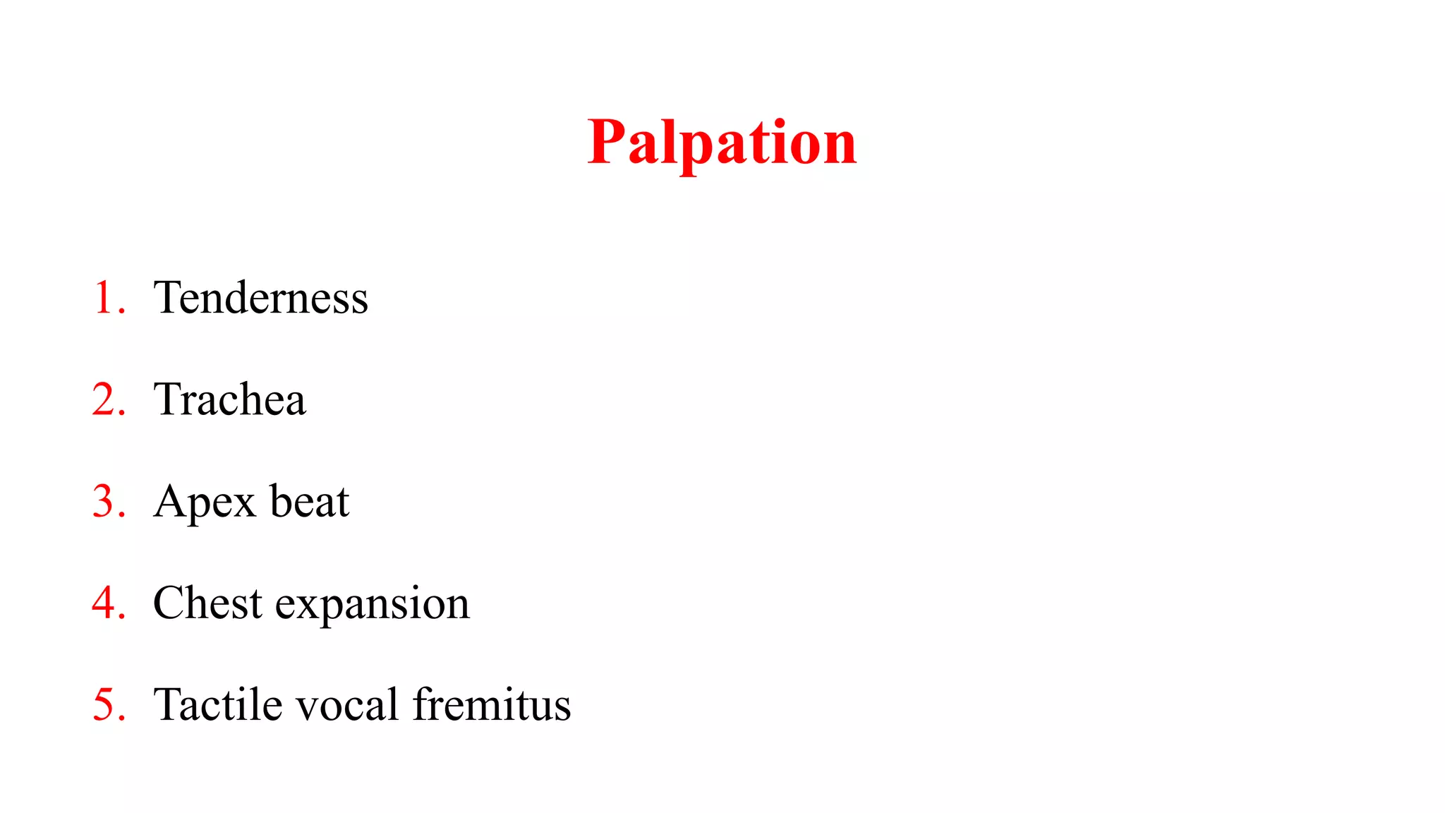
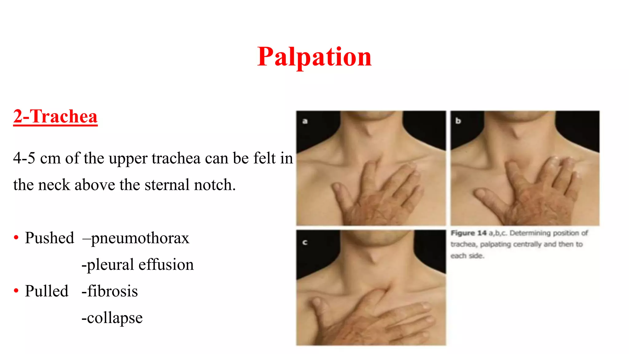
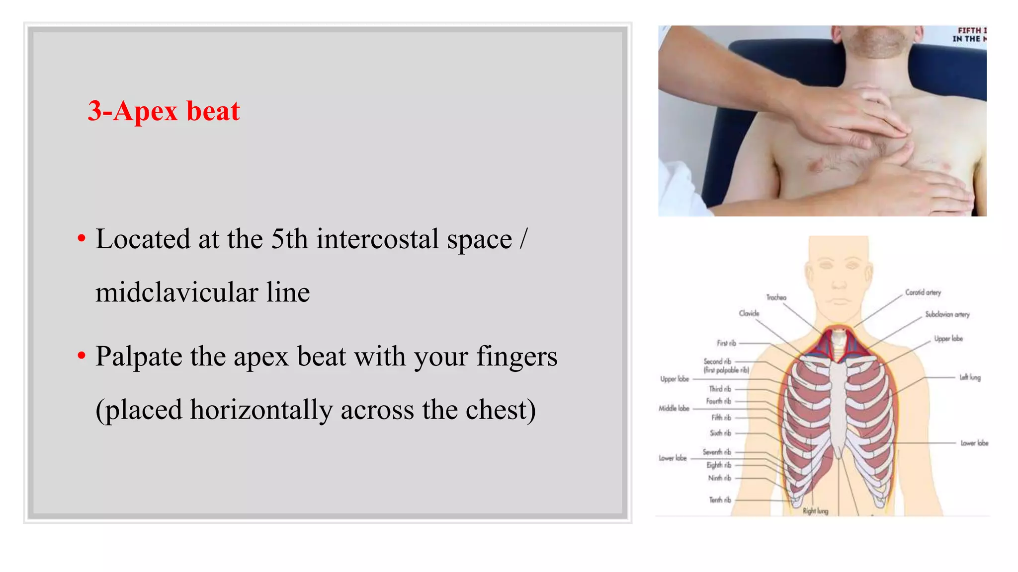
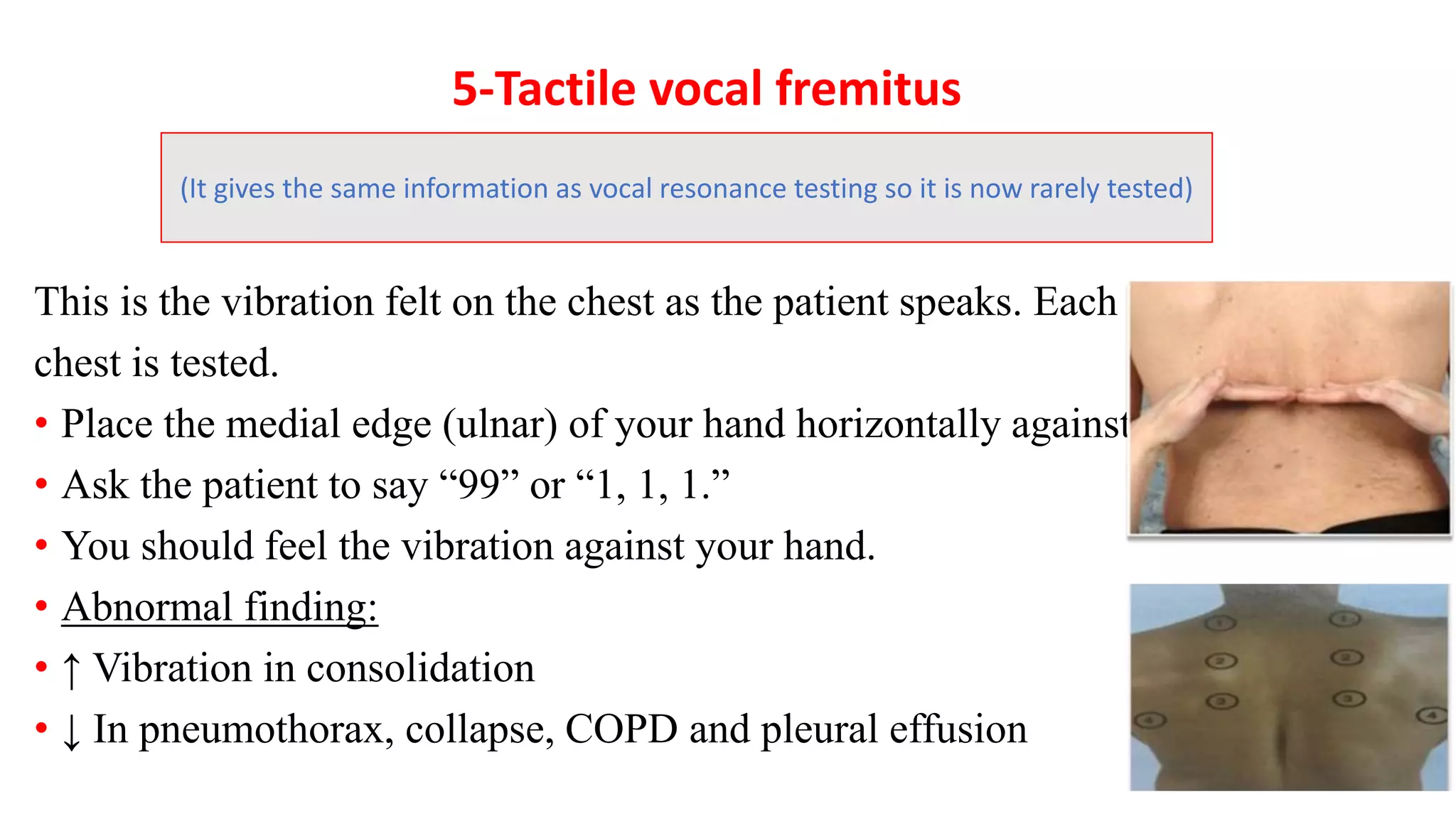
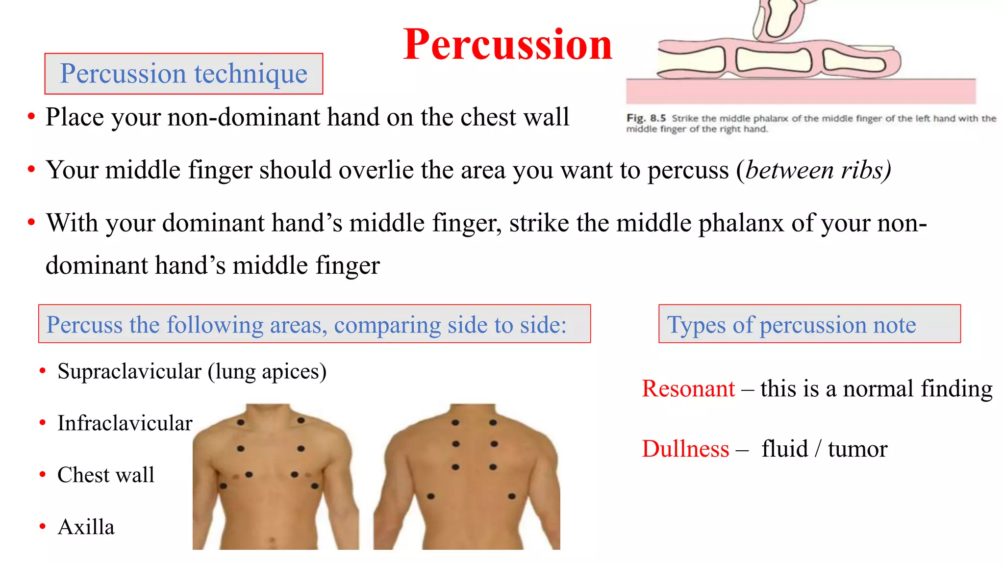
2. RWIIPPPPEE
6. Position the patient on the bed .
7. Pain(ask if the patient if he has any pain ).
8. Privacy .
9. Explain the examination .
10. Expose adequately .
1. Right side .
2. Wash or Warm hands .
3. Introduce yourself .
4. Identity of patient ( Name /DOB) .
5. Permission (gain consent ) .
3. • P - Position the patient. In this examination the
patient should be supine and reclined at sitting position
or 45 degrees.
• E - Expose the necessary parts of the patient.
Ideally the patient should be undressed from the
waist up, taking care to ensure the patient is not
cold .
4. Respiratory General Appearance
Evidence of respiratory distress (Includes all of the following steps.)
1. Observe rate, rhythm, depth, and effort of breathing
2. Assess patient’s color, looking for cyanosis
3. Listen to patient’s breathing for audible wheezing
4. Look for use of accessory muscles (Look at neck and supraclavicular area.)
5. Note any retraction of the intercostal muscles during inspiration (Check
lower interspaces anteriorly and posteriorly.)
Stand in front of the patient and OBSERVE
10. Inspection of the chest
1. Shape of the chest (normal or there is deformity)
2. Scars
3. Apical pulsation and intercostal spaces
4. Movement of the chest ASK THE PATIENT to take
deep breath and observe chest wall, (symmetrical or
a symmetrical movement)
5. Breathing pattern (abdominothoracic in male
thoracoabdominal in female)
13. Palpation
2-Trachea
4-5 cm of the upper trachea can be felt in
the neck above the sternal notch.
• Pushed –pneumothorax
-pleural effusion
• Pulled -fibrosis
-collapse
14. 3-Apex beat
• Located at the 5th intercostal space /
midclavicular line
• Palpate the apex beat with your fingers
(placed horizontally across the chest)
15. 4-Chest expansion
• Place your hands on the patient’s chest, inferior to the
nipples
• Wrap your fingers around either side of the chest
• Bring your thumbs together in the midline, so that they
touch
• Ask patient to take a deep breath
• Observe movement of your thumbs, they should move
apart equally
• If one of your thumbs moves less, this suggests
reduced expansion on that side
Abnormal Chest expansion:
Less than 2 cm
Reduced expansion can be caused by
lung collapse / pneumonia
16. 5-Tactile vocal fremitus
This is the vibration felt on the chest as the patient speaks. Each part of the
chest is tested.
• Place the medial edge (ulnar) of your hand horizontally against the chest.
• Ask the patient to say “99” or “1, 1, 1.”
• You should feel the vibration against your hand.
• Abnormal finding:
• ↑ Vibration in consolidation
• ↓ In pneumothorax, collapse, COPD and pleural effusion
(It gives the same information as vocal resonance testing so it is now rarely tested)
18. Percussion
• Place your non-dominant hand on the chest wall
• Your middle finger should overlie the area you want to percuss (between ribs)
• With your dominant hand’s middle finger, strike the middle phalanx of your non-
dominant hand’s middle finger
Percussion technique
• Supraclavicular (lung apices)
• Infraclavicular
• Chest wall
• Axilla
Percuss the following areas, comparing side to side:
Resonant – this is a normal finding
Dullness – fluid / tumor
Types of percussion note
19. Introduction to Auscultation
• Auscultation is the term for listening to the internal sounds of the body
• Auscultation is performed for the purposes of examining:
1. The circulatory system
2. The respiratory system
3. The gastrointestinal system (bowel sounds).
21. The bell is designed to hear low pitched sounds
The diaphragm is designed to hear high pitched
sounds.
How are these worn?
These should be worn ……..
!!!!!
facing forward .
23. Chest Auscultation
• Listen one full respiration at each spot
• Compare one side to the other
• Listen for the breath sounds and any added sounds.
• (“keep going, in and out”) will help.
Ladder pattern
24. Auscultation involves
1. Listening to the sounds generated by breathing
2. Listening for any adventitious (added) sounds
3. Listening to the sounds of the patient’s spoken as they are transmitted through
the chest wall. (vocal resonance)
25. 1- Breath sounds (lung sound)
• Produced by airflow in the large airways and larynx
and altered by passage through the small airways
before reaching the stethoscope.
Reduced sound:
• Local : Effusion, tumor, pneumothorax, pneumonia.
• Global : Asthma
Vesicular breathing:
26. Bronchial breathing:
• caused by ↑ density of matter in the peripheral lung, allowing sound
from the larynx to the stethoscope unchanged, has a hollow, blowing
quality, heard equally in inspiration and expiration, often with a brief
pause between.
• A similar sound can be heard by listening over the trachea in the neck.
• Bronchial breathing is heard over consolidation, lung abscess at the
chest wall, and with dense fibrosis.
27. • Sound like the pop of cereal.
• They are heard in inspiration.
• Crackles are often a normal finding at the lung bases , they will clear after asking
the patient to cough
• Causes include fluid or infection.
• Musical sounds caused by narrowed airways.
• It is heard easier in expiration
Crackles (crepitations, rales):
Wheeze (rhonchi):
2- Added sounds
28. 3- Vocal resonance
• Auscultatory equivalent of vocal fremitus
• Ask the patient to say “99” or 1, 1, 1” and listen over
the same areas as before.
29. Inspection:
• Shape of the chest or deformity
• Movement of the chest (symmetrical or
asymmetrical) and Breathing pattern
• Scars
• Apical pulsation.
• Intercostal space
Palpation:
• Position of trachea
• Apex beat
• Chest expansion
• Local rib tenderness
• Vocal fremitus
Percussion:
• Percussion note
• Percussion technique
Auscultation:
• Breath sound
• Added sound (rhonchi, crepitation)
• Vocal sound