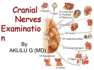
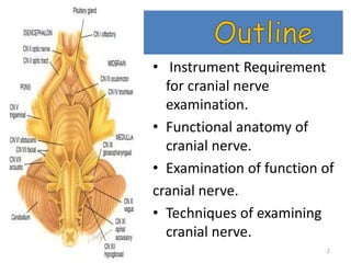
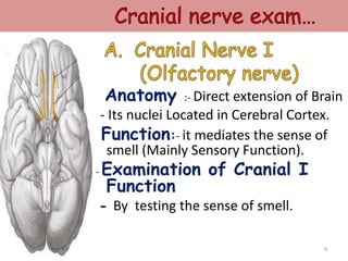
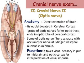
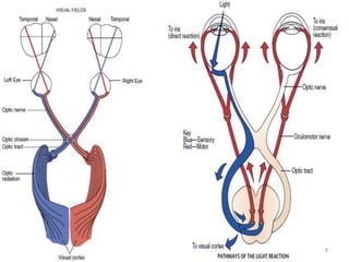
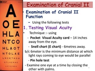
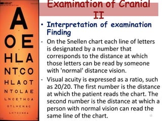
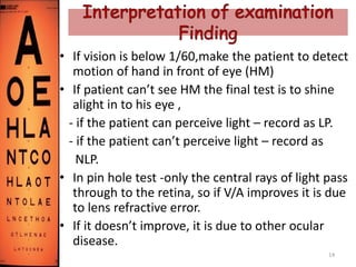
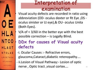
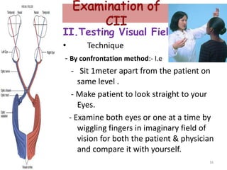
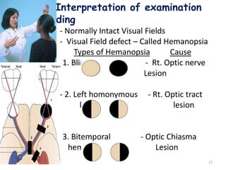
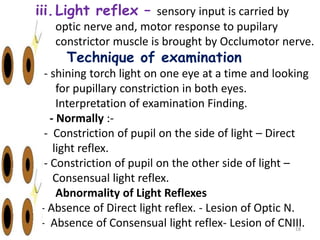
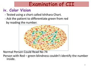
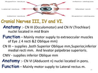
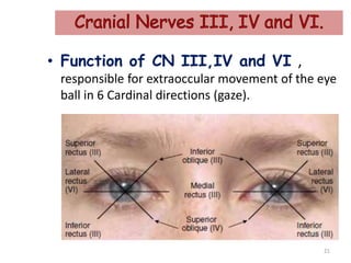
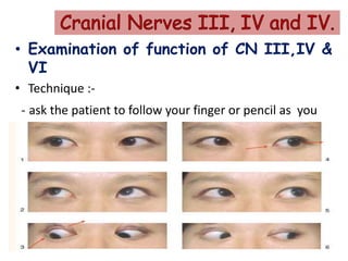
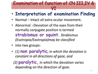
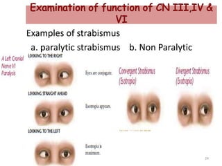
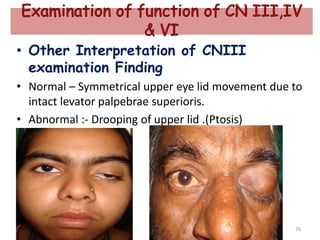
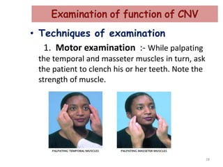
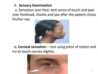
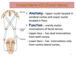
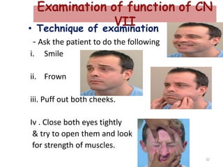
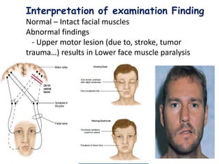
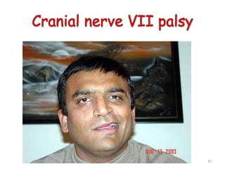
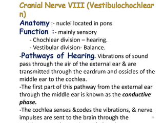
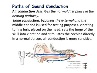
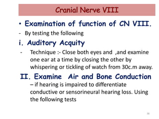
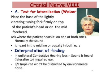
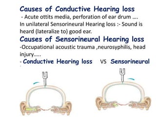
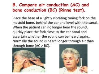
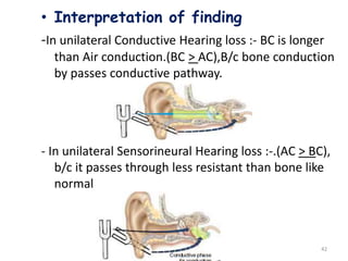
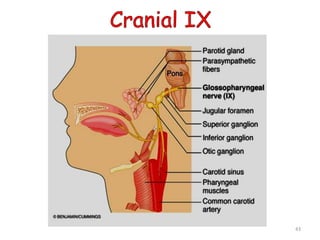
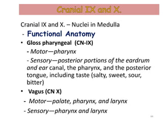
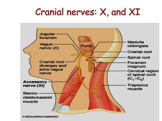
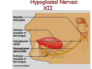
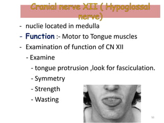
2. • Instrument Requirement
for cranial nerve
examination.
• Functional anatomy of
cranial nerve.
• Examination of function of
cranial nerve.
• Techniques of examining
cranial nerve.
2
4. Anatomy :- Direct extension of Brain
- Its nuclei Located in Cerebral Cortex.
Function:- it mediates the sense of
smell (Mainly Sensory Function).
- Examination of Cranial I
Function
- By testing the sense of smell.
4
5. • Technique of Examination.
-test the sense of smell
using substance with familiar
and non irritating odor.
- Use piece of soap, fruit or
other sbs…..
- First be sure that each nasal passage is open
by compressing one side of the nose &
asking the patient to sniff through the other.
-The patient should then close both eyes.
- Occlude one nostril and test smell in the
other with familiar substance. 5
6. • Interpretation of examination
Finding.
• Normal – Cranial nerve I is Intact .
• Abnormal – Loss of sense of smell
(Ansomia)
• Some causes of Ansomia
(DDX)
- Nasal obstruction due to common cold.
- tumors of olfactory groove (meningoma,
frontal glioma)
- Head injury 6
7. Anatomy :- Direct extension of Brain
- Its nuclei Located in Cerebral Cortex.
- group of optic nerves forms optic tract,
ends in optic lobe of cerebral cortex.
- Some of optic nerve fibers synapse with
occlumotor nerve at Edinger westphal
nucleus in midbrain.
- Function: it takes visual sensory in put
to midbrain and optic cortex for
interpretation of visual impulse. 7
9. • Examination of Cranial II
Function
- Using the following tests
1. Testing Visual Acuity
• Technique :- using
- Pocket Visual Acuity card – 14 inches
away from the eye.
- Snell chart (E chart) - 6metres away.
b/c 6meter is the minimum distance at which
light rays coming to eye would be parallel
- Pin hole test
- Examine one eye at a time by closing the
other with palms.
9
12. • Interpretation of examination
Finding
- On the Snellen chart each line of letters
is designated by a number that
corresponds to the distance at which
those letters can be read by someone
with 'normal' distance vision.
- Visual acuity is expressed as a ratio, such
as 20/20. The first number is the distance
at which the patient reads the chart. The
second number is the distance at which a
person with normal vision can read the
same line of the chart. 12
13. • Interpretation of examination
Finding
-On the Snellen chart each line of letters is
designated by a number that corresponds to
the distance at which those letters can be read
by someone with 'normal' distance vision.
-Visual acuity is expressed as a ratio, such as
6/60.
-The first number is the distance at which the
patient reads the chart.
-The second number is the distance at which a
person with normal vision can read the same
line of the chart.
- The normal person can read the line 13
14. • If vision is below 1/60,make the patient to detect
motion of hand in front of eye (HM)
• If patient can’t see HM the final test is to shine
alight in to his eye ,
- if the patient can perceive light – record as LP.
- if the patient can’t perceive light – record as
NLP.
• In pin hole test -only the central rays of light pass
through to the retina, so if V/A improves it is due
to lens refractive error.
• If it doesn’t improve, it is due to other ocular
disease.
14
15. • Visual acuity defects are recorded in ratio using
abbreviation (OD- oculus dexter or Rt Eye ,OS -
oculus sinister or Lt eye),& OU- oculus Unita
(Both Eyes).
• V/A of < 3/60 in the better eye with the best
possible correction – is Legally Blind.
• DDx for causes of Visual acuity
defects
-i. Ocular Causes :- Refractive errors,
glaucoma,Cataract,diabetic retenopathy….
- ii.Lesion of Visual Pathway:- Lesion of optic
nerve , Optic tract ,visual cortex…. 15
16. II.Testing Visual Fields
• Technique
- By confrontation method:- I.e
- Sit 1meter apart from the patient on
same level .
- Make patient to look straight to your
Eyes.
- Examine both eyes or one at a time by
wiggling fingers in imaginary field of
vision for both the patient & physician
and compare it with yourself.
16
17. Interpretation of examination
Finding
- Normally Intact Visual Fields
- Visual Field defect – Called Hemanopsia
Types of Hemanopsia Cause
- 1. Blind Rt Eye - Rt. Optic nerve
Lesion
- 2. Left homonymous - Rt. Optic tract
He hemianopsia lesion
- 3. Bitemporal - Optic Chiasma
heminaopsia Lesion
17
18. iii.Light reflex – sensory input is carried by
optic nerve and, motor response to pupilary
constrictor muscle is brought by Occlumotor nerve.
- Technique of examination
- shining torch light on one eye at a time and looking
for pupillary constriction in both eyes.
- Interpretation of examination Finding.
- Normally :-
- Constriction of pupil on the side of light – Direct
light reflex.
- Constriction of pupil on the other side of light –
Consensual light reflex.
- Abnormality of Light Reflexes
- Absence of Direct light reflex. - Lesion of Optic N.
- Absence of Consensual light reflex- Lesion of CNIII.
18
19. iv. Color Vision
- Tested using a chart called Ishihara Chart.
- Ask the patient to differentiate green from red
by reading the number.
Normal Person Could Read No 74
Person with Red – green blindness couldn’t identify the number
inside.
19
20. Anatomy :- CN III (Occulomotor) and CN IV (Trochlear)
nuclei located in mid Brain
Function – Mainly motor supply to extraoccular muscles
of Eye .( 4 recti &2 Oblique mm)
CN III – supplies ,both Superior Oblique mm,Superior,inferior
medial recti mm. And levator palpebrae superioris.
CNIV - supplies inferior Oblique mm
Anatomy :- CN VI (Abducent n) nuclei located in pons.
Function – Mainly motor supply to Lateral rectus m.
20
21. • Function of CN III,IV and VI ,
responsible for extraoccular movement of the eye
ball in 6 Cardinal directions (gaze).
21
22. • Examination of function of CN III,IV &
VI
• Technique :-
- ask the patient to follow your finger or pencil as you
sweep through the six cardinal directions of gaze .
22
23. • Interpretation of examination Finding
• Normal – Intact all extra ocular movement.
• Abnormal :-Deviation of the eyes from their
normally conjugate position is termed
strabismus or squint. Strabismus
(Esotropia/Exotropia)may be classified
• into two groups:
• (1) non paralytic, in which the deviation is
constant in all directions of gaze, and
(2) paralytic, in which the deviation varies
depending on the direction of gaze.
23
26. • Other Interpretation of CNIII
examination Finding
• Normal – Symmetrical upper eye lid movement due to
intact levator palpebrae superioris.
• Abnormal :- Drooping of upper lid .(Ptosis)
26
27. Anatomy :- Nuclei Located in Pons
Function :- Mainly sensory innervations to face
via its three main division (Ophthalmic,
maxillary,& Mandibular branches)
:- sensory innervations to cornea with
motor response via facial nerve.
:- Motor supply to muscles of mastication
(temporal and masseter Muscles)
27
28. • Techniques of examination
1. Motor examination :- While palpating
the temporal and masseter muscles in turn, ask
the patient to clench his or her teeth. Note the
strength of muscle.
28
29. . II. Sensory Examination
a. Sensation over face:-test sense of touch and pain
over forehead, cheeks and jaw after the patient closes
his/her eye.
b. Corneal sensation :- test using piece of cotton and
try to touch cornea slightly.
29
30. • Interpretation of examination
Finding
- loss of sensation (paresthesia)
- exaggerated sensory perception (Hypersthesia)
- Diminished corneal reflex
- Deviation of jaw to side of lesion when mouth
opens.
• Some of causes for abnormal
finding (DDX)
- Brain stem lesion
- Pontine lesion
- Infection of trigeminal ganglion 30
31. • Anatomy:- Upper nuclei located in
cerebral cortex and Lower nuclei
located in Pons
• Function :- mainly motor
innervations of facial nerves.
- Upper face :- has dual innervations
from both cortex.
- Lower face:-- has innervations only
from contra lateral cortex..
31
32. • Technique of examination
- Ask the patient to do the following
i. Smile
ii. Frown
iii. Puff out both cheeks.
Iv . Close both eyes tightly
& try to open them and look
for strength of muscles.
32
33. Interpretation of examination Finding
Normal – Intact facial muscles
Abnormal findings
- Upper motor lesion (due to, stroke, tumor
trauma…) results in Lower face muscle paralysis
33
34. Interpretation of examination Finding
• Lower motor lesion (due to, Viral infection
of facial nerve, Parotid tumor …) results in
all facial muscle paralysis.
34
36. Anatomy :- nuclei located in pons
Function :- mainly sensory
- Chochlear division – hearing.
- Vestibular division- Balance.
-Pathways of Hearing. Vibrations of sound
pass through the air of the external ear & are
transmitted through the eardrum and ossicles of the
middle ear to the cochlea.
-The first part of this pathway from the external ear
through the middle ear is known as the conductive
phase.
-The cochlea senses &codes the vibrations, & nerve
impulses are sent to the brain through the 36
37. Paths of Sound Conduction
Air conduction describes the normal first phase in the
hearing pathway.
bone conduction, bypasses the external and the
middle ear and is used for testing purposes. vibrating
tuning fork, placed on the head, sets the bone of the
skull into vibration and stimulates the cochlea directly.
In a normal person, air conduction is more sensitive.
37
38. • Examination of function of CN VIII.
- By testing the following
i. Auditory Acquity
- Technique :- Close both eyes and ,and examine
one ear at a time by closing the other by
whispering or tickling of watch from 30c.m away.
II. Examine Air and Bone Conduction
– if hearing is impaired to differentiate
conductive or sensorineural hearing loss. Using
the following tests
38
39. • A. Test for lateralization (Weber test).
Place the base of the lightly
vibrating tuning fork firmly on top
of the patient’s head or on the mid
forehead.
Ask where the patient hears it: on one or both sides.
Normally the sound
• is heard in the midline or equally in both ears
• Interpretation of finding
- In unilateral Conductive Hearing loss :- Sound is heard
(lateralize to) impaired ear.
- B/c Impaired won’t be distracted by environmental
noise.
39
40. 40
Causes of Conductive Hearing loss
- Acute ottits media, perforation of ear drum ….
In unilateral Sensorineural Hearing loss :- Sound is
heard (lateralize to) good ear.
Causes of Sensorineural Hearing loss
-Occupational acoustic trauma ,neurosyphilis, head
injury…..
- Conductive Hearing loss VS Sensorineural
loss
41. B. Compare air conduction (AC) and
bone conduction (BC) (Rinne test).
Place the base of a lightly vibrating tuning fork on the
mastoid bone, behind the ear and level with the canal.
When the patient can no longer hear the sound,
quickly place the fork close to the ear canal and
ascertain whether the sound can be heard again..
Normally the sound is heard longer through air than
through bone (AC > BC).
41
42. 42
• Interpretation of finding
-In unilateral Conductive Hearing loss :- BC is longer
than Air conduction.(BC > AC),B/c bone conduction
by passes conductive pathway.
- In unilateral Sensorineural Hearing loss :-.(AC > BC),
b/c it passes through less resistant than bone like
normal
44. Cranial IX and X. – Nuclei in Medulla
- Functional Anatomy
• Gloss pharyngeal (CN-IX)
- Motor—pharynx
- Sensory—posterior portions of the eardrum
and ear canal, the pharynx, and the posterior
tongue, including taste (salty, sweet, sour,
bitter)
• Vagus (CN X)
- Motor—palate, pharynx, and larynx
- Sensory—pharynx and larynx
44
45.
46. Examination of function of CN IX
&X
• Examination of function of CN IX
&X
- Technique:- examine the following
- Palatal movement (tell the patient to say
“Aha”)
- Check gag reflex with spatula
- Notice phonation
• Abnormal findings – Deviation of palatal
movement, absent gag reflex.
• Causes : Head trauma, Medullary lesions
46
47. • Function
- Motor—the sternomastoid and upper portion
of the trapizus.
• Examination of function of CN XI
- Examine rotation of head & neck against resistant
Loss of Resistance :- sternomastoid muscle
weakness
47
48. - Examine Shrugging of shoulder against
resistant.
- Loss of Resistance :- trapizus muscle
weakness
48
49.
50. - nuclie located in medulla
- Function :- Motor to Tongue muscles
- Examination of function of CN XII
- Examine
- tongue protrusion ,look for fasciculation.
- Symmetry
- Strength
- Wasting
50