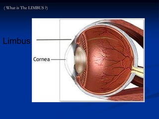
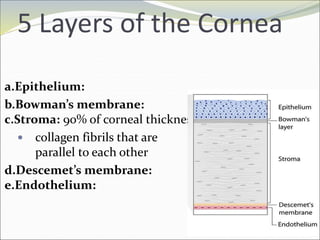
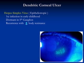
Mohamed Ahmed
ELShafie
MD, HMS alumni
Anatomy of the Cornea
Anterior 1/6 of outer coat
Curved & Dom shaped
Fibrous, Transparent & No BVs
• Measured by corneal Caliber or
Pentacam corneal topography
• Important in Congenital
Glaucoma
Keratometry
Limbus
( What is The LIMBUS ?)
5 Layers of the Cornea
a.Epithelium:
b.Bowman’s membrane:
c.Stroma: 90% of corneal thickness
 collagen fibrils that are
parallel to each other
d.Descemet’s membrane:
e.Endothelium:
NERVE SUPPLY OF THE CORNEA
5TH C.N
OPHTH. division NASOCILIARY N 2 Long CILIARY N
PAIN & COLD & SUPERFICIAL TOUCH
Clinical tests
Corneal senstion test
= corneal reflex test
= Blinking reflex
• Aim
• Technique
• result
• Causes of lost corneal reflex
• Receprors:
Keratitis: HSV,HZV, Keratomalacia,
Corneal scars
High IOP
Topical anaeathesia
CL
• Afferent:
• Center :
• Efferent:
5th n. lesion
Occipital cortex lesion
7thh n. lesion (facial palsy)
Corneal reflex
SUPPURATIVE SUPERFICIAL
KERATITS
(CORNEAL ULCERS)
DEFINITION
Localized Necrosis of Sup. Stroma
with destruction of overlying Epith.
Symptoms
Stitching
Referred to the eye brow
Because of:
• corneal edema
• accompanying iridocyclitis
Lacrimation
MANAGEMENT OF CORNEAL ULCERS
INVESTIGATIONS + TREATMENT
A) Corneal Scrapping ( Culture & Sensitivity )
Gram Stain for Bacteria
Geimsa Stain for Trachoma & Acanthamoeba
Silver Stain for Fungi
B) Local ttt (1) Atropine sulphate 1%
(3) Bandage or Dark Glasses
(4) Counter irritant
(2) Dressings ( Antibiotic dps & oint )
C) Systemic ttt
Antibiotics Analgesics
Vitamins A & C
Acanthamoeba keratitis
 Aetiology
Protozoa ( Tap water and Swimming pools )
70% of cases are C L wearers
 Clinical Picture
Punctate or Dendritic K
Partial or Complete ring of Infiltration
Dendritic Corneal Ulcer
Herpes Simplex Virus ( Epitheliotropic )
1ry infection in early childhood
Dormant in 5th Ganglion
Recurrence with body resistance
Herpes Zoster Ophthalmicus
Varicella-Zoster (Neurotropic) Virus
Old age - Immunity
Clinical Picture :
Lids : Dermatoblepharitis ( pain and rash )
Keratitis : ( Hutchinson’s rule )
Epithelial Keratitis ( Punctate or dendritic )
Interstitial Keratitis
Ulcer with Lagophthalmos
 Bell’s phenomena
 Symptoms
as usual corneal ulcer ( of vision is not marked..why?)
 Signs
Incomplete lid closure
Ciliary injection & +ve flurorescein
Ulcer in lower 1/3 with straight upper border
 Treatment
Usual ttt
Methyl cellulose drops 0.5% several times/day
Degenerative Conditions
ARCUS SENELIS
Bilateral peripheral Fatty degeneration
Common in old age
Symptoms non
Signs
Clear zone between the opacity and Limbus (Lucid interval of vogt)
NB ARCUS JUVENILIS may occur in hyperlipidemia or juv. DM
• Sympyoms: Stitching pain, lacrimation, photophobia,
..
• Treatment: FB removal (Search for other FBs)
Definition
Progressive conical distortion of the
cornea with:
• irregular astigmatism
• axial stromal thinning
• apical protrusion
• increasing myopia.
KERATOPLASTY
 Aim: Replacing the opaque part by a clear cadaveric cornea
 Types:
- Lamellar ( Superficial )
- Deep ( Penetrating )
NB: Both of them may be partial or total
 Indications:
- Optical a) Central corneal opacities
b) Keratoconus
- Therapeutic a) Resistant corneal ulcer
b) Corneal fistula
‫المليون؟‬ ‫سيربح‬ ‫من‬
Examination of
cornea reveals: 15-$1
Million
14-$500,000
13-$250,000
12-$125,000
11-$64,000
10-$32,000
9-$16,000
8-$8,000
7-$4,000
6-$2,000
5-$1,000
4-$500
3-$300
2-$200
1-$100
B-Anterior
staphyloma
C-Arcus senilis
D-
Decematocele
A-Nebula
C-Arcus senilis
15-$million
14-$500,000
13-$250,000
12-$125,000
11-$64,000
10-$32,000
9-$16,000
8-$8,000
7-$4,000
6-$2,000
5-$1,000
4-$500
3-$300
2-$200
1-$100
Examination of cornea reveals:
A-Nebula
B-Leucoma
C-
Decematocele
D-
keratoconus
B-Leucoma
Examination of cornea reveals:
A-Nebula
B-Leucoma
adherent
C-Leucoma
non-adherent
D-
Keratectasia
15-$million
14-$500,000
13-$250,000
12-$125,000
11-$64,000
10-$32,000
9-$16,000
8-$8,000
7-$4,000
6-$2,000
5-$1,000
4-$500
3-$300
2-$200
1-$100
A-Nebula
Leucoma adherent
THANK YOU

Cornea.ppt