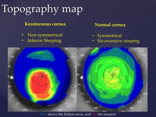
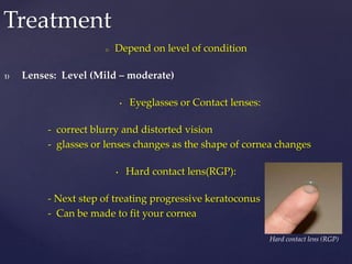
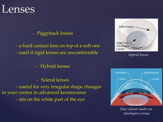
{
Understanding Keratoconus
Saleh Hassan
May 2018
Background
o Cornea: transparent front part of the eye that deflects light rays
which focused into a point of sharp focus. For a good vision the
focus point must be on retina
o Keratoconus is a disorder of the eye which results in progressive
thinning of the cornea and bulging like a cone
o Changing the shape of the cornea brings light rays out of focus
o As a result, your vision is blurry and
distorted, making daily tasks like reading
or driving difficult
Normal eye Keratoconus
KeratoconusNormal
Blur vision Distorted vision
o Unknown
o It is believed that genetics, the environment and the endocrine
system all play a role in keratoconus
Causes
Source of causes: https://www.nkcf.org
 Normal eye Keratoconus
• Collagen fibers
(tiny fibers hold cornea in place
and keep it from bulging)
• Weak collagen fibres
• Protective antioxidants
(molecules that inhibit the oxidation)
(Oxidation is a chemical reaction produce radicals
absorbed from environment Ex: O*,
leading to chain reactions
that may affect signal transduction
and damage corneal cells)
• Protective antioxidants
o Early stages
• mild blurring of vision
• slightly distorted vision, where straight lines look bent or wavy
• increased sensitivity to light and glare
• eye redness or swelling
Symptoms
o Later stages
• more blurry and distorted vision
• increased astigmatism
(when your eye cannot focus as well as it should)
• not being able to wear contact lenses
o Keratoconus affects both eyes,
and can lead to very different vision between the two eyes
o Measuring the shape of cornea by several devices
o Step1 Slit-lamp
determines the damage of cornea
o Step2 Corneal Topography
more accurate details and showing
how the disease is progressing
Diagnosis
o There are three levels of severity when it comes to Keratoconus and it
determined by several parameters
1) Testing Steepness of greatest Curvature
40-45D is mild
52 D is considered advance
Above 52D is severe
2) Thickness of the Cornea
Mild- 506µm
Advanced- Less than 446µm
3) Morphology of the Cone
If the cone is sagging it too is an indicator of the
advancing of the disease.
Parameters
Keratoconus cornea
• Non symmetrical
• Inferior Steeping
Normal cornea
• Symmetrical
• No excessive steeping
Topography map
Blue shows the flattest areas, and red the steepest
o Depend on level of condition
1) Lenses: Level (Mild – moderate)
• Eyeglasses or Contact lenses:
- correct blurry and distorted vision
- glasses or lenses changes as the shape of cornea changes
• Hard contact lens(RGP):
- Next step of treating progressive keratoconus
- Can be made to fit your cornea
Treatment
Hard contact lens (RGP)
• Piggyback lenses
- a hard contact lens on top of a soft one
- used if rigid lenses are uncomfortable
• Hybrid lenses
• Scleral lenses
- useful for very irregular shape changes
in your cornea in advanced keratoconus
- sits on the white part of the eye
Lenses
Hybrid lenses
How scleral vaults on
misshapen cornea
• Small device implanted in the eye to correct
vision
• Procedure:
Making small incision in the
cornea and inserts two crescent or semi-
circular shaped ring segments between the
layers of the corneal stroma , one on each side
of the pupil
• Uses:
Treatment of mild– moderate
keratoconus
Near sighted vision
Surgery: Corneal Insert
A pair of corneal rings after
insertion into the cornea
Stroma
 Outcomes:
- short-terms
central flattening of the cornea were reported in all of the studies
• keratometric was reduced to (2.14 to 9.60 D)
- medium and long-terms
ICRS did not stop cone progression
• Useful for improving the contact lens tolerance for the residual refractive
error.
• Carrasquillo et al found an 81% improvement in contact lens tolerance
after Intacs implantation in keratoconus and post-LASIK ectasia
 Complications
Rare. Its reversible. No tissues need to be removed
Surgery: Corneal Insert
Source: http://eyewiki.aao.org
o Required when damage is severe
o It’s a surgical procedure to replace part of your cornea with corneal tissue
from a donor
o Procedure
Eye drops to help you to relax
The central damage part is removed
The healthy donor cornea is securely sewn into place
o Outcomes
Generally is very successful
o Complications
New cornea rejection
Poor vision
Astigmatism
Inability to wear contact lenses
Infection
Surgery: Cornea transplant
Corneal transplantation
Background
• Cross-link is a bond that links a polymer chains to another whether its
synthetic or natural such as Protein
• In chemistry when polymer chains are cross-linked, the materiel is
rigified
• Collagen fibers are fibers that hold cornea together made of proteins
How it works
• Collagens + Riboflavin(Vitamin B2)
Surgery: Collagen cross-linking
Activation
UV
New bonds between
collagen fibers
• The result is a cornea that is stiffer and more resistant to any change in
its shape
Aim
• Stop the progression & prevent from getting worse
Procedure
• the epithelium is first disrupted
• Riboflavin (vitamin B2) is then applied to the eye
• eye is exposed to UV light
Outcomes
• Success rate is more than 95% for epi-off treatment(epi is removed by
cotton bud then the normal procedure. thin bandage contact lens is
placed on the eye at the end for 24–72 hours)
Complications
• Small risk of infection
• The procedure don’t causing bleeding due to affected cornea dosent have
blood
• The UV only penetrate the cornea not deeper structure of the eye
Corneal cross-linking
Source: https://visioneyeinstitute.com.au

Keratoconus presentation

  • 1.
  • 2.
    Background o Cornea: transparentfront part of the eye that deflects light rays which focused into a point of sharp focus. For a good vision the focus point must be on retina o Keratoconus is a disorder of the eye which results in progressive thinning of the cornea and bulging like a cone o Changing the shape of the cornea brings light rays out of focus o As a result, your vision is blurry and distorted, making daily tasks like reading or driving difficult Normal eye Keratoconus
  • 3.
  • 4.
    o Unknown o Itis believed that genetics, the environment and the endocrine system all play a role in keratoconus Causes Source of causes: https://www.nkcf.org  Normal eye Keratoconus • Collagen fibers (tiny fibers hold cornea in place and keep it from bulging) • Weak collagen fibres • Protective antioxidants (molecules that inhibit the oxidation) (Oxidation is a chemical reaction produce radicals absorbed from environment Ex: O*, leading to chain reactions that may affect signal transduction and damage corneal cells) • Protective antioxidants
  • 5.
    o Early stages •mild blurring of vision • slightly distorted vision, where straight lines look bent or wavy • increased sensitivity to light and glare • eye redness or swelling Symptoms o Later stages • more blurry and distorted vision • increased astigmatism (when your eye cannot focus as well as it should) • not being able to wear contact lenses o Keratoconus affects both eyes, and can lead to very different vision between the two eyes
  • 6.
    o Measuring theshape of cornea by several devices o Step1 Slit-lamp determines the damage of cornea o Step2 Corneal Topography more accurate details and showing how the disease is progressing Diagnosis o There are three levels of severity when it comes to Keratoconus and it determined by several parameters
  • 7.
    1) Testing Steepnessof greatest Curvature 40-45D is mild 52 D is considered advance Above 52D is severe 2) Thickness of the Cornea Mild- 506µm Advanced- Less than 446µm 3) Morphology of the Cone If the cone is sagging it too is an indicator of the advancing of the disease. Parameters
  • 8.
    Keratoconus cornea • Nonsymmetrical • Inferior Steeping Normal cornea • Symmetrical • No excessive steeping Topography map Blue shows the flattest areas, and red the steepest
  • 9.
    o Depend onlevel of condition 1) Lenses: Level (Mild – moderate) • Eyeglasses or Contact lenses: - correct blurry and distorted vision - glasses or lenses changes as the shape of cornea changes • Hard contact lens(RGP): - Next step of treating progressive keratoconus - Can be made to fit your cornea Treatment Hard contact lens (RGP)
  • 10.
    • Piggyback lenses -a hard contact lens on top of a soft one - used if rigid lenses are uncomfortable • Hybrid lenses • Scleral lenses - useful for very irregular shape changes in your cornea in advanced keratoconus - sits on the white part of the eye Lenses Hybrid lenses How scleral vaults on misshapen cornea
  • 11.
    • Small deviceimplanted in the eye to correct vision • Procedure: Making small incision in the cornea and inserts two crescent or semi- circular shaped ring segments between the layers of the corneal stroma , one on each side of the pupil • Uses: Treatment of mild– moderate keratoconus Near sighted vision Surgery: Corneal Insert A pair of corneal rings after insertion into the cornea Stroma
  • 12.
     Outcomes: - short-terms centralflattening of the cornea were reported in all of the studies • keratometric was reduced to (2.14 to 9.60 D) - medium and long-terms ICRS did not stop cone progression • Useful for improving the contact lens tolerance for the residual refractive error. • Carrasquillo et al found an 81% improvement in contact lens tolerance after Intacs implantation in keratoconus and post-LASIK ectasia  Complications Rare. Its reversible. No tissues need to be removed Surgery: Corneal Insert Source: http://eyewiki.aao.org
  • 13.
    o Required whendamage is severe o It’s a surgical procedure to replace part of your cornea with corneal tissue from a donor o Procedure Eye drops to help you to relax The central damage part is removed The healthy donor cornea is securely sewn into place o Outcomes Generally is very successful o Complications New cornea rejection Poor vision Astigmatism Inability to wear contact lenses Infection Surgery: Cornea transplant Corneal transplantation
  • 14.
    Background • Cross-link isa bond that links a polymer chains to another whether its synthetic or natural such as Protein • In chemistry when polymer chains are cross-linked, the materiel is rigified • Collagen fibers are fibers that hold cornea together made of proteins How it works • Collagens + Riboflavin(Vitamin B2) Surgery: Collagen cross-linking Activation UV New bonds between collagen fibers • The result is a cornea that is stiffer and more resistant to any change in its shape
  • 15.
    Aim • Stop theprogression & prevent from getting worse Procedure • the epithelium is first disrupted • Riboflavin (vitamin B2) is then applied to the eye • eye is exposed to UV light Outcomes • Success rate is more than 95% for epi-off treatment(epi is removed by cotton bud then the normal procedure. thin bandage contact lens is placed on the eye at the end for 24–72 hours) Complications • Small risk of infection • The procedure don’t causing bleeding due to affected cornea dosent have blood • The UV only penetrate the cornea not deeper structure of the eye Corneal cross-linking Source: https://visioneyeinstitute.com.au