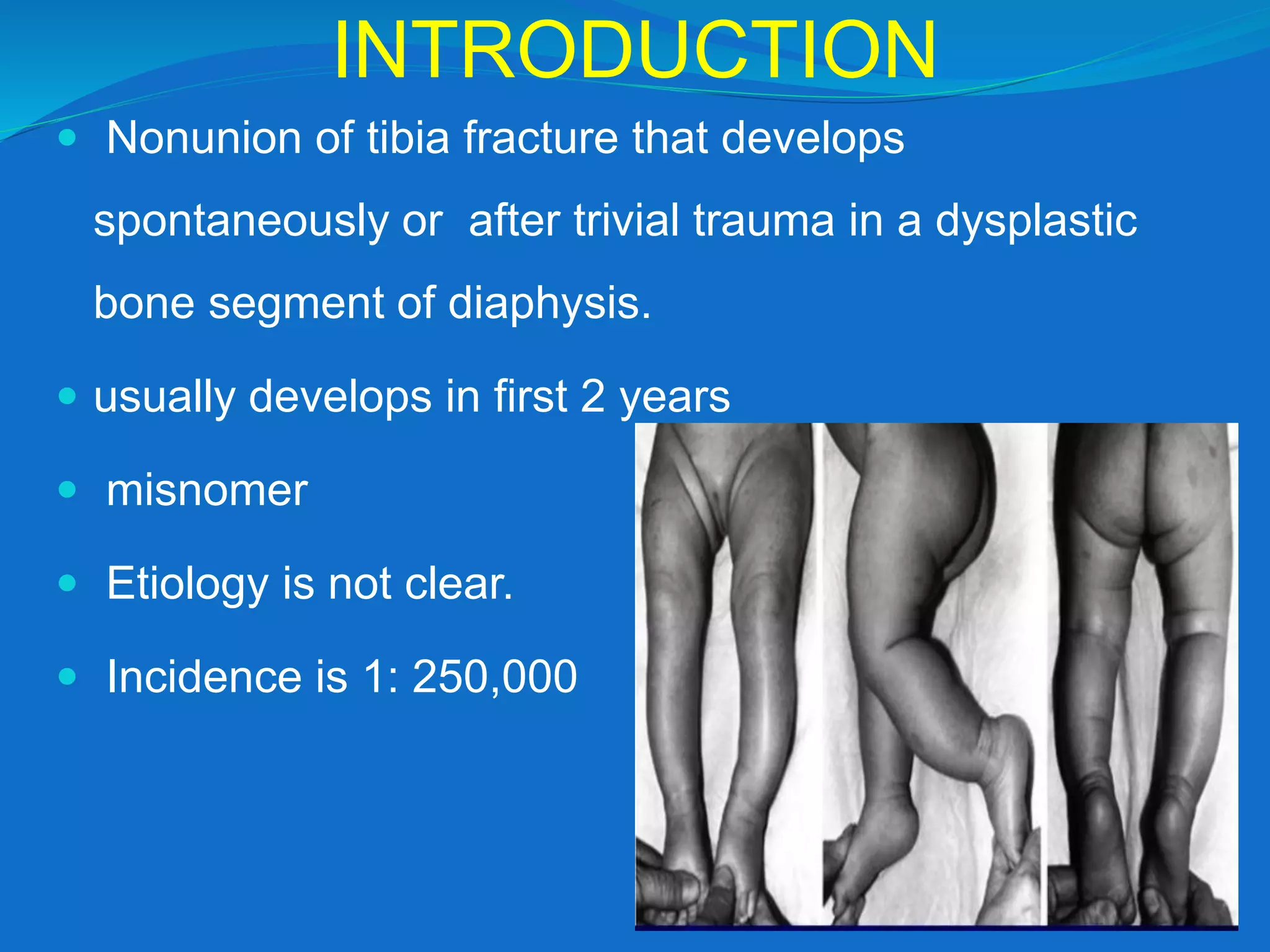
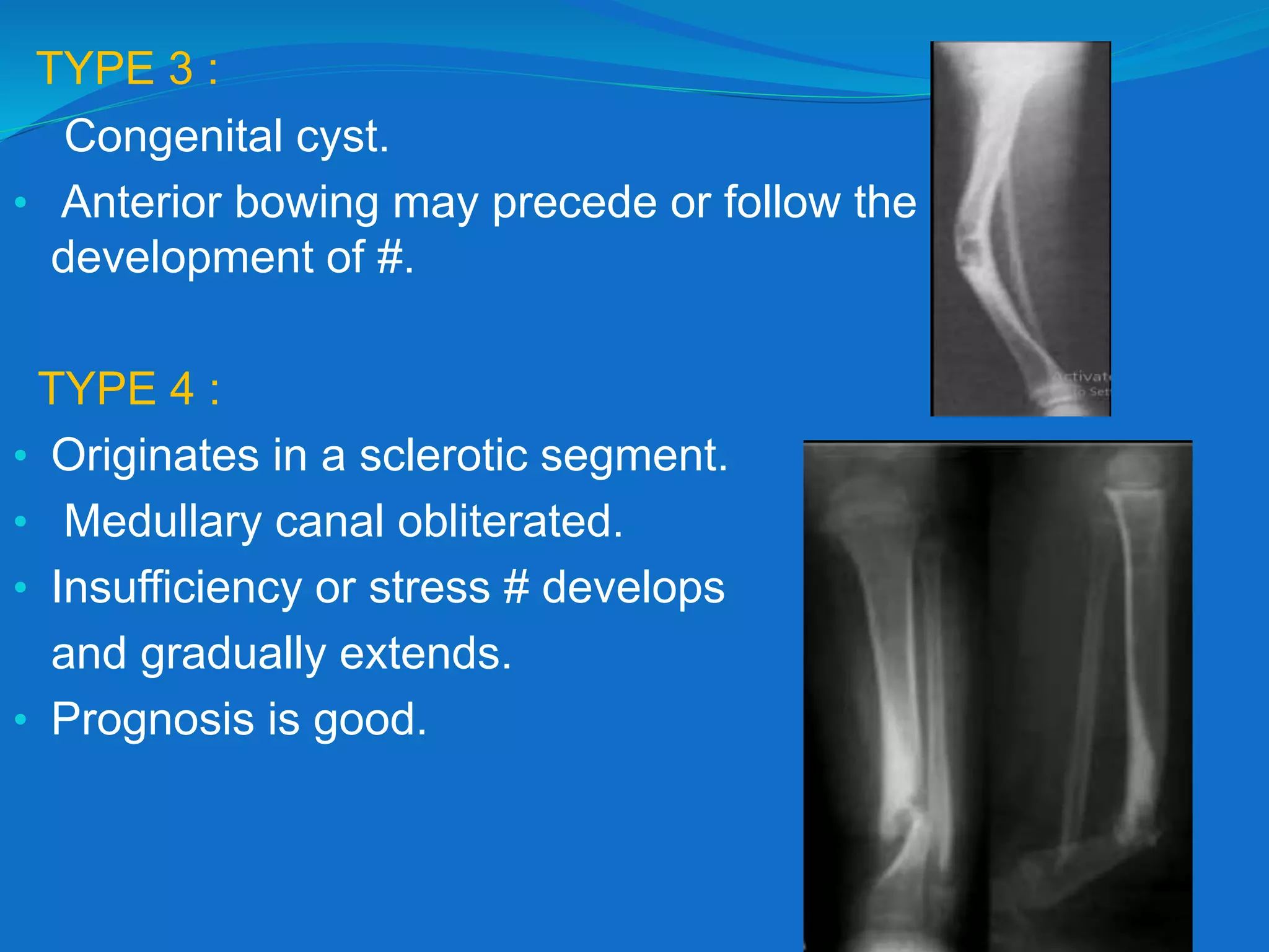
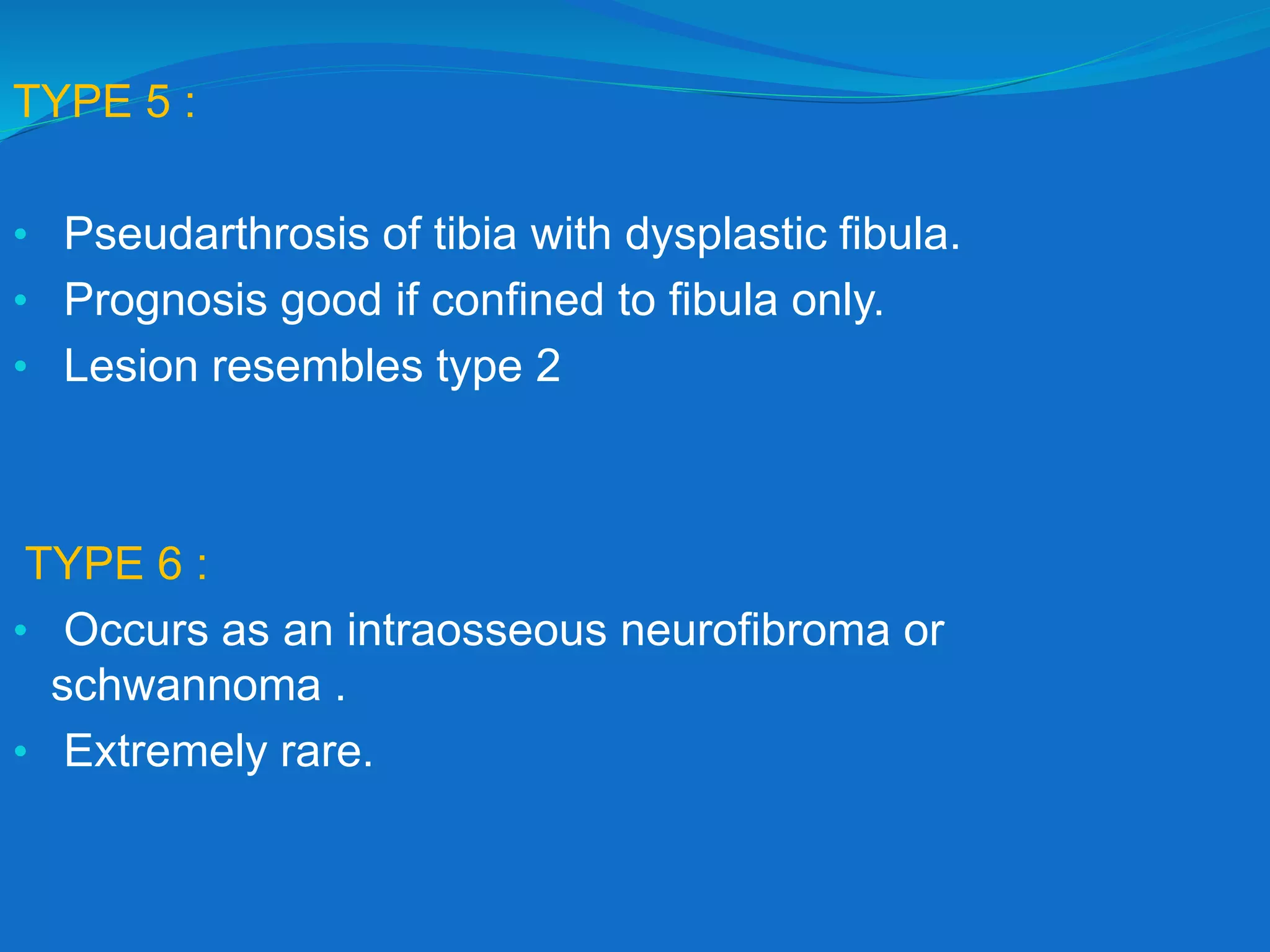
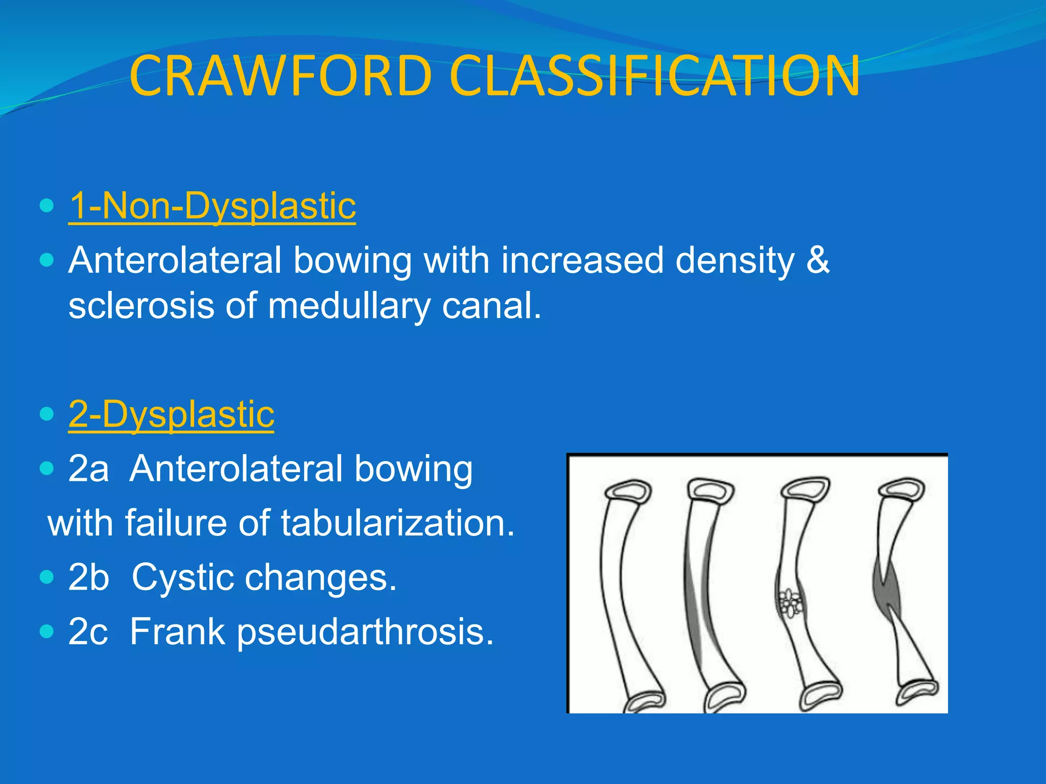
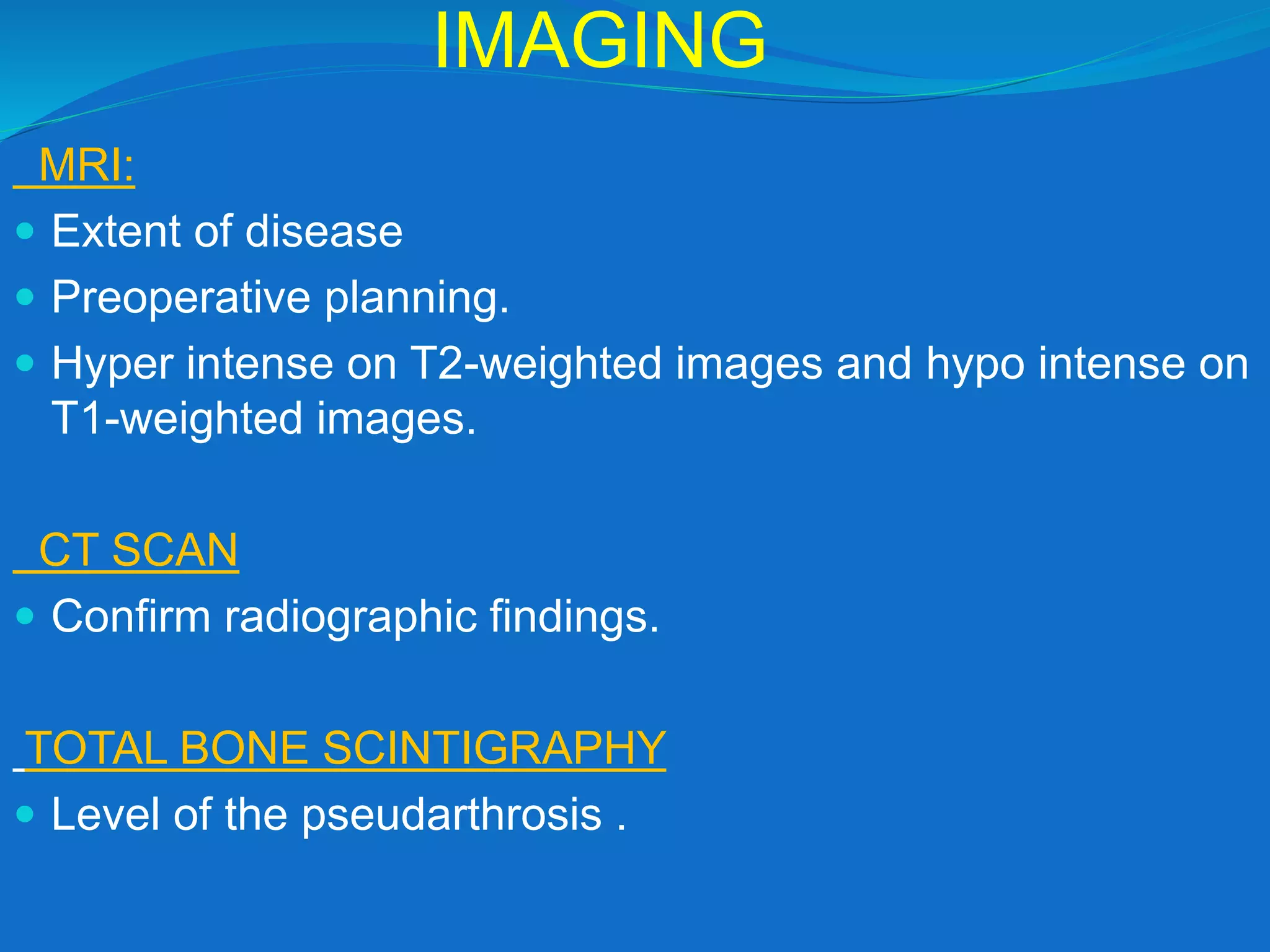
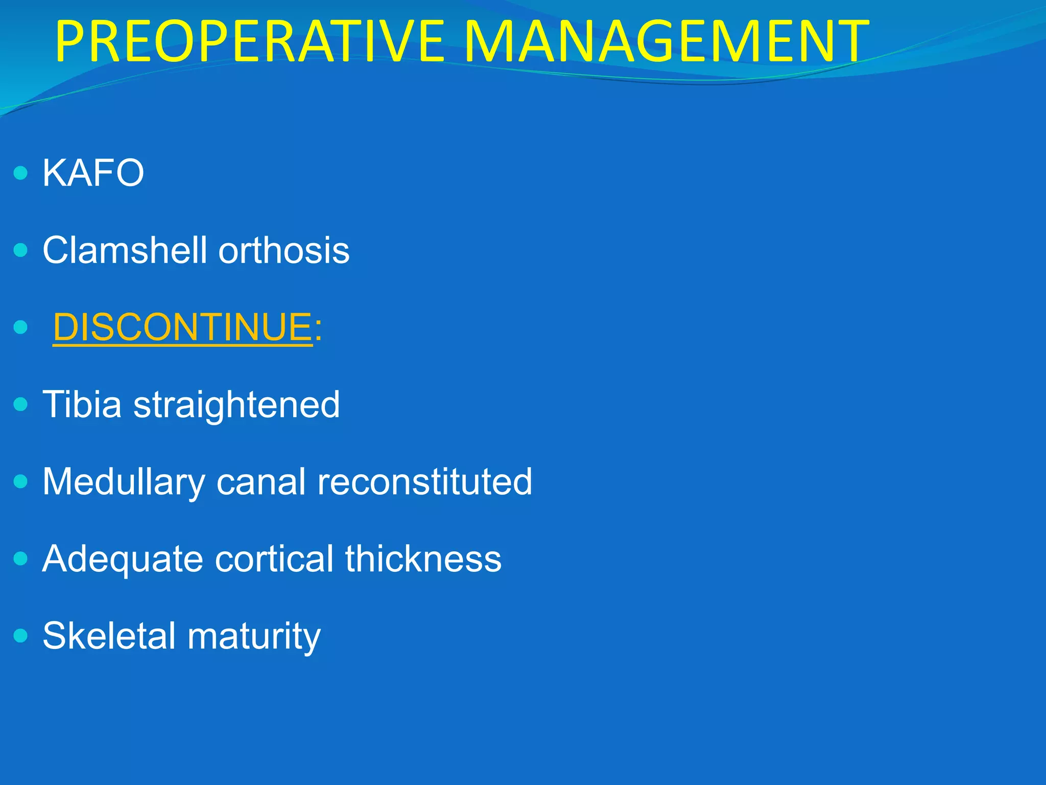
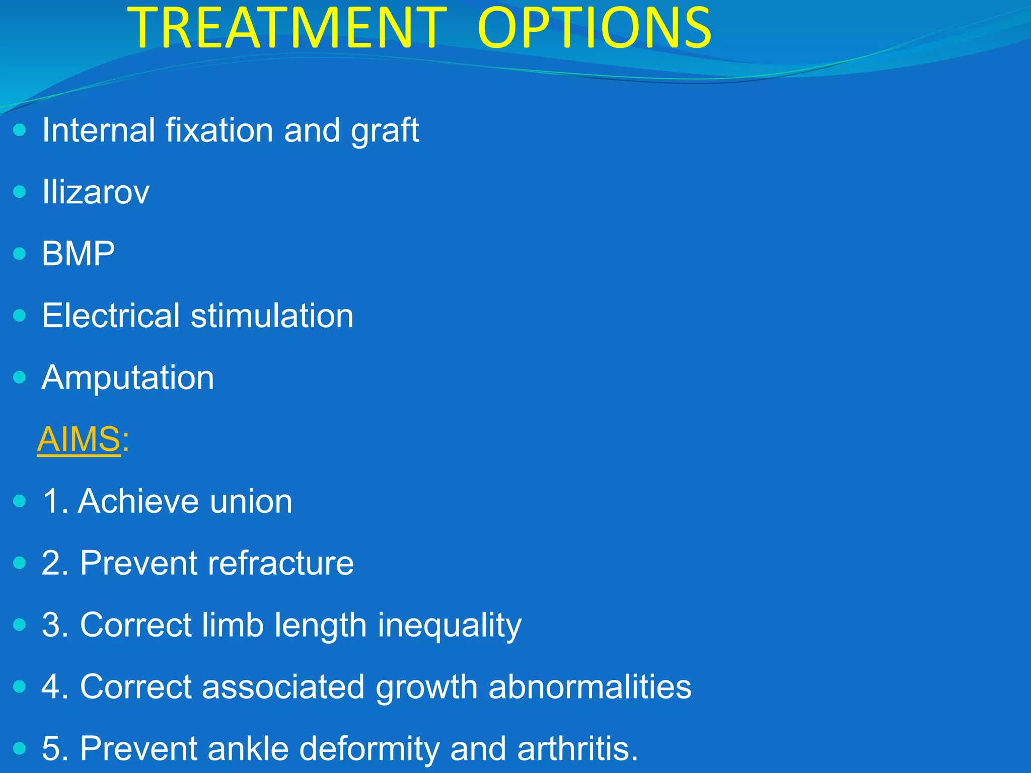
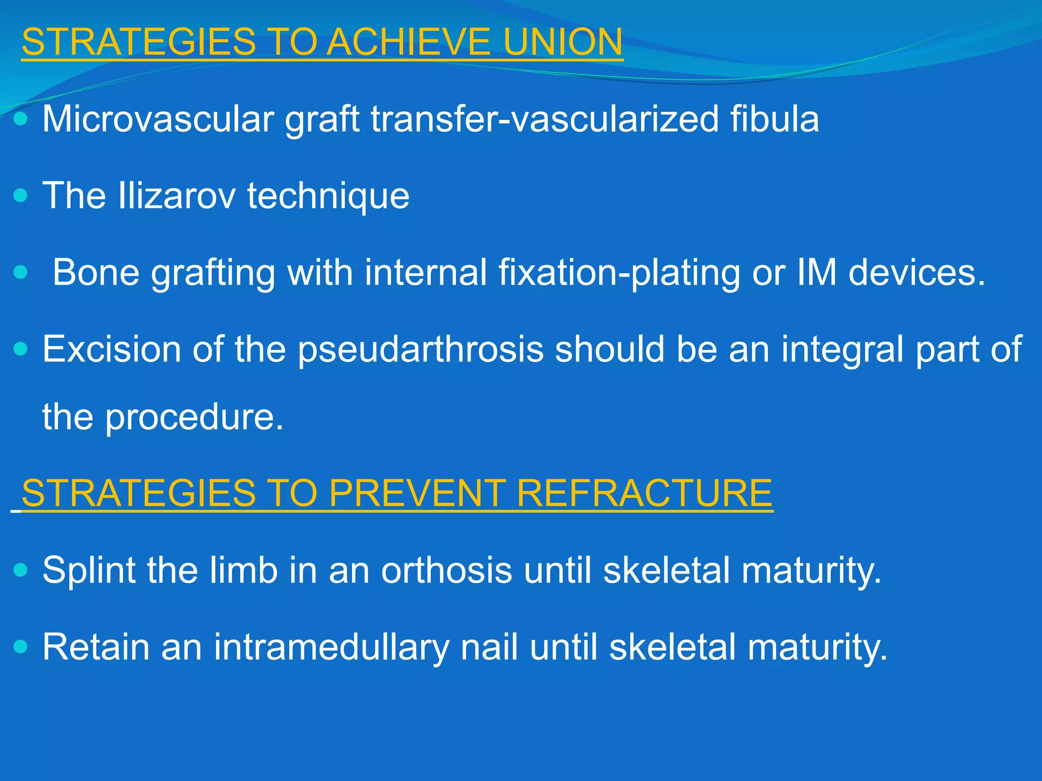
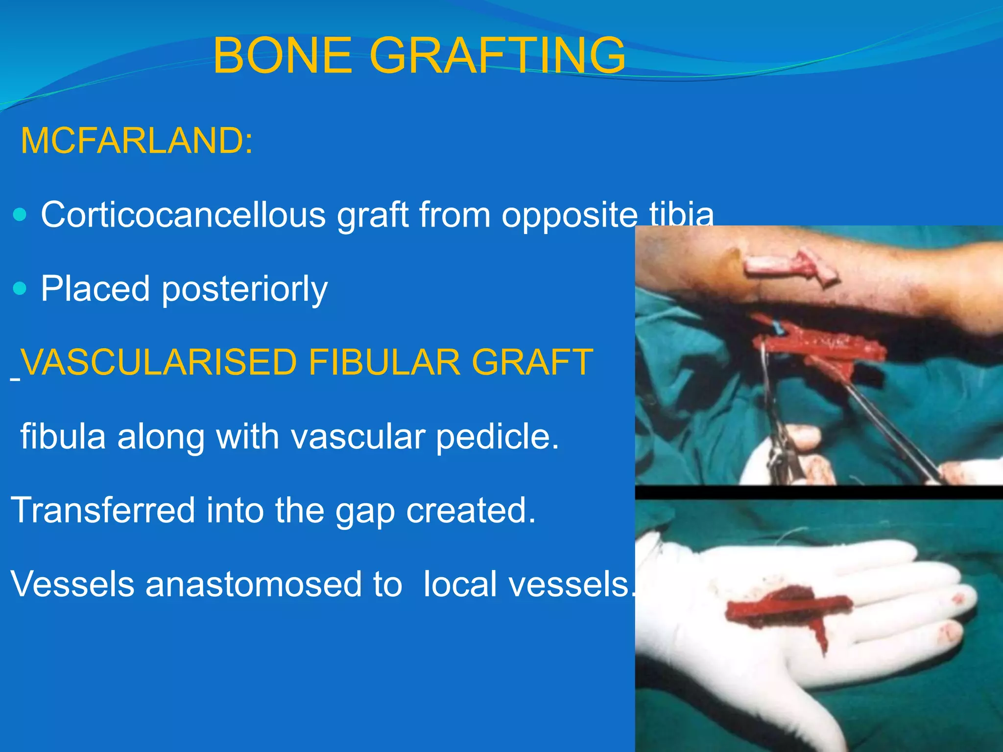
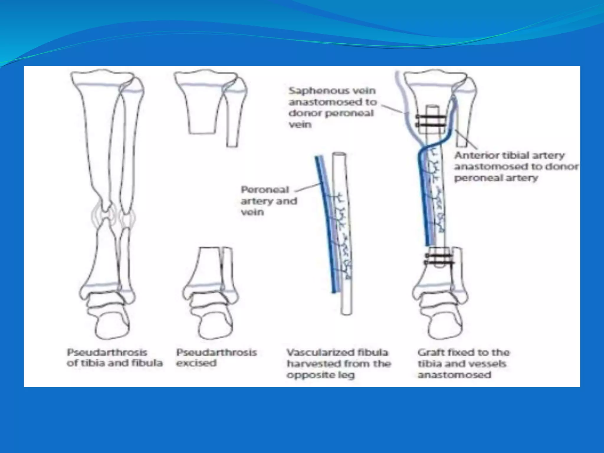
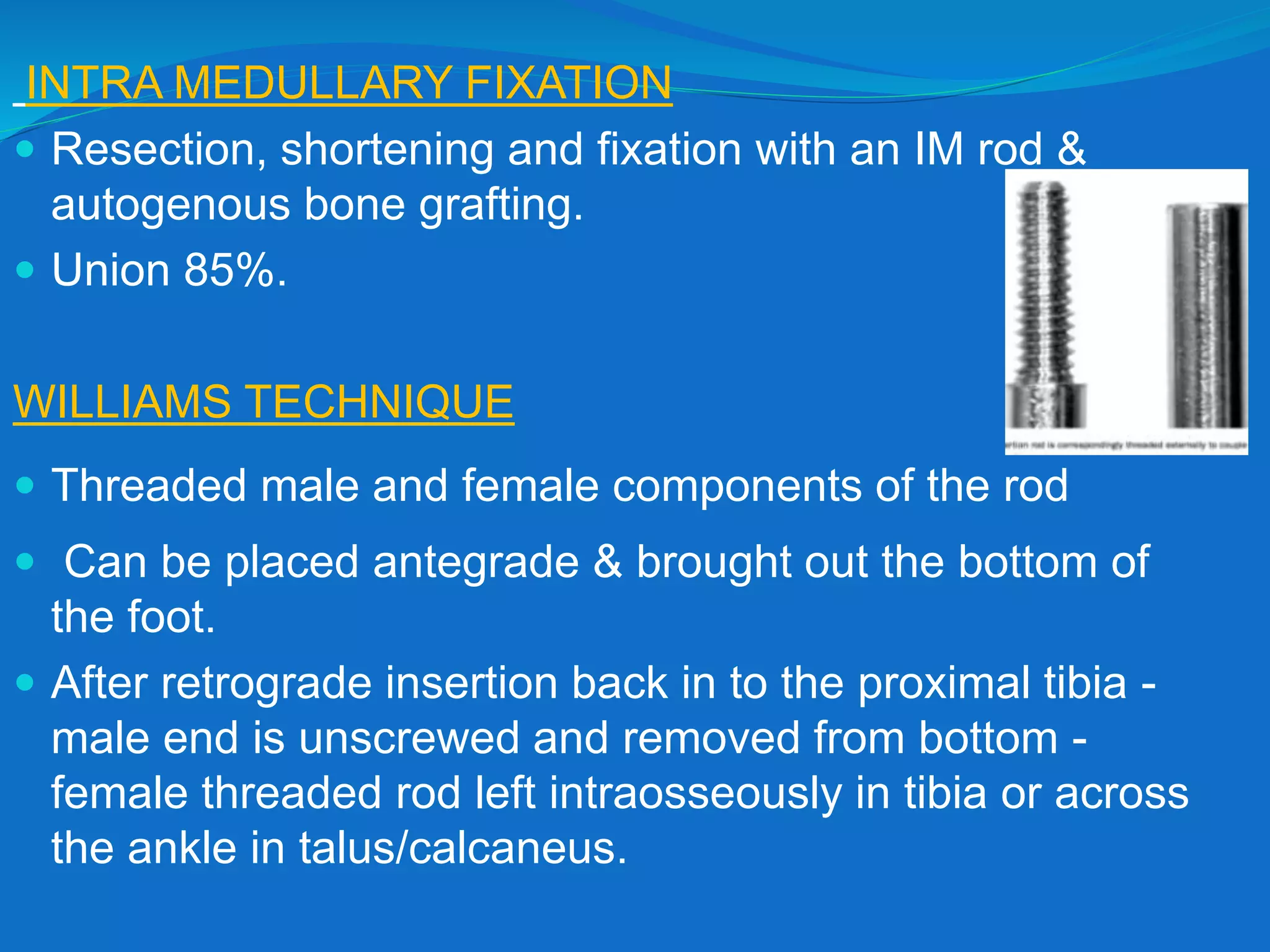
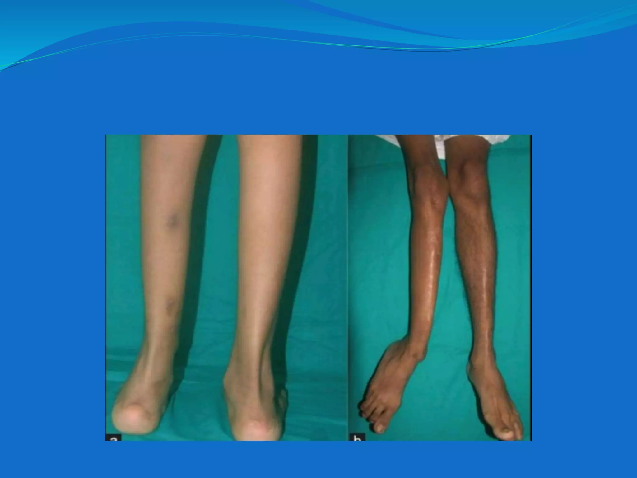
CONGENITAL
PSEUDOARTHROSIS OF
TIBIA
Dr. Bipul Borthakur (Professor)
Dept of Orthopaedics, SMCH
INTRODUCTION
 Nonunion of tibia fracture that develops
spontaneously or after trivial trauma in a dysplastic
bone segment of diaphysis.
 usually develops in first 2 years
 misnomer
 Etiology is not clear.
 Incidence is 1: 250,000
 Strong association with NF type 1.
 55% of cases associated with NF.
 Some authors: association in nearly every instance.
 Fibrous dysplasia: 15% of patients with anterolateral
bowing.
NEUROFIBROMATOSIS
 NF-1: due to mutation on the gene coding for
NEUROFIBROMIN on chromosome 17.
 It negatively regulates Ras activity.
 Affects Ras-dependent MAPK( mitogen activated protein
kinase) activity-essential for osteoclast function & survival.
•
•6 or more café-au-lait macules
• Axillary or inguinal freckling.
• 2 or more neurofibromas or 1 plexiform neurofibroma.
• 2 or more Lisch nodules.
•Optic glioma.
•Distinctive osseous lesions.
•Family history
•Recent studies :Hyperplasia of fibroblasts
•Osteolytic fibromatosis
•Paley et al: it is not bony but periosteal
POINTS FAVOURING:
•Thick & harmartomatous periosteum
•Avascular & atrophic changes in bone
•Failure of remodelling
CLASSIFICATION
No universal classification
CAMURATI-1930
ADGLEY -1952
BOYD -1958
APOIL -1970
ANDERSON-1973
CRAWFORD-1986
CRAWFORD-1999
BOYD & ANDERSON commonly used
BOYD CLASSIFICATION
TYPE 1: Anterior bowing, defect at birth,
other congenital abnormalities
TYPE2: Mc type
 Anterior bowing & a hourglass constriction.
 Spontaneous #s or after minor trauma.
 Commonly occurs before 2 years
 HIGH RISK TIBIA.
 Associated with NF-1
 Poorest prognosis
TYPE 3 :
Congenital cyst.
• Anterior bowing may precede or follow the
development of #.
TYPE 4 :
• Originates in a sclerotic segment.
• Medullary canal obliterated.
• Insufficiency or stress # develops
and gradually extends.
• Prognosis is good.
TYPE 5 :
• Pseudarthrosis of tibia with dysplastic fibula.
• Prognosis good if confined to fibula only.
• Lesion resembles type 2
TYPE 6 :
• Occurs as an intraosseous neurofibroma or
schwannoma .
• Extremely rare.
CRAWFORD CLASSIFICATION
 1-Non-Dysplastic
 Anterolateral bowing with increased density &
sclerosis of medullary canal.
 2-Dysplastic
 2a Anterolateral bowing
with failure of tabularization.
 2b Cystic changes.
 2c Frank pseudarthrosis.
DIAGNOSIS
CLINICAL FEATURES:
 cutaneous signs of NF
 anterolateral bowing of tibia.
 bowing usually at junction of middle & distal third.
 Associated with skin dimple, limb shortening, dysplasia
of fibula & ankle valgus.
 Usually unilateral.
IMAGING
MRI:
 Extent of disease
 Preoperative planning.
 Hyper intense on T2-weighted images and hypo intense on
T1-weighted images.
CT SCAN
 Confirm radiographic findings.
TOTAL BONE SCINTIGRAPHY
 Level of the pseudarthrosis .
PREOPERATIVE MANAGEMENT
 KAFO
 Clamshell orthosis
 DISCONTINUE:
 Tibia straightened
 Medullary canal reconstituted
 Adequate cortical thickness
 Skeletal maturity
TREATMENT OPTIONS
 Internal fixation and graft
 Ilizarov
 BMP
 Electrical stimulation
 Amputation
AIMS:
 1. Achieve union
 2. Prevent refracture
 3. Correct limb length inequality
 4. Correct associated growth abnormalities
 5. Prevent ankle deformity and arthritis.
STRATEGIES TO ACHIEVE UNION
 Microvascular graft transfer-vascularized fibula
 The Ilizarov technique
 Bone grafting with internal fixation-plating or IM devices.
 Excision of the pseudarthrosis should be an integral part of
the procedure.
STRATEGIES TO PREVENT REFRACTURE
 Splint the limb in an orthosis until skeletal maturity.
 Retain an intramedullary nail until skeletal maturity.
 STRATEGIES FOR DEALING WITH SHORTENING OF
THE LIMB
Minimize the extent of shortening.
Limb equalization procedures
STRATEGIES FOR MINIMIZING VALGUS DEFORMITY
OF THE ANKLE
Ensure union of fibular pseudarthrosis.
Retaining an IM rod that crosses ankle.
BONE GRAFTING
MCFARLAND:
 Corticocancellous graft from opposite tibia
 Placed posteriorly
VASCULARISED FIBULAR GRAFT
fibula along with vascular pedicle.
Transferred into the gap created.
Vessels anastomosed to local vessels.
 Procedure of choice for gaps > 3cm.
 92%- 95% union rate
 Refracture.
 Langenskiold procedure to prevent ankle valgus.
 In addition weakness may ensue in the donor leg due to
resection of origins of flexor muscles.
 PERIOSTEAL GRAFTING: has been tried
INTRA MEDULLARY FIXATION
 Resection, shortening and fixation with an IM rod &
autogenous bone grafting.
 Union 85%.
WILLIAMS TECHNIQUE
 Threaded male and female components of the rod
 Can be placed antegrade & brought out the bottom of
the foot.
 After retrograde insertion back in to the proximal tibia -
male end is unscrewed and removed from bottom -
female threaded rod left intraosseously in tibia or across
the ankle in talus/calcaneus.
ILIZAROV TECHNIQUE
 Provides excellent stability
 Complete resection of pseudo-arthrotic part.
 Enables weight bearing which aids healing
 Compression of pseudo-arthrosis
 Limb lengthening procedures can be done
BONE MORPHOGENIC PROTEIN
 BMP2
 BMP7
 ELECTRICAL STIMULATION
 Limited to the earlier phases when union is the primary
goal.
AMPUTATION – Maybe the better option in some
situations
McCARTHY CRITERIA
 Failure after 3 surgeries
 LLD 5cm or more
 Deformed foot
 Prolonged hospitalization
 High medical costs
COMPLICATIONS
1. REFRACTURE
 14% to 60%.
 Anatomic alignment minimizes the risk.
 IM rod and external bracing -protection against re-
fractures.
2. MALALIGNMENT
 Procurvatum
 valgus deformity
3. LIMB LENGTH DISCREPANCY
 Residual limb length discrepancy common
 Growth abnormalities noted with CPT.
4. ANKLE VALGUS
 Progressive ankle valgus is a problematic
postoperative donor-site morbidity of a vascularized
fibular graft in children.
 Tibiofibular metaphyseal synostosis (Langenskold
procedure) useful.
5. ANKLE STIFFNESS
 Progressively regresses after IM removed from
ankle.
 Pain secondary to degenerative changes of ankle-
limitation of activity and shoe modification.
 Severe pain – ankle arthrodesis
CONCLUSION
 Challenging for the surgeon
 Poor tendency to heal
 Excision of the hamartomatous tissue and pathological
periosteum is the KEY.
“yogasthaḥ kuru karmāṇi saṅgaṃ tyaktvā dhanañjaya
siddhyasiddhyoḥ samo bhūtvā samatvaṃ yoga ucyate”
By being established in Yoga, O Dhananjaya, undertake actions,
casting off attachment
and remaining equipoised in success and failure. Equanimity is
called Yoga.
THANK YOU.

Congenital pseudoarthrosis of tibia

  • 1.
    CONGENITAL PSEUDOARTHROSIS OF TIBIA Dr. BipulBorthakur (Professor) Dept of Orthopaedics, SMCH
  • 2.
    INTRODUCTION  Nonunion oftibia fracture that develops spontaneously or after trivial trauma in a dysplastic bone segment of diaphysis.  usually develops in first 2 years  misnomer  Etiology is not clear.  Incidence is 1: 250,000
  • 3.
     Strong associationwith NF type 1.  55% of cases associated with NF.  Some authors: association in nearly every instance.  Fibrous dysplasia: 15% of patients with anterolateral bowing.
  • 4.
    NEUROFIBROMATOSIS  NF-1: dueto mutation on the gene coding for NEUROFIBROMIN on chromosome 17.  It negatively regulates Ras activity.  Affects Ras-dependent MAPK( mitogen activated protein kinase) activity-essential for osteoclast function & survival.
  • 5.
    • •6 or morecafé-au-lait macules • Axillary or inguinal freckling. • 2 or more neurofibromas or 1 plexiform neurofibroma. • 2 or more Lisch nodules. •Optic glioma. •Distinctive osseous lesions. •Family history
  • 6.
    •Recent studies :Hyperplasiaof fibroblasts •Osteolytic fibromatosis •Paley et al: it is not bony but periosteal POINTS FAVOURING: •Thick & harmartomatous periosteum •Avascular & atrophic changes in bone •Failure of remodelling
  • 7.
    CLASSIFICATION No universal classification CAMURATI-1930 ADGLEY-1952 BOYD -1958 APOIL -1970 ANDERSON-1973 CRAWFORD-1986 CRAWFORD-1999 BOYD & ANDERSON commonly used
  • 8.
    BOYD CLASSIFICATION TYPE 1:Anterior bowing, defect at birth, other congenital abnormalities TYPE2: Mc type  Anterior bowing & a hourglass constriction.  Spontaneous #s or after minor trauma.  Commonly occurs before 2 years  HIGH RISK TIBIA.  Associated with NF-1  Poorest prognosis
  • 9.
    TYPE 3 : Congenitalcyst. • Anterior bowing may precede or follow the development of #. TYPE 4 : • Originates in a sclerotic segment. • Medullary canal obliterated. • Insufficiency or stress # develops and gradually extends. • Prognosis is good.
  • 10.
    TYPE 5 : •Pseudarthrosis of tibia with dysplastic fibula. • Prognosis good if confined to fibula only. • Lesion resembles type 2 TYPE 6 : • Occurs as an intraosseous neurofibroma or schwannoma . • Extremely rare.
  • 11.
    CRAWFORD CLASSIFICATION  1-Non-Dysplastic Anterolateral bowing with increased density & sclerosis of medullary canal.  2-Dysplastic  2a Anterolateral bowing with failure of tabularization.  2b Cystic changes.  2c Frank pseudarthrosis.
  • 12.
    DIAGNOSIS CLINICAL FEATURES:  cutaneoussigns of NF  anterolateral bowing of tibia.  bowing usually at junction of middle & distal third.  Associated with skin dimple, limb shortening, dysplasia of fibula & ankle valgus.  Usually unilateral.
  • 13.
    IMAGING MRI:  Extent ofdisease  Preoperative planning.  Hyper intense on T2-weighted images and hypo intense on T1-weighted images. CT SCAN  Confirm radiographic findings. TOTAL BONE SCINTIGRAPHY  Level of the pseudarthrosis .
  • 14.
    PREOPERATIVE MANAGEMENT  KAFO Clamshell orthosis  DISCONTINUE:  Tibia straightened  Medullary canal reconstituted  Adequate cortical thickness  Skeletal maturity
  • 15.
    TREATMENT OPTIONS  Internalfixation and graft  Ilizarov  BMP  Electrical stimulation  Amputation AIMS:  1. Achieve union  2. Prevent refracture  3. Correct limb length inequality  4. Correct associated growth abnormalities  5. Prevent ankle deformity and arthritis.
  • 16.
    STRATEGIES TO ACHIEVEUNION  Microvascular graft transfer-vascularized fibula  The Ilizarov technique  Bone grafting with internal fixation-plating or IM devices.  Excision of the pseudarthrosis should be an integral part of the procedure. STRATEGIES TO PREVENT REFRACTURE  Splint the limb in an orthosis until skeletal maturity.  Retain an intramedullary nail until skeletal maturity.
  • 17.
     STRATEGIES FORDEALING WITH SHORTENING OF THE LIMB Minimize the extent of shortening. Limb equalization procedures STRATEGIES FOR MINIMIZING VALGUS DEFORMITY OF THE ANKLE Ensure union of fibular pseudarthrosis. Retaining an IM rod that crosses ankle.
  • 18.
    BONE GRAFTING MCFARLAND:  Corticocancellousgraft from opposite tibia  Placed posteriorly VASCULARISED FIBULAR GRAFT fibula along with vascular pedicle. Transferred into the gap created. Vessels anastomosed to local vessels.
  • 19.
     Procedure ofchoice for gaps > 3cm.  92%- 95% union rate  Refracture.  Langenskiold procedure to prevent ankle valgus.  In addition weakness may ensue in the donor leg due to resection of origins of flexor muscles.  PERIOSTEAL GRAFTING: has been tried
  • 21.
    INTRA MEDULLARY FIXATION Resection, shortening and fixation with an IM rod & autogenous bone grafting.  Union 85%. WILLIAMS TECHNIQUE  Threaded male and female components of the rod  Can be placed antegrade & brought out the bottom of the foot.  After retrograde insertion back in to the proximal tibia - male end is unscrewed and removed from bottom - female threaded rod left intraosseously in tibia or across the ankle in talus/calcaneus.
  • 22.
    ILIZAROV TECHNIQUE  Providesexcellent stability  Complete resection of pseudo-arthrotic part.  Enables weight bearing which aids healing  Compression of pseudo-arthrosis  Limb lengthening procedures can be done
  • 23.
    BONE MORPHOGENIC PROTEIN BMP2  BMP7  ELECTRICAL STIMULATION  Limited to the earlier phases when union is the primary goal.
  • 24.
    AMPUTATION – Maybethe better option in some situations McCARTHY CRITERIA  Failure after 3 surgeries  LLD 5cm or more  Deformed foot  Prolonged hospitalization  High medical costs
  • 25.
    COMPLICATIONS 1. REFRACTURE  14%to 60%.  Anatomic alignment minimizes the risk.  IM rod and external bracing -protection against re- fractures.
  • 26.
    2. MALALIGNMENT  Procurvatum valgus deformity 3. LIMB LENGTH DISCREPANCY  Residual limb length discrepancy common  Growth abnormalities noted with CPT.
  • 27.
    4. ANKLE VALGUS Progressive ankle valgus is a problematic postoperative donor-site morbidity of a vascularized fibular graft in children.  Tibiofibular metaphyseal synostosis (Langenskold procedure) useful.
  • 29.
    5. ANKLE STIFFNESS Progressively regresses after IM removed from ankle.  Pain secondary to degenerative changes of ankle- limitation of activity and shoe modification.  Severe pain – ankle arthrodesis
  • 30.
    CONCLUSION  Challenging forthe surgeon  Poor tendency to heal  Excision of the hamartomatous tissue and pathological periosteum is the KEY.
  • 31.
    “yogasthaḥ kuru karmāṇisaṅgaṃ tyaktvā dhanañjaya siddhyasiddhyoḥ samo bhūtvā samatvaṃ yoga ucyate” By being established in Yoga, O Dhananjaya, undertake actions, casting off attachment and remaining equipoised in success and failure. Equanimity is called Yoga. THANK YOU.