congenital cytomegalovirus infection is a major problem in children. severe morbidity also in some cases mortality can occur due to this infection. this presentation has highlighted updates on this topic in short.
Congenital CMV infection is a leading cause of sensorineural hearing loss and mental retardation. Approximately 0.15-2% of live births are affected. While most infections are asymptomatic, 10% of symptomatic newborns die and survivors often have lifelong disabilities. Congenital CMV is transmitted from mother to fetus via the placenta or during birth if the mother is infected. Young children are the primary transmitters of CMV through bodily fluids. Treatment with ganciclovir may help reduce hearing loss in symptomatic newborns but is associated with significant toxicity. Prevention through hygiene and education is important since there is no vaccine.
This document provides information on cytomegalovirus (CMV), a herpes virus. Some key points:
- CMV was first isolated in 1956 from infants and causes lifelong latent infection. It is a major cause of disease in transplant recipients.
- CMV infection is generally asymptomatic in healthy individuals but can cause disease in the immunocompromised via direct tissue damage or indirect immune effects.
- Diagnosis involves detecting viral DNA, antigens, or culture. Treatment includes antivirals like ganciclovir or valganciclovir.
- Prevention strategies for transplant recipients include preemptive therapy based on viral monitoring or prophylaxis for high risk groups to reduce disease and
Approach to congenital cmv infection in newbornJigar Patel
This document provides an overview of congenital cytomegalovirus (CMV) infection. CMV is a herpesvirus that can be transmitted from mother to fetus. Approximately 40,000 infants are born with congenital CMV annually in the US, with 10-15% experiencing hearing loss. Symptomatic infants may experience jaundice, hepatosplenomegaly, or hearing loss at birth. Treatment involves antiviral therapy with ganciclovir or valganciclovir for symptomatic infants or those with hearing loss or central nervous system involvement to improve outcomes. Adverse effects require monitoring bloodwork during the recommended 6 month course of treatment.
This document discusses infectious mononucleosis and Epstein-Barr virus (EBV). It states that EBV is spread through oral secretions and is a cause of infectious mononucleosis. Infectious mononucleosis presents as enlarged lymph nodes, sore throat, fever, and extreme tiredness. EBV is also associated with African Burkitt's lymphoma, nasopharyngeal carcinoma, and B cell lymphomas. Treatment for infectious mononucleosis involves rest and supportive care.
Hepatitis is inflammation of the liver that can be caused by five primary viruses. Hepatitis A and E viruses are transmitted through the fecal-oral route while hepatitis B, C, and D viruses are transmitted parenterally or sexually. Hepatitis B, C, and D can result in chronic infection. Most cases of acute viral hepatitis resolve without treatment but hepatitis B, C, and D pose long term risks of cirrhosis and liver cancer if infection is chronic. Diagnosis involves serologic testing to detect viral antigens and antibodies. Treatment focuses on supportive care although antivirals exist for chronic hepatitis B and C. Prevention emphasizes vaccination for hepatitis A and B.
Cytomegalovirus is a herpesvirus that commonly infects humans. It can cause enlarged cells (cytomegalic inclusion disease) and poses a risk for severe infections in infants during pregnancy or birth as well as immunosuppressed individuals. The virus replicates slowly in human fibroblasts and establishes lifelong latent infections. Primary infection is usually asymptomatic but can resemble mononucleosis. Congenital infection may cause death, growth problems, or long-term neurological and vision issues in infants. Polymerase chain reaction testing and antigen detection are now used to diagnose active cytomegalovirus infections.
Congenital rubella syndrome is caused when a pregnant woman is infected with rubella virus, usually in the first trimester of pregnancy. It can cause deafness, eye abnormalities, and congenital heart defects in the fetus. While the exact molecular mechanisms are unclear, rubella virus is known to have an apoptotic effect on certain cell types. Introduction of the rubella vaccine has dramatically reduced the number of babies born with congenital rubella syndrome.
Approach to a child with HepatosplenomegalySunil Agrawal
This document discusses hepatosplenomegaly, or the enlargement of the liver and spleen. It begins with an introduction and overview of hepatomegaly and splenomegaly. It then covers the various causes of hepatosplenomegaly including infections, hematological disorders, vascular congestion, tumors and infiltrations, storage disorders, and miscellaneous causes. The document provides details on evaluating a patient's history, physical examination findings, investigations, and treatment strategies for hepatosplenomegaly in both children and neonates. It concludes with references for further information.
Hi Guys,
This presentation talks about Tuberculosis diagnosed in mother in the antenatal period, its treatment, implications on mother and fetus, the various protocols available currently regarding the neonatal management . Special focus being in major issues like breastmilk feeding, BCG, AKT prophylaxis, mother-child isolation.
Hope you find it useful.
P.S. - Please checkout my youtube channel - 'NEONATOHUB' & Facebook page 'Neonatohub' for lectures on neonatology.
Hemolytic uremic syndrome (HUS) is a disease characterized by hemolytic anemia, low platelet count, and kidney failure. It predominantly affects children and can be caused by infections from E. coli or pneumococcal bacteria or complement factor abnormalities. The typical pathophysiology involves Shiga toxin or other bacterial toxins damaging endothelial cells and platelets. Treatment involves supportive care, antibiotics for infections, plasma therapy for complement abnormalities, and long-term prognosis depends on severity and treatment.
This document provides an overview of sickle cell disease (SCD), including its definition, epidemiology, pathogenesis, clinical presentation, diagnosis, treatment, and counseling. SCD is caused by a genetic mutation resulting in abnormal hemoglobin called HbS. It most commonly presents as painful vasoclusive crises, acute chest syndrome, splenic sequestration, or aplastic crises. Treatment involves management of symptoms, antibiotics, hydroxyurea, blood transfusions, and counseling to enable informed family planning decisions.
Neonatal anaemia: overview of pathophysiology, clinical approaches and compre...Gabriel Shamavu
This document provides an outline and introduction to the topics of neonatal anemia. It discusses physiological anemia of newborns, anemia of prematurity, and pathologic anemia. For physiological anemia, it explains the normal developmental process of erythropoiesis and hemoglobin production during fetal development and after birth. It also describes the typical decline in hemoglobin levels seen in both term and preterm infants in the first few weeks of life. For anemia of prematurity, it notes this is an exaggerated form of physiological anemia seen in very preterm infants less than 32 weeks gestation. The document lays the foundation for further discussion of these topics in neonatal anemia.
Malaria is a mosquito-borne infectious disease caused by Plasmodium parasites. It is widespread in tropical and subtropical regions, with P. falciparum being the most dangerous species. The parasite has a complex life cycle involving transmission between humans and female Anopheles mosquitoes. Malaria symptoms include fever, chills, and fatigue in cyclical patterns. It remains a major global health problem, with hundreds of millions of cases and over one million deaths per year. Definitive diagnosis requires microscopic examination of blood smears to identify the malaria parasites.
This neonatal case presentation involves a 16 day old infant admitted to the NICU with jaundice since birth and vomiting for 1 day. Investigations revealed severe anemia, indirect hyperbilirubinemia, increased reticulocyte count, and elevated LDH. The infant received a PRBC transfusion and was discharged with improved Hb. The clinical picture is suggestive of hemolytic anemia, possibly due to fetomaternal hemorrhage given the maternal history of blood transfusions in the second trimester.
Non-typhoidal Salmonellosis, is one of the most common and widely distributed foodborne diseases, with tens of millions of human cases occurring worldwide every year.
In the United States, the incidence of NTS infection has doubled in the past 2 decades.
In 2009 there were 14 million cases of NTS.
Vitamin K deficiency in newborns can cause a condition called haemorrhagic disease of the newborn (HDN) where there is bleeding due to a lack of vitamin K dependent clotting factors. Newborns are especially vulnerable because of minimal vitamin K transfer from mother and lack of intestinal bacteria. HDN presents as bleeding from the GI tract, skin, or brain. It is classified as early, classical or late-onset depending on timing. Treatment involves vitamin K supplementation while serious or intracranial bleeding may require transfusions. Prophylactic vitamin K shots at birth can prevent most cases of HDN.
Here are some key points regarding the feasibility of bacteriological diagnosis in children with TB:
- Sputum induction or gastric lavage are generally required to obtain specimens from children, as they typically cannot produce sputum on demand. This requires specialized equipment and trained personnel.
- Even with induced sputum or gastric lavage, specimen quality and volume may be low, reducing the sensitivity of bacteriological tests.
- Young children especially may not be able to cooperate with procedures like sputum induction.
- Extrapulmonary TB is more common in children than adults, so specimens from sites like lymph nodes, cerebrospinal fluid, etc. need to be obtained invasively via procedures like biopsy or lumbar puncture
Tuberculosis is caused by Mycobacterium tuberculosis and is a chronic infectious disease characterized by vague symptoms and a protracted course. India accounts for one third of the global TB burden, with 15 million infected people in India and 3-4 million of those being children. Tuberculosis most commonly enters the body through inhalation and can spread through droplets or ingestion. Primary infection typically occurs in the lungs or lymph nodes and may heal or progress to more serious complications affecting multiple organs if not contained. Common symptoms in children include failure to thrive, fever, and painless lymphadenopathy.
Idiopathic thrombocytopenic purpura (ITP) is an autoimmune disorder characterized by low platelet counts and bruising. It most commonly affects children ages 1-4 years old within 4 weeks of a viral infection. An autoantibody develops against platelets, causing their destruction by the spleen. Most cases of childhood ITP are mild and resolve spontaneously within 6 months. Treatment involves observation for mild cases or corticosteroids, IVIG, or anti-D therapy for more severe cases. Splenectomy is reserved for chronic or uncontrolled cases.
This document provides statistics on the global HIV epidemic in 2018 from UNAIDS as well as information on HIV in India. Some key points:
- 37.9 million people globally were living with HIV in 2018. 1.7 million became newly infected that year while 23.3 million were accessing antiretroviral therapy.
- India has the third largest HIV epidemic in the world. In 2015, the national adult prevalence was 0.26%. Prevalence is highest in certain states like Mizoram (2.04%) and Manipur (1.43%).
- Children account for 6.54% of total PLHIV in India. Early infant diagnosis, appropriate infant feeding and prophylaxis
This document summarizes key information about infectious mononucleosis caused by Epstein-Barr virus (EBV). It was first described in 1920 and causes fever, pharyngitis, and adenopathy. Diagnosis involves detecting heterophile antibodies or testing for EBV-specific antibodies. Physical exam may show lymphadenopathy, hepatosplenomegaly, or periorbital edema. Laboratory tests include complete blood count showing lymphocytosis and atypical lymphocytes as well as elevated liver enzymes. Specific antibody testing confirms diagnosis and distinguishes between acute and past EBV infection.
This document provides information on evaluating and diagnosing children with recurrent pneumonia. It discusses obtaining a thorough medical history including details of pneumonia episodes, risk factors, and associated symptoms. A complete physical exam focuses on the respiratory system and signs of underlying conditions. Initial tests include a CBC, chest X-ray, and Mantoux test. Further tests like CT, PFTs, bronchoscopy may help identify structural abnormalities, aspiration, or immunodeficiencies causing recurrent pneumonia. Treatment involves managing the specific illness and providing nutritional/respiratory support.
The document discusses tuberculosis in children, including its epidemiology, etiology, clinical features, diagnosis, and management. It notes that tuberculosis is endemic in Pakistan, with over 200,000 new cases annually. Children under 15 account for 20% of cases. The causative agent is Mycobacterium tuberculosis. Clinical features vary depending on the site of infection, and may include cough, fever, lymph node enlargement, and meningitis. Diagnosis involves tuberculin tests, chest X-rays, and culture of fluid/tissue samples. Standard drug regimens include isoniazid and rifampin for 6-12 months. Prevention involves BCG vaccination, contact screening, and prophylactic treatment of
Neonatal sepsis is a clinical syndrome of bacteremia and infection in infants under 4 weeks of age. Common causes are E. coli, Group B Streptococcus, and Listeria. It can be early-onset from transmission during birth or late-onset from hospital-acquired infections. Symptoms are non-specific but include respiratory distress, feeding issues, and temperature instability. Diagnosis involves blood, urine and CSF cultures. Treatment is antibiotics like ampicillin and gentamicin for 10-14 days along with supportive care. Prevention includes good antenatal care, treating maternal infections, early breastfeeding and infection control policies in the NICU.
Congenital syphilis is an infection transmitted from mother to fetus during pregnancy. It can occur at any stage of maternal infection. Without treatment, it can cause complications like stillbirth, neonatal death, and long term effects on bones, teeth, eyes and brain. Diagnosis involves serologic testing of both mother and infant. Treatment is with penicillin to prevent transmission and complications in the newborn. Careful follow up is needed to monitor treatment response and detect any late manifestations.
This document discusses special situations and adverse events following immunization. It provides guidance on vaccinating preterm/low birth weight infants, those receiving corticosteroids or immunosuppressive therapy, children with malignancies, congenital immunodeficiencies, chronic diseases, allergies, bleeding disorders, or acute illness. It recommends that most vaccines can be administered according to chronological age for preterm/low birth weight infants. It also provides specific guidance on contraindications and precautions for different groups.
Common TORCH Infection that affect pediatricsznfq8kmwhz
The document provides an overview of TORCH infections that can be transmitted from mother to fetus during pregnancy, including Toxoplasmosis, Other (syphilis), Rubella, Cytomegalovirus (CMV), and Herpes simplex virus (HSV). It discusses the epidemiology, pathophysiology, distinguishing clinical features, recommended workup, and prognosis for each infection. Key points include the importance of prenatal screening and treatment to prevent fetal transmission and long term sequelae. Maternal education on immunizations, safe sexual practices, and food safety are emphasized to reduce risk of TORCH infections during pregnancy.
"Congenital Cytomegalovirus Infection, Update on Treatment"Basem Hamed
Cytomegalovirus (CMV) is the leading cause of congenital viral infection and can cause hearing loss and neurological disabilities. Prenatal screening is not routinely recommended but may be offered to pregnant women with symptoms suggestive of CMV infection. Diagnosis involves testing for IgM, IgG avidity, and PCR of amniotic fluid. Prognosis depends on infection timing and severity of ultrasound findings, with termination of pregnancy considered for severe cases. Prevention focuses on handwashing and avoiding contact with young children's bodily fluids.
This slide contains clinical features, perinatal and post natal diagnosis of congenital torch infection in fetus and neonates, and management of congenital toxaplasma, rubella, CMV, Herpes simplex, varicella, and other infections.
1. The document discusses several systemic viral infections that can present with or without an exanthem. These include measles, rubella, parvovirus B19, human herpesvirus 6 and 7, chickenpox, shingles, mumps, influenza, infectious mononucleosis caused by Epstein-Barr virus, cytomegalovirus, and viral hemorrhagic fevers.
2. The infections are described in terms of their causative virus, clinical features, complications, diagnosis, and management including treatment and prevention through vaccination or antiviral therapy.
3. Prevention is focused on vaccination programs and hygiene measures to limit transmission through respiratory droplets or other bodily fluids.
A brief description of very common infection caused by the virus: Cytomegalovirus. Typically affects infants, and pregnant ladies. Features in HIV patients. Transmitted by saliva, fomites and at the time of delivery. Helpful for medical students, doctors, pediatricians, gynecologists, dermatologists. Useful for exams USMLE, FCPS, MCPS and MRCP, MD students.
TORCH infections refer to a group of congenital infections caused by Toxoplasma gondii, other agents (syphilis), Rubella virus, Cytomegalovirus, and Herpes simplex virus. These infections can be transmitted prenatally, perinatally, or postnatally and may cause fetal and neonatal morbidity and mortality. While the TORCH acronym was originally used to group these five infections, it has become obsolete due to the increasing number of pathogens that can cause congenital infections. Diagnosis of suspected TORCH infections in infants involves clinical examination, laboratory tests, and imaging based on the specific symptoms presented. Timely diagnosis is important for treatment and management.
This document discusses TORCH infections, which are a group of infections that can be transmitted from mother to fetus during pregnancy and cause congenital infections. It notes that a newborn male is under evaluation for being small for gestational age with thrombocytopenia, which raises suspicion for TORCH infections. The document then summarizes each infection: Toxoplasmosis can cause chorioretinitis, hydrocephalus, and intracranial calcifications. Syphilis presents with snuffles and can cause perinatal death if untreated. Rubella causes sensorineural hearing loss, cataracts, and cardiac defects. Cytomegalovirus commonly causes asymptomatic infection but can later cause hearing loss. Her
This document provides an overview of congenital infections caused by cytomegalovirus (CMV) and Toxoplasma gondii. It discusses the properties and transmission of CMV, which can cause birth defects if transmitted from mother to fetus. Clinical manifestations range from asymptomatic infection to organ damage in immunocompromised individuals. Diagnosis involves virus detection and antibody testing. Treatment focuses on antiviral drugs for severe cases. Toxoplasmosis is caused by the parasite T. gondii and transmitted through cats, meat, and congenitally. It can cause abortion or symptoms in susceptible hosts. Diagnosis is usually serological and treatment includes sulfa drugs and pyrimethamine.
Evaluation of Infection In Pregnancy
- Some infections can be transmitted from mother to fetus and cause serious issues. Rarely, serious maternal illness can impact the fetus as well.
- Intra-amniotic infections from bacteria in the vaginal flora can cause infections like sepsis and meningitis in newborns.
- Common organisms that cause neonatal sepsis include Streptococcus agalactiae (group B strep), E. coli, coagulase-negative staphylococci, and Staphylococcus aureus. Screening and treatment is recommended for some high risk infections.
This document discusses perinatal infections, providing details on several pathogens that can cause congenital infections including Toxoplasma gondii, rubella virus, cytomegalovirus, herpes simplex virus, and Treponema pallidum (syphilis). It describes the transmission, signs/symptoms, diagnosis, and treatment of infections caused by each pathogen. Key points include that primary maternal infection with toxoplasmosis, rubella virus, or herpes is highest risk for fetal transmission and infection earlier in pregnancy leads to worse outcomes. Diagnosis involves pathogen detection and serologic testing, while treatment focuses on antimicrobials and supportive care of the newborn.
Perinatal infections can cause significant morbidity and mortality if not properly diagnosed and treated. Some important perinatal infections discussed in the document include toxoplasmosis, rubella, cytomegalovirus, herpes simplex virus, and syphilis. For each infection, the document outlines the causative agent, route of transmission, signs and symptoms in both the mother and infant, diagnostic testing, and treatment recommendations. Preventing transmission requires screening, counseling, and treatment as indicated for at-risk mothers and newborns.
Microbiology of Cytomegalovirus (Herpes).pptxNawangSherpa6
The Presentation here is about Medically important Cytomegalovirus. How does it infect the Human host? What are it's clinical manifestations and How can we diagnose for their infection and potential application for other studies.
TORCH infections refer to a group of viral, bacterial and protozoan infections that can be transmitted from mother to fetus during pregnancy, potentially causing fetal anomalies or loss. These include toxoplasmosis, caused by the protozoan Toxoplasma gondii; rubella virus; cytomegalovirus; and herpes simplex virus. Screening and treatment of maternal infections can help prevent fetal transmission and complications, but often has limited impact if transmission has already occurred. Common TORCH infections, their signs, risks in pregnancy, methods of diagnosis and treatment approaches were discussed in the document.
This document summarizes various viral and protozoal infections that can occur during pregnancy including rubella, measles, influenza, chickenpox, cytomegalovirus, parvovirus, mumps, herpes simplex virus, and HIV. For each infection, the document discusses the causative virus, clinical features, effects on pregnancy, methods of diagnosis, and management approaches. Complications of congenital infections are also outlined. The management of HIV positive pregnancies including antiretroviral therapy and approaches to reduce mother-to-child transmission are described in detail.
This document provides information on nursing care for HIV/AIDS children. It discusses HIV transmission, clinical manifestations at different stages, opportunistic infections, antiretroviral therapy, nutrition management, immunizations, and nursing assessments and interventions. Key points include transmission from mother to child, asymptomatic onset but development of symptoms over time, opportunistic infections as immune system weakens, antiretroviral therapy to suppress virus, and holistic nursing care to support health and development.
This document discusses infections during pregnancy and their effects. It covers how pregnancy induces immunological changes that affect a woman's ability to fight certain infections. The fetus and newborn also have immature immune systems. Several viral infections are then examined in more detail, including varicella-zoster virus, influenza, and rubella. For each, the document outlines how maternal infection can impact the fetus and neonate, as well as recommendations for diagnosis and management.
This document provides background information on maternal infections in pregnancy. It discusses various viral, bacterial, fungal, and parasitic infections that can affect both mother and baby during pregnancy. Some key points:
- Infections are a major cause of morbidity and mortality for both mothers and babies in developing countries. Common infections in Ghana include malaria, Group B Streptococcus, and other bacterial infections.
- Pregnancy increases susceptibility to certain infections due to immune system changes that help tolerate the fetus. The placenta can also serve as a site of infection for some pathogens.
- Major viral infections discussed include chickenpox, influenza, rubella, cytomegalovirus, and HIV. Bacterial infections
Similar to Congenital cytomegalovirus infection (20)
Portal biliopathy is defined as abnormalities in the intrahepatic and extrahepatic biliary tract, gallbladder and cystic duct secondary to portal hypertension
The document summarizes the case of a 10-year-old boy admitted with fever, jaundice, and abdominal pain. Key findings include intermittent high fever for 4 days, jaundice for 4 days, and occasional right upper abdominal pain. He had a similar prior episode 2 months ago. Examination found him febrile but otherwise stable. Tests showed elevated liver enzymes and bilirubin. Ultrasound and MRCP found a fusiform dilated common bile duct suggestive of a choledochal cyst. He was started on antibiotics and vitamin K. His fever subsided after a few days but jaundice remained. A pediatric surgery consultation was requested.
Functional GI disorder is common in children. Rome IV criteria are helpful in differentiating organic causes from functional disorders. this presentation shows the difference between Rome III and IV criteria.
intravenous fluid and electrolytes are important topics in medical science. potassium is one of the vital electrolytes of the human body. this presentation has a discussion on several iv fluids and potassium balance and also how to manage the potassium imbalance.
This document provides information about oxygen therapy for children. It defines oxygen and why oxygen therapy is used to treat conditions caused by low blood oxygen levels. It describes the different modes of oxygen delivery including nasal prongs, masks, and ventilators. It covers indications for oxygen therapy, sources of oxygen like cylinders, and important considerations like humidification and hazards of oxygen toxicity from high concentrations over long periods.
Atrial septal defect (ASD) is an abnormal opening in the wall separating the left and right atria of the heart. There are several types of ASDs including secundum, ostium primum, sinus venosus, and coronary sinus defects. ASDs are usually diagnosed through echocardiography which can determine the size and location of the defect. Small, asymptomatic ASDs may not require treatment, but larger defects with evidence of right heart strain often warrant closure either through open heart surgery or a nonsurgical approach using an implantable device delivered through catheters. Both methods effectively close the defect to prevent long-term complications like heart failure and pulmonary hypertension.
Dr. Maimuna Sayeed presented the case of an 11-month-old boy with breathing difficulties for 2 days, vomiting since 3 months of age, and dribbling of urine since birth. Examination found the boy to be dyspneic, pale, and hypertensive with ballotable kidneys and palpable bladder. Investigations supported chronic kidney disease stage 5 due to posterior urethral valve, with hydronephrosis, hydroureters, and vesicoureteral reflux seen on imaging. The boy was managed conservatively with antibiotics and peritoneal dialysis, showing improvement over follow-ups. Surgery consultation was planned to address the underlying valve abnormality.
Dr. Maimuna Sayeed presented the case of 2 year old Diya who presented with rashes on her body for 2 months along with swelling all over her body for 5 days and decreased urination. Her physical exam showed purpuric rashes, edema, and joint tenderness. Tests showed mild anemia, proteinuria, and normal electrolytes. Her ultrasound was normal. The provisional diagnosis was Henoch-Schönlein purpura with nephritis based on her rash, joint pain, abdominal pain, edema, and proteinuria. She was started on medications but had worsening symptoms on follow-up, so additional medications and a renal biopsy were planned.
The patient, an 11-year-old boy, presented with weakness of all four limbs and difficulty swallowing over the past 10 days. Examination found symmetrical weakness, hypotonia, diminished reflexes, and facial nerve palsy. Investigations including CSF analysis and nerve conduction study supported a diagnosis of Guillain-Barré syndrome. He was treated with IVIG and made gradual improvement over his hospital stay, with resolution of accompanying hypertension. He was discharged with advice for outpatient physiotherapy follow up.
- The patient Laam, a 9 month old boy, presented with recurrent seizures for 1 month with delayed developmental milestones.
- His neurological exam and investigations including EEG and CT brain were suggestive of West Syndrome with bilateral cerebral atrophy possibly due to birth asphyxia.
- He was started on ACTH and antihypertensive treatment. Follow up showed improved seizure control but persistent hypertension requiring dose adjustment. Eye evaluation found pale optic discs and chorioretinal changes.
The document discusses the anatomy and functions of the brain ventricles. It describes the four ventricles - the left and right lateral ventricles, the third ventricle, and the fourth ventricle. The ventricles are lined with ependymal cells and filled with cerebrospinal fluid (CSF), which is produced by the choroid plexus. CSF circulates through the ventricles, provides cushioning and protection to the brain, and is absorbed by the arachnoid granulations. Increased CSF pressure can cause conditions like hydrocephalus and papilledema. A lumbar puncture is described as a method to examine CSF in different medical conditions.
Md. Huzaifa, a 6-year-old boy, presented with 2 months of fever, multiple nodular swellings, left testicular swelling, and gradual pallor. On examination, he had generalized lymphadenopathy, hepatosplenomegaly, proptosis, and left testicular swelling. Blood tests found pancytopenia and 80% blasts. Bone marrow biopsy revealed 80% lymphoblasts. He was diagnosed with acute lymphoblastic leukemia (B-cell lineage). He received supportive care and chemotherapy. Follow up showed improvement with chemotherapy continuation planned.
1) The document summarizes a clinical meeting presentation about conjoined twin infants born at 35 weeks gestation who were attached at the head.
2) On examination, the twins were generally healthy and developing appropriately except for being jaundiced and experiencing intermittent low urine output.
3) Over the course of their hospital stay, their jaundice and urine output were monitored and treated conservatively with phototherapy and IV fluids respectively. Electrolyte abnormalities were also corrected.
The document describes the case of a 31⁄2 year old boy who was admitted with a 1 year history of painless rectal bleeding after bowel movements. On examination, he appeared well with no abnormalities found. A colonoscopy revealed a pedunculated polyp 20cm from the anal verge, which was removed via colonoscopic polypectomy.
Rafin, a 40 day old male infant, presented with projectile vomiting since 17 days of age. Examination revealed a soft abdomen with an olive-sized mass palpable in the left upper quadrant. Imaging showed hypertrophy of the pyloric muscle, consistent with infantile hypertrophic pyloric stenosis. He was treated with intravenous fluids and electrolyte replacement. After correction of dehydration and electrolyte abnormalities, the patient was referred to pediatric surgery for pyloromyotomy to treat the underlying pyloric stenosis.
This document provides a case scenario of a 4-week-old male infant presenting with cyanosis after feeding or crying. On examination, the infant has purplish lips, hands and feet, a grade III/VI systolic murmur, and tests revealing low oxygen levels that increase slightly with oxygen. The document discusses possible diagnoses of congenital cyanotic heart disease such as tetralogy of Fallot and asks what the diagnosis could be. It then provides an overview of tetralogy of Fallot and its characteristics.
Halima, an 11-year-old girl with consanguineous parents, presented with not growing well, gradual pallor, and abdominal distension for 7 years. On examination, she was severely pale with facial dysmorphism and hepatosplenomegaly. Her history included repeated blood transfusions. She was diagnosed with hereditary hemolytic anemia. The seminar discussed thalassemia, including the types of thalassemia, clinical features, investigations, complications, and management with a focus on blood transfusions and chelation therapy.
A case of a 3 month old boy with jaundice and pale stool is presented. On examination, he was icteric with hepatomegaly but no other abnormalities. Laboratory tests found direct hyperbilirubinemia. The objectives of the discussion are to understand neonatal cholestasis, evaluate cases, understand the differential diagnosis, and discuss treatment options. Neonatal cholestasis is prolonged conjugated hyperbilirubinemia beyond the first 14 days of life. Causes include extrahepatic conditions like biliary atresia or intrahepatic conditions like idiopathic neonatal hepatitis. Evaluation and management aim to identify treatable causes and prevent progression of liver disease.
Muktapishti is a traditional Ayurvedic preparation made from Shoditha Mukta (Purified Pearl), is believed to help regulate thyroid function and reduce symptoms of hyperthyroidism due to its cooling and balancing properties. Clinical evidence on its efficacy remains limited, necessitating further research to validate its therapeutic benefits.
NVBDCP.pptx Nation vector borne disease control programSapna Thakur
NVBDCP was launched in 2003-2004 . Vector-Borne Disease: Disease that results from an infection transmitted to humans and other animals by blood-feeding arthropods, such as mosquitoes, ticks, and fleas. Examples of vector-borne diseases include Dengue fever, West Nile Virus, Lyme disease, and malaria.
- Video recording of this lecture in English language: https://youtu.be/kqbnxVAZs-0
- Video recording of this lecture in Arabic language: https://youtu.be/SINlygW1Mpc
- Link to download the book free: https://nephrotube.blogspot.com/p/nephrotube-nephrology-books.html
- Link to NephroTube website: www.NephroTube.com
- Link to NephroTube social media accounts: https://nephrotube.blogspot.com/p/join-nephrotube-on-social-media.html
Title: Sense of Smell
Presenter: Dr. Faiza, Assistant Professor of Physiology
Qualifications:
MBBS (Best Graduate, AIMC Lahore)
FCPS Physiology
ICMT, CHPE, DHPE (STMU)
MPH (GC University, Faisalabad)
MBA (Virtual University of Pakistan)
Learning Objectives:
Describe the primary categories of smells and the concept of odor blindness.
Explain the structure and location of the olfactory membrane and mucosa, including the types and roles of cells involved in olfaction.
Describe the pathway and mechanisms of olfactory signal transmission from the olfactory receptors to the brain.
Illustrate the biochemical cascade triggered by odorant binding to olfactory receptors, including the role of G-proteins and second messengers in generating an action potential.
Identify different types of olfactory disorders such as anosmia, hyposmia, hyperosmia, and dysosmia, including their potential causes.
Key Topics:
Olfactory Genes:
3% of the human genome accounts for olfactory genes.
400 genes for odorant receptors.
Olfactory Membrane:
Located in the superior part of the nasal cavity.
Medially: Folds downward along the superior septum.
Laterally: Folds over the superior turbinate and upper surface of the middle turbinate.
Total surface area: 5-10 square centimeters.
Olfactory Mucosa:
Olfactory Cells: Bipolar nerve cells derived from the CNS (100 million), with 4-25 olfactory cilia per cell.
Sustentacular Cells: Produce mucus and maintain ionic and molecular environment.
Basal Cells: Replace worn-out olfactory cells with an average lifespan of 1-2 months.
Bowman’s Gland: Secretes mucus.
Stimulation of Olfactory Cells:
Odorant dissolves in mucus and attaches to receptors on olfactory cilia.
Involves a cascade effect through G-proteins and second messengers, leading to depolarization and action potential generation in the olfactory nerve.
Quality of a Good Odorant:
Small (3-20 Carbon atoms), volatile, water-soluble, and lipid-soluble.
Facilitated by odorant-binding proteins in mucus.
Membrane Potential and Action Potential:
Resting membrane potential: -55mV.
Action potential frequency in the olfactory nerve increases with odorant strength.
Adaptation Towards the Sense of Smell:
Rapid adaptation within the first second, with further slow adaptation.
Psychological adaptation greater than receptor adaptation, involving feedback inhibition from the central nervous system.
Primary Sensations of Smell:
Camphoraceous, Musky, Floral, Pepperminty, Ethereal, Pungent, Putrid.
Odor Detection Threshold:
Examples: Hydrogen sulfide (0.0005 ppm), Methyl-mercaptan (0.002 ppm).
Some toxic substances are odorless at lethal concentrations.
Characteristics of Smell:
Odor blindness for single substances due to lack of appropriate receptor protein.
Behavioral and emotional influences of smell.
Transmission of Olfactory Signals:
From olfactory cells to glomeruli in the olfactory bulb, involving lateral inhibition.
Primitive, less old, and new olfactory systems with different path
Adhd Medication Shortage Uk - trinexpharmacy.comreignlana06
The UK is currently facing a Adhd Medication Shortage Uk, which has left many patients and their families grappling with uncertainty and frustration. ADHD, or Attention Deficit Hyperactivity Disorder, is a chronic condition that requires consistent medication to manage effectively. This shortage has highlighted the critical role these medications play in the daily lives of those affected by ADHD. Contact : +1 (747) 209 – 3649 E-mail : sales@trinexpharmacy.com
ABDOMINAL TRAUMA in pediatrics part one.drhasanrajab
Abdominal trauma in pediatrics refers to injuries or damage to the abdominal organs in children. It can occur due to various causes such as falls, motor vehicle accidents, sports-related injuries, and physical abuse. Children are more vulnerable to abdominal trauma due to their unique anatomical and physiological characteristics. Signs and symptoms include abdominal pain, tenderness, distension, vomiting, and signs of shock. Diagnosis involves physical examination, imaging studies, and laboratory tests. Management depends on the severity and may involve conservative treatment or surgical intervention. Prevention is crucial in reducing the incidence of abdominal trauma in children.
micro teaching on communication m.sc nursing.pdfAnurag Sharma
Microteaching is a unique model of practice teaching. It is a viable instrument for the. desired change in the teaching behavior or the behavior potential which, in specified types of real. classroom situations, tends to facilitate the achievement of specified types of objectives.
Ozempic: Preoperative Management of Patients on GLP-1 Receptor Agonists Saeid Safari
Preoperative Management of Patients on GLP-1 Receptor Agonists like Ozempic and Semiglutide
ASA GUIDELINE
NYSORA Guideline
2 Case Reports of Gastric Ultrasound
2. Introduction
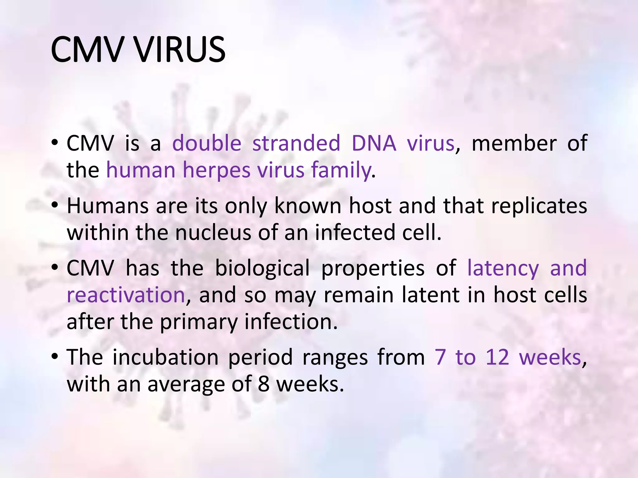
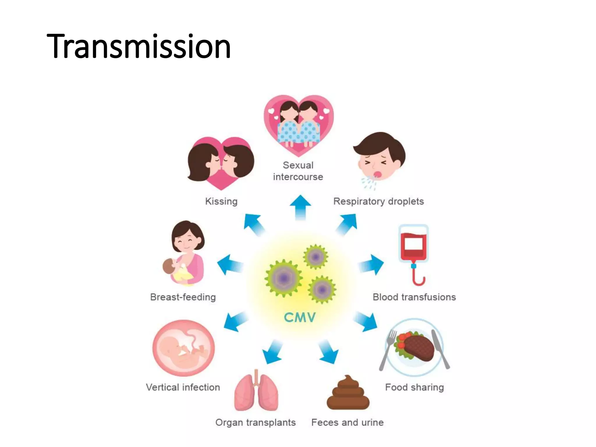
• Congenital cytomegalovirus (CMV) infection is
common worldwide.
• It is one of the leading cause of non-hereditary
sensorineural hearing loss (SNHL) and can cause
other long-term neuro-developmental disabilities,
including cerebral palsy, intellectual disability, vision
impairment, and seizures.
3. CMV infection in newborns and
immunocompromised children
Ranges
• Asymptomatic or mild
disease in
immunologically
normal hosts
• Severe and potentially
life-threatening disease
in
immunocompromised
children.
4. CMV VIRUS
• CMV is a double stranded DNA virus, member of
the human herpes virus family.
• Humans are its only known host and that replicates
within the nucleus of an infected cell.
• CMV has the biological properties of latency and
reactivation, and so may remain latent in host cells
after the primary infection.
• The incubation period ranges from 7 to 12 weeks,
with an average of 8 weeks.
5. EPIDEMIOLOGY
• In developed countries, CMV seroprevalence in
women of reproductive age ranges from 50 to 85%.
• In developing countries the seroprevalence approximates
100%.
• Incidence of congenital CMV infection in developed
countries is 0.6% to 0.7% of all live births.
8. Vertical transmission-
• Ante-partum:
Newborns may be acquired congenitally through
trans-placental transmission.
• Intra-partum:
Contaminated blood and genital secretions during
delivery
• Post-partum:
Via breast milk after delivery.
9. TERMINOLOGY
• The term "symptomatic" refers to infants with one
or more symptoms at birth.
• The term "asymptomatic" refers to infants with no
apparent symptoms at birth, although some of
these infants may develop hearing loss or subtle
symptoms later in life.
• The term "asymptomatic with isolated hearing
loss" refers to infants with isolated hearing loss at
birth but no other symptoms.
15. Life-threatening disease
Approximately 8 to 10 percent of newborns with
symptomatic congenital CMV infection have severe,
life-threatening disease.
Risk for mortality
• Premature infants and
• Infants with primary immune disorders of T cells or
natural killer cells
17. Liver function test abnormalities are characterized by
• Aspartate aminotransferase,
• Alanine aminotransferase and (usually <3 folds elevated)
• Bilirubin- normal or only mild-to-moderately elevated.
Liver biopsy reveal-
• Giant cell transformation
• Presence of large intracellular inclusion bodies in bile duct
epithelium , occasionally in hepatocyte or kupffer cells and
intracytoplasmic inclusion bodies in hepatocytes
• Bile stasis, inflammation, fibrosis and bile duct
proliferation
18. CMV Colitis
• Gastrointestinal involvement with CMV is
uncommon in immunocompetent hosts, while it
can cause significant morbidity and mortality in
immunocompromised host
• CMV colitis is due to the reactivation of a latent
infection in immunosuppressed patients
19. The clinical manifestation of CMV colitis is associated with
• low-grade fever,
• weight loss,
• anorexia,
• malaise,
• abdominal pain,
• Watery diarrhea,
• Hematochezia,
• Extensive mucosal hemorrhage and perforation related to
CMV infection can be life-threatening complications
21. Clinical suspicion —
Congenital CMV should be suspected in the following
clinical scenarios:
Newborns with signs and symptoms consistent
with congenital CMV disease.
• Microcephaly
• Small for gestational age (SGA)
• Thrombocytopenia
• Hepatosplenomegaly
• Jaundice or direct hyperbilirubinaemia at birth
22. Newborns with abnormal neuroimaging consistent
with CMV.
• Periventricular calcification
• Lenticulostriate vasculopathy
• White matter disease
• Ventriculomegaly
• Migrational abnormalities (polymicrogyria)
• Periventricular leukomalacia
23. Newborns born to mothers with known or
suspected CMV infection during pregnancy.
• Maternal seroconversion during pregnancy
• Presumptive maternal primary CMV infection (CMV IgG
and IgM positive)
• A mononucleosis like illness during pregnancy
Newborns who have documented sensorineural
hearing loss (SNHL).
Immune-compromised newborns.
24. Approach to testing
• Laboratory diagnosis of congenital CMV infection is
accomplished by isolation or molecular detection of
CMV from urine or saliva samples collected within
the first three weeks of life.
26. Virologically-proven congenital CMV infection can
be diagnosed on the basis of any of the following:
• Detection of CMV via viral culture in urine or saliva
samples obtained within the first three weeks of
life.
• Detection of CMV via shell vial assay in urine or
saliva samples obtained within the first three weeks
of life, with a positive confirmatory test (either viral
culture or PCR).
27. • Detection of CMV via PCR in urine, saliva, or blood
samples obtained within the first three weeks of
life, confirmed on repeat testing.
• Detection of CMV via PCR in the newborn screening
dried blood spot, with a positive confirmatory test
(either viral culture or PCR) if the infant is less than
three weeks old.
28. Possible congenital CMV infection –A diagnosis of
"possible" congenital CMV infection may be made if
all of the following criteria are met:
• One or more signs or symptoms of congenital CMV.
• Other conditions that cause these abnormalities
have been excluded.
• CMV is detected in urine or saliva samples (via viral
culture, shell vial assay, or PCR) or CMV IgG
antibody is detected in the blood after the first
three weeks of life, up to one year of age.
30. POST-DIAGNOSIS EVALUATION
• Infants with virologically-confirmed congenital
cytomegalovirus (CMV) infection should be
evaluated for evidence of organ involvement. And
do baseline tests prior starting antiviral therapy.
• A thorough physical, and neurologic and neuro-
developmental examination, including:
• Measurements of weight
• Length
• Head circumference (to detect microcephaly).
32. • Neuroimaging-
Ultrasonography of brain, computed tomography
(CT) or magnetic resonance imaging (MRI)
Findings:
Periventricular calcification
Lenticulostriate vasculopathy
White matter disease
Ventriculomegaly
Migrational abnormalities (e.g. focal polymicrogyria,
pachygyria, lissencephaly)
Periventricular leukomalacia and cystic abnormalities
33. CT MRI
Advantage:
• more sensitive in detecting
ventriculomegaly or
calcifications
Advantage:
• more sensitive in detecting
structural abnormalities.
Disadvantage:
• It exposes the infant to
ionizing radiation
Disadvantage:
• Infant may require
sedation/ anesthesia
• Longer procedure
34. • Hearing evaluation
• Ophthalmology evaluation
• CMV DNAemia by quantitative polymerase chain
reaction (PCR) to establish baseline level of viremia
37. Supportive measures
For the symptomatic neonate with severe or life-
threatening disease may include:
• Provision of oxygen and mechanical ventilator
support
• Fluid maintenance
• Control of seizures
• Transfusion of platelets and other blood products
• Antimicrobial treatment for secondary bacterial
infections
• Nutritional support
39. Whom to treat
• Symptomatic infection— infants with virologically-
confirmed congenital CMV and at least one end-
organ symptom.
• Primary immunodeficiency — Antiviral therapy is
also provided to infants with primary
immunodeficiency, regardless of the degree of
symptoms.
• Asymptomatic infection with isolated hearing loss
40. NOT generally initiate antiviral therapy in the
following circumstances because there is insufficient
evidence of benefit to outweigh the potential risks:
• In utero – Maternal antiviral therapy for prevention
or treatment of fetal CMV is generally not
recommended.
• Asymptomatic newborns with normal hearing
41. Timing —
• Antiviral therapy should be initiated as soon as
virological testing is confirmed.
• The effectiveness of antiviral therapy in congenital
CMV disease have found a benefit when treatment
is initiated within the first 30 days of life.
42. Antiviral regimen —
The choice of initial
antiviral agent (IV
ganciclovir versus oral
valganciclovir) depends
upon the severity of
disease.
43. Ganciclovir
Dose:
6 mg/kg per dose administered intravenously every
12 hours
Route: IV
Duration: two to six weeks
Infants may be transitioned to oral valganciclovir if
clinically stable and able to take oral medications,
usually after two to six weeks.
44. • Viral sepsis-like
syndrome
• Pneumonitis
• Myocarditis
• Severe hepatitis
• Enterocolitis
• Severe and refractory
thrombocytopenia
• Sight-threatening retinitis
• Severe neurologic disease
• Underlying primary
immune disorder (e.g.,
severe combined
immunodeficiency [SCID])
regardless of degree of
symptoms
Indication:
Life-threatening disease —with any of the following:
45. Valganciclovir
Dose:
16 mg/kg per dose administered orally every 12
hours
The dose should be increased with infant weight gain
to maintain a dose of 16 mg/kg per dose.
Route: Oral
Duration: 6months
46. Indication:
Non-life-threatening disease — those that meet ALL
of the following criteria:
• Well appearing or only mildly symptomatic, being
managed in the newborn nursery or at home
• Stable respiratory status (in room air or low
supplemental oxygen)
• Tolerating oral feeds
• Stable and gaining weight
47. • These infants generally lack severe end-organ
involvement.
• Examples of non-life-threatening disease include
• microcephaly or intracranial calcifications without
seizures or encephalopathy;
• jaundice or hepatosplenomegaly without severe
hepatitis or refractory thrombocytopenia;
• and isolated hearing loss.
48. Adverse effects
associated with ganciclovir and valganciclovir treatment
include:
• Neutropenia (ANC falls below 500/microL)
It is rarely severe and usually resolves with holding the
antiviral for one to seven days .
Following discontinuation of therapy, the ANC is
monitored two to three times per week, and once it has
recovered to >1000/microL, antiviral therapy is
reinstituted at the previous dose.
Treatment discontinuation may be necessary if
neutropenia recurs.
49. • Thrombocytopenia (platelets <50,000 cells/microL)
Worsening thrombocytopenia during antiviral
therapy while CMV DNAemia levels are declining
suggests drug-induced thrombocytopenia.
• Hepatotoxicity
Drugs need to be discontinued if transaminases rise
to >250 int. units/L with no alternative cause.
Transaminase levels should then be monitored
weekly and when they decline, antiviral therapy can
be restarted.
50. • Nephrotoxicity
The dose of ganciclovir and valganciclovir should be
adjusted in renal failure.
• IV catheter events
Extravasation of the intravenous ganciclovir solution
may produce local reaction, ulcers, and scarring.
51. Monitoring
Parameters Schedule
CBC Weekly for 6 weeks→ every 2-
4weeks
LFT
aspartate aminotransferase,
alanine aminotransferase,
total and direct bilirubin
Weekly initially→ monthly
RFT
BUN and
creatinine
Weekly initially→ 3 monthly
52. Treatment response — can be assessed by
monitoring clinical symptoms and level of viremia.
• Clinical symptoms are monitored with:
oRegular general physical examination
oNeurologic examination
oHearing evaluation every three to six months
oEye exam every three to six months; more frequent
evaluations are required in infants with chorioretinitis
• Level of viremia is measured with quantitative CMV
DNA PCR (CMV DNAemia) in whole blood or
plasma.
53. Treatment failure — Evidence of treatment failure
includes any of the following:
• Progressive end-organ disease despite adequate
treatment.
• Rising CMV DNAemia levels after two weeks of
treatment.
• Sustained increase in CMV DNAemia levels after an
initial decline.
54. Alternative antiviral agents
These agents may be used in select cases if there is-
• Suspected or confirmed ganciclovir-resistance
• Significant toxicity is encountered during ganciclovir
or valganciclovir therapy.
55. Foscarnet
Dose:
60 mg/kg per dose administered intravenously every
eight hours for two to three weeks
Followed by maintenance therapy at a dose of 90
mg/kg per dose once daily for two to three weeks.
Route : IV
56. Toxicity:
• Renal insufficiency and
• Electrolyte derangements
ohypocalcemia or hypercalcemia,
ometabolic acidosis,
ohyper-/hypophosphatemia,
ohypokalemia,
ohypomagnesemia, and
ohyponatremia
57. Monitoring at least twice weekly:
• BUN,
• S. creatinine, and
• Serum electrolytes
• S. calcium,
• S. magnesium, and
• S. phosphorus levels
58. Cidofovir
Dose:
• 5 mg/kg per dose administered intravenously every
seven days for two weeks, then every other week
for an additional four weeks.
• An alternative dosing regimen of 1 mg/kg per dose
three times per week has also been used
Route : IV
60. Monitoring:
• serum BUN
• creatinine
• calcium
• magnesium
• phosphorous
• uric acid levels
• urine protein measurement
• liver function tests and
• complete blood count with differential.
61.
62. Special circumstances
Chorioretinitis
• In addition to treatment with IV ganciclovir ,infants
require consultation with a pediatric ophthalmologist
or retinal specialist.
• Monitoring of the retinal lesions every 7 to 10 days is
recommended.
• For infants with severe sight-threatening CMV-
associated chorioretinitis, intravitreal antiviral
therapy may be required.
66. All children with congenital CMV infection (including
those who are symptomatic at birth and those who are
asymptomatic), should be followed throughout
childhood and into adulthood for evidence of long-term
effects of CMV.
• Hearing loss
Hearing evaluations are recommended every three to six
months during the first three years of life, and annually
thereafter until at least 18 years of age
Hearing aids may be needed and if progression to
profound hearing loss occurs, cochlear implantation may
be indicated.
67. • Central nervous system manifestations
Central nervous system (CNS) manifestations
(intellectual disability, cerebral palsy, and seizures)
may require special education services and speech,
language, occupational, and/or physical therapy
• Eye disease
Chorioretinitis, retinopathy, optic atrophy, and
strabismus require specialized management.
68. • Dental disease
Congenital CMV is associated with hypoplasia and
hypo-calcification of tooth enamel, affecting mainly
primary dentition. Regular dental visits are an
important component .
• Liver disease
Chronic hepatitis as a result of congenital CMV
disease is uncommon. Abnormal liver function tests
usually resolve during the first few weeks of life.
Hepatomegaly usually resolves by three months.
69. • Infants with congenital CMV and persistent liver
dysfunction or persistent direct hyperbilirubinemia
should be evaluated for biliary atresia or alpha 1-
antitrypsin deficiency, since CMV and another
diagnosis affecting liver function may coexist in the
same infant, and present a diagnostic challenge
71. Prevention of neonatal transmission — Preventive
measures to reduce transmission of CMV to
newborns include:
• Use of CMV-seronegative or leukocyte-reduced
blood for extremely preterm infants, which
essentially eliminates the risk of transfusion-related
CMV infection
• Treatment of maternal breast milk by
freezing/thawing or pasteurization.
72. Personal protective measures to reduce the risk of
family transmission of the virus can be instituted in
pregnant women or those trying to become
pregnant, especially those women who are CMV
seronegative
• Avoiding kissing young toddlers and children on or
near the mouth
• Not sharing eating or drinking utensils, drinks, or
food with toddlers or young children
73. • Careful handwashing or using
gloves after wiping noses,
drool, and changing diapers.
• Avoiding intimate contact
during the communicable
period with a sexual partner if
the partner has documented
CMV mononucleosis.
74. • Blood products and organ donors — Blood
products given to newborns (especially preterm
infants), pregnant women, and immuno-
compromised patients should be from CMV-
seronegative donors or leuko-reduced.
75. • Prophylaxis — Passive immunoprophylaxis with
intravenous immune globulin or CMV
hyperimmune globulin may prevent acquisition of
serious CMV disease in hematopoietic stem cell
and solid organ transplant recipients at high risk for
acquiring CMV disease.
76. • Vaccines — A variety of experimental CMV vaccines
have been evaluated in clinical trials; however,
none are licensed.
77. References
• UpToDate
• Liver disease in children –by Frederick J. Suchy,
Ronald J. Sokol, William F. Balistreri
• Walker’s pediatric gastrointestinal disease
• Nakase H, Herfarth H: Cytomegalovirus Colitis,
Cytomegalovirus Hepatitis and Systemic
Cytomegalovirus Infection: Common Features and
Differences. Inflamm Intest Dis 2016;1:15-23. doi:
10.1159/000443198