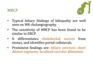
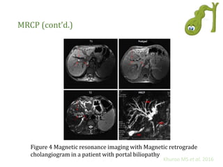
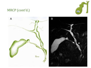
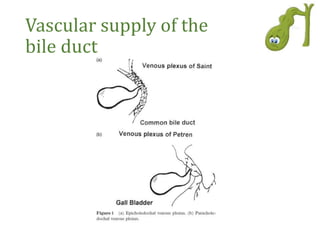
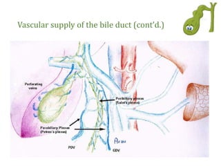
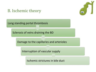
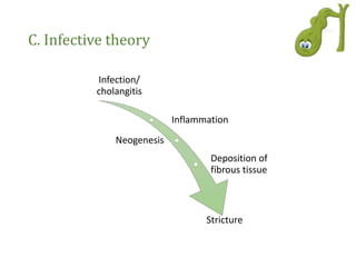
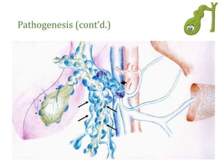
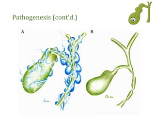
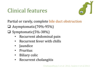
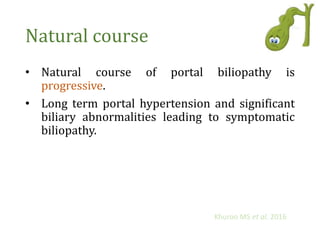
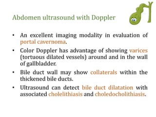
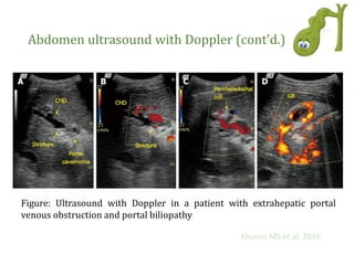
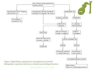
Portal Biliopathy
Presenter:
Dr. Maimuna Sayeed
Resident Phase B (year 4)
Paediatric Gastroenterology and Nutrition
BSMMU
Outline
• Definition
• Vascular supply of the bile duct
• Etiology
• Pathogenesis
• Classification
• Clinical feature
• Natural course
• Investigation
• Treatment
• Complication
Definition
Portal biliopathy is define as abnormalities in the
intrahepatic and extrahepatic biliary tract,
gallbladder and cystic duct secondary to portal
hypertension.
Portal biliopathy refers to cholangiographic
abnormalities which occur in patients with portal
cavernoma.
Khuroo MS et al. 2016
Chattopadhyay S et al. 2012
Incidence
• Extrahepatic portal vein obstruction (81%-
100%)
• Non-cirrhotic portal fibrosis (9%-40%)
• Cirrhosis of the liver (0%-33%)
• Congenital hepatic fibrosis
Chattopadhyay S et al. 2012, Suárez V et al.2013
Vascular supply of the
bile duct
Vascular supply of the bile duct (cont’d.)
Pathogenesis
There are three main theories for the pathogenesis
of portal biliopathy
A. Compression theory
B. Ischemic theory
C. Infective theory
Chattopadhyay S et al. 2012
A. Compression theory
Long standing EHPVO
Replacement of PV by large
collaterals along CBD
Cavernomatous
transformation of PV
Compress pliable CBD
Long standing EHPVO
Vascular neogenesis and
formation of tumor like CT
Encase CBD/Cause
angulation of BD
B. Ischemic theory
Long standing portal thrombosis
Sclerosis of veins draining the BD
Damage to the capillaries and arterioles
Interruption of vascular supply
Ischemic strictures in bile duct
C. Infective theory
Infection/
cholangitis
Inflammation
Neogenesis
Deposition of
fibrous tissue
Stricture
Pathogenesis (cont’d.)
Classification
I II IIIa IIIb
:
Suárez V et al.2013
Clinical features
Partial or rarely, complete bile duct obstruction
 Asymptomatic(70%-95%)
 Symptomatic(5%-38%)
• Recurrent abdominal pain
• Recurrent fever with chills
• Jaundice
• Pruritus
• Biliary colic
• Recurrent cholangitis
Chattopadhyay S et al. 2012, Suárez V et al.2013
Natural course
• Natural course of portal biliopathy is
progressive.
• Long term portal hypertension and significant
biliary abnormalities leading to symptomatic
biliopathy.
Khuroo MS et al. 2016
Investigation
Investigation
Liver function tests:
• Serum bilirubin-direct
• Serum alkaline phosphatase
• Serum aminotransferase
• Serum albumin
• Prothrombin time
Biliary cirrhosis
Biliary imaging
↑
↑
↑
↓
↑
Investigation(cont’d.)
Abdomen ultrasound with Doppler
Endoscopic retrograde
cholangiopancreatography (ERCP)
Magnetic resonance
cholangiopancreatography (MRCP)
Endoscopic ultrasonography
Abdomen ultrasound with Doppler
• An excellent imaging modality in evaluation of
portal cavernoma.
• Color Doppler has advantage of showing varices
(tortuous dilated vessels) around and in the wall
of gallbladder.
• Bile duct wall may show collaterals within the
thickened bile ducts.
• Ultrasound can detect bile duct dilatation with
associated cholelithiasis and choledocholithiasis.
Abdomen ultrasound with Doppler (cont’d.)
Figure: Ultrasound with Doppler in a patient with extrahepatic portal
venous obstruction and portal biliopathy
Khuroo MS et al. 2016
ERCP
• Gold standard for defining the biliary changes
of portal biliopathy.
• It has both diagnostic and therapeutic role.
Khuroo MS et al. 2016
ERCP (cont’d.)
Diagnostic role:
To see changes in the bile ducts, this includes-
• Single or multiple smooth strictures of varying
length and degree,
• Saccular dilatations,
• Indentations,
• Dilated intrahepatic bile duct radicles,
• Displacement of bile ducts, and
• Filling defects in the CBD which may be due to
stones or varices.
ERCP (cont’d.)
Therapeutic role:
This includes-
• Removal of CBD stones,
• Relief of cholangitis, and
• Dilatation of dominant strictures with stenting.
ERCP (cont’d.)
Figure: Endoscopic retrograde cholangiograms in 8 patients of portal biliopathy
depicting spectrum of cholangiographic abnormalities of portal biliopathy
Khuroo MS et al. 2016
MRCP
• Typical biliary findings of biliopathy are well
seen on MR cholangiography.
• The sensitivity of MRCP has been found to be
similar to ERCP.
• It differentiates choledochal varices from
stones, and identifies portal collaterals.
• Prominent findings are: biliary stricture, short
dilated segments, localized saccular dilatation.
MRCP (cont’d.)
Figure 4 Magnetic resonance imaging with Magnetic retrograde
cholangiogram in a patient with portal biliopathy
Khuroo MS et al. 2016
MRCP (cont’d.)
Endoscopic ultrasonography
• To the differentiation between CBD varices,
stones, and tumors when other imaging
modalities are not clear.
• Portal cavernoma and its anatomy can be well
defined.
• These collaterals appear as multiple vascular
channels in and/or around the extrahepatic
biliary tract.
• Paracholedochal, epicholedochal, intra-
choledochal and subepithelial varices can be
well seen and differentiated.
Endoscopic ultrasonography (cont’d.)
Figure: Radial endosonography images in portal biliopathy
M. S. Sarma et al. 2018
Treatment
Treatment
Asymptomatic patients:
Need no active intervention.
Treatment (cont’d.)
Symptomatic patients:
 Medical management
Ursodeoxycholic acid (10-15mg/kg per day)
Suárez et al. 2013, Khuroo MS et al. 2016
Treatment (cont’d.)
 Endotherapy
Indicated:
• CBD stones
• Cholangitis
• Shunt surgery is not feasible
Chattopadhyay S et al. 2012
Treatment (cont’d.)
Complications of endotherapy:
1. Filling defects seen on imaging may be due to
varices → bleeding during attempted clearance
2. Venous collaterals in the region of the ampulla
of Vater → bleeding during papillotomy
3. Stents become blocked frequently requiring
multiple changes → risk of bleeding
Chattopadhyay S et al. 2012
Treatment (cont’d.)
 Surgical management:
Portosystemic shunt surgery is the treatment of
choice.
Advantages:
1. Prevents variceal bleeding
2. Biliary bypass possible
Chattopadhyay S et al. 2012
Treatment (cont’d.)
Approached in a stepwise fashion:
Managing
biliary
strictures
and
stones
Portal
decompression
should be done
to reverse
biliary
abnormalities
Biliary
obstruction
should be
relieved by
biliary
diversion at
surgery
Khuroo MS et al. 2016
Complications
• Cholestasis
• Recurrent cholangitis
• Biliary sludge
• Gall stone
• Secondary biliary cirrhosis (2%-4%)
Suárez et al. 2013,
Figure: Algorithmic approach to management of portal
biliopathy in patients with non-cirrhotic portal hypertension
Chattopadhyay S et al. 2012
Key points
 Portal biliopathy (PB) is the late complication
of portal hypertension.
 More commonly seen in patients with
extrahepatic portal venous obstruction.
 External pressure over the bile ducts from
biliary collaterals and/or ischaemic injury of
bile ducts during portal vein thrombosis seems
to be the main mechanism responsible for the
development of PB.
Key points (cont’d.)
 Symptoms are associated with higher age,
longer duration of disease.
 MRCP is the choice of investigation.
 Endotherapy is the preferred treatment for
patients with CBD stones, cholangitis or patients
with dominant biliary stricture, but without a
shuntable vein.
 Portosystemic shunt should be performed in
patients with dominant biliary strictures with a
shuntable vein.
 Rarely, second stage biliary bypass may be
required.
Thank you

Portal biliopathy

  • 1.
    Portal Biliopathy Presenter: Dr. MaimunaSayeed Resident Phase B (year 4) Paediatric Gastroenterology and Nutrition BSMMU
  • 2.
    Outline • Definition • Vascularsupply of the bile duct • Etiology • Pathogenesis • Classification • Clinical feature • Natural course • Investigation • Treatment • Complication
  • 3.
    Definition Portal biliopathy isdefine as abnormalities in the intrahepatic and extrahepatic biliary tract, gallbladder and cystic duct secondary to portal hypertension. Portal biliopathy refers to cholangiographic abnormalities which occur in patients with portal cavernoma. Khuroo MS et al. 2016 Chattopadhyay S et al. 2012
  • 4.
    Incidence • Extrahepatic portalvein obstruction (81%- 100%) • Non-cirrhotic portal fibrosis (9%-40%) • Cirrhosis of the liver (0%-33%) • Congenital hepatic fibrosis Chattopadhyay S et al. 2012, Suárez V et al.2013
  • 5.
    Vascular supply ofthe bile duct
  • 6.
    Vascular supply ofthe bile duct (cont’d.)
  • 7.
    Pathogenesis There are threemain theories for the pathogenesis of portal biliopathy A. Compression theory B. Ischemic theory C. Infective theory Chattopadhyay S et al. 2012
  • 8.
    A. Compression theory Longstanding EHPVO Replacement of PV by large collaterals along CBD Cavernomatous transformation of PV Compress pliable CBD Long standing EHPVO Vascular neogenesis and formation of tumor like CT Encase CBD/Cause angulation of BD
  • 9.
    B. Ischemic theory Longstanding portal thrombosis Sclerosis of veins draining the BD Damage to the capillaries and arterioles Interruption of vascular supply Ischemic strictures in bile duct
  • 10.
  • 11.
  • 12.
  • 13.
    Classification I II IIIaIIIb : Suárez V et al.2013
  • 14.
    Clinical features Partial orrarely, complete bile duct obstruction  Asymptomatic(70%-95%)  Symptomatic(5%-38%) • Recurrent abdominal pain • Recurrent fever with chills • Jaundice • Pruritus • Biliary colic • Recurrent cholangitis Chattopadhyay S et al. 2012, Suárez V et al.2013
  • 15.
    Natural course • Naturalcourse of portal biliopathy is progressive. • Long term portal hypertension and significant biliary abnormalities leading to symptomatic biliopathy. Khuroo MS et al. 2016
  • 16.
  • 17.
    Investigation Liver function tests: •Serum bilirubin-direct • Serum alkaline phosphatase • Serum aminotransferase • Serum albumin • Prothrombin time Biliary cirrhosis Biliary imaging ↑ ↑ ↑ ↓ ↑
  • 18.
    Investigation(cont’d.) Abdomen ultrasound withDoppler Endoscopic retrograde cholangiopancreatography (ERCP) Magnetic resonance cholangiopancreatography (MRCP) Endoscopic ultrasonography
  • 19.
    Abdomen ultrasound withDoppler • An excellent imaging modality in evaluation of portal cavernoma. • Color Doppler has advantage of showing varices (tortuous dilated vessels) around and in the wall of gallbladder. • Bile duct wall may show collaterals within the thickened bile ducts. • Ultrasound can detect bile duct dilatation with associated cholelithiasis and choledocholithiasis.
  • 20.
    Abdomen ultrasound withDoppler (cont’d.) Figure: Ultrasound with Doppler in a patient with extrahepatic portal venous obstruction and portal biliopathy Khuroo MS et al. 2016
  • 21.
    ERCP • Gold standardfor defining the biliary changes of portal biliopathy. • It has both diagnostic and therapeutic role. Khuroo MS et al. 2016
  • 22.
    ERCP (cont’d.) Diagnostic role: Tosee changes in the bile ducts, this includes- • Single or multiple smooth strictures of varying length and degree, • Saccular dilatations, • Indentations, • Dilated intrahepatic bile duct radicles, • Displacement of bile ducts, and • Filling defects in the CBD which may be due to stones or varices.
  • 23.
    ERCP (cont’d.) Therapeutic role: Thisincludes- • Removal of CBD stones, • Relief of cholangitis, and • Dilatation of dominant strictures with stenting.
  • 24.
    ERCP (cont’d.) Figure: Endoscopicretrograde cholangiograms in 8 patients of portal biliopathy depicting spectrum of cholangiographic abnormalities of portal biliopathy Khuroo MS et al. 2016
  • 25.
    MRCP • Typical biliaryfindings of biliopathy are well seen on MR cholangiography. • The sensitivity of MRCP has been found to be similar to ERCP. • It differentiates choledochal varices from stones, and identifies portal collaterals. • Prominent findings are: biliary stricture, short dilated segments, localized saccular dilatation.
  • 26.
    MRCP (cont’d.) Figure 4Magnetic resonance imaging with Magnetic retrograde cholangiogram in a patient with portal biliopathy Khuroo MS et al. 2016
  • 27.
  • 28.
    Endoscopic ultrasonography • Tothe differentiation between CBD varices, stones, and tumors when other imaging modalities are not clear. • Portal cavernoma and its anatomy can be well defined. • These collaterals appear as multiple vascular channels in and/or around the extrahepatic biliary tract. • Paracholedochal, epicholedochal, intra- choledochal and subepithelial varices can be well seen and differentiated.
  • 29.
    Endoscopic ultrasonography (cont’d.) Figure:Radial endosonography images in portal biliopathy M. S. Sarma et al. 2018
  • 30.
  • 31.
  • 32.
    Treatment (cont’d.) Symptomatic patients: Medical management Ursodeoxycholic acid (10-15mg/kg per day) Suárez et al. 2013, Khuroo MS et al. 2016
  • 33.
    Treatment (cont’d.)  Endotherapy Indicated: •CBD stones • Cholangitis • Shunt surgery is not feasible Chattopadhyay S et al. 2012
  • 34.
    Treatment (cont’d.) Complications ofendotherapy: 1. Filling defects seen on imaging may be due to varices → bleeding during attempted clearance 2. Venous collaterals in the region of the ampulla of Vater → bleeding during papillotomy 3. Stents become blocked frequently requiring multiple changes → risk of bleeding Chattopadhyay S et al. 2012
  • 35.
    Treatment (cont’d.)  Surgicalmanagement: Portosystemic shunt surgery is the treatment of choice. Advantages: 1. Prevents variceal bleeding 2. Biliary bypass possible Chattopadhyay S et al. 2012
  • 36.
    Treatment (cont’d.) Approached ina stepwise fashion: Managing biliary strictures and stones Portal decompression should be done to reverse biliary abnormalities Biliary obstruction should be relieved by biliary diversion at surgery Khuroo MS et al. 2016
  • 37.
    Complications • Cholestasis • Recurrentcholangitis • Biliary sludge • Gall stone • Secondary biliary cirrhosis (2%-4%) Suárez et al. 2013,
  • 38.
    Figure: Algorithmic approachto management of portal biliopathy in patients with non-cirrhotic portal hypertension Chattopadhyay S et al. 2012
  • 39.
    Key points  Portalbiliopathy (PB) is the late complication of portal hypertension.  More commonly seen in patients with extrahepatic portal venous obstruction.  External pressure over the bile ducts from biliary collaterals and/or ischaemic injury of bile ducts during portal vein thrombosis seems to be the main mechanism responsible for the development of PB.
  • 40.
    Key points (cont’d.) Symptoms are associated with higher age, longer duration of disease.  MRCP is the choice of investigation.  Endotherapy is the preferred treatment for patients with CBD stones, cholangitis or patients with dominant biliary stricture, but without a shuntable vein.  Portosystemic shunt should be performed in patients with dominant biliary strictures with a shuntable vein.  Rarely, second stage biliary bypass may be required.
  • 41.