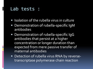
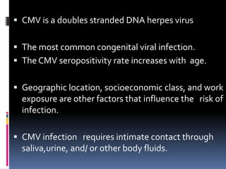
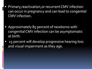
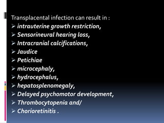
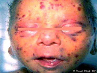
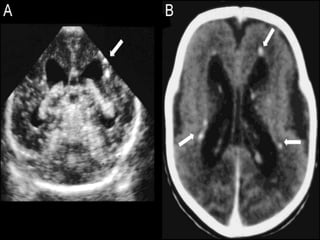
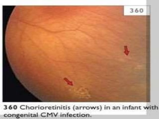
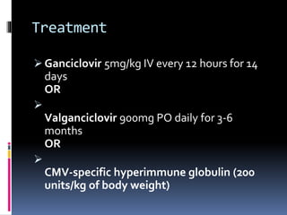
ASHLY ALEX
 TORCH complex is a medical acronym for a set
of perinatal infections .
 TheTORCH infections can lead to severe fetal
anomalies or even fetal loss.
 They are a group of viral, bacterial, and
protozoan infections that gain access to the
fetal blood stream transplacentally via the
chrionic villi.
 Hematogenous transmission may occur at
anytime during gestation or occasionally at
the time of delivery via maternal-to-fetal
transfusion.
 The capitalization"TORCH”consists of:
 T–TOXOPLASMOSIS
 O _Other infections (Syphilis,Varicella
Zoster, ParvovirusB- 19, HEP B)
 R–RUBELLA
 C–CYTOMEGALOVIRUS
 H–HERPES SIMPLEXVIRUS–2
 Toxoplasmosis is a disease caused by an
intracellular parasite TOXOPLASMAGONDII.
 Human acquisition of the infection occurs
by:
Oocyst contaminated soil,salads,vegetables.
Ingestion of raw or undercooked meat
containing tissue cysts (Sheep,pigs and
rabbits are the most common meat sources).
Out breaks of toxoplasmosis have also been
linked to the consumption of unfiltered water
 •Primary maternal infection in pregnancy–
 Infection rate higher with infection in 3rd
trimester.
 Fetal death higher with infection in 1st
trimester
Signs and sympoms
 Infected Pregnant women : usually no clinical
manifestation.
 Although some may have a mild
mononucleosis-like syndrome, regional
lymphadenopathy, or occasionally
chorioretinitis.
 Similarly, infected neonates are usually
asymptomatic at birth.
 Manifestations may include :
Prematurity
Intrauterine growth restriction
Jaundice
Hepatosplenomegaly
Myocarditis
Pneumonitis
Various rashes
anemia, thrombocytopenia,
 and abnormal CSF findings (Mononuclear
CSF pleocytosis or elevatedCSF protein)
The classic triad of findings :
chorioretinitis,
 hydrocephalus, and
 intracranial calcifications
Diagnosis
 Serial IgG measurement (for maternal
infection)
 Amniotic fluid PCR (for fetal infection)
 Serologic testing, brain imaging, CSF analysis
and ophthalmologic evaluation (for neonatal
infection),
 and PCR testing of various body fluids or
tissues
Treatment
 •In PREGNANT WOMEN with an established
recent infection, SPIRAMYCIN (3g daily in
divided doses) should be given.
In neonates :
 Pyrimethamine: 50mg twice daily for 2 days
then 50mg daily.
PLUS
Sulfadiazine: 75mg/kg/daily in two divided doses
for 2 days then 50mg/kg/twice daily
PLUS
Folinic Acid: 10-20mg daily
Rubella
 It is caused by rubella virus,Rubivirus genus and
familyTogaviridae.
 Intrauterine infection with rubella virus is referred
to as congenital rubella infection (CRI) or
syndrome.
 Infection with rubella earlier in pregnancy(1st
trimester ) cause worse prognosis and neonatal
complications.
 The virus can be transmitted to the fetus through
the placenta and is capable of causing serious
congenital defects, abortions, and stillbirths.
 In the baby
 Infection in weeks 8-10 of pregnancy results
in damage in up to 90% of surviving infants.
Multiple defects are then common.
 The risk of damage reduces to 10-20% if the
infection is in weeks 11-16 of pregnancy.
 Fetal damage is rare over 18 weeks of
gestation.
 Transmission to the fetus occurs via maternal
hematogenous spread to the placenta.
 It typically occurs 5-7 days after maternal
inoculation.
 After the virus invades the placental barrier,
it spreads throughout the fetus via their
vascular system.
 The congenital defects that result from
infection is secondary to the cytopathical
damage ensued to the blood vessels.
 This in turn results in ischemia of the affected
organs .
Clinical features
Transient :
 Intrauterine growth restriction.
 Thrombocytopenic purpura (25% - 'blueberry
skin').
 Haemolytic anaemia.
 Hepatosplenomegaly.
 Jaundice (common).
 Radiolucent bone disease (20%).
 Meningoencephalitis (25%) +/- neurological
sequelae
 Developmental:
 Sensorineural deafness 80% .
 General learning disability (55%).
 Insulin-dependent diabetes (20%, immune-
mediated but often delayed to adolescence or
adulthood).
 'Late-onset' disease at 3-12 months with rash,
diarrhoea, pneumonitis and high mortality.
 Permanent:
 Congenital heart disease (commonly patent
ductus arteriosus or peripheral pulmonary artery
stenosis).
 Eye defects including cataracts, congenital
glaucoma, pigmentary retinopathy (50% - so-
called 'salt and pepper'), severe myopia,
microphthalmia.
 Microcephaly.
 The risk of maternal-fetal transmission is the
greatest in the first 10 days after gestation
 cardiac and eye defects typically resulting
when maternal infection occurs prior to 8
week.
 Hearing loss is typically observed in
infections up to 18 weeks of gestation
Lab tests :
 Isolation of the rubella virus in culture
 Demonstration of rubella-specific IgM
antibodies
 Demonstration of rubella-specific IgG
antibodies that persist at a higher
concentration or longer duration than
expected from mere passive transfer of
maternal antibodies
 Detection of rubella virus RNA by reverse-
transcriptase polymerase chain reaction
Treatment
 Supportive care and surveillance is the only
recommended option available at this time.
 Close monitoring within the first 6 to 12 months of
life is recommended; particularly for the evaluation
of hearing impairment .
 Prevention is considered the most important aspect
as far as the management of CRI concerned.
 Preventive measures include recommended
immunizations, testing of pregnant women for
rubella immunity and proper counseling regarding
avoiding exposure.
 CMV is a doubles stranded DNA herpes virus
 The most common congenital viral infection.
 The CMV seropositivity rate increases with age.
 Geographic location, socioeconomic class, and work
exposure are other factors that influence the risk of
infection.
 CMV infection requires intimate contact through
saliva,urine, and/ or other body fluids.
 Possible routes of transmission include
sexual contact,
organ transplantation,
transplacental transmission,
transmission via breastmilk,and
blood transfusion(rare).
 Primary,reactivation,or recurrent CMV infection
can occur in pregnancy and can lead to congenital
CMV infection.
 Approximately 85 percent of newborns with
congenital CMV infection can be asymptomatic
at birth.
 15 percent will develop progressive hearing loss
and visual impairment as they age.
Transplacental infection can result in :
 intrauterine growth restriction,
 Sensorineural hearing loss,
 Intracranial calcifications,
 Jaudice
 Petichiae
 microcephaly,
 hydrocephalus,
 hepatosplenomegaly,
 Delayed psychomotor development,
 Thrombocytopenia and/
 Chorioretinitis .
 Vertical transmission of CMV can occur at any
stage of pregnancy.
 Severe sequelae are more common with
infection in the 1st trimester.
 The overall risk of infection is greatest in
the 3rd trimester.
 The risk of transmission to the fetus in
primary infection is 30%-40%.
Diagosis In foetus
Amniocentisis : viral culture and PCR
Ultrasound
Treatment
 Ganciclovir 5mg/kg IV every 12 hours for 14
days
OR

Valganciclovir 900mg PO daily for 3-6
months
OR

CMV-specific hyperimmune globulin (200
units/kg of body weight)
 Herpes simplex virus (HSV) infection during
pregnancy can pose a serious threat to the
developing fetus and the newborn infant.
 Transmission typically occurs via direct contact
between the neonate and an infected maternal
genital tract.
 If the primary HSV infection was acquired during
pregnancy, then the risk of transmission is
greater as compared with reactivation of a
previous infection.
 incidence of neonatal HSV infection ranges from
1 in 3200 to 1 in 10,000 births .
 HSV is a member of the Herpes viridae family of
viruses
 Enters the host through the inoculation of oral,
genital, or conjunctival mucosa.
 Inoculation also can occur through breaks in the
skin.
 Dissemination of the virus eventually allows the
virus to reach the dorsal root ganglia, where it
remains dormant for the rest of the host’s life.
 Antiviral drugs do not affect latent HSV infection
and therefore infection is life-long
 Intrauterine HSV is a rare occurrence and most likely
is caused by maternal viremia associated with
primary infection during pregnancy.
Intrauterine infection is associated with
 hydropsfetalis and
 in-utero fetal demise.
The characteristic triad noted at birth includes
skin lesions consistent of vesicles,
ulcerations or scarring ,
eye damage and
CNS abnormalities, such as hydranencephaly and
microcephaly.
 Clinical manifestation can arise any time during
the first six weeks of life, but usually occurs
within the first month of life .
Classic CSF findings include :
 a mononuclear cell pleocytosis,
 normal or slightly low glucose concentration
 and moderately elevated protein level.
Electroencephalogram (EEG) is often abnormal
from early on in the disease and may show focal
or multifocal periodic epileptic form discharges
. Neuroimaging studies may show parenchymal
brain edema, hemorrhage or destructive lesions
in the temporal frontal, parietal or brainstem
regions in the brain
Diagnosis
 Diagnosis of neonatal HSV infection can be
established through any of the following
methods:
 Isolation of HSV in culture
 Detection of DNA via PCR assays
 Detection of HSV specific antigens using
rapid direct immunofluorescence or enzyme
immunoassays.
Treatment
 Suppressive viral therapy from 36 weeks
until delivery
 Valacyclovir 500 mg orally BD OR
 Acyclovir 400 mg orallyTDS.
Cesarean section is recommended for all
women in labor with active genital lesions
or prodromal symptoms such as vulvar pain
Others are :
Disease Trasmission Symptoms Diagnosis Treatment
Varicella zooster Ist 20 weeks of
pregnancy
Cicatricial
lesions ,limb
hypoplasia,micr
ocepaly ,hydrops
etc
Prenatal
Ultrasound
andMRI
Acyclovir
Parvo virus b19 1st 2 trimester of
pregnancy
ProdromalFever,
Slapped cheek
rash anemia etc
Serological
PCR
Symptomatic
relief
Syphilis
(Trepinoma
palidum )
Severe outcome
after 4 weeks
Miscarrage
,prematurity
,still birth,
hepatosplenome
galy,bullous
rash,pneumonia
Serological tests Benzathine
Penicillin G IM
Hep b transmission
specially in the
third trimester.
jaundice ,rashes
PAN ,Vomiting
,dark urine etc
Serological tests Lamivudine
,interferon alpha
entecavir
presentation2-150208112421-conversion-gate02.pdf

presentation2-150208112421-conversion-gate02.pdf

  • 1.
  • 2.
     TORCH complexis a medical acronym for a set of perinatal infections .  TheTORCH infections can lead to severe fetal anomalies or even fetal loss.  They are a group of viral, bacterial, and protozoan infections that gain access to the fetal blood stream transplacentally via the chrionic villi.  Hematogenous transmission may occur at anytime during gestation or occasionally at the time of delivery via maternal-to-fetal transfusion.
  • 3.
     The capitalization"TORCH”consistsof:  T–TOXOPLASMOSIS  O _Other infections (Syphilis,Varicella Zoster, ParvovirusB- 19, HEP B)  R–RUBELLA  C–CYTOMEGALOVIRUS  H–HERPES SIMPLEXVIRUS–2
  • 5.
     Toxoplasmosis isa disease caused by an intracellular parasite TOXOPLASMAGONDII.  Human acquisition of the infection occurs by: Oocyst contaminated soil,salads,vegetables. Ingestion of raw or undercooked meat containing tissue cysts (Sheep,pigs and rabbits are the most common meat sources). Out breaks of toxoplasmosis have also been linked to the consumption of unfiltered water
  • 7.
     •Primary maternalinfection in pregnancy–  Infection rate higher with infection in 3rd trimester.  Fetal death higher with infection in 1st trimester
  • 8.
    Signs and sympoms Infected Pregnant women : usually no clinical manifestation.  Although some may have a mild mononucleosis-like syndrome, regional lymphadenopathy, or occasionally chorioretinitis.  Similarly, infected neonates are usually asymptomatic at birth.
  • 9.
     Manifestations mayinclude : Prematurity Intrauterine growth restriction Jaundice Hepatosplenomegaly Myocarditis Pneumonitis Various rashes anemia, thrombocytopenia,  and abnormal CSF findings (Mononuclear CSF pleocytosis or elevatedCSF protein)
  • 10.
    The classic triadof findings : chorioretinitis,  hydrocephalus, and  intracranial calcifications
  • 13.
    Diagnosis  Serial IgGmeasurement (for maternal infection)  Amniotic fluid PCR (for fetal infection)  Serologic testing, brain imaging, CSF analysis and ophthalmologic evaluation (for neonatal infection),  and PCR testing of various body fluids or tissues
  • 14.
    Treatment  •In PREGNANTWOMEN with an established recent infection, SPIRAMYCIN (3g daily in divided doses) should be given. In neonates :  Pyrimethamine: 50mg twice daily for 2 days then 50mg daily. PLUS Sulfadiazine: 75mg/kg/daily in two divided doses for 2 days then 50mg/kg/twice daily PLUS Folinic Acid: 10-20mg daily
  • 15.
  • 16.
     It iscaused by rubella virus,Rubivirus genus and familyTogaviridae.  Intrauterine infection with rubella virus is referred to as congenital rubella infection (CRI) or syndrome.  Infection with rubella earlier in pregnancy(1st trimester ) cause worse prognosis and neonatal complications.  The virus can be transmitted to the fetus through the placenta and is capable of causing serious congenital defects, abortions, and stillbirths.
  • 17.
     In thebaby  Infection in weeks 8-10 of pregnancy results in damage in up to 90% of surviving infants. Multiple defects are then common.  The risk of damage reduces to 10-20% if the infection is in weeks 11-16 of pregnancy.  Fetal damage is rare over 18 weeks of gestation.
  • 18.
     Transmission tothe fetus occurs via maternal hematogenous spread to the placenta.  It typically occurs 5-7 days after maternal inoculation.  After the virus invades the placental barrier, it spreads throughout the fetus via their vascular system.  The congenital defects that result from infection is secondary to the cytopathical damage ensued to the blood vessels.  This in turn results in ischemia of the affected organs .
  • 19.
    Clinical features Transient : Intrauterine growth restriction.  Thrombocytopenic purpura (25% - 'blueberry skin').  Haemolytic anaemia.  Hepatosplenomegaly.  Jaundice (common).  Radiolucent bone disease (20%).  Meningoencephalitis (25%) +/- neurological sequelae
  • 20.
     Developmental:  Sensorineuraldeafness 80% .  General learning disability (55%).  Insulin-dependent diabetes (20%, immune- mediated but often delayed to adolescence or adulthood).  'Late-onset' disease at 3-12 months with rash, diarrhoea, pneumonitis and high mortality.
  • 21.
     Permanent:  Congenitalheart disease (commonly patent ductus arteriosus or peripheral pulmonary artery stenosis).  Eye defects including cataracts, congenital glaucoma, pigmentary retinopathy (50% - so- called 'salt and pepper'), severe myopia, microphthalmia.  Microcephaly.
  • 23.
     The riskof maternal-fetal transmission is the greatest in the first 10 days after gestation  cardiac and eye defects typically resulting when maternal infection occurs prior to 8 week.  Hearing loss is typically observed in infections up to 18 weeks of gestation
  • 25.
    Lab tests : Isolation of the rubella virus in culture  Demonstration of rubella-specific IgM antibodies  Demonstration of rubella-specific IgG antibodies that persist at a higher concentration or longer duration than expected from mere passive transfer of maternal antibodies  Detection of rubella virus RNA by reverse- transcriptase polymerase chain reaction
  • 26.
    Treatment  Supportive careand surveillance is the only recommended option available at this time.  Close monitoring within the first 6 to 12 months of life is recommended; particularly for the evaluation of hearing impairment .  Prevention is considered the most important aspect as far as the management of CRI concerned.  Preventive measures include recommended immunizations, testing of pregnant women for rubella immunity and proper counseling regarding avoiding exposure.
  • 28.
     CMV isa doubles stranded DNA herpes virus  The most common congenital viral infection.  The CMV seropositivity rate increases with age.  Geographic location, socioeconomic class, and work exposure are other factors that influence the risk of infection.  CMV infection requires intimate contact through saliva,urine, and/ or other body fluids.
  • 29.
     Possible routesof transmission include sexual contact, organ transplantation, transplacental transmission, transmission via breastmilk,and blood transfusion(rare).
  • 30.
     Primary,reactivation,or recurrentCMV infection can occur in pregnancy and can lead to congenital CMV infection.  Approximately 85 percent of newborns with congenital CMV infection can be asymptomatic at birth.  15 percent will develop progressive hearing loss and visual impairment as they age.
  • 31.
    Transplacental infection canresult in :  intrauterine growth restriction,  Sensorineural hearing loss,  Intracranial calcifications,  Jaudice  Petichiae  microcephaly,  hydrocephalus,  hepatosplenomegaly,  Delayed psychomotor development,  Thrombocytopenia and/  Chorioretinitis .
  • 32.
     Vertical transmissionof CMV can occur at any stage of pregnancy.  Severe sequelae are more common with infection in the 1st trimester.  The overall risk of infection is greatest in the 3rd trimester.  The risk of transmission to the fetus in primary infection is 30%-40%.
  • 36.
    Diagosis In foetus Amniocentisis: viral culture and PCR Ultrasound
  • 38.
    Treatment  Ganciclovir 5mg/kgIV every 12 hours for 14 days OR  Valganciclovir 900mg PO daily for 3-6 months OR  CMV-specific hyperimmune globulin (200 units/kg of body weight)
  • 40.
     Herpes simplexvirus (HSV) infection during pregnancy can pose a serious threat to the developing fetus and the newborn infant.  Transmission typically occurs via direct contact between the neonate and an infected maternal genital tract.  If the primary HSV infection was acquired during pregnancy, then the risk of transmission is greater as compared with reactivation of a previous infection.  incidence of neonatal HSV infection ranges from 1 in 3200 to 1 in 10,000 births .
  • 41.
     HSV isa member of the Herpes viridae family of viruses  Enters the host through the inoculation of oral, genital, or conjunctival mucosa.  Inoculation also can occur through breaks in the skin.  Dissemination of the virus eventually allows the virus to reach the dorsal root ganglia, where it remains dormant for the rest of the host’s life.  Antiviral drugs do not affect latent HSV infection and therefore infection is life-long
  • 42.
     Intrauterine HSVis a rare occurrence and most likely is caused by maternal viremia associated with primary infection during pregnancy. Intrauterine infection is associated with  hydropsfetalis and  in-utero fetal demise. The characteristic triad noted at birth includes skin lesions consistent of vesicles, ulcerations or scarring , eye damage and CNS abnormalities, such as hydranencephaly and microcephaly.
  • 43.
     Clinical manifestationcan arise any time during the first six weeks of life, but usually occurs within the first month of life . Classic CSF findings include :  a mononuclear cell pleocytosis,  normal or slightly low glucose concentration  and moderately elevated protein level. Electroencephalogram (EEG) is often abnormal from early on in the disease and may show focal or multifocal periodic epileptic form discharges . Neuroimaging studies may show parenchymal brain edema, hemorrhage or destructive lesions in the temporal frontal, parietal or brainstem regions in the brain
  • 46.
    Diagnosis  Diagnosis ofneonatal HSV infection can be established through any of the following methods:  Isolation of HSV in culture  Detection of DNA via PCR assays  Detection of HSV specific antigens using rapid direct immunofluorescence or enzyme immunoassays.
  • 47.
    Treatment  Suppressive viraltherapy from 36 weeks until delivery  Valacyclovir 500 mg orally BD OR  Acyclovir 400 mg orallyTDS. Cesarean section is recommended for all women in labor with active genital lesions or prodromal symptoms such as vulvar pain
  • 48.
    Others are : DiseaseTrasmission Symptoms Diagnosis Treatment Varicella zooster Ist 20 weeks of pregnancy Cicatricial lesions ,limb hypoplasia,micr ocepaly ,hydrops etc Prenatal Ultrasound andMRI Acyclovir Parvo virus b19 1st 2 trimester of pregnancy ProdromalFever, Slapped cheek rash anemia etc Serological PCR Symptomatic relief Syphilis (Trepinoma palidum ) Severe outcome after 4 weeks Miscarrage ,prematurity ,still birth, hepatosplenome galy,bullous rash,pneumonia Serological tests Benzathine Penicillin G IM Hep b transmission specially in the third trimester. jaundice ,rashes PAN ,Vomiting ,dark urine etc Serological tests Lamivudine ,interferon alpha entecavir