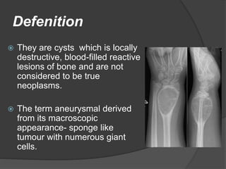
Defenition
 They are cysts which is locally
destructive, blood-filled reactive
lesions of bone and are not
considered to be true
neoplasms.
 The term aneurysmal derived
from its macroscopic
appearance- sponge like
tumour with numerous giant
cells.
Etiology
 It was first recognized by Jaffe and
Lichtenstein(1942)
 True etiology of ABC is not known.
 It is not a cyst and is called an aneurysm
(quite wrongly) only due to characteristic
of expanding the bone into “aneurysm”
shape and that it is filled with blood
Incidence and
Demographics
 ABC is found at any age
 Around 75% before 20 years and rare after
30 years.
 Female : Male :: 2:1
 Any bone may be involved, but the most
common locations include the proximal
humerus , distal femur , proximal tibia, and
spine
 Around 12-30% of ABCs involve spine that
represents only 1.4% of Primary vertebral
column tumours
Primary ABCs
 They are driven by upregulation of
the ubiquitin-specific
protease USP6 (Tre2) gene on 17p13
when combined by a translocation with a
promoter gene.
 Most commonly described
translocation t(16;17)(q22;p13) leading
to juxtaposition of promoter region
CDH11 on 16q22
Secondary ABCs
 Not considered a neoplasm because no
known translocation has been identified
 It can be associated with other tumours.
 Associated with other tumours (30% of
times)
 Giant cell tumor (GCT) of bone
 Fibrous dysplasia
 Chondroblastoma
 Osteoblastoma
 Nonossifying fibroma (NOF)
 Fibrous histiocytoma
 Chondromyxoid fibroma
 Simple bone cyst
 Giant cell reparative granuloma
 Telangiectatic osteosarcoma
Epidemiology
 75% of patients are < 20 yrs.
 Anatomical location
 ~15% seen in spine
 >60% in long bones (Femur and tibia being
most common)
 51% occurrs in the lower extremities, 22.5% in
upper extremities
 Usually in Metaphysis
 Metatarsal and Calcaneus are the most
common locations in the foot
Epidemiology
 Spine :
 Most commonly in
thoracolumbar region
 Cervical spine involved in 30-
40%
 60 % of anuerysmal bone cyst
occur in posterior elements
 Half of the cases involve more
than 1 vertebra
Presentation
 Patients usually presents with pain ,mass, a
pathological fracture or a combination of all in
the affected area.
 Neurological symptoms may develop involving
nerve typically in spine.
 Other findings include:
 Deformity
 Decreased movement ,weakness or stiffness
 Warmth over affected area
Clinical Features
 Patients commonly have pain and swelling at the
site
 Neurological symptoms may be seen as primary
complaint in ABCs affecting the vertebrae
 Unusually patients may present with a pathological
fracture at the site of ABC
Gross Pathology
 Specimens reveal a
thin osseous shell
surrounding a
honeycombed
sponge-like mass
filled with blood
Histology
 ABCs consists of blood-filled
spaces variable size that are
separated by connective tissue
containing trabeculae of bone
oseoid tissue and osteoclast giant
cells.
 They are not lined by endothelium.
 Cavity lining by numerous benign
giant cells / spindle cells / thin
strands of woven (new) bone
present
Development of ABC
 It follows 3 stages
 Initial phase :
○ Osteolysis without peculiar findings.
 Growth phase:
○ Rapid increase in size of osseous erosion.
○ Enlargement of involved bone.
○ Formation of shell around central part of
lesion.
 Stabilization phase
○ Fully developed radiological pattern.
Imaging Modalities
 X-Ray
 CT scan
 MRI
 Nuclear Imaging
 Angiography
X-Ray Appearance
 ABC is normally placed in the
metaphysis and appears as a osteolytic
lesion. The periosteum is elevated and
the eroded to a thin margin.
 The expansile nature of the lesion is
often refleced by a "blown-out" or "soap
bubble” appearance.
 The lesion rarely penetrates the articular
surface or growth plate.
Evolution of ABCs
(Radiologically)
 Incipient phase (incidentally found): There is a small
nonexpansile intramedullary lytic lesion with distinctive
elevation of periosteum
 Growth phase: Rapid growth and lysis of bone that present
as cortical blowout in “accelerated” phenomenon of ABC
The Codman’s triangles may be prominent at the ends
 Stable phase: Expanded bone with a “shell” around the
lesion along with trabeculations coursing within it (coarse
soap-bubble appearance)
Evolution of ABCs
 Stable phase: Expanded bone with a “shell” around the lesion
along with trabeculations coursing within it (coarse soap-bubble
appearance)
 The Healing Cyst:(self-resolution or
following treatment)shows progressive
ossification, resulting in a coarsely
trabeculated bony mass often with
mineralized matrix
 The cyst may invaginate into normal bone
and to epiphysis through growth plate or
extend into soft tissue
Capanna et al. described five“morphologic types”
based on the radiographic findings:
 Type I: Central metaphyseal lesion well-contained within bone with intact
bone cortices.
 Type II: Lesion gives an inflated appearance to bone with cortical thinning.
 Type III: Eccentric metaphyseal location with unaffected cortex.
 Type IV: Subperiosteal extension but cortex intact rarely seen in the
diaphysis.
 Type V: Metadiaphyseal location blowout appearance, cortical breach
present and cyst may invaginate into nearby bone
Capanna et al. Classification based on
Morphology
Computed Tomography
(CT)
 Cross-sectional CT is the most useful
imaging examination, It can demonstrate
the intraosseous and extraosseous lesion.
 CT can be used to determine the nature of
the matrix of especially when tumors are in
complex locations, such as facial skeleton,
spine, thoracic cage, and pelvis.
 Spinal CT can demonstrate stenosis of the
spinal canal due to involvement of the
posterior elements.
MRI
MRI can demonstrate the characteristic fluid-fluid levels exquisitely, as
well as identify the presence of a solid component and concerning
features suggesting an aneurysmal bone cyst-like appearance of
another tumor entity.
MRI
Demonstrates the typical blood fluid levels seen on
an MRI scan of ABC
Treatment
 The treatment options are multiple and best one
for a patient needs to be individualized
 The inactive cysts have complete periosteal shell
and sclerotic bone margins
 The active cysts have incomplete shell and
aggressive lesions have indefinite margins
 Inactive lesions :spontaneous regression seen
 Active and aggressive ABCs- NEED TREATMENT
Percutaneous methods
 For Inducing sclerosis in the cyst and secondary
mineralization (healing)
○ They have advantage particularly in the juxtaphyseal location
and difficult surgical sites
○ Alcoholic solution of zein has shown good result
○ Side effects :percutaneous fistulation, local abscess
formation, and sometimes embolization is seen.
 Healing takes 6–18 months depending on the size of initial lesion
 Percutaneous methods not to be used in patients
 with rapid expansion
 impending fractures
 patients allergic to drug.
 Percutaneous
sclerotherapy with
Polidocanol is a safe
alternative to conventional
surgery for the treatment of
an aneurysmal bone cyst.
It can be used at
surgically-inaccessible
sites and treatment can be
performed on an out-
patient basis.
Demineralized bone particle
 It is instilled into the lesion as a paste of allogenic
bone powder and autogenous bone marrow to
induce healing.
 No curettage or extensive surgery is done
 It is expected that the bone grafting material
promotes ossification at a pace faster that the
native rate of ABC expansion
Curettage and Bone
Grafting
 It had been the gold standard of treatment that is
still used to compare efficacy of other methods
 It is supplemented with the use of high-speed burr,
or extended curettage with adjuvants [peroxide,
phenol, cryotherapy, zinc chloride,hypochlorite,
PMMA to reduce recurrence rate
 Selective arterial embolization has been used for
reducing the blood loss in large cysts but should
not be used to rely upon for inducing healing
 Growth plate can get damaged while treating the
juxtaphyseal lesions
Curettage and Bone
Grafting
Curettage and Bone
Grafting
Complication of curettage and bone grafting for ABC : The growth plate got
damaged during the index lesion leading to premature fusion and growth
abnormalities of the bone
Marginal en bloc resection
 It can be done in
expendable bones (fibula,
clavicle, rib, pubic ramus)
 It has got the lowest
recurrence rates.
 Sub periosteal resection is
possible in children which
can be simultaneously
reconstructed with
fibula
Radiation therapy
 It has been found effective in inducing healing and
ossification
 It should be abandoned for high likelihood of
inducing radiation sarcoma
Solid ABCs
 A third variant called “solid” ABC have been
described(coined by Sanerkin et al.) in 1983
Containing , lacy, chondroid like material seen in
conventional ABC, but without the typical vascular
and cyst like cavities.
Represent 5–10% of the ABCs
 Nevertheless a fourth variant called the “soft tissue
ABCs” has also been reported
REFERENCE
 Campbell’s Operative Orthopaedics
 Apley & Solomon’s System of
Orthopaedics and Trauma
 Essential Orthopaedics by Manish Kumar
Varshney
 Orthobullets.com
THANK YOU

Aneurysmal bone cyst.pptx

  • 2.
    Defenition  They arecysts which is locally destructive, blood-filled reactive lesions of bone and are not considered to be true neoplasms.  The term aneurysmal derived from its macroscopic appearance- sponge like tumour with numerous giant cells.
  • 3.
    Etiology  It wasfirst recognized by Jaffe and Lichtenstein(1942)  True etiology of ABC is not known.  It is not a cyst and is called an aneurysm (quite wrongly) only due to characteristic of expanding the bone into “aneurysm” shape and that it is filled with blood
  • 4.
    Incidence and Demographics  ABCis found at any age  Around 75% before 20 years and rare after 30 years.  Female : Male :: 2:1  Any bone may be involved, but the most common locations include the proximal humerus , distal femur , proximal tibia, and spine  Around 12-30% of ABCs involve spine that represents only 1.4% of Primary vertebral column tumours
  • 5.
    Primary ABCs  Theyare driven by upregulation of the ubiquitin-specific protease USP6 (Tre2) gene on 17p13 when combined by a translocation with a promoter gene.  Most commonly described translocation t(16;17)(q22;p13) leading to juxtaposition of promoter region CDH11 on 16q22
  • 6.
    Secondary ABCs  Notconsidered a neoplasm because no known translocation has been identified  It can be associated with other tumours.
  • 7.
     Associated withother tumours (30% of times)  Giant cell tumor (GCT) of bone  Fibrous dysplasia  Chondroblastoma  Osteoblastoma  Nonossifying fibroma (NOF)  Fibrous histiocytoma  Chondromyxoid fibroma  Simple bone cyst  Giant cell reparative granuloma  Telangiectatic osteosarcoma
  • 8.
    Epidemiology  75% ofpatients are < 20 yrs.  Anatomical location  ~15% seen in spine  >60% in long bones (Femur and tibia being most common)  51% occurrs in the lower extremities, 22.5% in upper extremities  Usually in Metaphysis  Metatarsal and Calcaneus are the most common locations in the foot
  • 9.
    Epidemiology  Spine : Most commonly in thoracolumbar region  Cervical spine involved in 30- 40%  60 % of anuerysmal bone cyst occur in posterior elements  Half of the cases involve more than 1 vertebra
  • 10.
    Presentation  Patients usuallypresents with pain ,mass, a pathological fracture or a combination of all in the affected area.  Neurological symptoms may develop involving nerve typically in spine.  Other findings include:  Deformity  Decreased movement ,weakness or stiffness  Warmth over affected area
  • 11.
    Clinical Features  Patientscommonly have pain and swelling at the site  Neurological symptoms may be seen as primary complaint in ABCs affecting the vertebrae  Unusually patients may present with a pathological fracture at the site of ABC
  • 12.
    Gross Pathology  Specimensreveal a thin osseous shell surrounding a honeycombed sponge-like mass filled with blood
  • 13.
    Histology  ABCs consistsof blood-filled spaces variable size that are separated by connective tissue containing trabeculae of bone oseoid tissue and osteoclast giant cells.  They are not lined by endothelium.  Cavity lining by numerous benign giant cells / spindle cells / thin strands of woven (new) bone present
  • 14.
    Development of ABC It follows 3 stages  Initial phase : ○ Osteolysis without peculiar findings.  Growth phase: ○ Rapid increase in size of osseous erosion. ○ Enlargement of involved bone. ○ Formation of shell around central part of lesion.  Stabilization phase ○ Fully developed radiological pattern.
  • 15.
    Imaging Modalities  X-Ray CT scan  MRI  Nuclear Imaging  Angiography
  • 16.
    X-Ray Appearance  ABCis normally placed in the metaphysis and appears as a osteolytic lesion. The periosteum is elevated and the eroded to a thin margin.  The expansile nature of the lesion is often refleced by a "blown-out" or "soap bubble” appearance.  The lesion rarely penetrates the articular surface or growth plate.
  • 18.
    Evolution of ABCs (Radiologically) Incipient phase (incidentally found): There is a small nonexpansile intramedullary lytic lesion with distinctive elevation of periosteum  Growth phase: Rapid growth and lysis of bone that present as cortical blowout in “accelerated” phenomenon of ABC The Codman’s triangles may be prominent at the ends  Stable phase: Expanded bone with a “shell” around the lesion along with trabeculations coursing within it (coarse soap-bubble appearance)
  • 19.
    Evolution of ABCs Stable phase: Expanded bone with a “shell” around the lesion along with trabeculations coursing within it (coarse soap-bubble appearance)  The Healing Cyst:(self-resolution or following treatment)shows progressive ossification, resulting in a coarsely trabeculated bony mass often with mineralized matrix  The cyst may invaginate into normal bone and to epiphysis through growth plate or extend into soft tissue
  • 20.
    Capanna et al.described five“morphologic types” based on the radiographic findings:  Type I: Central metaphyseal lesion well-contained within bone with intact bone cortices.  Type II: Lesion gives an inflated appearance to bone with cortical thinning.  Type III: Eccentric metaphyseal location with unaffected cortex.  Type IV: Subperiosteal extension but cortex intact rarely seen in the diaphysis.  Type V: Metadiaphyseal location blowout appearance, cortical breach present and cyst may invaginate into nearby bone
  • 21.
    Capanna et al.Classification based on Morphology
  • 22.
    Computed Tomography (CT)  Cross-sectionalCT is the most useful imaging examination, It can demonstrate the intraosseous and extraosseous lesion.  CT can be used to determine the nature of the matrix of especially when tumors are in complex locations, such as facial skeleton, spine, thoracic cage, and pelvis.  Spinal CT can demonstrate stenosis of the spinal canal due to involvement of the posterior elements.
  • 24.
    MRI MRI can demonstratethe characteristic fluid-fluid levels exquisitely, as well as identify the presence of a solid component and concerning features suggesting an aneurysmal bone cyst-like appearance of another tumor entity.
  • 25.
    MRI Demonstrates the typicalblood fluid levels seen on an MRI scan of ABC
  • 26.
    Treatment  The treatmentoptions are multiple and best one for a patient needs to be individualized  The inactive cysts have complete periosteal shell and sclerotic bone margins  The active cysts have incomplete shell and aggressive lesions have indefinite margins  Inactive lesions :spontaneous regression seen  Active and aggressive ABCs- NEED TREATMENT
  • 27.
    Percutaneous methods  ForInducing sclerosis in the cyst and secondary mineralization (healing) ○ They have advantage particularly in the juxtaphyseal location and difficult surgical sites ○ Alcoholic solution of zein has shown good result ○ Side effects :percutaneous fistulation, local abscess formation, and sometimes embolization is seen.  Healing takes 6–18 months depending on the size of initial lesion  Percutaneous methods not to be used in patients  with rapid expansion  impending fractures  patients allergic to drug.
  • 28.
     Percutaneous sclerotherapy with Polidocanolis a safe alternative to conventional surgery for the treatment of an aneurysmal bone cyst. It can be used at surgically-inaccessible sites and treatment can be performed on an out- patient basis.
  • 29.
    Demineralized bone particle It is instilled into the lesion as a paste of allogenic bone powder and autogenous bone marrow to induce healing.  No curettage or extensive surgery is done  It is expected that the bone grafting material promotes ossification at a pace faster that the native rate of ABC expansion
  • 30.
    Curettage and Bone Grafting It had been the gold standard of treatment that is still used to compare efficacy of other methods  It is supplemented with the use of high-speed burr, or extended curettage with adjuvants [peroxide, phenol, cryotherapy, zinc chloride,hypochlorite, PMMA to reduce recurrence rate  Selective arterial embolization has been used for reducing the blood loss in large cysts but should not be used to rely upon for inducing healing  Growth plate can get damaged while treating the juxtaphyseal lesions
  • 31.
  • 32.
    Curettage and Bone Grafting Complicationof curettage and bone grafting for ABC : The growth plate got damaged during the index lesion leading to premature fusion and growth abnormalities of the bone
  • 33.
    Marginal en blocresection  It can be done in expendable bones (fibula, clavicle, rib, pubic ramus)  It has got the lowest recurrence rates.  Sub periosteal resection is possible in children which can be simultaneously reconstructed with fibula
  • 34.
    Radiation therapy  Ithas been found effective in inducing healing and ossification  It should be abandoned for high likelihood of inducing radiation sarcoma
  • 35.
    Solid ABCs  Athird variant called “solid” ABC have been described(coined by Sanerkin et al.) in 1983 Containing , lacy, chondroid like material seen in conventional ABC, but without the typical vascular and cyst like cavities. Represent 5–10% of the ABCs  Nevertheless a fourth variant called the “soft tissue ABCs” has also been reported
  • 36.
    REFERENCE  Campbell’s OperativeOrthopaedics  Apley & Solomon’s System of Orthopaedics and Trauma  Essential Orthopaedics by Manish Kumar Varshney  Orthobullets.com
  • 37.