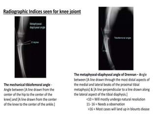
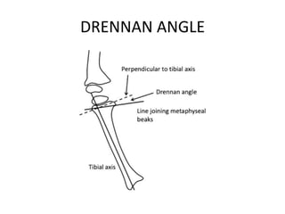
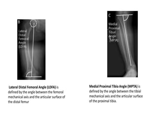
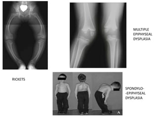
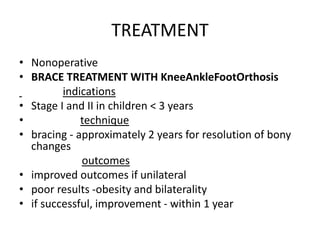
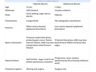
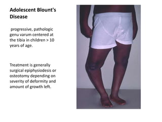
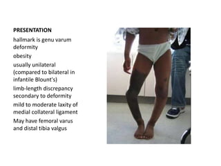
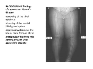
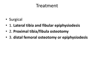
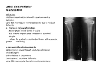
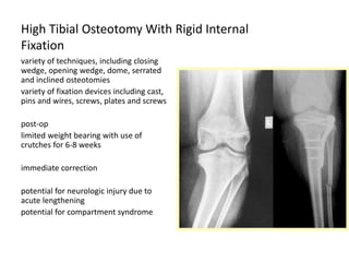
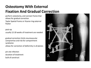
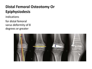
Blount's disease is a progressive deformity of the knee characterized by bowing of the legs (genu varum). It is classified into infantile and adolescent types. Infantile Blount's disease presents between ages 2-5 and is often bilateral, while adolescent Blount's disease presents after age 10 and is usually unilateral. Radiographs show abnormalities of the proximal tibial epiphysis. Treatment involves bracing for mild cases and osteotomy or epiphysiodesis surgery for more severe deformities, with the goal of correcting the varus alignment through a valgus procedure. Prognosis is best when treated early before significant bony changes occur.
Perthes disease is a rare childhood condition that affects the hip. It occurs when the blood supply to the rounded head of the femur (thighbone) is temporarily disrupted. Without an adequate blood supply, the bone cells die, a process called avascular necrosis.
Ozempic: Preoperative Management of Patients on GLP-1 Receptor Agonists Saeid Safari
Preoperative Management of Patients on GLP-1 Receptor Agonists like Ozempic and Semiglutide
ASA GUIDELINE
NYSORA Guideline
2 Case Reports of Gastric Ultrasound
- Video recording of this lecture in English language: https://youtu.be/lK81BzxMqdo
- Video recording of this lecture in Arabic language: https://youtu.be/Ve4P0COk9OI
- Link to download the book free: https://nephrotube.blogspot.com/p/nephrotube-nephrology-books.html
- Link to NephroTube website: www.NephroTube.com
- Link to NephroTube social media accounts: https://nephrotube.blogspot.com/p/join-nephrotube-on-social-media.html
Recomendações da OMS sobre cuidados maternos e neonatais para uma experiência pós-natal positiva.
Em consonância com os ODS – Objetivos do Desenvolvimento Sustentável e a Estratégia Global para a Saúde das Mulheres, Crianças e Adolescentes, e aplicando uma abordagem baseada nos direitos humanos, os esforços de cuidados pós-natais devem expandir-se para além da cobertura e da simples sobrevivência, de modo a incluir cuidados de qualidade.
Estas diretrizes visam melhorar a qualidade dos cuidados pós-natais essenciais e de rotina prestados às mulheres e aos recém-nascidos, com o objetivo final de melhorar a saúde e o bem-estar materno e neonatal.
Uma “experiência pós-natal positiva” é um resultado importante para todas as mulheres que dão à luz e para os seus recém-nascidos, estabelecendo as bases para a melhoria da saúde e do bem-estar a curto e longo prazo. Uma experiência pós-natal positiva é definida como aquela em que as mulheres, pessoas que gestam, os recém-nascidos, os casais, os pais, os cuidadores e as famílias recebem informação consistente, garantia e apoio de profissionais de saúde motivados; e onde um sistema de saúde flexível e com recursos reconheça as necessidades das mulheres e dos bebês e respeite o seu contexto cultural.
Estas diretrizes consolidadas apresentam algumas recomendações novas e já bem fundamentadas sobre cuidados pós-natais de rotina para mulheres e neonatos que recebem cuidados no pós-parto em unidades de saúde ou na comunidade, independentemente dos recursos disponíveis.
É fornecido um conjunto abrangente de recomendações para cuidados durante o período puerperal, com ênfase nos cuidados essenciais que todas as mulheres e recém-nascidos devem receber, e com a devida atenção à qualidade dos cuidados; isto é, a entrega e a experiência do cuidado recebido. Estas diretrizes atualizam e ampliam as recomendações da OMS de 2014 sobre cuidados pós-natais da mãe e do recém-nascido e complementam as atuais diretrizes da OMS sobre a gestão de complicações pós-natais.
O estabelecimento da amamentação e o manejo das principais intercorrências é contemplada.
Recomendamos muito.
Vamos discutir essas recomendações no nosso curso de pós-graduação em Aleitamento no Instituto Ciclos.
Esta publicação só está disponível em inglês até o momento.
Prof. Marcus Renato de Carvalho
www.agostodourado.com
New Drug Discovery and Development .....NEHA GUPTA
The "New Drug Discovery and Development" process involves the identification, design, testing, and manufacturing of novel pharmaceutical compounds with the aim of introducing new and improved treatments for various medical conditions. This comprehensive endeavor encompasses various stages, including target identification, preclinical studies, clinical trials, regulatory approval, and post-market surveillance. It involves multidisciplinary collaboration among scientists, researchers, clinicians, regulatory experts, and pharmaceutical companies to bring innovative therapies to market and address unmet medical needs.
Title: Sense of Taste
Presenter: Dr. Faiza, Assistant Professor of Physiology
Qualifications:
MBBS (Best Graduate, AIMC Lahore)
FCPS Physiology
ICMT, CHPE, DHPE (STMU)
MPH (GC University, Faisalabad)
MBA (Virtual University of Pakistan)
Learning Objectives:
Describe the structure and function of taste buds.
Describe the relationship between the taste threshold and taste index of common substances.
Explain the chemical basis and signal transduction of taste perception for each type of primary taste sensation.
Recognize different abnormalities of taste perception and their causes.
Key Topics:
Significance of Taste Sensation:
Differentiation between pleasant and harmful food
Influence on behavior
Selection of food based on metabolic needs
Receptors of Taste:
Taste buds on the tongue
Influence of sense of smell, texture of food, and pain stimulation (e.g., by pepper)
Primary and Secondary Taste Sensations:
Primary taste sensations: Sweet, Sour, Salty, Bitter, Umami
Chemical basis and signal transduction mechanisms for each taste
Taste Threshold and Index:
Taste threshold values for Sweet (sucrose), Salty (NaCl), Sour (HCl), and Bitter (Quinine)
Taste index relationship: Inversely proportional to taste threshold
Taste Blindness:
Inability to taste certain substances, particularly thiourea compounds
Example: Phenylthiocarbamide
Structure and Function of Taste Buds:
Composition: Epithelial cells, Sustentacular/Supporting cells, Taste cells, Basal cells
Features: Taste pores, Taste hairs/microvilli, and Taste nerve fibers
Location of Taste Buds:
Found in papillae of the tongue (Fungiform, Circumvallate, Foliate)
Also present on the palate, tonsillar pillars, epiglottis, and proximal esophagus
Mechanism of Taste Stimulation:
Interaction of taste substances with receptors on microvilli
Signal transduction pathways for Umami, Sweet, Bitter, Sour, and Salty tastes
Taste Sensitivity and Adaptation:
Decrease in sensitivity with age
Rapid adaptation of taste sensation
Role of Saliva in Taste:
Dissolution of tastants to reach receptors
Washing away the stimulus
Taste Preferences and Aversions:
Mechanisms behind taste preference and aversion
Influence of receptors and neural pathways
Impact of Sensory Nerve Damage:
Degeneration of taste buds if the sensory nerve fiber is cut
Abnormalities of Taste Detection:
Conditions: Ageusia, Hypogeusia, Dysgeusia (parageusia)
Causes: Nerve damage, neurological disorders, infections, poor oral hygiene, adverse drug effects, deficiencies, aging, tobacco use, altered neurotransmitter levels
Neurotransmitters and Taste Threshold:
Effects of serotonin (5-HT) and norepinephrine (NE) on taste sensitivity
Supertasters:
25% of the population with heightened sensitivity to taste, especially bitterness
Increased number of fungiform papillae
Lung Cancer: Artificial Intelligence, Synergetics, Complex System Analysis, S...Oleg Kshivets
RESULTS: Overall life span (LS) was 2252.1±1742.5 days and cumulative 5-year survival (5YS) reached 73.2%, 10 years – 64.8%, 20 years – 42.5%. 513 LCP lived more than 5 years (LS=3124.6±1525.6 days), 148 LCP – more than 10 years (LS=5054.4±1504.1 days).199 LCP died because of LC (LS=562.7±374.5 days). 5YS of LCP after bi/lobectomies was significantly superior in comparison with LCP after pneumonectomies (78.1% vs.63.7%, P=0.00001 by log-rank test). AT significantly improved 5YS (66.3% vs. 34.8%) (P=0.00000 by log-rank test) only for LCP with N1-2. Cox modeling displayed that 5YS of LCP significantly depended on: phase transition (PT) early-invasive LC in terms of synergetics, PT N0—N12, cell ratio factors (ratio between cancer cells- CC and blood cells subpopulations), G1-3, histology, glucose, AT, blood cell circuit, prothrombin index, heparin tolerance, recalcification time (P=0.000-0.038). Neural networks, genetic algorithm selection and bootstrap simulation revealed relationships between 5YS and PT early-invasive LC (rank=1), PT N0—N12 (rank=2), thrombocytes/CC (3), erythrocytes/CC (4), eosinophils/CC (5), healthy cells/CC (6), lymphocytes/CC (7), segmented neutrophils/CC (8), stick neutrophils/CC (9), monocytes/CC (10); leucocytes/CC (11). Correct prediction of 5YS was 100% by neural networks computing (area under ROC curve=1.0; error=0.0).
CONCLUSIONS: 5YS of LCP after radical procedures significantly depended on: 1) PT early-invasive cancer; 2) PT N0--N12; 3) cell ratio factors; 4) blood cell circuit; 5) biochemical factors; 6) hemostasis system; 7) AT; 8) LC characteristics; 9) LC cell dynamics; 10) surgery type: lobectomy/pneumonectomy; 11) anthropometric data. Optimal diagnosis and treatment strategies for LC are: 1) screening and early detection of LC; 2) availability of experienced thoracic surgeons because of complexity of radical procedures; 3) aggressive en block surgery and adequate lymph node dissection for completeness; 4) precise prediction; 5) adjuvant chemoimmunoradiotherapy for LCP with unfavorable prognosis.
5. synonyms
• Infantile tibia vara
• Erlachers disease
• Blount-barber disease
• Subepiphyseal osteochondropathy
• Non rachitic bow legs
• Osteitis deformans tibia
6.
7. • Secondary effects
• -internal torsion of tibia
• -insufficient ossification of the medial portion
of medial tibial condyle
• Streched LCL
8. ANATOMY
• Genu varum is a normal physiologic process in
children
• genu varum (bowed legs) is normal in
children less than 2 years
• genu varum migrates to a neutral at ~ 14 months
• continues on to a peak genu valgum (knocked
knees) at ~ 3 years of age
• genu valgum then migrates back to normal
physiologic valgus(<15) at ~ 7 years of age
9.
10.
11. Types
• 2 types
• 1-Infantile-begins before 8 years of age
• 2-Adolescent- begins after 10 years of age but
before skeletal maturity
• A) b/w 10 and 13 years -partial closure of
epiphysis - trauma/infection
• B) b/w 10 and 13 years –black and obese
children-no distinct cause
12. INFANTILE BLOUNTS DISEASE
• progressive pathologic genu varum centered
at the tibia in children 2 to 5 years of age.
• Diagnosis- presence of a genu
varum/flexion/internal rotation deformity and
confirmed radiographically with an
increased metaphyseal-diaphyseal angle.
• Treatment - bracing to surgery (patient age,
severity of deformity, and presence of a
physeal bar.)
13. ETIOLOGY
Idiopathic
more common
male > female
bilateral in 50%
Altered enchondral
ossification
Risk factors
overweight children
early walkers (< 1 year)
Hispanic and African
American
14. Pathophysiology
• multifactorial -but related to mechanical
overload in genetically susceptible individuals
• excessive medial pressure produces an
osteochondrosis of the medial proximal tibial
physis and epiphysis
• (osteochondrosis can progress to a physeal
bar)
15. • Intra articular pathology
• Enlargement and hypermobility of medial
meniscus
• Depression of anterior aspect of medial
femoral condyle
• Depression of posteromedial plateau of tibia
16.
17. type I to IV consist of increasing medial metaphyseal beaking and sloping
type V and VI have an epiphyseal-metaphyseal bony bridge (congenital bar
across physis)
At stage 6 – medial portion of epiphysis fuses at 90 degree downward angle
18. PRESENTATION
• Physical examination
• genu varum/flexion/internal rotation deformity
• usually b/l in infants
• lateral thrust on walking
• Waddling gait
• may exhibit positive 'cover-up test'
• often associated with internal tibial torsion
• leg length discrepancy
19.
20. • usually NO tenderness, restriction of motion, effusion
• Bony hard non tender prominence –palpable at the
medial epiphysiometaaphyseal junction
• SIFFERT KATZ sign-as the extended knee is slightly
flexed---medial femoral condyle engages tibial medial
plateau depression and sublaxates posteromedially
(varus instability at 15 degree knee flexion)
21. • Long standing neglected cases’
• -slight flexion deformity added
• Unstable joint-lax ligaments
• Medial compartment OA knee
22. IMAGING
Radiographs
• standing long-cassette AP radiograph of both lower
extremities
• ensure that patella are facing forwards for evaluation
(commonly associated with internal tibial torsion)
– findings suggestive of Blounts disease
• medial and posterior sloping of proximal tibial
epiphysis(short ,thin,wedged,irregular)
• varus focused at proximal tibia
• severe deformity(asymmetric bowing)
• sharp angular deformity(progressive)
32. TREATMENT
• Nonoperative
• BRACE TREATMENT WITH KneeAnkleFootOrthosis
indications
• Stage I and II in children < 3 years
• technique
• bracing - approximately 2 years for resolution of bony
changes
outcomes
• improved outcomes if unilateral
• poor results -obesity and bilaterality
• if successful, improvement - within 1 year
35. OPERATIVE
• PROXIMAL TIBIA/FIBULA VALGUS OSTEOTOMY
• overcome the varus/flexion/internal rotation deformity
indications
• Stage I and II in children > 3 years
• Stage III, IV, V, VI
• age ≥ 4y (all stages)
• failure of brace treatment
• progressive deformity
• metaphyseal-diaphyseal angles > 20 degrees
outcomes
• risk of recurrence is significantly lessened if performed
before 4 years of age
36. OSTEOTOMY
GOALS OF CORRECTION
• overcorrect into 10-15° of valgus because
medial physeal growth abnormalities persist
• distal segment is fixed in valgus, external
rotation and lateral translation
45. • PHYSEAL BAR RESECTION
• indication
• at least 4y of growth remaining
• technique
• perform together with osteotomy
• interpositional material is usually fat or PMMA
46. COMPLICATIONS
• 1.Compartment syndrome (with high
tibial/fibular osteotomy)-prophylactic release of
anterior compartment
• 2.CPN palsy
• 3.anterior tibia artery injury
4.Recurrence of tibial vara
• severe cases-- physeal bar-- progressive varus
after a well executed proximal tibial valgus
osteotomy--- require a lateral tibial
hemiepiphysiodesis or physeal bar resection
47. Physeal bar
• MC cause of recurrence
• Greene criteria(CT scan)
• 1.age>5
• 2.medial physeal slope 50-70 degree
• 3.langenskiold grade 4 or more’
• 4.body weight >95 th percentile
• 5.black girls who meet previous criteria
48. PROGNOSIS
• Best outcomes
• early diagnosis and unloading of the medial
joint with either bracing or an osteotomy
• Young children with stage II and stage IV can
have spontaneous correction
49. •Infantile versus Adolescent Blount's
•Infantile Blounts •Adolescent Blounts
•Age •2-5yrs •>10yrs
•Bilaterally •50% bilateral •Usually unilateral
•Risks
•Early walking, large stature,
obesity
•Obesity
•Classification •Langenskiold •No radiographic classification
•Severity
•More severe physeal/
epiphyseal disturbance
•Less severe physeal/ epiphyseal
disturbance
•Bone Involvement
•Proximal medial tibia physis,
producing genu varus, flexion,
internal rotation, AND may have
compensatory distal femoral
VALGUS
•Proximal tibia physis, AND may have
distal femoral VARUS and distal tibia
valgus
•Natural History
•Self-limited - stage II and IV can
exhibit spontaneous resolution
•Progressive, never resolves
spontaneously (thus bracing unlikely to
work)
•Treatment options •Bracing and surgery •Surgery only
50. Adolescent Blount's
Disease
progressive, pathologic
genu varum centered at
the tibia in children > 10
years of age.
Treatment is generally
surgical epiphysiodesis or
osteotomy depending on
severity of deformity and
amount of growth left.
51. ETIOLOGY
• Less common
• less severe
• more likely to be unilateral
• more likely to have femoral deformity
• obesity
• African-American descent
• Pathophysiology-dyschondrosis of medial
physis of proximal tibia – multifactorial-
mechanical overload in genetically
susceptible individuals
52. PRESENTATION
hallmark is genu varum
deformity
obesity
usually unilateral
(compared to bilateral in
infantile Blount's)
limb-length discrepancy
secondary to deformity
mild to moderate laxity of
medial collateral ligament
May have femoral varus
and distal tibia valgus
53. RADIOGRAPHIC findings
s/o adolescent Blount's
disease
narrowing of the tibial
epiphysis
widening of the medial
tibial growth plate
occasional widening of the
lateral distal femoral physis
metaphyseal breaking less
commonly seen with
adolescent Blount's
55. Lateral tibia and fibular
epiphysiodesis
indications
mild to moderate deformity with growth remaining
outcomes
up to 25% may require formal osteotomy due to residual
deformity
a. transient hemiepiphysiodesis
tether physis with 8-plates or staple
may remove implant once correction is achieved
simple
allows for gradual correction is children with adequate
growth remaining
b. permanent hemiepiphysiodesis
obliteration of physis through small, lateral incision
limited surgery
overcorrection is uncommon
cannot correct rotational deformity
up to 25% may require formal corrective osteotomy
56. Proximal tibia/fibula osteotomy
indications
more severe cases in the skeletally mature
- valgus producing tibial osteotomy and plating
-external fixation
goals of correction
overcorrection to valgus not indicated (as is the case in infantile Blount's)
strive for neutral mechanical axi
57. High Tibial Osteotomy With Rigid Internal
Fixation
variety of techniques, including closing
wedge, opening wedge, dome, serrated
and inclined osteotomies
variety of fixation devices including cast,
pins and wires, screws, plates and screws
post-op
limited weight bearing with use of
crutches for 6-8 weeks
immediate correction
potential for neurologic injury due to
acute lengthening
potential for compartment syndrome
58. Osteotomy With External
Fixation And Gradual Correction
perform osteotomy, and connect frame that
allows for gradual correction
Taylor Spatial Frame or Ilizarov ring external
fixator
post-op
usually 12-18 weeks of treatment are needed
gradual correction limits neurovascular
compromise and risk for compartment
syndrome
allows for correction of deformity in all planes
pin site infection
duration of treatment
bulk of construct
59. Distal Femoral Osteotomy Or
Epiphysiodesis
indications
for distal femoral
varus deformity of 8
degrees or greater
60.
61. REFERENCES
• Campbell’s Operative Orthopaedics 14th
edition (2021)
• Apley's System of Orthopaedics and Fractures
• TUREK orthopedic principles and application
• https://pubmed.ncbi.nlm.nih.gov/
• https://www.orthobullets.com/