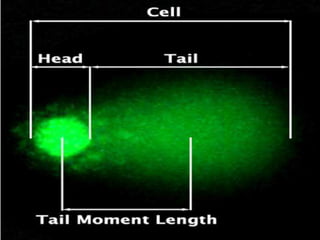
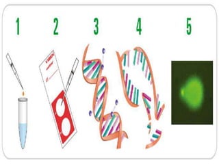
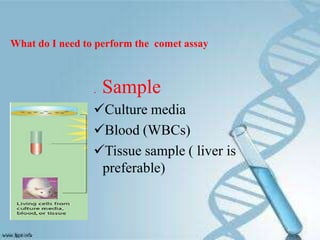
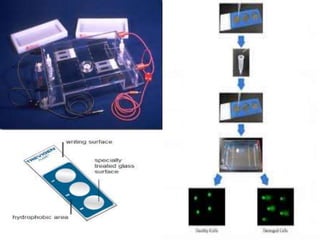
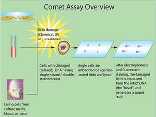
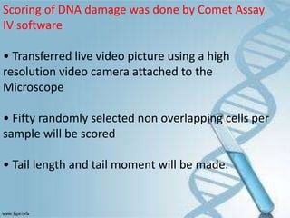
Comet Assay
Single cell gel electrophoresis (SCGE)
BY
A.MANOJ KUMAR
II M.Sc (BIO-TECHNOLOGY)
INTRODUCTION
Single cell gel electrophoresis
(Comet assay) is a sensitive and
rapid method for measuring DNA
damage and repair in single
eukaryotic cell.
Swedish researchers Ostling &
Johansson developed this technique
in 1984
PRINCIPLE
•The basis for this assay is that
loops of DNA containing a break
lose their supercoiling and become
free to extend toward the anode
when exposed to current during
electrophoresis at high pH.
•The results appear as structures
resembling comets observed by
fluorescence microscopy.
Comet contain a distinct head
and tail.
• The head is composed of
intact DNA, while the tail consists
of damaged or broken pieces of
DNA.
• The intensity of the comet tail
relative to the head reflects the
number of DNA breaks.
. Sample
Culture media
Blood (WBCs)
Tissue sample ( liver is
preferable)
What do I need to perform the comet assay
Comet assay Kit:
1.Comet Agarose.
2.Hemogenizing solution.
3.Lysis Solution.
4.Alkaline unwinding Solution, pH> 13.
5.SYBR Green staining Solution.
6.Neutralizing Solution.
7.Comet assay slides
1.Fluorescence microscope.
2.Comet assay scoring software
3.Laboratory hemogenizer.
4.Centrifuge.
5.Water bath.
6.Horizontal electrophoresis tank.
PROCEDURE
1.Liver tissue must be fresh.
2. Preparation of lysing solution : 40ml of lysing solution +4ml
of DMSO.
3. Homogenization of sample.
4. The homogenized liver tissue centrifuged, the supernatant
discardes,
resuspende the cells at 1X 10⁵ cells/ml in ice cold PBS.
6. Melt agarose
7. Cells will be combined with melting agarose then
immediately pipette 75 μl onto Comet Slides
8. Slides will be placed at 4°C in the refrigerator for 30
minutes.
9. Immerse slides in prechilled Lysing Solution at 4°C for 60
minutes
10. Slides will immersed in freshly prepared alkaline solution,
pH>13 then left it for 20 minutes at room temperature
11. Slide will be put in a horizontal electrophoresis apparatus.
The Alkaline Solution will be carefully poured until level just
covers samples
12. Adjust the apparatus on 21 Vol and 300 m.A.
13.Electrophoresis will be performed for 20 minutes
14. Slides will be neutralized by dipping several times in
distalled water , then immerse it in 70% ethanol for 5
minutes
14. The slides will be stained with 50 μl of diluted SYBR Green
,then examined by fluorescence microscope
Scoring of DNA damage was done by Comet Assay
IV software
• Transferred live video picture using a high
resolution video camera attached to the
Microscope
• Fifty randomly selected non overlapping cells per
sample will be scored
• Tail length and tail moment will be made.
• Image analysis of histopathological glass slide
stained with H&E to study different components
of the tissue.
• Analysis of cytopathological image for studying
the cells.
• Immunofluorescence image analysis using
molecular markers based on chromogen dyes
(e.g., DAB) or fluorescent dyes.
APPLICATION
• Genotoxicity testing.
It is provides a set of information about safety and
genotoxicity of newly developed pharmaceuticals
and chemicals.
Study of the protective effect of some
phytochemicals on genes when exposed to some
genotoxic insults.
This is one of the techniques used in the area of
cancer research for the evaluation of genotoxicity
and effectiveness of chemotherapy.
• Monitoring environmental contamination with
genotoxins.
• Human biomonitoring including:
Monitoring occupational exposure to
genotoxic chemicals or radiation.
Assessment of oxidative stress associated with
various human diseases.
Detection of DNA damage associated with
smoking.
• Nutritional Studies
The comet assay is ideal for investigating
nutrient or micronutrient effects at the level
of DNA damage in humans.
• Measuring DNA Repair
Commet assay is an important determinant
of individuals capacity for DNA repair and their
susceptibility to cancer.
Comet assay

Comet assay

  • 1.
    Comet Assay Single cellgel electrophoresis (SCGE) BY A.MANOJ KUMAR II M.Sc (BIO-TECHNOLOGY)
  • 2.
    INTRODUCTION Single cell gelelectrophoresis (Comet assay) is a sensitive and rapid method for measuring DNA damage and repair in single eukaryotic cell. Swedish researchers Ostling & Johansson developed this technique in 1984
  • 3.
    PRINCIPLE •The basis forthis assay is that loops of DNA containing a break lose their supercoiling and become free to extend toward the anode when exposed to current during electrophoresis at high pH. •The results appear as structures resembling comets observed by fluorescence microscopy.
  • 4.
    Comet contain adistinct head and tail. • The head is composed of intact DNA, while the tail consists of damaged or broken pieces of DNA. • The intensity of the comet tail relative to the head reflects the number of DNA breaks.
  • 7.
    . Sample Culture media Blood(WBCs) Tissue sample ( liver is preferable) What do I need to perform the comet assay
  • 8.
    Comet assay Kit: 1.CometAgarose. 2.Hemogenizing solution. 3.Lysis Solution. 4.Alkaline unwinding Solution, pH> 13. 5.SYBR Green staining Solution. 6.Neutralizing Solution. 7.Comet assay slides
  • 9.
    1.Fluorescence microscope. 2.Comet assayscoring software 3.Laboratory hemogenizer. 4.Centrifuge. 5.Water bath. 6.Horizontal electrophoresis tank.
  • 11.
    PROCEDURE 1.Liver tissue mustbe fresh. 2. Preparation of lysing solution : 40ml of lysing solution +4ml of DMSO. 3. Homogenization of sample. 4. The homogenized liver tissue centrifuged, the supernatant discardes, resuspende the cells at 1X 10⁵ cells/ml in ice cold PBS. 6. Melt agarose 7. Cells will be combined with melting agarose then immediately pipette 75 μl onto Comet Slides 8. Slides will be placed at 4°C in the refrigerator for 30 minutes. 9. Immerse slides in prechilled Lysing Solution at 4°C for 60 minutes
  • 12.
    10. Slides willimmersed in freshly prepared alkaline solution, pH>13 then left it for 20 minutes at room temperature 11. Slide will be put in a horizontal electrophoresis apparatus. The Alkaline Solution will be carefully poured until level just covers samples 12. Adjust the apparatus on 21 Vol and 300 m.A. 13.Electrophoresis will be performed for 20 minutes 14. Slides will be neutralized by dipping several times in distalled water , then immerse it in 70% ethanol for 5 minutes 14. The slides will be stained with 50 μl of diluted SYBR Green ,then examined by fluorescence microscope
  • 14.
    Scoring of DNAdamage was done by Comet Assay IV software • Transferred live video picture using a high resolution video camera attached to the Microscope • Fifty randomly selected non overlapping cells per sample will be scored • Tail length and tail moment will be made.
  • 15.
    • Image analysisof histopathological glass slide stained with H&E to study different components of the tissue. • Analysis of cytopathological image for studying the cells. • Immunofluorescence image analysis using molecular markers based on chromogen dyes (e.g., DAB) or fluorescent dyes.
  • 17.
    APPLICATION • Genotoxicity testing. Itis provides a set of information about safety and genotoxicity of newly developed pharmaceuticals and chemicals. Study of the protective effect of some phytochemicals on genes when exposed to some genotoxic insults. This is one of the techniques used in the area of cancer research for the evaluation of genotoxicity and effectiveness of chemotherapy. • Monitoring environmental contamination with genotoxins.
  • 18.
    • Human biomonitoringincluding: Monitoring occupational exposure to genotoxic chemicals or radiation. Assessment of oxidative stress associated with various human diseases. Detection of DNA damage associated with smoking.
  • 19.
    • Nutritional Studies Thecomet assay is ideal for investigating nutrient or micronutrient effects at the level of DNA damage in humans. • Measuring DNA Repair Commet assay is an important determinant of individuals capacity for DNA repair and their susceptibility to cancer.