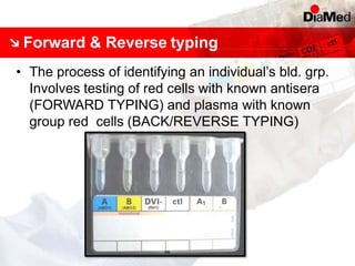
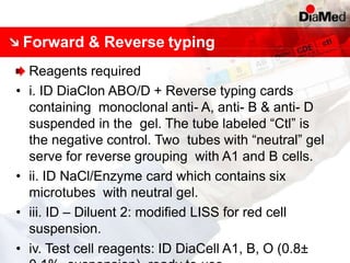
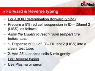
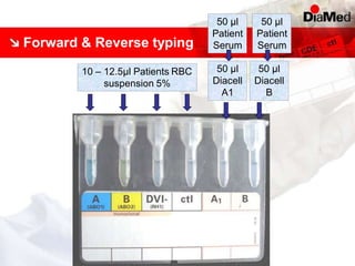
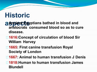
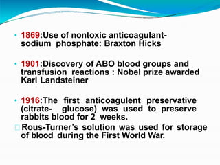
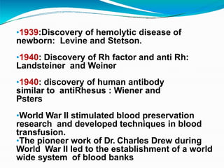
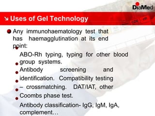
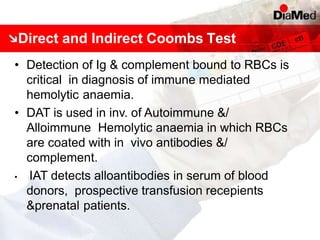
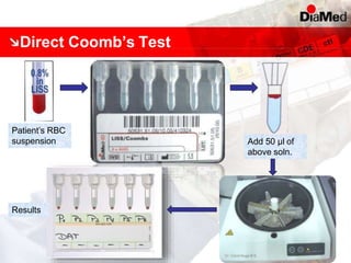
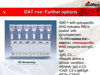
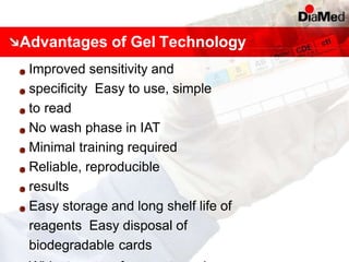
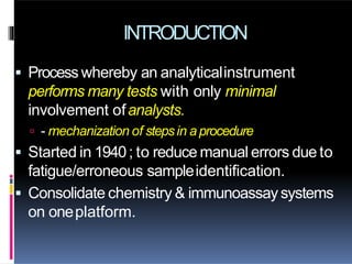
This document discusses gel technology used in blood banking and immunohaematology. It provides historical aspects of blood banking, basics of immunohaematology, principles of gel technology, grading of reactions, applications including blood grouping, antibody screening and identification, cross matching, and direct and indirect antiglobulin tests. It discusses the advantages of gel technology including improved sensitivity and specificity, ease of use, reliable and reproducible results. Literature on using gel technology for compatibility testing, diagnosis of autoimmune hemolytic anemia, and its increased sensitivity over conventional tube technique is also reviewed.
1) Over 60-80% of important medical decisions are based on laboratory test results, yet errors can occur throughout the testing process, from test ordering to result reporting.
2) The majority (90%) of errors occur in the pre-analytical phase involving test ordering, specimen collection, and handling. Common errors include mislabeling specimens, using improper collection methods, and failing to fast patients.
3) Both pre-analytical and post-analytical errors can significantly impact patient care through delays, unnecessary repeat testing, and potential harm if clinical decisions are made on inaccurate results. Reducing errors requires improved education, standardized procedures,
The document discusses the history and development of anticoagulants and blood preservatives used for collecting, storing, and transfusing blood and blood components. It describes the early methods used and key discoveries that expanded the shelf life of red blood cells and platelets. The document also outlines the various anticoagulant solutions, additive solutions, and storage conditions used to maintain viability and prevent clotting during the collection, storage, and transportation of whole blood and its components prior to transfusion.
This document discusses compatibility testing, also known as pre-transfusion testing, which involves procedures to select blood and components that will be safely transfused and will not cause the recipient's red blood cells to be destroyed. The key steps in compatibility testing include properly identifying the recipient's blood sample, checking for antibodies, determining blood types, screening for irregular antibodies, selecting compatible blood, and performing a cross-match test between the donor's red blood cells and the recipient's serum to ensure no reactions occur. The cross-match is the final compatibility test to verify ABO compatibility and detect any antibodies present in the recipient's serum.
Quality in clinical laboratory is a continuous journey of improving processes through team work, innovative solutions, regulatory compliance with final objective to meet the evolving needs of clinicians & patients.
This document provides an overview of antibody identification in blood banking. It discusses the key steps in performing an antibody panel, including using a panel of known red blood cells to test against a patient's unknown serum. The goal is to identify any unexpected antibodies in the patient's serum. It also covers interpreting panel results, such as ruling out non-reactive antigens and looking for a matching antigen pattern. Techniques for identifying multiple antibodies like selected cells, neutralization, and chemical treatments are also outlined.
Pretransfusion testing final- ab screening - NAGLAA MAKRAM Naglaa Makram
1. Antibody screening tests patient serum against reagent red blood cells to detect unexpected antibodies that could destroy transfused donor cells.
2. Screening cells must contain many common antigens and include some cells with homozygous antigen expression to detect weakly reacting antibodies.
3. A positive antibody screen requires antibody identification testing to determine the antibody specificity so that antigen-negative blood can be transfused.
The document discusses quality control and total quality management. It describes key thinkers in quality control like Deming, Juran, Crosby and their contributions. Some of the main points covered include defining quality as conformance to requirements, using statistical process control tools, and adopting the PDCA (plan-do-check-act) cycle. It also discusses the need for quality control in clinical laboratories to ensure accurate results and safe patient care. Quality assurance and quality control procedures help reduce errors and improve performance.
Gel technology provides an innovative approach to performing various tests in immunohaematology with improved sensitivity and specificity compared to conventional tube techniques. It involves centrifuging red blood cells through a gel column where agglutination reactions occur. The distribution of red blood cells throughout the column allows for easy grading of reaction strength. Gel technology is used for blood grouping, antibody screening and identification, compatibility testing, and other immunohaematology applications. It provides standardized, efficient and reliable results compared to conventional methods.
This document provides guidelines for point of care testing (POCT). It defines POCT as any test performed near the patient to enable immediate treatment decisions. The main laboratory is responsible for all POCT and must ensure results match central lab outputs. POCT devices must have internal quality controls, standard operating procedures, and staff training. Quality control practices depend on the technology but manufacturers' claims on precision are important due to challenges running controls. Participation in proficiency testing is required to monitor performance.
Use of laboratory instruments and specimen processing equipment to perform clinical laboratory assays with only minimal involvement of technologist .
Automation in clinical laboratory is a process by which analytical instruments perform many tests with the least involvement of an analyst.
The International Union of Pure and Applied Chemistry (IUPAC) define automation as "The replacement of human manipulative effort and facilities in the performance of a given process by mechanical and instrumental devices that are regulated by feedback of information so that an apparatus is self-monitoring or self adjusting”.
This document provides information about the Coombs test, which is used to detect antibody or complement coating of red blood cells. It describes the history and principles of the test, as well as the direct and indirect Coombs test procedures. The direct Coombs test detects in vivo coating of red blood cells and is used to diagnose conditions like hemolytic disease of the newborn. The indirect Coombs test detects in vitro coating of red cells and is used for compatibility testing and antibody screening. Factors affecting the tests and causes of false positive and negative results are also outlined.
Internal quality control in clinical laboratories hematology(2)NAZAR ABU-DULLA
This document discusses internal quality control in hematology laboratories. It begins with an introduction of the author, Nazar Ahmed Mohamed Abd-Alla, which notes his qualifications and experience in hematology and laboratory administration.
The document then outlines topics that will be covered, including quality definitions, types of errors, specimen handling, method selection, calibration, documentation, and quality programs. It discusses sources of errors like pre-analytical, analytical, and post-analytical errors. It also covers quality assurance, method validation, and specifications for proper specimen collection, transport, and acceptance or rejection. The goal is to provide reliable and accurate test results through effective quality control.
The document discusses the Rhesus blood group system. It describes the major Rh antigens, antibodies, and inheritance patterns. It explains the different nomenclature systems used including Fisher-Race, Wiener, Rosenfield, and ISBT. It discusses weak D phenotypes and antibodies such as anti-G, anti-f, anti-Cw, anti-V, and anti-VS. The Rh blood group is clinically significant in transfusion medicine due to its polymorphic nature and ability to cause hemolytic transfusion reactions or hemolytic disease of the fetus and newborn.
Osmotic fragility & rbc membrane defects 050916Anwar Siddiqui
This document discusses red blood cell membrane defects and osmotic fragility testing. It begins by introducing the structure and components of the red blood cell membrane, including integral proteins, lipids, and peripheral proteins that make up the cytoskeleton. Key membrane defects are then described, such as hereditary spherocytosis caused by weakened interactions between membrane proteins, and hereditary elliptocytosis caused by defects in spectrin. The document concludes by explaining how the osmotic fragility test measures red blood cell resistance to lysis in saline solutions of varying concentrations to evaluate membrane stability and defects.
LABORATORY INVESTIGATION OF TRANSFUSION REACTION CASESSadd Alias
The document provides information about investigating transfusion reaction cases in the laboratory. It discusses the learning outcomes, defines transfusion reaction, and outlines the role of the laboratory. It describes the initial measures taken before testing and the preliminary tests conducted, including clerical checks, visual checks, and serology checks. If these tests produce suspicious results, additional tests are done, such as redoing ABO and Rh grouping, antibody screening, and repeating compatibility testing. The document also discusses tests used to investigate specific transfusion reactions like TRALI, TACO, and acute hemolytic transfusion reaction.
What is quality?
Importance of a quality management system in the laboratory
Quality system essential elements
The history of development of quality principles
Discuss relationship of this quality model to ISO and CLSI standards
This document provides information about blood group antigens and the ABO blood group system. It discusses the key discoveries in blood grouping including the identification of the A, B, AB, and O blood groups. It describes Landsteiner's laws of blood grouping and the genetics underlying the ABO system. The antigens are sugars attached to red blood cells, with the A, B, and O groups depending on whether the A or B enzyme adds specific sugars to the H antigen. Factors affecting antigen-antibody reactions and the rare Bombay phenotype are also summarized.
Hema II Chapter11_ Automation in Hematologya.pptPravin Amabade
This document provides an overview of automated hematology analyzers. It discusses the principles of common analyzers, calibration procedures, quality control, and daily startup routines. The objectives are to describe hematology analyzers, explain calibration parameters and quality control, and discuss histogram interpretation and troubleshooting. Examples of specific analyzers are provided, such as the Cell-Dyn 1800, ABX Pentra 60 C+, and Sysmex.
1. Blood donation involves voluntarily donating blood or blood components which are then used for transfusions or to create medications. Donations can be of whole blood or specific components.
2. There are several types of donations including voluntary unpaid donations, donations from friends/family of patients, and paid donations. Donors can also donate for their own future planned medical procedures.
3. Prior to donation, potential donors undergo screening including testing for transmissible diseases and a medical history/physical exam to ensure the donation is safe. Donors must wait specified periods between donations depending on what they donate.
The document discusses automation in hematology. It describes how Wallace Coulter invented the first automated cell counter using electrical impedance to count and size cells. Automation provides advantages like speed, accuracy, and reduced labor but also has disadvantages like erroneous results. There are semi-automated and fully automated analyzers that use various principles like electrical impedance, light scatter, fluorescence, and electrical conductivity to measure cell parameters and provide diagnostic information. Modern analyzers can perform complete blood counts and immunophenotyping to aid in diagnosing conditions like leukemia.
1. Preanalytical errors from variables prior to laboratory testing can significantly impact hematological parameters. Patient-related factors like age, gender, medications, and circadian rhythms must be considered.
2. Proper collection, transport, storage, and handling of blood samples is important to avoid issues like hemolysis, clotting, and cell degradation. The volume of samples, mixing, and time delay before analysis can all affect test results.
3. Variables in venipuncture technique, use of anticoagulants, fasting status, blood lipids, glucose levels, and unexpected conditions may also introduce errors. Standardization is needed to reduce preanalytical variability.
This document discusses blood group systems, specifically ABO and Rh blood groups. It provides details on:
- The antigens found on red blood cell membranes that determine blood type
- Landsteiner's discovery of the ABO blood group system in 1900 and the four main blood types (A, B, AB, and O)
- The antigens and antibodies present in each blood type
- Rh blood group system including the Rho(D) antigen and typing only for Rho(D) to determine Rh status
- Techniques for blood typing including tube, slide, microplate, and newer gel/cassette methods
- Interpreting and resolving discrepancies in blood typing results
This document discusses gel card technology used in blood banking tests. It provides details on the history and development of gel cards, how they work, the types of tests they can be used for (including ABO typing, cross-matching, antibody screening), and their advantages over traditional tube-based methods such as improved sensitivity and reproducibility. Gel cards contain a gel matrix in microtubes that allows red blood cells to be separated based on agglutination during centrifugation, providing clear and standardized test results.
This document provides an overview of quality control in clinical biochemistry laboratories. It discusses that quality control aims to ensure test results are correct by minimizing errors. Errors can occur in the pre-analytical, analytical, and post-analytical phases. The pre-analytical phase, involving sample collection and handling, accounts for most errors. Laboratories use internal quality control methods like calibration, controls, and Levey-Jennings charts daily, as well as external quality assurance programs, to monitor performance and identify errors. Maintaining quality control is important for generating accurate, reliable test results.
Quality assurance in the department of clinical biochemistryDipesh Tamrakar
This document discusses quality assurance and control in clinical laboratories. It explains that quality control aims to ensure test results are correct by monitoring performance through tools like internal quality control and external quality assessment. The document outlines the pre-analytical, analytical and post-analytical phases of testing and discusses specific quality control procedures used at each stage like storage of controls, monitoring control data, and troubleshooting out of control errors. Westgard rules for determining if quality control is in or out of control are also explained.
Recent advances in blood banking include the introduction of gel technology for blood grouping and cross matching. This provides more sensitive, specific, and efficient testing compared to traditional methods. Automation has also increased at various levels of blood banking through use of systems like the automated immunohematology testing system ORTHO AUTOVUE INNOVA. Additionally, new technologies like apheresis allow for collection of individual blood components from a single donor rather than whole blood, reducing donor exposure and providing purer products.
Prof. heba raslan high sensitivity testing for paroxysmal nocturnal hemoglo...Hitham Esam
This document provides information on high sensitivity testing for Paroxysmal Nocturnal Hemoglobinuria (PNH) using flow cytometry. It discusses the disease mechanism and indications for testing. Key points include:
- PNH is a rare blood disease caused by a lack of GPI-anchored proteins on the surface of blood cells due to a genetic mutation, making red blood cells vulnerable to complement-mediated lysis.
- International guidelines have been developed for optimizing PNH testing using flow cytometry to detect clones of deficient blood cells through the use of GPI-linked antibodies and analysis of granulocytes, monocytes, and red blood cells.
- Validation methods including titration of reagents
This document is a report submitted by a student, Christian Chibuike Uzuh, on their six month industrial work experience at the Agege Local Government Primary Health Centre in Lagos, Nigeria under the Students’ Industrial Work Experience Scheme (SIWES). The report provides a brief history of the health centre, describes the various laboratory tests and procedures performed during the training including oral glucose tolerance tests, packed cell volume tests, blood grouping, and more. The student's training has helped bridge the gap between theoretical classroom learning and practical skills.
blood banking and components sept 2022.pptxvenugopal65248
This document provides an overview of blood banking and blood components. It discusses blood grouping systems like ABO and Rh, cross-matching processes, blood storage and components that can be transfused including red blood cells, platelets, fresh frozen plasma, and cryoprecipitate. It also covers topics like transfusion reactions, maximum blood order schedules, and risks associated with transfusions. The goal is to learn about blood banking processes and the uses of different blood components.
This document discusses compatibility testing, also known as pre-transfusion testing. It defines pre-transfusion testing as procedures required to select blood and components that will have acceptable survival when transfused and will not cause destruction of the recipient's red cells. The key steps are proper identification of the recipient's sample, ABO and Rh grouping, screening for irregular antibodies, cross-matching, and selecting a compatible donor. Compatibility testing is divided into pre-analytical, serological, and post-analytical phases. The serological phase includes ABO/Rh grouping, antibody screening and identification, and cross-matching to check for compatibility between the donor's cells and the recipient's serum.
Technological Advancements in transfusion medicinenehaSingh1543
Advances in transfusion medicine include non-invasive hemoglobin screening, additive solutions that extend blood storage, automated component separation, frozen red blood cell storage, and improved testing technologies. Precise molecular testing and nucleic acid testing have shortened window periods and improved detection of transfusion-transmitted pathogens. Automation has increased efficiency in processes like component separation and red blood cell washing. These technological advances aim to improve blood safety, availability, and management for patients.
The document discusses the Coombs test, which involves using Coombs serum (anti-human globulin) to detect sensitization of red blood cells. It describes the principles and procedures for both the direct and indirect Coombs test. The direct Coombs test detects antibodies or complement attached to red blood cells in vivo, while the indirect Coombs test detects circulating antibodies in the blood capable of attaching to red blood cells. The document provides detailed steps for performing both tests, and discusses interpretations and potential sources of false positive results.
Current Component Therapy by Diane Eklund, MDbloodbankhawaii
Lorem ipsum dolor sit amet, voluptaria percipitur has eu. Nibh iriure nostrud ei mea. Vel dicta voluptua convenire ei, id pro libris viderer. Pri et legendos atomorum, vel eu noster probatus menandri. Omnes possim ut eam, sed ea labore maiorum.
This document discusses automation in blood banking and two main automated methodologies: gel technology and solid phase technology. Gel technology uses a gel matrix in microtubes to trap agglutinated red blood cells during centrifugation. Solid phase technology coats antigens on microplate wells and detects antibodies. Automation provides standardized testing, reduces errors, and handles increasing workload. Both methods improve blood transfusion safety over manual methods.
Blood products include red blood cells, plasma, platelets, cryoprecipitate, and whole blood. Red cells are used to treat anemia and increase hemoglobin. Plasma is used for coagulopathy or low coagulation factors. Platelets are used for thrombocytopenia or platelet dysfunction with bleeding. Cryoprecipitate contains factors VIII, VWF, XIII, and fibrinogen for bleeding disorders. Whole blood transfusions are rarely used today. Compatibility testing and crossmatching are done to prevent hemolytic transfusion reactions. Complications include immune reactions, infections, and issues from large volume transfusions like coagulopathy, hypothermia, and electrolyte imbalances.
The document discusses the Coombs test, which involves using Coombs serum (anti-human globulin) to detect sensitization of red blood cells. It describes the principles and procedures for both the direct and indirect Coombs test. The direct Coombs test detects antibodies or complement attached to red blood cells in vivo, while the indirect test detects circulating antibodies in the blood. The procedures involve washing red blood cells, adding serum or plasma containing antibodies, and then adding Coombs serum to cause agglutination if sensitization is present. The tests are used to detect hemolytic disease of the newborn, transfusion reactions, drug sensitization, and autoimmune hemolytic anemia.
This document provides information on the preparation and separation of plasma and serum from whole blood. It explains that plasma can be separated from whole blood by transferring blood to a tube containing anticoagulant and centrifuging, while serum is separated from clotted whole blood. The document discusses proper storage methods for plasma and serum, such as refrigeration or freezing. It also outlines some common errors in plasma/serum preparation like hemolysis and provides tips to prevent hemolysis.
Classification of Thrombocyte Disorders Using Platelet Aggregation AssaysChristine Joyce Javier
IF YOU ARE GOING TO DOWNLOAD THIS FILE, PLEASE NOTIFY me by sending a message via Facebook.
It's a pleasure to help you through my presentation. Thank you so much!
NOTE: This presentation is in PDF format. If you want to have a copy of this file in PPT format, kindly message me thru Facebook also.
Use of flow cytometry in non neoplastic hematologic conditionsMuneerah Saeed
Flow cytometry is useful for evaluating several non-malignant hematologic disorders:
1. It can quantify reticulocytes, detect hemoglobin F, and diagnose paroxysmal nocturnal hemoglobinuria (PNH).
2. Platelet disorders like Bernard-Soulier syndrome and Glanzmann thrombasthenia can be diagnosed. Anti-platelet antibodies can also be detected.
3. Immunophenotyping of white blood cells allows identification of immunodeficiencies and lymphocyte subsets in conditions like HIV. Stem cells can be enumerated for transplantation.
This document provides guidelines for blood transfusion, including:
1. It discusses the selection and preparation of blood products such as whole blood, platelet concentrates, fresh frozen plasma, and packed red blood cells. Proper donor requirements, collection procedures, and storage conditions are outlined.
2. Indications, dosing, and expected responses to transfusions of various blood components are covered. Red blood cell and platelet transfusion thresholds and dosing are provided.
3. Safety procedures for blood typing, cross-matching, and transfusion monitoring are described. Special considerations for patients with conditions like autoimmune hemolytic anemia that could cause transfusion reactions are highlighted.
4. Two case illustrations demonstrate the
It contains indications of blood and blood products and perioperative blood therapy that we usually follow in Aiims Patna ..its is most recent one made in April 2020
Similar to Column agglutination gel techniques.pptx (20)
This particular slides consist of- what is Pneumothorax,what are it's causes and it's effect on body, risk factors, symptoms,complications, diagnosis and role of physiotherapy in it.
This slide is very helpful for physiotherapy students and also for other medical and healthcare students.
Here is a summary of Pneumothorax:
Pneumothorax, also known as a collapsed lung, is a condition that occurs when air leaks into the space between the lung and chest wall. This air buildup puts pressure on the lung, preventing it from expanding fully when you breathe. A pneumothorax can cause a complete or partial collapse of the lung.
Healthy Eating Habits:
Understanding Nutrition Labels: Teaches how to read and interpret food labels, focusing on serving sizes, calorie intake, and nutrients to limit or include.
Tips for Healthy Eating: Offers practical advice such as incorporating a variety of foods, practicing moderation, staying hydrated, and eating mindfully.
Benefits of Regular Exercise:
Physical Benefits: Discusses how exercise aids in weight management, muscle and bone health, cardiovascular health, and flexibility.
Mental Benefits: Explains the psychological advantages, including stress reduction, improved mood, and better sleep.
Tips for Staying Active:
Encourages consistency, variety in exercises, setting realistic goals, and finding enjoyable activities to maintain motivation.
Maintaining a Balanced Lifestyle:
Integrating Nutrition and Exercise: Suggests meal planning and incorporating physical activity into daily routines.
Monitoring Progress: Recommends tracking food intake and exercise, regular health check-ups, and provides tips for achieving balance, such as getting sufficient sleep, managing stress, and staying socially active.
We are one of the top Massage Spa Ajman Our highly skilled, experienced, and certified massage therapists from different corners of the world are committed to serving you with a soothing and relaxing experience. Luxuriate yourself at our spas in Sharjah and Ajman, which are indeed enriched with an ambiance of relaxation and tranquility. We could confidently claim that we are one of the most affordable Spa Ajman and Sharjah as well, where you can book the massage session of your choice for just 99 AED at any time as we are open 24 hours a day, 7 days a week.
Visit : https://massagespaajman.com/
Call : 052 987 1315
TEST BANK FOR Health Assessment in Nursing 7th Edition by Weber Chapters 1 - ...rightmanforbloodline
TEST BANK FOR Health Assessment in Nursing 7th Edition by Weber Chapters 1 - 34.
Let's Talk About It: Breast Cancer (What is Mindset and Does it Really Matter?)bkling
Your mindset is the way you make sense of the world around you. This lens influences the way you think, the way you feel, and how you might behave in certain situations. Let's talk about mindset myths that can get us into trouble and ways to cultivate a mindset to support your cancer survivorship in authentic ways. Let’s Talk About It!
This particular slides consist of- what is hypotension,what are it's causes and it's effect on body, risk factors, symptoms,complications, diagnosis and role of physiotherapy in it.
This slide is very helpful for physiotherapy students and also for other medical and healthcare students.
Here is the summary of hypotension:
Hypotension, or low blood pressure, is when the pressure of blood circulating in the body is lower than normal or expected. It's only a problem if it negatively impacts the body and causes symptoms. Normal blood pressure is usually between 90/60 mmHg and 120/80 mmHg, but pressures below 90/60 are generally considered hypotensive.
Michigan HealthTech Market Map 2024. Includes 7 categories: Policy Makers, Academic Innovation Centers, Digital Health Providers, Healthcare Providers, Payers / Insurance, Device Companies, Life Science Companies, Innovation Accelerators. Developed by the Michigan-Israel Business Accelerator
Joker Wigs has been a one-stop-shop for hair products for over 26 years. We provide high-quality hair wigs, hair extensions, hair toppers, hair patch, and more for both men and women.
Exploring the Benefits of Binaural Hearing: Why Two Hearing Aids Are Better T...Ear Solutions (ESPL)
Binaural hearing using two hearing aids instead of one offers numerous advantages, including improved sound localization, enhanced sound quality, better speech understanding in noise, reduced listening effort, and greater overall satisfaction. By leveraging the brain’s natural ability to process sound from both ears, binaural hearing aids provide a more balanced, clear, and comfortable hearing experience. If you or a loved one is considering hearing aids, consult with a hearing care professional at Ear Solutions hearing aid clinic in Mumbai to explore the benefits of binaural hearing and determine the best solution for your hearing needs. Embracing binaural hearing can lead to a richer, more engaging auditory experience and significantly improve your quality of life.
At Apollo Hospital, Lucknow, U.P., we provide specialized care for children experiencing dehydration and other symptoms. We also offer NICU & PICU Ambulance Facility Services. Consult our expert today for the best pediatric emergency care.
For More Details:
Map: https://cutt.ly/BwCeflYo
Name: Apollo Hospital
Address: Singar Nagar, LDA Colony, Lucknow, Uttar Pradesh 226012
Phone: 08429021957
Opening Hours: 24X7
4. Historic
aspects
Ancient Egyptians bathed in blood and
aristocrats consumed blood so as to cure
disease.
1616:Concept of circulation of blood Sir
William Harvey
1665: First canine transfusion Royal
Society of London
1667: Animal to human transfusion J Denis
1818:Human to human transfusion James
Blundell
5. • 1869:Use of nontoxic anticoagulant-
sodium phosphate: Braxton Hicks
• 1901:Discovery of ABO blood groups and
transfusion reactions : Nobel prize awarded
Karl Landsteiner
• 1916:The first anticoagulent preservative
(citrate- glucose) was used to preserve
rabbits blood for 2 weeks.
Rous-Turner’s solution was used for storage
of blood during the First World War.
6. •1939:Discovery of hemolytic disease of
newborn: Levine and Stetson.
•1940: Discovery of Rh factor and anti Rh:
Landsteiner and Weiner
•1940: discovery of human antibody
similar to antiRhesus : Wiener and
Psters
•World War II stimulated blood preservation
research and developed techniques in blood
transfusion.
•The pioneer work of Dr. Charles Drew during
World War II led to the establishment of a world
wide system of blood banks
7. •1950: Freeze drying of plasma New
concept of component therapy
•1959: First attempt of Bone Marrow
Transplant in France
•1978: CPDA-1 was developed, ↑ shelf
life of RBCs for 35 days
•1988: First umbilical cords blood
transplant
•1999: NAT for HIV and HCV
14. Effects of NormalSaline
The RBCs are surrounded by an electron cloud
Require longer incubation time for cells suspended in saline
medium.
Weak & incomplete antibodies get repelled and hence are
unable to cause agglutination
17. Gel Technology
• Innovative approach to red cell serology.
• Invented by Dr.Yves Lapierre of France in 1988.
• Developed to minimize problems associated
with conventional techniques of blood
grouping.
• Addresses the issues of standardization and
documentation with unmatched sensitivity,
specificity and efficiency.
• DiaMed-ID Microtyping system from DiaMed AG,
Switzerland in assocition with Dr. Lapierre, is
based on the Gel Tecnology.
18. Principle of Gel Technology
• Controlled centrifugation of RBCs through a gel
column
• Serum and cell reaction takes place in a microtube
.
• Six microtubes in a plastic card – easy handling.
• Microtube consists of a reaction chamber that
narrows to become a column with a conical
bottom.
• Reaction chamber is designed to allow prior
incubation of test serum and RBCs.
• Each column contains Sephacryl gel
20. Principle of Gel Technology
• Sephadex gel matrix acts as a sieve.
• Large aggutinates remain on or near the top of gel
interface.
• Smaller agglutinates pass partway through gel ,
depending on size.
• Unagglutinated cells pass to base of microtube to form a
button.
• Cells are always added prior to serum so that
serum does not come into contact with gel –
this eliminates the ‘WASH PHASE’ as in
conventional technique.
• Grading of reaction depending on the
22. INTERPRETATION OF
GEL TEST
4+
Solid band
of red cells
at top of
gel
3+
Agglutinated
red cells in
upper half
2+
Red cell
agglutinates
through
length
1+
Aggl. red
cell in
lower half
of gel col.
NEGA
TIVE
24. Uses of Gel Technology
Any immunohaematology test that
has haemagglutination at its end
point:
ABO-Rh typing, typing for other blood
group systems.
Antibody screening and
identification. Compatibility testing
– crossmatching. DAT/IAT, other
Coombs phase test.
Antibody classification- IgG, IgM, IgA,
complement…
25. Forward & Reverse typing
• The process of identifying an individual’s bld. grp.
Involves testing of red cells with known antisera
(FORWARD TYPING) and plasma with known
group red cells (BACK/REVERSE TYPING)
26. Forward & Reverse typing
Reagents required
• i. ID DiaClon ABO/D + Reverse typing cards
containing monoclonal anti- A, anti- B & anti- D
suspended in the gel. The tube labeled “Ctl” is
the negative control. Two tubes with “neutral” gel
serve for reverse grouping with A1 and B cells.
• ii. ID NaCl/Enzyme card which contains six
microtubes with neutral gel.
• iii. ID – Diluent 2: modified LISS for red cell
suspension.
• iv. Test cell reagents: ID DiaCell A1, B, O (0.8±
27. Forward & Reverse typing
• For ABO/D determination (forward typing)
• Prepare a 5% red cell suspension in ID – Diluent 2
(LISS) as follows:
• Allow the Diluent to reach room temperature
before use.
• 1. Dispense 500μl of ID – Diluent 2 (LISS) into a
clean test tube.
• 2. Add 25μL packed cells & mix gently.
• For Reverse typing
• Use Plasma or serum
31. • Determination of
ABO/Rh- antigen and
DAT.
• DAT on newborn blood
samples has become a
standard procedure,
since it is important to
know if the newborn’s
red cells have been
sensitised with
maternal antibodies in
utero.
ID-Card “ABO/Rh for newborns”
32. Cross Matching
Reagents required
• i. ID LISS/Coombs cards with six microtubes
containing polyspecific AHG (Anti- IgG+ C3d).
• ii. ID – Diluent 2: modified LISS for red cell
suspension.
Prepare a 0.8 -1.0% red
cell suspension in ID–
Diluent 2 (LISS) as
follows:
i.Dispense 1 mL of ID –
Diluent 2 into a clean
test tube.
ii.Add 10μL of packed
36. Direct and Indirect Coombs Test
• Detection of Ig & complement bound to RBCs is
critical in diagnosis of immune mediated
hemolytic anaemia.
• DAT is used in inv. of Autoimmune &/
Alloimmune Hemolytic anaemia in which RBCs
are coated with in vivo antibodies &/
complement.
• IAT detects alloantibodies in serum of blood
donors, prospective transfusion recepients
&prenatal patients.
38. DAT +ve: Further options
DC-Screening I
•DAT + with polyspecific
AHG indicates RBCs
coated with
Ig/complement.
•To differentiate the
reaction, monospecific
AHG reagents-anti IgG,-
IgA,
-IgM, C3….
•Results define a
clinical condition :
•WAIHA- IgG ± C3
•CAD- C3 ± IgM/IgA
39. DAT +ve: Further options….and more
DAT IgG1/IgG3
•Risk of hemolysis by
AIHA
/HDN depends on
amount of IgG &/
complement coated on
RBCs as well as on IgG
subclasses involved.
•IgG1 & IgG3
activate
complement.
•In DAT IgG1/IgG3 card
2 dilutions of both anti-
& anti-IgG3 are added to
gel to differentiate betwn
low &high risk for
40. DAT +ve: Further options….and more
DAT IgG-Dilution
•DAT IgG – Dilution card
prvides an indication of
clinical importance of DAT
positive results.
•The number of IgG
molecules per cell
influence the red cell
destruction seen in
AIHA,HDN and
Transfusion reactions.
41. Indirect Coomb’s Test
O cell
suspension Add 50 µl of
above soln.
Results
Add 25µl of
patient serum
Centrifuge Incubate
42. Review of Literature
• Kaur et al at Christian Medical College and
Hospital presented their experience with gel
based DiaMed ID microtyping system for
incompatibility testing over a year period and
noticed a startling 65 fold rise in the reported
no. of incompatibe units in 1 yr. which rose from
4 (0.02%) to 260 (1.6%) thus making
gel technology more sensitive than
CTT for crossmatching.
43. Review of Literature
• Role of Gel based technique for Coomb’s test
• M Jaiprakash, PK Gupta Harsh Kumar
• Dept. of Transfusion Medicine, AFMC, Pune
• AIM: Compare CTT & GT for Coomb’s test & to
evaluate their sensitivity & specificity.
• Result
s:
POSITIVITY CTT GTT
GT Sensitivity Specificity NPV
DATDAT 100% 6.1%97.3% 8
1
.
6
0
%
0
%
IAT IAT 100% 5.4%97.6% 6
1
.
6
0
%
0
%
• Conclusion: Gel Technology is a better
alternative to CTT.
44. Review of Literature
• Gel card in diagnosis of autoimmune
haemolytic anaemia.
• Renu Saxena et al at AIIMS, New Delhi.
• Aim: To compare the efficacy of Gel card
Coomb’s test with conventional Coomb’s test in
diagnosis of AIHA.
• Results: 13 out of 50 cases showed positive ICT
& DCT by both CTT & GT. Out of 13 cases 3
showed (+)DCT & (-)ICT by CTT but GT showed
(+)DCT & (+)ICT in all 3 cases.
• Conclusion: Simple, reliable and quick method
for detection of autoantibodies, antibodies
47. Particle Gel ImmunoAssay (PAGIA)
• Gel Technology now adopted with use of inert
polymer particles for detection of:
• Syphilis
• Paroxysmal Nocturnal Haemoglobinuria
• Leishmaniasis
• Sickle Hb Screening
48. Paroxysmal Nocturnal Hemoglobinuria
• Patients with PNH have a defective gene called
PIG-A, involved in the biosynthesis of glycosyl-
phosphatidylinositol (GPI).
• Without GPI, important regulatory proteins (e.g.
CD55 or “DAF”, and CD59 or “MIRL”) cannot
bind to the cell surface and protect blood cells
from attacks of complement.
• This may result in a break down of erythrocytes
and release of hemoglobin which causes the
urine to turn dark during an episode (or
“paroxysm”) of hemolysis, though this is not
found in all cases.
49. Paroxysmal Nocturnal Hemoglobinuria
Reagent1:
ID-PNH-Gel card with rabbit antiserum against
mouse immunoglobulines.
Reagent 2:
Monoclonal antibodies directed against DAF and
MIRL respectively, and negative control, ready-
to-use, 1.4 mL.
Additionally required: Diluent 2.
50. ID-PNH-Gel card with rabbit
antiserum against mouse
immunoglobulines.
Monoclonal
antibodies
directed against
DAF and MIRL
respectively, and
negative control,
ready-to- use,
1.4 mL.
51. Principle of PNH Gel Technology
• Monoclonal anti-DAF and anti-MIRL are incubated
with RBC suspension of suspected PNH cases
and then centrifuged through a microtube
containing rabbit antimouse antibody suspended
in a gel column.
• After centrifugation through the gel, cells
carrying antibodies, confirming the presence of
MIRL or DAF, will show a positive reaction.
This denotes that the patient does not have
PNH.
• Negative results or double population confirm the
presence of PNH.
56. Advantages of Gel Technology
Improved sensitivity and
specificity Easy to use, simple
to read
No wash phase in IAT
Minimal training required
Reliable, reproducible
results
Easy storage and long shelf life of
reagents Easy disposal of
biodegradable cards
57. Disadvantages of Gel Technology
Special centrifuge to accommodate the
microtubes cards.
Special incubators to incubate the microtube
cards. Pipette to dispose 25ul of serum.
Expensive.
58.
59.
60. INTRODUCTION
Process whereby an analyticalinstrument
performs many tests with only minimal
involvement ofanalysts.
- mechanization of stepsin aprocedure
Started in 1940; to reduce manual errors due to
fatigue/erroneous sampleidentification.
Consolidate chemistry & immunoassaysystems
on oneplatform.
62. Aut om
at ed i dent i f i cat i on m
et hods
Barcodes
A barcode comprises a series of vertical bars and
spaces arranged in various combinations to
represent different characters.
There are different barcode systems, each with
different rules governing the representation of the
characters. eg. CODABAR system with ABC
symbols
By combining the numbers, letters and other
characters, a series of barcodes can be built up to
represent donation numbers, blood groups and
various blood products.
In each instance an eye readable number or
description is included with the machine- readable
code.
Device which will interpret barcodes pass a beam of
light across the code making use of 2 levels of
63.
64.
65. Bl ood bank i nf or m
at i on syst em
s
Blood bank information systems are computer
systems that have been developed specifically to
assist the blood bank professionals in management
of the patients, donor and blood component
information.
Helps to assesstrends and decide future policies by
accessing the statisticalinformation.
Helps to correlate the laboratory data with donor
records and help to trace the donor records
following transfusionreactions.
66. BBISconsists of :
Hardware
Software: application software,operating
software and interfacesoftware
Users
FDA,AABBandCPAhave specific regulatory and
accredation requirments for BBIS
67.
68. Stages in Laboratory Investigations
Pre –analyticalstage
Analytical stage
Post –analyticalstage
69. PRE – ANALYTI CAL
STAG
ES IN LAB.
Sample Delivery-
Blood drawers or runners / courier facility
Pneumatic tube deliverysystem
Conveyers or tracksystem
Mobile robots
Sample processing –Threephases
Pre-centrifugation :- all measurements in <45min.
Centrifugation :- blood (Plasma/Serum),Urine &
Other body fluids (conc.particulate matter)
Post-centrifugation
75. ANALYTI CAL STAG
E
Tasksincluded –
Sample introduction & transport to
cuvet or dilutioncap.
Addition ofreagent
Mixing of sample& reagent
Incubation
Detection
Calculation
Readout & result reporting
76. PO
ST – AN
ALYTI C
AL STAG
E
Data processingi.e. –data acquisition, calculation,
Monitoring & displaying –charts & curves,
Performing statistics on patient & control value –
flags.
Analyzer computers have capacity to link to –
lab. Info.System
internet –companiesown manufacturing site –can
seereal time lab. data & can solve mechanical
problem in short duration.
77. AUTO
M
ATED SYSTEM D
ESI G
N
S
Total Laboratory Automation -
Japan,1980
Integration of several instruments =processing
specimen management +transportationsystems
+analyzers +digital interpretation +dispatch of
results.
creates inclusive, continuous networkmaking
each step in testingautomated.
78. AUTO
M
ATED SYSTEM D
ESI G
N
S
Chemical, hematological, coagulation & immuno -
histochemical studies at the same time on asample.
Advantage –reduction in –labeling errors,
turnaround times.
79. AUTO
M
ATED SYSTEM D
ESI G
N
S
Disadvantages –significant financialinvestment
- Increased floor space.
- highly technicalpersonnel
- Infrastructure remodeling,
- personnel teambuilding,
- Software interfacing.
80. Automated blood grouping
Groupometric:
Matte in 1963
It is an automated blood grouping machine
using multi-channel cuvettes for the reaction
mixtures
Electro-mechanical units: for agglutination
reactions
Electronic unit: for processing the results
of agglutination tests and print out of the
group.
Autoanalyzer grouping machine with
continuous flow system and laser scanner.
81. The ORTHO AUTOVUE INNOVA System is an automated
imunohematology testing system used for blood typing,
antibody screening and compatibility testing using
ORTHOBIOVUE System cassettes. The ORTHO
AUTOVUE INNOVA System is a computersoftware driven,
fully automated system which provides automated liquid
pipetting,cassette handling, incubation, centrifugation,
reaction grading and interpretation.
The software used with the AUTOVUE INNOVA can be
interfaced with a laboratory information
system for test data transfer.
82. APHRESIS
Apheresis is collection of anti-coagulated whole
blood from adonor, its separation into
components, retention of desired component
and return of remaining constituents backto the
donor with the help of automated cell separator
machines.
83. ADVANTAGES OF APHRESIS
Reducedmultiple donorexposure
Reduced risk ofalloimmunization
Reduced incidence oftransfusion transmitted
diseases
Full and effective transfusiondose
Purer product:
leucocyte reducedproducts
High qualityproduct
Fewerdonor reaction due to return of fluid
85. Automated separation techniqueby
centrifugation:
Centrifugal force separates blood into
different component depending upon the
specific gravity.
Blood is drawn from an a
u
t
o
m
a
t
i
cpump
Anticoagulant isadded tube blood is
pumped into roatating bowl,chamber in
which layering of components occursbased
on the density desired component
retained and rest returned to donor either
by continous flow or by intermittent flow.
86. Separation by MembraneFiltration:
Filtration of plasma through membrane which
allows collection of plasma from ahealthy
donor.
Membranes are arranged ashollow fibres
which expelsthe cellular elements in the flow
of blood.
Most commonlyused apheresis devices are:
Haemonetic corporation: Platelets,plasma,
leucocytes.
Baxter: Plasma, platelets, redcells,
leucocyte
Gambro: Plasma, platelets,leucocyte and
peripheral blood stemcells.
87. TECHNOLOGY FUCTIONN
HAEMONETIC Intermittent flow centrifuge separator.
Anticoagulant blood is pumped into rotating bowl
Incoming blood isseparated , red cells move to the
periphery and plasma to inside of rotating bowl and the white
cellsand plasma between red cellsand plasma
Using optical detectors and fluid surge elutriationprocess
desired component retained.
GAMBRO(Cobe) Continuous flow centrifuge cell separator where two arm
blood is drawn andreturned.
Here flat membrane is used to separate the cellsof blood from
plasma.
Allows lower WBCand RBCcontamination in platelets.
BAXTER Continuous flow technology.
CS3000 has two separation containers firstly for collection of
leucocyte reduced platelets and other for white cells (CS3000
plus).