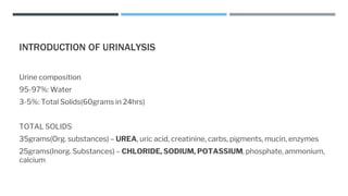
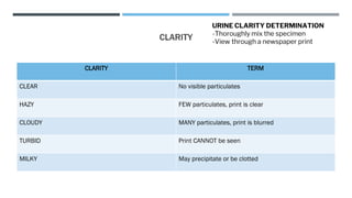
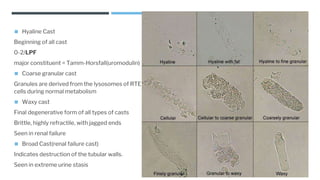
CLINICAL
MICROSCOPY
ANALYSIS OF URINE AND OTHER BODY FLUIDS
◼ URINE
◼ FECES
◼ CSF
◼ SYNOVIAL FLUID
◼ SEROUS FLUID
◼ AMNIOTIC FLUID
◼ SEMEN
INTRODUCTION OF URINALYSIS
Urine composition
95-97%: Water
3-5%: Total Solids(60grams in 24hrs)
TOTAL SOLIDS
35grams(Org. substances) – UREA, uric acid, creatinine, carbs, pigments, mucin, enzymes
25grams(Inorg. Substances) – CHLORIDE, SODIUM, POTASSIUM, phosphate, ammonium,
calcium
TYPES OF URINE SPECIMEN
RANDOM For routine and qualitative UA
FIRST MORNING Ideal specimen for routine UA.
Most concentrated, most acidic, well preservation of
cells and casts
MIDSTREAM CLEAN CATCH Routine screening and bacterial culture
CATHETERIZED Bacterial culture
SUPRAPUBIC ASPIRATION ANAEROBIC bacterial culture and urine cytology
SAMPLE CONTAINER
◼ Containers must be clean, dry, leak-proof.
◼ 10-15mL is required for routine urinalysis
◼ Tested within 2 hours of collection.
◼ Physical, chemical and microscopic
characteristics of a urine specimen begins to
change as soon as the urine is voided.
CHANGES IN AN UNPRESERVED URINE
INCREASED DARKENED/MODIFIED DECREASED LEAST AFFECTED
pH Color Clarity Protein
Bacteria Glucose
Odor RBC/WBC/Casts
Nitrite Trichomonas
PHYSICAL EXAMATION
◼ VOLUME
◼ COLOR
◼ CLARITY
◼ SPECIFIC GRAVITY
URINE VOLUME
◼ Normal urine output/24hrs = 600-2000mL
◼ Average Urine output/24hrs = 1200-1500mL
◼ Polyuria = 2000mL/24hrs(Henry’s), 2500mL/24hrs(Strasinger)
◼ Oliguria = <500mL(Henry’s), <400mL(Strasinger)
◼ Anuria = <100mL
URINE COLOR
◼ PIGMENTS:
1. UROCHROME(yellow) – major pigment
2. Uroerythrin(Pink)
3. Urobilin(Dark yellow/orange)
COLOR
COLOR CAUSE
COLORLESS/STRAW RECENT FLUID CONSUMPTION
PALE YELLOW POLYURIA/DI DI - Increases urine volume with low
specific gravity.
DARK YELLOW CONCENTRATED SPECIMEN
GREEN PSUEDOMONAS INFECTION Positive urine C/S.
RED/PINK/BROWN RBCs/Hemoglobin Beets
ORANGE BILIRUBIN Yellow foam after shaking the
specimen.
BLACK MELANIN
URINE COLOR DETERMINATION
-Look down through the container
against a white background.
CLARITY
CLARITY TERM
CLEAR No visible particulates
HAZY FEW particulates, print is clear
CLOUDY MANY particulates, print is blurred
TURBID Print CANNOT be seen
MILKY May precipitate or be clotted
URINE CLARITY DETERMINATION
-Thoroughly mix the specimen
-View through a newspaper print
SPECIFIC GRAVITY
◼ Refractometry – determines the dissolved particles in a specimen by measuring refractive index.
Refractive index is a comparison of the velocity of light in air with the velocity if light in a solution.
◼ Reagent strip - pka changes of a polyelectrolyte by ions present
◼ Osmolality – changes in colligative properties by particle number
PROPER HANDLING AND STORAGE OF REAGENT STRIPS
◼ Store with desiccant in an opaque, tightly closed
container.
◼ Store below 30C(room temperature); do not freeze.
◼ Don’t expose to volatile fumes
◼ Don’t use past expiration date
◼ Don’t use is chemical pads become discolored
◼ Remove strips immediately prior to use.
CHEMICAL EXAMINATION
◼ Glucose
◼ Bilirubin
◼ Ketones
◼ Specific Gravity
◼ pH
◼ Protein
◼ Blood
◼ Urobilinogen
◼ Nitrite
◼ Leukocytes
Reagent strip technique:
1. Dip the strip briefly into a WELL-MIXED
UNCENTRIFUGED sample.
1. Remove excess urine by touching the edge of
the strip to the container as the strip is
withdrawn.
1. Blot the edges on the strip on a absorbent
pad.
1. Wait for the specific reaction time to occur.
1. Compare the color reaction.
READING TIME PARAMETER PRINCIPLE POSITIVE COLOR
30secs
Glucose Double sequential enzyme rection Brown
Bilirubin Diazo reaction Violet
40secs Ketones Sodium nitroPrusside Purple
45secs S.G. pKa change of a polyelectrolyte Blue
60secs
Protein Protein(Sorensen’s) error of indicator Blue
pH Double indicator system Orange/Blue
Blood Pseudo peroxidase activity of Hgb Uniform
green/blue(Hgb/Mb)
Speckled(RBC)
Urobilinogen Ehrlich’s reaction Red
Nitrite Greiss reaction Uniform pink
120secs Leukocytes Leukocyte esterase Purple
GLUCOSE
◼ PRINCIPLE: Double sequential enzyme reaction
◼ Reagents: Glucose oxidase & Peroxidase
◼ Reaction time: 30secs
◼ Renal Glucose Threshold: 160-180mg/dL
◼ False (+) = Oxidizing agents, detergents
◼ False (-) = Ascorbic acid, improperly preserved specimen, low temp
BILIRUBIN
◼ Principle: Diazo reaction
◼ Reagent: 2,4-diclhloroaniline diazonium salt
◼ Reaction time: 30secs
◼ False (+) = Highly pigmented urine, indicant
◼ False (-) = exposure to light, ascorbic acid
KETONES
◼ Result from increased fat metabolism due to inability to inability to metabolize carbohydrates.
◼ PRINCIPLE: Sodium Nitroprusside Reaction(LEGAL’S TEST)
◼ Reagent: Sodium Nitroprusside
◼ Reaction time: 40secs
◼ KETONE BODIES:
a. 78% Beta-hydroxybutyric acid – major ketone but not detected in reagent pad
b. 20% Acetoacetic acid(AAA)/Diacetic acid – parent ketone that is detected in reagent pads
c. 2% Acetone
SPECIFIC GRAVITY
◼ PRINCIPLE: Change in pKa of polyelectrolytes.
◼ The polyelectrolyte ionizes, releasing H+ ions in proportion to the # of ions in the solution.
◼ Reagent: Poly(Methyl vinyl ether/maleic anhydride) bromthymol blue
◼ Reaction time: 45secs
◼ False (+) = high concentration of protein
◼ False (-) = alkaline urine
pH
◼ PRINCIPLE: Double indicator system
◼ Important in the identification of crystals and determination of unsatisfactory specimens
◼ Random = 4.5-8.0
◼ 1st morning = 5.0-6.0
◼ Unpreserved = 9.0
◼ Reagents: Methyl Red(pH 4.0-6.0) & Bromthymol blue(pH 6.0-9.0)
◼ Reaction time: 60secs
ACID URINE ALKALINE URINE
DM RENAL TUBULAR ACIDOSIS
STARVATION VEGETARIAN DIET
HIGH PROTEIN DIET AFTER MEALS(ALKALINE TIDE)
CRANBERRY JUICE VOMITING
PROTEIN
◼ PRINCIPLE: Protein error of indicators(SORENSEN’S)
◼ Indicator is sensitive to ALBUMIN
◼ Reagent: Tetrabromphenol blue
◼ Reaction time: 60secs
BLOOD
◼ PRINCIPLE: Pseudoperoxidase activity of the hemoglobin
◼ Reagent: Diisopropylbenzene dehydroperoxidase tertramethylbenzidine
◼ Reaction time: 60secs
◼ False (+) = oxidizing agents, menstrual contamination, bacterial peroxidase
◼ False (-) = Crenated rbcs, formalin, Ascorbic acid, unmixed specimen
HEMATURIA HEMOGLOBINURIA MYOGLOBINURIA
Cloudy red urine Clear red urine Clear red(reddish-brown) urine
Seen in: Glomerulonephritis, renal
calculi, tumors, strenuous exercise,
trauma
Seen in: Intravascular hemolysis,
transfusion reactions, hemolytic
anemia, severe burns
Seen in: Rhabdomyolysis, muscular
trauma, CRUSH syndrome
Microscopic exam: Intact RBCs Heme portion of the myoglobin is toxic
to the renal tubules
UROBILINOGEN
◼ PRINCIPLE: Ehrlich reaction
◼ Bile pigment that result from hemoglobin degradation
◼ Reagent: p-dimethylaminobenzaldehyde (PDAB/Ehrlich reagent)
◼ Reaction time: 60secs
◼ False (+) = porphobilinogen, pigmented urine
◼ False (-) = old specimen, formalin
NITRITE
◼ PRINCIPLE: Greiss Reaction
◼ Rapid screening test for urinary tract infection or bacteriuria
◼ Reagent: p-arsanilic acid, tetrahydrobenzo-quinoline-3-ol
◼ Reaction time: 60secs
◼ False (+) = improperly preserved specimen
◼ False (-) = ascorbic acid, large quantities of bacteria converting nitrite to nitrogen
LEUKOCYTE
◼ PRINCIPLE: Leukocyte esterase reaction
◼ Significant in UTI and screening for urine culture specimen.
◼ Reagent: Derivatized pyrrole amino acid ester, Diazonium salt
◼ Reaction time: 120secs
◼ False (+) = oxidizing reagent, formalin
◼ False (-) = Ascorbic acid
◼ Leukocyte with no esterase – Lymphocyte
AUTION
Principle: Reflectance photometry
• Light reflection from the test pads
decreases in proportion to the intensity
of the color produced by the
concentration of the test substance.
• The darker the color of the reagent pad,
the lesser the light reflection(vice versa)
• Reaction time of all chemical parameter
is 60secs.
MICROSCOPIC EXAMINATION
◼ RBC
0-2/HPF
Hypertonic urine – Crenate
Hypotonic urine – Swell
-associated with glomerular membrane
damage
• Yeast
Small refractile oval structures that may or
may not bud.
Can be confused with RBC.
Qualitative(few, many, etc.) report.
◼ WBC
0-5/HPF
Increased number indicated presence of an infection or
inflammation(PYURIA)
a.) Neutrophils
Granulated and multilobed(predominant)
Hypotonic urine – swell and granules undergo Brownian
movement(Glitter cells)
b.) Eosionphils
Stained with HANSEL Stain
Associated with drug induced interstitial nephritis
• Squamous Epithelial Cell
Largest cell w/ abundant, irregular
cytoplasm and prominent nucleus.
Can serve as a good reference for
focusing of the microscope.
Qualitative
• Transitional Epithelial Cells(Urothelial)
spherical, polyhedral or caudate with centrally
located nucleus
Seen in Catheterized patients
Derived from the linings of renal pelvis, calycles,
ureter,
• Renal Tubular Epithelial cell
Most clinically significant epithelial cell
Originally from the nephrons.
Rectangular, polyhedral, cuboidal or columnar
with an eccentric nucleus.
Indicated tubular damage.
◼ Bacteria
Cocci/bacilli
Most common cause of UTI – E. coli
Qualitative report
◼ Trichomonas vaginalis
Most frequent parasite encountered in the
urine
Pear-shaped flagellate with jerky motility
Agent of ping-pong disease
Qualitative report
◼ Schistosoma haematobium
Blood fluke with a terminal spine
Causes hematuria
Associated with bladder cancer
◼ Enterobius vermicularis
most common fecal contaminant
◼ Hyaline Cast
Beginning of all cast
0-2/LPF
major constituent = Tamm-Horsfall(uromodulin)
◼ Coarse granular cast
Granules are derived from the lysosomes of RTE
cells during normal metabolism
◼ Waxy cast
Final degenerative form of all types of casts
Brittle, highly refractile, with jagged ends
Seen in renal failure
◼ Broad Cast(renal failure cast)
Indicates destruction of the tubular walls.
Seen in extreme urine stasis
CRYSTALS
◼ Formed by precipitation of urine solutes(salts, org. compounds, meds)
◼ Factors that contribute to crystal formation:
a. temperature
b. Solute concentration
c.pH
ACIDIC URINE
Crystal Info significance
Amorphous urates -Brink dust/yellow brown granules
-Pink sediments
--
Uric Acid -purine metabolism
-whetstone, lemon-shape
-most pleomorphic
Gout
Lesch-Nyhan syndrome
Calcium Oxalate 1. Dihydrate (Wheddelite)
envelope
2. Monohydrate (Whewellite)
Oval/Dumbell
Ethylene glycol poisoning
(anti-freeze reagent)
Calcium sulfate “Cigarette-butt: appearance --
ALKALINE URINE
Crystal Info Significance
Amorphous phosphate Granular in appearance
White precipitate
--
Ammonium biurate Thorny-apples
indicated old sample
--
Triple phosphate/STRUVITE/
Magnesium ammonium
phosphate
Prism-shape, Coffin-lid
Fern leaf
Calcium Phosphate/Apatite Flat plates, thin prisms --
Calcium carbonate Dumbell
ABNORMAL CRYSTALS
Cystine Hexagonal plates
Mistaken as uric acid crystal
Cholesterol Rectangular plates w/ notch in one or
many corners(staircase pattern)
Radiographic Dye Similar to cholesterol crystals
To differentiate:
-Check patient history
-correlation w/ other UA results
SG=>1.040 using Refrectometer
Bilirubin -Clumped needles or granules with
bright yellow color
-(+) Bilirubin reagent strip
-LIVER DISEASE
Leucine -Yellow-brown spheres with concentric
circles & radial striations
-Precipitated w/ tyrosine after adding
ALCOHOL
-Sofa pillow
-LIVER DISEASE
Tyrosine -Colorless to yellow needles in clumps
or rosettes
-LIVER DISEASE
◼ URINALYSIS RESULT:
Yellow/Hazy
pH - 5.5
SG – 1.015
CHON – Neg
Glucose- Neg
Blood – Neg
RBC- 0-1/hpf
WBC – 0-2/hpf
Ammonium biurate – Few
Mucus Threads- Few
Which result is inconsistent ?
A patient was rush to the Emergency room from a crush injury. The UA result of the patient was:
-Gross examination
Red/Clear
-Chemical examination
pH 6.0
SG – 1.010
Protein – Neg
Blood- 2+
Glucose – Neg
-Microscopic Examination
RBC – 0
Wbc – 0-2/hpf
What is the reason why the Chemical strip and microscopic exam does not correlate ?
THANK YOU!

Clinical microscopy [Autosaved].pptx

  • 1.
  • 2.
    ANALYSIS OF URINEAND OTHER BODY FLUIDS ◼ URINE ◼ FECES ◼ CSF ◼ SYNOVIAL FLUID ◼ SEROUS FLUID ◼ AMNIOTIC FLUID ◼ SEMEN
  • 3.
    INTRODUCTION OF URINALYSIS Urinecomposition 95-97%: Water 3-5%: Total Solids(60grams in 24hrs) TOTAL SOLIDS 35grams(Org. substances) – UREA, uric acid, creatinine, carbs, pigments, mucin, enzymes 25grams(Inorg. Substances) – CHLORIDE, SODIUM, POTASSIUM, phosphate, ammonium, calcium
  • 4.
    TYPES OF URINESPECIMEN RANDOM For routine and qualitative UA FIRST MORNING Ideal specimen for routine UA. Most concentrated, most acidic, well preservation of cells and casts MIDSTREAM CLEAN CATCH Routine screening and bacterial culture CATHETERIZED Bacterial culture SUPRAPUBIC ASPIRATION ANAEROBIC bacterial culture and urine cytology
  • 5.
    SAMPLE CONTAINER ◼ Containersmust be clean, dry, leak-proof. ◼ 10-15mL is required for routine urinalysis ◼ Tested within 2 hours of collection. ◼ Physical, chemical and microscopic characteristics of a urine specimen begins to change as soon as the urine is voided.
  • 6.
    CHANGES IN ANUNPRESERVED URINE INCREASED DARKENED/MODIFIED DECREASED LEAST AFFECTED pH Color Clarity Protein Bacteria Glucose Odor RBC/WBC/Casts Nitrite Trichomonas
  • 7.
    PHYSICAL EXAMATION ◼ VOLUME ◼COLOR ◼ CLARITY ◼ SPECIFIC GRAVITY
  • 8.
    URINE VOLUME ◼ Normalurine output/24hrs = 600-2000mL ◼ Average Urine output/24hrs = 1200-1500mL ◼ Polyuria = 2000mL/24hrs(Henry’s), 2500mL/24hrs(Strasinger) ◼ Oliguria = <500mL(Henry’s), <400mL(Strasinger) ◼ Anuria = <100mL
  • 9.
    URINE COLOR ◼ PIGMENTS: 1.UROCHROME(yellow) – major pigment 2. Uroerythrin(Pink) 3. Urobilin(Dark yellow/orange)
  • 10.
    COLOR COLOR CAUSE COLORLESS/STRAW RECENTFLUID CONSUMPTION PALE YELLOW POLYURIA/DI DI - Increases urine volume with low specific gravity. DARK YELLOW CONCENTRATED SPECIMEN GREEN PSUEDOMONAS INFECTION Positive urine C/S. RED/PINK/BROWN RBCs/Hemoglobin Beets ORANGE BILIRUBIN Yellow foam after shaking the specimen. BLACK MELANIN URINE COLOR DETERMINATION -Look down through the container against a white background.
  • 11.
    CLARITY CLARITY TERM CLEAR Novisible particulates HAZY FEW particulates, print is clear CLOUDY MANY particulates, print is blurred TURBID Print CANNOT be seen MILKY May precipitate or be clotted URINE CLARITY DETERMINATION -Thoroughly mix the specimen -View through a newspaper print
  • 12.
    SPECIFIC GRAVITY ◼ Refractometry– determines the dissolved particles in a specimen by measuring refractive index. Refractive index is a comparison of the velocity of light in air with the velocity if light in a solution. ◼ Reagent strip - pka changes of a polyelectrolyte by ions present ◼ Osmolality – changes in colligative properties by particle number
  • 13.
    PROPER HANDLING ANDSTORAGE OF REAGENT STRIPS ◼ Store with desiccant in an opaque, tightly closed container. ◼ Store below 30C(room temperature); do not freeze. ◼ Don’t expose to volatile fumes ◼ Don’t use past expiration date ◼ Don’t use is chemical pads become discolored ◼ Remove strips immediately prior to use.
  • 14.
    CHEMICAL EXAMINATION ◼ Glucose ◼Bilirubin ◼ Ketones ◼ Specific Gravity ◼ pH ◼ Protein ◼ Blood ◼ Urobilinogen ◼ Nitrite ◼ Leukocytes Reagent strip technique: 1. Dip the strip briefly into a WELL-MIXED UNCENTRIFUGED sample. 1. Remove excess urine by touching the edge of the strip to the container as the strip is withdrawn. 1. Blot the edges on the strip on a absorbent pad. 1. Wait for the specific reaction time to occur. 1. Compare the color reaction.
  • 15.
    READING TIME PARAMETERPRINCIPLE POSITIVE COLOR 30secs Glucose Double sequential enzyme rection Brown Bilirubin Diazo reaction Violet 40secs Ketones Sodium nitroPrusside Purple 45secs S.G. pKa change of a polyelectrolyte Blue 60secs Protein Protein(Sorensen’s) error of indicator Blue pH Double indicator system Orange/Blue Blood Pseudo peroxidase activity of Hgb Uniform green/blue(Hgb/Mb) Speckled(RBC) Urobilinogen Ehrlich’s reaction Red Nitrite Greiss reaction Uniform pink 120secs Leukocytes Leukocyte esterase Purple
  • 16.
    GLUCOSE ◼ PRINCIPLE: Doublesequential enzyme reaction ◼ Reagents: Glucose oxidase & Peroxidase ◼ Reaction time: 30secs ◼ Renal Glucose Threshold: 160-180mg/dL ◼ False (+) = Oxidizing agents, detergents ◼ False (-) = Ascorbic acid, improperly preserved specimen, low temp
  • 17.
    BILIRUBIN ◼ Principle: Diazoreaction ◼ Reagent: 2,4-diclhloroaniline diazonium salt ◼ Reaction time: 30secs ◼ False (+) = Highly pigmented urine, indicant ◼ False (-) = exposure to light, ascorbic acid
  • 18.
    KETONES ◼ Result fromincreased fat metabolism due to inability to inability to metabolize carbohydrates. ◼ PRINCIPLE: Sodium Nitroprusside Reaction(LEGAL’S TEST) ◼ Reagent: Sodium Nitroprusside ◼ Reaction time: 40secs ◼ KETONE BODIES: a. 78% Beta-hydroxybutyric acid – major ketone but not detected in reagent pad b. 20% Acetoacetic acid(AAA)/Diacetic acid – parent ketone that is detected in reagent pads c. 2% Acetone
  • 19.
    SPECIFIC GRAVITY ◼ PRINCIPLE:Change in pKa of polyelectrolytes. ◼ The polyelectrolyte ionizes, releasing H+ ions in proportion to the # of ions in the solution. ◼ Reagent: Poly(Methyl vinyl ether/maleic anhydride) bromthymol blue ◼ Reaction time: 45secs ◼ False (+) = high concentration of protein ◼ False (-) = alkaline urine
  • 20.
    pH ◼ PRINCIPLE: Doubleindicator system ◼ Important in the identification of crystals and determination of unsatisfactory specimens ◼ Random = 4.5-8.0 ◼ 1st morning = 5.0-6.0 ◼ Unpreserved = 9.0 ◼ Reagents: Methyl Red(pH 4.0-6.0) & Bromthymol blue(pH 6.0-9.0) ◼ Reaction time: 60secs ACID URINE ALKALINE URINE DM RENAL TUBULAR ACIDOSIS STARVATION VEGETARIAN DIET HIGH PROTEIN DIET AFTER MEALS(ALKALINE TIDE) CRANBERRY JUICE VOMITING
  • 21.
    PROTEIN ◼ PRINCIPLE: Proteinerror of indicators(SORENSEN’S) ◼ Indicator is sensitive to ALBUMIN ◼ Reagent: Tetrabromphenol blue ◼ Reaction time: 60secs
  • 22.
    BLOOD ◼ PRINCIPLE: Pseudoperoxidaseactivity of the hemoglobin ◼ Reagent: Diisopropylbenzene dehydroperoxidase tertramethylbenzidine ◼ Reaction time: 60secs ◼ False (+) = oxidizing agents, menstrual contamination, bacterial peroxidase ◼ False (-) = Crenated rbcs, formalin, Ascorbic acid, unmixed specimen HEMATURIA HEMOGLOBINURIA MYOGLOBINURIA Cloudy red urine Clear red urine Clear red(reddish-brown) urine Seen in: Glomerulonephritis, renal calculi, tumors, strenuous exercise, trauma Seen in: Intravascular hemolysis, transfusion reactions, hemolytic anemia, severe burns Seen in: Rhabdomyolysis, muscular trauma, CRUSH syndrome Microscopic exam: Intact RBCs Heme portion of the myoglobin is toxic to the renal tubules
  • 23.
    UROBILINOGEN ◼ PRINCIPLE: Ehrlichreaction ◼ Bile pigment that result from hemoglobin degradation ◼ Reagent: p-dimethylaminobenzaldehyde (PDAB/Ehrlich reagent) ◼ Reaction time: 60secs ◼ False (+) = porphobilinogen, pigmented urine ◼ False (-) = old specimen, formalin
  • 24.
    NITRITE ◼ PRINCIPLE: GreissReaction ◼ Rapid screening test for urinary tract infection or bacteriuria ◼ Reagent: p-arsanilic acid, tetrahydrobenzo-quinoline-3-ol ◼ Reaction time: 60secs ◼ False (+) = improperly preserved specimen ◼ False (-) = ascorbic acid, large quantities of bacteria converting nitrite to nitrogen
  • 25.
    LEUKOCYTE ◼ PRINCIPLE: Leukocyteesterase reaction ◼ Significant in UTI and screening for urine culture specimen. ◼ Reagent: Derivatized pyrrole amino acid ester, Diazonium salt ◼ Reaction time: 120secs ◼ False (+) = oxidizing reagent, formalin ◼ False (-) = Ascorbic acid ◼ Leukocyte with no esterase – Lymphocyte
  • 26.
    AUTION Principle: Reflectance photometry •Light reflection from the test pads decreases in proportion to the intensity of the color produced by the concentration of the test substance. • The darker the color of the reagent pad, the lesser the light reflection(vice versa) • Reaction time of all chemical parameter is 60secs.
  • 27.
    MICROSCOPIC EXAMINATION ◼ RBC 0-2/HPF Hypertonicurine – Crenate Hypotonic urine – Swell -associated with glomerular membrane damage • Yeast Small refractile oval structures that may or may not bud. Can be confused with RBC. Qualitative(few, many, etc.) report.
  • 28.
    ◼ WBC 0-5/HPF Increased numberindicated presence of an infection or inflammation(PYURIA) a.) Neutrophils Granulated and multilobed(predominant) Hypotonic urine – swell and granules undergo Brownian movement(Glitter cells) b.) Eosionphils Stained with HANSEL Stain Associated with drug induced interstitial nephritis
  • 29.
    • Squamous EpithelialCell Largest cell w/ abundant, irregular cytoplasm and prominent nucleus. Can serve as a good reference for focusing of the microscope. Qualitative
  • 30.
    • Transitional EpithelialCells(Urothelial) spherical, polyhedral or caudate with centrally located nucleus Seen in Catheterized patients Derived from the linings of renal pelvis, calycles, ureter, • Renal Tubular Epithelial cell Most clinically significant epithelial cell Originally from the nephrons. Rectangular, polyhedral, cuboidal or columnar with an eccentric nucleus. Indicated tubular damage.
  • 31.
    ◼ Bacteria Cocci/bacilli Most commoncause of UTI – E. coli Qualitative report
  • 32.
    ◼ Trichomonas vaginalis Mostfrequent parasite encountered in the urine Pear-shaped flagellate with jerky motility Agent of ping-pong disease Qualitative report
  • 33.
    ◼ Schistosoma haematobium Bloodfluke with a terminal spine Causes hematuria Associated with bladder cancer
  • 34.
    ◼ Enterobius vermicularis mostcommon fecal contaminant
  • 35.
    ◼ Hyaline Cast Beginningof all cast 0-2/LPF major constituent = Tamm-Horsfall(uromodulin) ◼ Coarse granular cast Granules are derived from the lysosomes of RTE cells during normal metabolism ◼ Waxy cast Final degenerative form of all types of casts Brittle, highly refractile, with jagged ends Seen in renal failure ◼ Broad Cast(renal failure cast) Indicates destruction of the tubular walls. Seen in extreme urine stasis
  • 36.
    CRYSTALS ◼ Formed byprecipitation of urine solutes(salts, org. compounds, meds) ◼ Factors that contribute to crystal formation: a. temperature b. Solute concentration c.pH
  • 37.
    ACIDIC URINE Crystal Infosignificance Amorphous urates -Brink dust/yellow brown granules -Pink sediments -- Uric Acid -purine metabolism -whetstone, lemon-shape -most pleomorphic Gout Lesch-Nyhan syndrome Calcium Oxalate 1. Dihydrate (Wheddelite) envelope 2. Monohydrate (Whewellite) Oval/Dumbell Ethylene glycol poisoning (anti-freeze reagent) Calcium sulfate “Cigarette-butt: appearance --
  • 38.
    ALKALINE URINE Crystal InfoSignificance Amorphous phosphate Granular in appearance White precipitate -- Ammonium biurate Thorny-apples indicated old sample -- Triple phosphate/STRUVITE/ Magnesium ammonium phosphate Prism-shape, Coffin-lid Fern leaf Calcium Phosphate/Apatite Flat plates, thin prisms -- Calcium carbonate Dumbell
  • 39.
    ABNORMAL CRYSTALS Cystine Hexagonalplates Mistaken as uric acid crystal Cholesterol Rectangular plates w/ notch in one or many corners(staircase pattern) Radiographic Dye Similar to cholesterol crystals To differentiate: -Check patient history -correlation w/ other UA results SG=>1.040 using Refrectometer
  • 40.
    Bilirubin -Clumped needlesor granules with bright yellow color -(+) Bilirubin reagent strip -LIVER DISEASE Leucine -Yellow-brown spheres with concentric circles & radial striations -Precipitated w/ tyrosine after adding ALCOHOL -Sofa pillow -LIVER DISEASE Tyrosine -Colorless to yellow needles in clumps or rosettes -LIVER DISEASE
  • 41.
    ◼ URINALYSIS RESULT: Yellow/Hazy pH- 5.5 SG – 1.015 CHON – Neg Glucose- Neg Blood – Neg RBC- 0-1/hpf WBC – 0-2/hpf Ammonium biurate – Few Mucus Threads- Few Which result is inconsistent ?
  • 42.
    A patient wasrush to the Emergency room from a crush injury. The UA result of the patient was: -Gross examination Red/Clear -Chemical examination pH 6.0 SG – 1.010 Protein – Neg Blood- 2+ Glucose – Neg -Microscopic Examination RBC – 0 Wbc – 0-2/hpf What is the reason why the Chemical strip and microscopic exam does not correlate ?
  • 43.