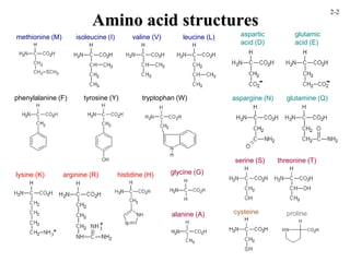
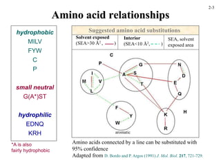
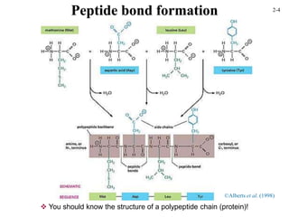
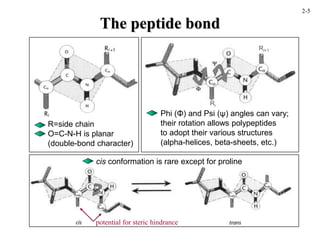
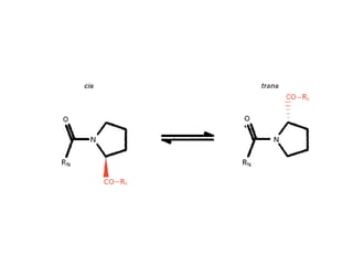
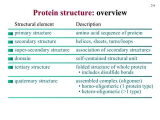
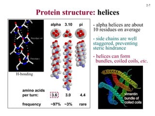
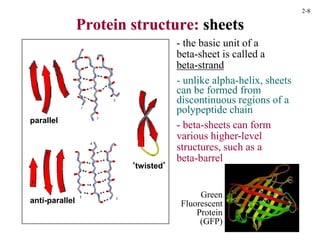
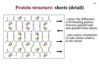
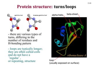
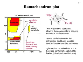
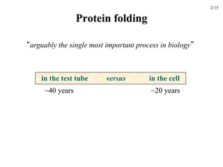
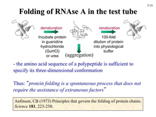
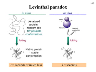
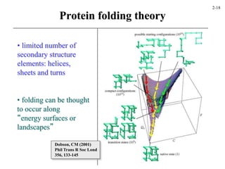
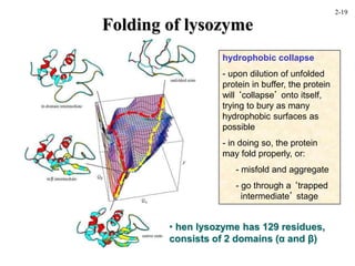
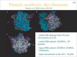
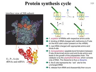
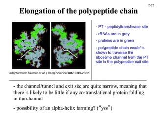
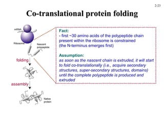
This document discusses protein structure and synthesis. It begins by describing the primary, secondary, tertiary, and quaternary structures of proteins. This includes the structures of alpha helices, beta sheets, turns, and domains. It then discusses protein translation, noting that proteins begin folding as they emerge from the ribosome in a co-translational manner. The final section discusses protein folding and some of the challenges of the folding process.
Proteins are complex organic compounds made of carbon, hydrogen, oxygen, nitrogen, and sometimes sulfur or phosphorus. They are essential to all living organisms and perform many important functions. The document discusses protein structure from primary to quaternary levels. It also covers protein synthesis, folding, and methods to study folding both in vitro and in vivo. Molecular chaperones and proper cellular conditions help proteins fold into their native states.
Dystrophin is a high molecular weight cytoskeletal protein that localizes to the cytoplasmic face of the sarcolemma. It has four domains - an actin binding domain, a central rod domain composed of spectrin-like repeats, a cysteine-rich domain, and a carboxy-terminal domain. Dystrophin forms the dystrophin-glycoprotein complex with other proteins like dystroglycans and sarcoglycans to connect the actin cytoskeleton to the extracellular matrix. Mutations in dystrophin cause Duchenne/Becker muscular dystrophy by disrupting this connection and leading to muscle degeneration.
This document discusses the structure of proteins at different levels, including primary, secondary, tertiary and quaternary structure. It explains that primary structure is the amino acid sequence, and secondary structure includes alpha helices and beta sheets formed by hydrogen bonding. Tertiary structure involves the folding of secondary structure elements into the final three-dimensional shape. The document outlines methods for determining primary structure and describes concepts like the Ramachandran plot that show allowed phi and psi angles. It provides examples of common motifs in tertiary structure and defines domains.
The document discusses the amino acids that make up proteins and their properties. It describes:
1) Hydrophilic amino acids that interact with solvent through ionic and hydrogen bonding, though some can be buried inside proteins forming salt bridges.
2) Hydrophobic amino acids that tend to form the core of proteins and be buried within the folded structure, though some residues can be exposed.
3) Small neutral amino acids that have less preference for being solvent-exposed or buried.
This document summarizes the structural organization of proteins from primary to quaternary structure. It discusses that proteins have a unique amino acid sequence specified by genes which form the primary structure. The primary structure is stabilized by peptide bonds. Secondary structure involves folding into shapes like alpha helices and beta sheets held by hydrogen bonds. Tertiary structure involves the 3D shaping of the entire polypeptide chain through various interactions. Quaternary structure involves multiple polypeptide subunits combining to form a functional protein. Overall the document provides an overview of the hierarchical structural organization of proteins from sequence to final 3D shape.
This document discusses various structural motifs found in proteins, including super-secondary structures composed of combinations of secondary structures. It describes helix motifs like helix-turn-helix and helix-loop-helix, sheet motifs like beta hairpins and Greek key, and mixed motifs like beta-alpha-beta and Rossmann fold. It also covers transmembrane motifs like helix bundles and beta barrels, as well as other motifs like EF-hand, leucine zipper, zinc finger, and TIM barrel fold. The document provides examples of proteins containing these motifs and their biological roles.
Prebiotic Pyrite Chemistry Molecular Scaffold & Catalyst 1 21Heather Jordan
Jordan-Ohmoto model of abiogenesis whereby framboidal pyrite serves as a photocatalytic scaffold in crucial prebiotic chemical reactions such as liposome nucleation, NTP hydrolysis, and peptide synthesis.
Proteins are composed of amino acids bonded together in chains called polypeptides. There are four levels of protein structure: primary, secondary, tertiary, and quaternary. The primary structure is the sequence of amino acids in the polypeptide chain. The secondary structure describes local folding patterns like alpha helices and beta sheets formed by hydrogen bonds. Tertiary structure refers to the overall 3D shape formed by interactions between amino acids distant in the chain. Quaternary structure involves the interaction of multiple polypeptide chains. Changes in protein structure can alter its function.
The document discusses the primary structure of proteins, which is the specific sequence of amino acids. It explains that genetic diseases can result from abnormal amino acid sequences that cause improper folding and loss of function. Determining the normal and mutated protein sequences can help diagnose or study the disease. It then goes on to describe various methods used to determine a protein's primary structure, including identifying amino acid composition through chromatography, N-terminal sequencing using Edman reagent, cleaving large proteins into fragments, and DNA sequencing to translate the nucleotide sequence into the amino acid sequence.
Protein structures determine their functions. There are four levels of protein structure:
1) Primary structure is the amino acid sequence
2) Secondary structure involves local patterns like alpha helices and beta sheets
3) Tertiary structure describes the overall 3D shape formed by secondary structures
4) Quaternary structure refers to the arrangement of multiple polypeptide chains
The most common secondary structures are alpha helices, stabilized by hydrogen bonds between amino acids i and i+4, and beta sheets formed by hydrogen bonding between strands. Protein structure enables functions like catalysis, transport, and information transfer.
The document discusses the structure of proteins at various levels of organization:
- Proteins are composed of amino acids linked together by peptide bonds to form polypeptide chains. The sequence and interactions of these chains determine the protein's structure.
- There are four levels of protein structure - primary, secondary, tertiary, and quaternary. Secondary structure includes alpha helices and beta sheets formed by hydrogen bonding between amino acids in the chain. Tertiary structure describes the overall 3D shape formed by interactions between amino acid side chains. Quaternary structure involves the interaction of multiple polypeptide chains.
- Protein structure enables proteins to perform their diverse functions through processes like enzyme catalysis, oxygen transport, and providing structure
Proteins are polymers made up of amino acid chains that form specific structures. There are four levels of protein structure:
1. Primary structure is the amino acid sequence.
2. Secondary structures include alpha helices and beta sheets formed by hydrogen bonds between amino acids.
3. Tertiary structure is the three dimensional folding of secondary structures.
4. Quaternary structure occurs in proteins made of multiple polypeptide chains that aggregate. The document discusses these levels of structure in detail, focusing on alpha helices and beta sheets as common secondary structures stabilized by hydrogen bonding.
The document discusses the tertiary structure of proteins, which is the three-dimensional structure of a single polypeptide chain that results from folding and interactions between different parts of the amino acid sequence. It describes the various non-covalent interactions like hydrogen bonds, ionic bonds, hydrophobic interactions, and disulfide bonds that stabilize tertiary structure. The tertiary structure is more conserved than primary structure and is determined globally by various interactions that optimize thermodynamic stability.
Proteins play key roles in living systems through catalysis, transport, and information transfer. They have a hierarchical structure including primary, secondary, tertiary, and quaternary levels. The primary structure is the amino acid sequence, and higher levels of organization are determined by the primary structure. Protein folding and interactions between residues determine the final 3D tertiary and quaternary structures, which are critical for protein function. Misfolded proteins can cause diseases.
Peptide and polypeptide, protein structure.pptxRASHMI M G
peptide and polypeptide-meaning and definition, peptide bond, protein structures- primary, secondary, tertiary, supersecondary, quaternary structures denaturation and solubilities of proteins.
The document discusses several key topics regarding proteins and enzymes:
1. It describes the primary structure of proteins and how their amino acid sequence determines their function. It also discusses polymorphic proteins and early protein sequencing methods.
2. Protein folding and the factors that influence a protein's three-dimensional structure are summarized, including non-covalent bonds, domains, and common structural motifs like alpha helices and beta sheets.
3. The roles of different protein types like globular, fibrous, and assembly proteins are highlighted. Key fibrous proteins like collagen and elastin are also mentioned.
The document discusses protein structure at multiple levels of organization. It describes the 20 amino acids that make up proteins and how they can be categorized based on properties like size and affinity for water. It then explains how amino acids join together through peptide bonds to form the primary structure of a protein as a linear sequence. Secondary structures like alpha helices and beta sheets involve hydrogen bonding between amino acids to create regular local structures. Tertiary structure refers to the overall 3D shape formed by packing and arrangement of secondary structures. There are two main types of tertiary structure - globular proteins that are soluble and membrane proteins that exist in cell membranes.
Peptides and proteins chemistry veterinary.pdfTatendaMageja
This document discusses peptides and proteins. It begins by explaining that peptides and proteins are made of amino acids linked by peptide bonds. It then describes different types of secondary protein structures, including alpha helices and beta sheets. Alpha helices form coiled structures stabilized by hydrogen bonds between amino acids spaced four residues apart. Beta sheets consist of extended peptide chains stabilized by hydrogen bonds between adjacent strands running in opposite directions. The document also discusses different classes of proteins based on shape and function.
Pengetahuan struktur, bentuk dan sintesa proteinSiti Julaiha
The document discusses the four levels of protein structure: primary, secondary, tertiary, and quaternary. It explains that proteins are made of amino acids that are linked together via peptide bonds. The order and sequence of amino acids determines the primary structure. Hydrogen bonding leads to the formation of regular structures like alpha helices and beta sheets, which make up the secondary structure. Tertiary structure refers to the overall three-dimensional shape of the protein, which is stabilized by interactions between amino acid side chains. Some proteins have quaternary structure consisting of multiple polypeptide subunits.
Proteins and their biological structuresHelao Silas
1. Hemoglobin transports oxygen in red blood cells through a cooperative binding mechanism between its four subunits. Each subunit contains a heme group that reversibly binds oxygen.
2. In tissues, higher carbon dioxide and hydrogen ion concentrations cause hemoglobin to release oxygen. However, in the lungs where oxygen levels are high, hemoglobin becomes saturated with oxygen.
3. Sickle cell anemia results from a mutation where glutamate is replaced by valine in the beta chain of hemoglobin. This causes deoxygenated hemoglobin to polymerize and distort red blood cells into a sickle shape, blocking blood flow.
The document summarizes key aspects of protein structure and function. It discusses the building blocks of proteins, amino acids, and how they combine through peptide bonds to form protein primary structures. It then describes the four levels of protein structure - primary, secondary, tertiary, and quaternary - focusing on common secondary structures like alpha helices and beta sheets, and how their packing forms tertiary and quaternary structures. Experimental methods for determining protein structures like X-ray crystallography and NMR are also summarized.
This document provides an overview of protein structure, including levels of structure and classification. It discusses the importance of protein structure in determining function. The primary levels of structure are defined as primary (amino acid sequence), secondary (local folding patterns like alpha helices and beta sheets), tertiary (packing of secondary structures), and quaternary (assembly of protein chains). Protein structures can be classified based on their secondary structure composition as all-alpha, all-beta, alpha/beta, or alpha+beta. Domains are compact folding units associated with function.
Introduction:
Protein
Protein motif.
2. History:
3. A brief overview of protein structure.
4. The Structural Classification of Protein(SCOP):
All α.
All β
α/β
α+β
5.The super secondary structure.
6. Rules for formation of Protein Motifs.
7. Structural motifs.
8. Some Common Protein Motifs:
β-hairpin.
β-meander.
Alpha-alpha corner.
Helix-turn-helix motif.
β-α-β motif.
β-sandwich.
β-barrel.
Greek key.
The Jellyroll topology.
Omega loop.
Zinc finger motif.
9. Conclusion.
10. References.
The document discusses various topics related to protein structure and function. It defines different types of bonds in proteins including peptide bonds, disulfide bonds, and hydrogen bonds. It describes the 20 common amino acids that make up proteins and different secondary structures such as alpha helices and beta sheets. It discusses the four levels of protein structure - primary, secondary, tertiary, and quaternary structure. It also covers protein folding driven by hydrophobic interactions and hydrogen bonding, as well as denaturation of proteins.
This document discusses lipids, which are concentrated energy molecules that serve several functions in biology. Lipids include fats, oils, waxes, and hormones. They are used for energy storage and provide twice the energy of carbohydrates. Lipids also make up cell membranes and help cushion and insulate organs. Saturated fats from animals are solid at room temperature and contribute to heart disease, while unsaturated fats from plants and fish are liquid and are a healthier choice. Cell membranes contain phospholipids that form a barrier for the cell, with hydrophilic heads on the outside and hydrophobic tails on the inside.
This document discusses lipids and membranes. It describes the basic structures of lipids like fatty acids, glycerophospholipids, sphingolipids, and cholesterol. These lipids can assemble into structures like micelles and bilayers in aqueous environments due to their amphipathic nature. Bilayers allow for the formation of cell membranes. Membranes contain proteins that can be integral, peripheral, or lipid-anchored. Lipid composition and proteins influence membrane properties like fluidity.
The document discusses the evolution of cell membranes from early RNA molecules clinging to clay particles to the modern fluid mosaic model. Key events include the formation of lipid bilayers that separated internal and external chemistry, allowing more efficient reactions. Experiments showed lipids spontaneously forming enclosed compartments and lipid bilayers with integral membrane proteins that gave membranes a mosaic-like structure. The fluid mosaic model proposes membranes are fluid with lipids and proteins able to diffuse freely within the plane of the bilayer. Transport proteins like channels and carriers allow selective permeability while pumps use ATP to transport molecules against gradients.
This document provides information about lipids and fatty acids. It defines lipids as biomolecules that contain fatty acids or a steroid nucleus and are soluble in organic solvents but not water. There are different types of lipids containing fatty acids, including waxes, fats and oils (triacylglycerols), glycerophospholipids, and prostaglandins. Fatty acids are long-chain carboxylic acids that can be saturated or unsaturated. Fats and oils are esters of glycerol and three fatty acids called triacylglycerols. Unsaturated fatty acids have kinks that prevent close packing, giving oils and unsaturated fats lower melting points than saturated fats. Hydrogen
The octapeptide contains the amino acids A, C, D, G, L, M, S. Enzyme digestion and mass spectrometry identify the fragments D-C-M, A-S, C-M-A, S-G-A, and L-D. This information determines the primary structure is L-A-G-S-D-C-M-A. Secondary structure is based on bond rotations forming elements like alpha helices and beta pleated sheets. Tertiary structure describes the overall shape from peptide chain folding while quaternary structure involves interactions of multiple protein subunits.
This chapter discusses protein therapeutics including recombinant proteins and monoclonal antibodies. It provides examples of recombinant proteins approved for human use to treat disorders like hemophilia, diabetes, and cystic fibrosis. The chapter outlines different expression systems used to produce recombinant proteins, including bacteria, yeast, insect, and mammalian cells. It also describes the structure of antibodies and the development of monoclonal antibodies as therapeutic agents, from mouse antibodies to humanized antibodies to reduce immunogenicity.
Proteins are made up of chains of amino acids and are essential to many bodily functions. Amino acids link together through peptide bonds and proteins fold into complex three-dimensional shapes that determine their specific roles. Both insufficient and excessive protein intake can be harmful, so a balanced diet containing moderate protein is recommended.
This document provides an overview of amino acids, peptides, and proteins. It discusses the 20 standard amino acids, including their structures, properties, and classifications. Peptide bond formation between amino acids is described. Peptides are defined as short chains of amino acids, with examples of peptide functions. Proteins are introduced as longer polymers made up of amino acids that may also contain cofactors or modifications. The learning goals cover the key aspects of amino acid and peptide structures and properties.
Protein folding is the process by which a protein goes from an unfolded state to its biologically active three-dimensional structure. It is important to understand protein folding to help predict protein structures from sequence alone and to understand diseases caused by protein misfolding. Proteins typically fold through progressive formation of native-like structures rather than through a random search. Molecular chaperones help other proteins fold within cells. Misfolded proteins can form amyloid fibrils associated with diseases. Computational methods aim to predict protein structures from sequence using fragment libraries and modeling protein energy landscapes. Protein design techniques aim to computationally modify protein sequences to achieve desired stabilities, functions, and binding properties.
This document discusses protein classification and structure. It defines protein classification as grouping proteins based on structure, function or size. Proteins can have domains and subunits. They come in globular, fibrous, and other shapes. Proteins are linked within and between polypeptide chains using covalent bonds. Proteins bind other molecules specifically through interactions like ionic bonds. Binding allows proteins to regulate activity and form complexes with other molecules like opsins.
Heat shock proteins (HSPs) help other proteins properly fold and function. HSP90 and HSP70 are molecular chaperones that work sequentially to fold proteins in the cytoplasm. Misfolded proteins can cause disease. HSP90 helps buffer hidden genetic variations but under stress these variations are expressed and can lead to morphological changes. HSP90 is highly conserved across species and plays a role in evolution by allowing traits to change in response to stress. Current research studies HSP90 to better understand protein misfolding diseases.
1. Enzymes are biological catalysts that lower the activation energy of reactions and increase reaction rates. They are often proteins that contain cofactors.
2. Enzymes are classified based on the type of reaction they catalyze, such as oxidation-reduction, hydrolysis, or transfer of chemical groups. Common enzyme names end in "-ase".
3. The lock and key model describes how enzymes bind specifically to substrates in their active sites to form enzyme-substrate complexes. In the induced fit model, the enzyme structure changes to better fit the substrate.
Protein structures are classified to generate overviews of structure types and detect evolutionary relationships. Major classification schemes include SCOP, CATH, and FSSP. SCOP classifies proteins into classes, folds, superfamilies, and families based on structural and sequence similarities. CATH also uses a hierarchical system of classes, architectures, topologies, and superfamilies. FSSP provides fully automated and updated structural alignments and classifications.
This document discusses proteins from a chemist's perspective. It describes how proteins are made of amino acids, with 20 standard types but only 9 being essential. The unique side groups of each amino acid determine their individual properties. Protein structure and function depend on the specific amino acid sequence. Amino acids are linked through peptide bonds to form proteins. The document also covers protein digestion and roles of proteins in the body.
The document discusses heat shock proteins (Hsps), Hsp90 inhibitors, and protein degradation. It provides background on protein degradation mechanisms and heat shock proteins. Hsp90 plays a key role in cancer cell survival by regulating oncogenic signaling proteins. Hsp90 inhibitors like geldanamycin and 17-AAG bind Hsp90's ATP binding site, altering its function and inducing degradation of client proteins, stopping cancer cell growth. The paper found that tumor Hsp90 exclusively exists in active multichaperone complexes, conferring higher binding affinity for 17-AAG compared to normal cell Hsp90. This activated conformation in tumor cells represents a unique drug target.
This document summarizes protein therapeutics and provides a pharmacological classification. It notes that the human genome contains 25,000-40,000 genes that can undergo alternative splicing and post-translational modifications, resulting in a very high number of functionally distinct proteins. Protein therapeutics are classified into 4 groups based on their function: group I includes proteins with enzymatic or regulatory activity, group II targets specific molecules or organisms, group III are protein vaccines, and group IV are protein diagnostics. The document outlines some advantages of protein therapeutics but also challenges including solubility, immune response, stability, and costs.
1. Proteins are made up of amino acids and take on specific three-dimensional structures that dictate their function. Determining a protein's structure is important for understanding its role in biological processes.
2. There are several methods for determining and predicting protein structure, including X-ray crystallography, NMR, and computational methods like homology modeling or ab initio structure prediction.
3. Protein structure is hierarchical, ranging from secondary structure like alpha helices and beta sheets to the overall fold classified in databases like SCOP and CATH. Predicting secondary structure is easier than predicting a protein's full three-dimensional structure.
Amino acids are organic compounds that contain an amino group and a carboxyl group. There are 20 different amino acids that serve as the building blocks of proteins. Amino acids link together through peptide bonds to form polypeptide chains that fold into complex three-dimensional protein structures. Proteins serve essential functions and 10 of the 20 amino acids must be obtained through diet as humans cannot synthesize them. Common protein tests identify the presence of proteins using reactions that detect peptide bonds, amino acid side chains, or disulfide bridges.
- Proteins are made up of amino acids, which are the building blocks. There are 20 different amino acids, some are essential and must be obtained through diet.
- The specific sequence of amino acids determines the 3D shape of a protein and its function. Denaturation occurs when proteins lose their shape due to heat, acid, etc.
- Proteins serve many important functions in the body including structure, enzymes, transport, hormones, antibodies, and more. An inadequate intake can result in the body breaking down its own proteins to obtain energy.
This document summarizes different classes of proteolytic enzymes (proteases) including serine, cysteine, aspartic acid, and metallo proteases. It describes their catalytic mechanisms and examples of each class. Key points are that serine proteases use a catalytic triad of serine, histidine, and aspartate residues in their mechanisms, while cysteine proteases employ a catalytic cysteine and histidine. Aspartic and metallo proteases also have distinct catalytic residues and mechanisms. The roles of proteases in digestion, lysosomes, and other cellular processes are overviewed.
Gemma Wean- Nutritional solution for Artemiasmuskaan0008
GEMMA Wean is a high end larval co-feeding and weaning diet aimed at Artemia optimisation and is fortified with a high level of proteins and phospholipids. GEMMA Wean provides the early weaned juveniles with dedicated fish nutrition and is an ideal follow on from GEMMA Micro or Artemia.
GEMMA Wean has an optimised nutritional balance and physical quality so that it flows more freely and spreads readily on the water surface. The balance of phospholipid classes to- gether with the production technology based on a low temperature extrusion process improve the physical aspect of the pellets while still retaining the high phospholipid content.
GEMMA Wean is available in 0.1mm, 0.2mm and 0.3mm. There is also a 0.5mm micro-pellet, GEMMA Wean Diamond, which covers the early nursery stage from post-weaning to pre-growing.
DECODING THE RISKS - ALCOHOL, TOBACCO & DRUGS.pdfDr Rachana Gujar
Introduction: Substance use education is crucial due to its prevalence and societal impact.
Alcohol Use: Immediate and long-term risks include impaired judgment, health issues, and social consequences.
Tobacco Use: Immediate effects include increased heart rate, while long-term risks encompass cancer and heart disease.
Drug Use: Risks vary depending on the drug type, including health and psychological implications.
Prevention Strategies: Education, healthy coping mechanisms, community support, and policies are vital in preventing substance use.
Harm Reduction Strategies: Safe use practices, medication-assisted treatment, and naloxone availability aim to reduce harm.
Seeking Help for Addiction: Recognizing signs, available treatments, support systems, and resources are essential for recovery.
Personal Stories: Real stories of recovery emphasize hope and resilience.
Interactive Q&A: Engage the audience and encourage discussion.
Conclusion: Recap key points and emphasize the importance of awareness, prevention, and seeking help.
Resources: Provide contact information and links for further support.
Hypertension and it's role of physiotherapy in it.Vishal kr Thakur
This particular slides consist of- what is hypertension,what are it's causes and it's effect on body, risk factors, symptoms,complications, diagnosis and role of physiotherapy in it.
This slide is very helpful for physiotherapy students and also for other medical and healthcare students.
Here is summary of hypertension -
Hypertension, also known as high blood pressure, is a serious medical condition that occurs when blood pressure in the body's arteries is consistently too high. Blood pressure is the force of blood pushing against the walls of blood vessels as the heart pumps it. Hypertension can increase the risk of heart disease, brain disease, kidney disease, and premature death.
Comprehensive Rainy Season Advisory: Safety and Preparedness Tips.pdfDr Rachana Gujar
The "Comprehensive Rainy Season Advisory: Safety and Preparedness Tips" offers essential guidance for navigating rainy weather conditions. It covers strategies for staying safe during storms, flood prevention measures, and advice on preparing for inclement weather. This advisory aims to ensure individuals are equipped with the knowledge and resources to handle the challenges of the rainy season effectively, emphasizing safety, preparedness, and resilience.
The facial nerve, also known as cranial nerve VII, is one of the 12 cranial nerves originating from the brain. It's a mixed nerve, meaning it contains both sensory and motor fibres, and it plays a crucial role in controlling various facial muscles, as well as conveying sensory information from the taste buds on the anterior two-thirds of the tongue.
Can coffee help me lose weight? Yes, 25,422 users in the USA use it for that ...nirahealhty
The South Beach Coffee Java Diet is a variation of the popular South Beach Diet, which was developed by cardiologist Dr. Arthur Agatston. The original South Beach Diet focuses on consuming lean proteins, healthy fats, and low-glycemic index carbohydrates. The South Beach Coffee Java Diet adds the element of coffee, specifically caffeine, to enhance weight loss and improve energy levels.
Michigan HealthTech Market Map 2024. Includes 7 categories: Policy Makers, Academic Innovation Centers, Digital Health Providers, Healthcare Providers, Payers / Insurance, Device Companies, Life Science Companies, Innovation Accelerators. Developed by the Michigan-Israel Business Accelerator
LGBTQ+ Adults: Unique Opportunities and Inclusive Approaches to CareVITASAuthor
This webinar helps clinicians understand the unique healthcare needs of the LGBTQ+ community, primarily in relation to end-of-life care. Topics include social and cultural background and challenges, healthcare disparities, advanced care planning, and strategies for reaching the community and improving quality of care.
Chandrima Spa Ajman is one of the leading Massage Center in Ajman, which is open 24 hours exclusively for men. Being one of the most affordable Spa in Ajman, we offer Body to Body massage, Kerala Massage, Malayali Massage, Indian Massage, Pakistani Massage Russian massage, Thai massage, Swedish massage, Hot Stone Massage, Deep Tissue Massage, and many more. Indulge in the ultimate massage experience and book your appointment today. We are confident that you will leave our Massage spa feeling refreshed, rejuvenated, and ready to take on the world.
Visit : https://massagespaajman.com/
Call : 052 987 1315
This particular slides consist of- what is Pneumothorax,what are it's causes and it's effect on body, risk factors, symptoms,complications, diagnosis and role of physiotherapy in it.
This slide is very helpful for physiotherapy students and also for other medical and healthcare students.
Here is a summary of Pneumothorax:
Pneumothorax, also known as a collapsed lung, is a condition that occurs when air leaks into the space between the lung and chest wall. This air buildup puts pressure on the lung, preventing it from expanding fully when you breathe. A pneumothorax can cause a complete or partial collapse of the lung.
The best massage spa Ajman is Chandrima Spa Ajman, which was founded in 2023 and is exclusively for men 24 hours a day. As of right now, our parent firm has been providing massage services to over 50,000+ clients in Ajman for the past 10 years. It has about 8+ branches. This demonstrates that Chandrima Spa Ajman is among the most reasonably priced spas in Ajman and the ideal place to unwind and rejuvenate. We provide a wide range of Spa massage treatments, including Indian, Pakistani, Kerala, Malayali, and body-to-body massages. Numerous massage techniques are available, including deep tissue, Swedish, Thai, Russian, and hot stone massages. Our massage therapists produce genuinely unique treatments that generate a revitalized sense of inner serenely by fusing modern techniques, the cleanest natural substances, and traditional holistic therapists.
Let's Talk About It: Breast Cancer (What is Mindset and Does it Really Matter?)bkling
Your mindset is the way you make sense of the world around you. This lens influences the way you think, the way you feel, and how you might behave in certain situations. Let's talk about mindset myths that can get us into trouble and ways to cultivate a mindset to support your cancer survivorship in authentic ways. Let’s Talk About It!
1. Proteins: structure, translation, etc.
Structure of proteins
- amino acids, peptide bond, primary-quaternary structures, disulfide bond
Protein synthesis
-protein translation, co-translational folding, stalling, etc.
Protein folding and unfolding
- Levinthal paradox, acquisition of native structure, loss of structure
2-1
3. hydrophobic
MILV
FYW
C
P
small neutral
G(A*)ST
hydrophilic
EDNQ
KRH
Amino acid relationships
*A is also
fairly hydrophobic
Suggested amino acid substitutions
Amino acids connected by a line can be substituted with
95% confidence
Adapted from D. Bordo and P. Argos (1991) J. Mol. Biol. 217, 721-729.
Solvent exposed
(SEA>30 Å2 , )
Interior
(SEA<10 Å2, )
SEA, solvent
exposed area
aromatic
2-3
5. The peptide bond
R=side chain
O=C-N-H is planar
(double-bond character)
Phi (Φ) and Psi (ψ) angles can vary;
their rotation allows polypeptides
to adopt their various structures
(alpha-helices, beta-sheets, etc.)
Ri+1
Ri
cis conformation is rare except for proline
potential for steric hindrance
2-5
6.
7. Protein structure: overview
Structural element Description
primary structure amino acid sequence of protein
secondary structure helices, sheets, turns/loops
super-secondary structure association of secondary structures
domain self-contained structural unit
tertiary structure folded structure of whole protein
• includes disulfide bonds
quaternary structure assembled complex (oligomer)
• homo-oligomeric (1 protein type)
• hetero-oligomeric (>1 type)
2-6
8. Protein structure: helices
alpha 3.10 pi
amino acids
per turn: 3.6 3.0 4.4
frequency ~97% ~3% rare
- alpha helices are about
10 residues on average
- side chains are well
staggered, preventing
steric hindrance
- helices can form
bundles, coiled coils, etc.
H-bonding
2-7
9. Protein structure: sheets
- the basic unit of a
beta-sheet is called a
beta-strand
- unlike alpha-helix, sheets
can be formed from
discontinuous regions of a
polypeptide chain
- beta-sheets can form
various higher-level
structures, such as a
beta-barrel
anti-parallel
parallel
‘twisted’
Green
Fluorescent
Protein
(GFP)
2-8
10. Protein structure: sheets (detail)
‘twisted’
- notice the difference
in H-bonding pattern
between parallel and
anti-parallel beta-sheets
- also notice orientation
of side chains relative
to the sheets
2-9
11. Protein structure: turns/loops
ribonuclease A
- there are various types of
turns, differing in the
number of residues and
H-bonding pattern
- loops are typically longer;
they are often called coils
and do not have a
‘regular’,
or repeating, structure loop
(usually exposed on surface)
alpha-helix beta-sheet
2-10
12. Ramachandran plot
Phi (Φ)
Psi (ψ)
- Phi (Φ) and Psi (ψ) rotate,
allowing the polypeptide to assume
its various conformations
- some conformations of the
polypeptide backbone result in
steric hindrance and are disallowed
- glycine has no side chain and is
therefore conformationally highly
flexible (it is often found in turns)
no steric
clashes
permitted
if atoms are
more closely
spaced
2-11
13. Types of non-covalent interactions
interaction nature
bond
length
“bond”
strength example
ionic
(salt bridge)
electrostatic 1.8-4.0 Å
(3.0-10 Å
for like
charges)
1-6
kcal/mol
positive: K, R, H,
N-terminus
negative: D, E,
C-terminus
hydrophobic entropy - 2-3 hydrophobic side chains
(M,I,L,V,F,W,Y,A,C,P)
H-bond H-bonding 2.6-3.5 2-10 H donor, O acceptor
van der
Waals
attraction/
repulsion
2.8-4.0 <1 closely-spaced atoms;
if too close, repulsion
aromatic-
aromatic
p-p 4.5-7.0 1-2 F,W,Y (stacked)
aromatic-
amino group
H-bonding 2.9-3.6 2.7-4.9 N-H donor to F,W,Y
these all contribute to some extent to protein structure & stability;
- important to understand extremophilic (or any other) proteins
2-12
14. Protein-solvent interactions
hydrophilic amino acids (D, E, K, R, H, N, Q)
- these amino acids tend to interact extensively with solvent in
context of the folded protein; the interaction is mostly ionic and H-
bonding
- there are instances of hydrophilic residues being buried in the
interior of the protein; often, pairs of these residues form salt
bridges
hydrophobic amino acids (M, I, L, V, F, W, Y, A*, C, P)
- these tend to form the ‘core’ of the protein, i.e., are buried
within the folded protein; some hydrophobic residues can be
entirely (or partially) exposed
small neutral amino acids (G, A*, S, T)
- less preference for being solvent-exposed or not
2-13
15. The disulfide bond
protein protein
+
protein protein
• disulfide bond formation is a covalent modification; the
oxidation reaction can either be intramolecular (within the same
protein) or inter-molecular (within different proteins, e.g.,
antibody light and heavy chains). The reaction is reversible.
- most disulfide-bonded proteins are extracellular
(e.g. lysozyme contains four disulfide bonds);
the conditions inside the cytosol are reducing,
meaning that the cysteines are usually in reduced form
- cellular enzymes (protein disulfide isomerases) assist
many proteins in forming proper disulfide bond(s)
oxidation
reduction
+ 2 H+
+ 2 e-
2-14
16. Protein folding
“arguably the single most important process in biology”
in the test tube versus in the cell
~40 years ~20 years
2-15
17. Folding of RNAse A in the test tube
denaturation renaturation
Incubate protein
in guanidine
hydrochloride
(GuHCl)
or urea
100-fold
dilution of protein
into physiological
buffer
Anfinsen, CB (1973) Principles that govern the folding of protein chains.
Science 181, 223-230.
- the amino acid sequence of a polypeptide is sufficient to
specify its three-dimensional conformation
Thus: “protein folding is a spontaneous process that does not
require the assistance of extraneous factors”
(aggregation)
2-16
19. • folding can be thought
to occur along
“energy surfaces or
landscapes”
• limited number of
secondary structure
elements: helices,
sheets and turns
Protein folding theory
Dobson, CM (2001)
Phil Trans R Soc Lond
356, 133-145
2-18
20. Folding of lysozyme
• hen lysozyme has 129 residues,
consists of 2 domains (α and β)
hydrophobic collapse
- upon dilution of unfolded
protein in buffer, the protein
will ‘collapse’ onto itself,
trying to bury as many
hydrophobic surfaces as
possible
- in doing so, the protein
may fold properly, or:
- misfold and aggregate
- go through a ‘trapped
intermediate’ stage
2-19
21. Protein synthesis: the ribosome
Yusupov et al. (2001) Science 292, 883.
- whole 70S ribosome from Thermus
thermophilus at 5.5Å
- small (30S) subunit: 16S RNA, ~20
proteins
- large (50S) subunit: 23S RNA, 5S RNA,
>30 proteins
- high concentration in the cell (~ 50 μM)
2-20
22. Protein synthesis cycle
interface view of 50S subunit
E-, P-, A-site
tRNAs and mRNA
1. acylation of tRNAs with respective amino acids
2. binding of tRNA charged with methionine to P-site
on the AUG start codon (present on the mRNA)
3. next tRNA charged with appropriate amino acid
binds A-site
4. transpeptidation (peptide bond formation) between
P-site (N-terminal) amino acid and A-site amino acid
leads to the growth of the polypeptide chain. The
catalysis is by the peptidyltransferase, which consists
only of RNA. The ribosome is thus a ribozyme.
5. the E-site represents the ‘exit’ site for the
uncharged tRNA
6. release from tRNA and disassembly then occurs
2-21
23. Elongation of the polypeptide chain
adapted from Selmer et al. (1999) Science 286: 2349-2352
- PT = peptidyltransferase site
- rRNAs are in grey
- proteins are in green
- polypeptide chain model is
shown to traverse the
ribosome channel from the PT
site to the polypeptide exit site
- the channel/tunnel and exit site are quite narrow, meaning that
there is likely to be little if any co-translational protein folding
in the channel
- possibility of an alpha-helix forming? (“yes”)
2-22
24. Co-translational protein folding
folding
assembly
Fact:
- first ~30 amino acids of the polypeptide chain
present within the ribosome is constrained
(the N-terminus emerges first)
Assumption:
as soon as the nascent chain is extruded, it will start
to fold co-translationally (i.e., acquire secondary
structures, super-secondary structures, domains)
until the complete polypeptide is produced and
extruded
2-23
25. Observing co-translational folding
N-terminal
domain
(~22 kDa)
C-terminal
domain
(~40 kDa)
Experiment:
1. translate firefly luciferase RNA in vitro in the
presence of 35S-methionine for 2 min
2. Prevent re-initiation of translation with
aurintricarboxylic acid (ATCA): ‘synchronizing’
3. at set timepoints, quench translation, incubate with
proteinase K (digests unstructured/non-compact
regions in proteins, but not folded domains/proteins)
4. add denaturing (SDS) buffer, then perform SDS-
PAGE (polyacrylamide gel electrophoresis)
5. dry gel, observe by autoradiography
Firefly
Luciferase
(62 kDa)
3
Result:
4 5 6 7 8 10 12
no
ProK
with
ProK
min
60 kDa
40 kDa
20 kDa
60 kDa
40 kDa
20 kDa
2
3 4 5 6 7 8 10 12 min
2
2-24
26. Antibiotics & protein synthesis
antibiotic effect
cyclohexamide
inhibits the eukaryotic peptidyltransferase;
prevents release of the polypeptide chain. Can
be used to isolate ribosome-nascent chain
complexes
chloramphenicol inhibits the prokaryotic peptidyltransferase
puromycin
causes premature chain termination and release
from ribosome. Puromycin is similar to a
tyrosyl-tRNA and acts as a substrate during
elongation. Once added to the carboxyl end of
the nascent chain, protein synthesis is aborted
tetracycline inhibits aminoacyl tRNA binding to the A-site
kanamycin causes misreading of the mRNA
streptomycin causes misreading of the mRNA
antibiotics can be useful tools for manipulating translation, folding
2-25
27. ssrA RNA in bacteria
Solution:
- SsrA, or 10SA RNA is a small RNA (363 nt)
that resembles a tRNA and can be charged
with alanine. It is placed into the
peptidyltransferase site by the protein SsrB
- SsrA can be used as a template, and codes
a peptide, ANDENYALAA
- the fusion protein containing this sequence
is recognized and degraded by the ClpAP or
ClpPX proteases
Problem:
- turnover (degradation) of mRNA occurs
very quickly in bacteria, and the 3’ end of
the mRNA has a higher probability of being
degraded first
- if the stop codon is removed, there are no
signals for mRNA release from the
ribosome, and the mRNA will stall
2-26
28. Nascent chain stalling in eukaryotes
- can make proteins that are of a defined length by translating
an RNA that is truncated at the 3’ end (i.e., has no stop codon)
Steps:
1. linearize a vector encoding a gene of interest using a restriction
enzyme, such that the cut is precisely where you want the
polypeptide to end (before the stop codon)
2. make RNA using nucleotides and polymerase enzyme
3. add to an in vitro translation system (rabbit reticulocyte lysate),
which has all of the required components to translate the RNA
4. if the RNA is not truncated, the full-length protein will be made
and released; if the RNA is truncated, it will remain bound to the
ribosome
Note: the protein can be labeled this way with 35S-methionine;
co-translational folding still takes place
2-27
29. Chain stalling: in practice
Goal: show that firefly luciferase can adopt a folded, functional
conformation co-translationally
Experiment:
1. prepare DNA construct that encodes firefly luciferase and an extra 35
amino acids at its C-terminus
2. digest construct such that the last 2 amino acids and the stop codon are
removed
3. prepare RNA using polymerase and nucleotides
4. in vitro translate the RNA in rabbit reticulocyte lysate
5. assay for firefly luciferase activity (light emission at 560 nm occurs when
luciferin substrate is oxidatively decarboxylated)
Fact: only full-length firefly luciferase is functional
Problem? Hint: does this experiment show physiological relevance?
2-28
30. Protein folding:
in 3 different environments
• ex vivo refolding rabbit reticulocyte lysate
- rabbit reticulocyte lysate is an abundant source of molecular
chaperones, many of which are ATP-dependent
• in vitro folding environments
- protein folding (from denaturant), when possible, requires the
proper environment:
proper pH, salts, concentration of protein, temperature,
stabilizing agents (e.g., other proteins, glycerol, etc.)
• in vivo folding
- molecular chaperones, protein folding catalysts, proper redox
environment, availability of binding partners
2-29
31. Following the acquisition
of (native) structure
denaturation renaturation native
structure?
• regain of 2º, 3º and 4º structures
- by circular dichroism and
fluorescence measurements
- by other criteria (e.g., native gel
electrophoresis, SEC,
protease sensitivity assays, etc.)
• regain of activity
- activity not necessarily enzymatic
Circular
dichroism
unfolding
refolding
2-30
32. Acquisition of native structure:
examples
• actin
- chemically denatured actin can be refolded by incubating it in
rabbit reticulocyte lysate; native gel electrophoresis, and
binding to DNAse I is used to assess folding
• various small proteins (RNAse A, lysozyme, etc.)
- can be denatured chemically and refolded simply by dilution
of the denaturing agent; activity assays are available, but
folding can be monitored using spectroscopic techniques
• other
- small-angle light x-ray scattering (SAXS), NMR are some
other techniques used to monitor protein folding
2-31
33. Protein denaturants
• high temperatures
- cause protein unfolding, aggregation
• low temperatures
- some proteins are sensitive to cold denaturation
• heavy metals (e.g., lead, cadmium, etc.)
- highly toxic; efficiently induce the ‘stress response’
• proteotoxic agents (e.g., alcohols, cross-linking agents, etc.)
• oxygen radicals, ionizing radiation
- cause permanent protein damage
• chaotropes (urea, guanidine hydrochloride, etc.)
- highly potent at denaturing proteins;
often used in protein folding studies
2-32
34. Following the loss of structure
• loss of secondary structure
- the far-UV circular dichroism spectrum of a protein changes
at the so-called ‘melting temperature’ or Tm
- fluorescence characteristics will likely also change
• loss of tertiary structure
- the far- and near-UV circular dichroism spectra of a protein
change, but the Tm of both spectra may be different
- fluorescence characteristics will likely also change
• loss of activity
- the activity of a protein can be monitored over time
• aggregation
- can measure light scattering (e.g., at 320 nm) spectrophoto-
metrically, or by detecting the protein in a precipitate
2-33
35. Loss of structure: example
folded
unfolded
intermediate
Far-UV
spectrum
Fluorescence
spectrum
Noland et al. (1999) Biochemistry 38, 16136.
native
unfolded
2M
urea
Urea (M)
chymotrypsin
0
no
0
Yes
1
Yes
2
Yes
Bacterial luciferase (α subunit)
2-34