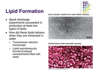
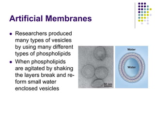
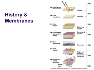
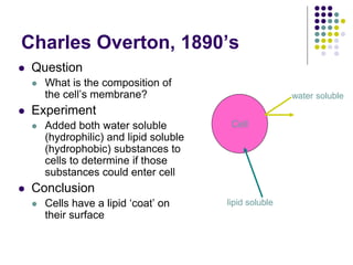
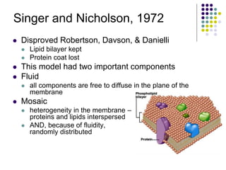
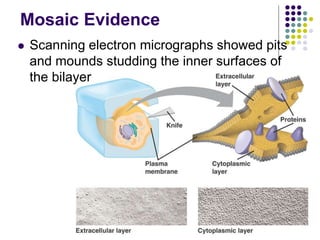
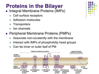
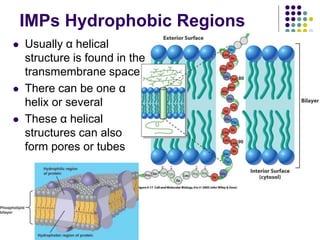
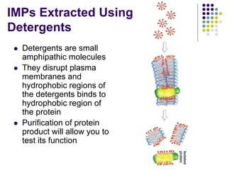
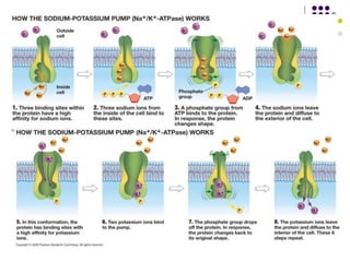
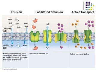
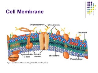
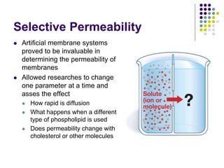
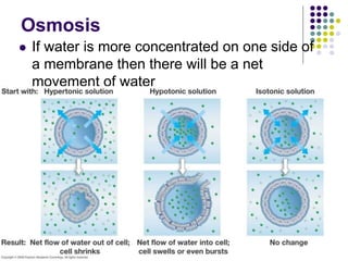
2. Evolution of the Membrane
The evolution of the plasma membrane was a
momentous event because it separated life
from non-life
Before the cell RNA molecules clung to clay
particles, building copies as nucleotides
washed over them randomly
3. Lipid Membrane
The formation of the membrane performed
three important tasks
Separated the chemical composition of the inside
from the chemical composition of the outside
Chemical reactions became more efficient as
reactants could collide more frequently
Membrane could form a collective barrier
4. Lipid Formation
Spark discharge
experiments succeeded in
production at least two
types of lipids
How did these lipids behave
when they are immersed in
water
Transmission electron
microscope
Lipids spontaneously
formed enclosed
compartments filled with
water
5. What Characterizes
a Lipid
Lipids are defined by a
physical property
Solubility in water
Not strictly characterized
by structure
Therefore the structure of
lipids varies widely
Hydrocarbons
Hydrophobic
6. Lipids Found In Cells
There are three types of lipids found in cells
Lipids for energy storage
Triacylglycerols
Lipids for cell membranes
Phospholipids
Lipids for steroids
Cholesterol, estrogen, & testosterone
9. Steroids
Family of lipids that has
a four ring structure
Cholesterol is an
important membrane
component of plasma
membranes is some
organisms
Others are important
hormone signals
10. Phospholipids
Structure
Glycerol linked to
phosphate group
Glycerol linked to two
fatty acids
For Archaea bacteria
Glycerol linked to
phosphate group and two
isoprene units
13. Lipids & Disease
Membrane lipids undergo constant metabolic
turnover
Breakdown is performed by hydrolytic enzymes in
lysosomes
Impaired degradation by a defect enzyme leads to
the accumulation of partial breakdown products
Brain, liver & spleen
Genetic basis for Niemann-Pick disease and Tay-
Sachs disease
Metal retardation, paralysis, blindness, early death
14. Lipids in Membranes
In order to spontaneously form a lipid bilayer
lipid must have
Charges and polar bonds in the head region to
interact with water
Long fatty acid tails to interact with each other
Amphipathic
15. Lipid Bilayer
Formation & Energy
Lipid structures form
spontaneously
No energy input is required
Energy Concepts
Independent phospholipids are unstable in water high
potential energy
Hydrophobic tails disrupt hydrogen bonds
When tails interact with one another reach a lower potential
energy state
Formation of these structures clearly decreases entropy
But overall ∆H outweighs ∆S leading to a negative ∆G
17. Artificial Membranes
Researchers produced
many types of vesicles
by using many different
types of phospholipids
When phospholipids
are agitated by shaking
the layers break and re-
form small water
enclosed vesicles
18. Evolution of Membrane Models
Today cell membranes are characterized by
what is known as a fluid mosaic model
Over 100 years of research was performed
before this model
Historical Perspectives
20. Cell
water soluble
lipid soluble
Charles Overton, 1890’s
Question
What is the composition of
the cell’s membrane?
Experiment
Added both water soluble
(hydrophilic) and lipid soluble
(hydrophobic) substances to
cells to determine if those
substances could enter cell
Conclusion
Cells have a lipid ‘coat’ on
their surface
21. Gorter and Grendel, 1925
Langmuir trough
Question:
What is the arrangement
of lipids in the plasma
membrane
Experiment
Measure surface area of
RBC
Measure surface area of
lipid monolayer
Compare areas
Why use RBCs
22. Gorter and Grendel, Results
Results:
Surface area of monolayer = 2x the SA of RBCs, therefore lipid is oriented as a bilayer
Conclusions:
23. Gorter and Grendel,
Conclusion
Lipids are two layers thick in
the membrane.
bilayer
Proposed that lipids were in
bilayer with polar groups
toward the aqueous
compartments and non-
polar fatty acid parts toward
the center of the bilayer
Phospholipids can rotate,
diffuse and flip in the lipid
sea
25. Davson and Danielli
1st model
Evidence
surface tension of oil droplets is high
surface tension of cell membranes is low
using starfish eggs
Surface tension of oil droplets coated with protein
is low
26. Davson and Danielli
2nd model
Realized that there was a problem with the
Davson/Danielli 1st model
Transport
Model was revised to
allow for pores
28. Singer and Nicholson, 1972
Disproved Robertson, Davson, & Danielli
Lipid bilayer kept
Protein coat lost
This model had two important components
Fluid
all components are free to diffuse in the plane of the
membrane
Mosaic
heterogeneity in the membrane –
proteins and lipids interspersed
AND, because of fluidity,
randomly distributed
29. Evidence for Fluid Mosaic
Mixing of fluorescently tagged proteins on
hybrid cells
Frye & Edidin, 1970
Florescence recovery after photobleaching
FRAP
Mosaic
Freeze fracture
30. Frye and Edidin, 1970
Journal of cell science
Used fluorescently labeled antibodies
Fluorophore
31. Membrane proteins
Mouse cell
Human cell
Hybrid cell
Mixed
proteins
after
1 hour
+
Frye and Edidin, 1970
Explanations
Proteins are free to diffuse in the membrane
Newly synthesized membrane proteins are
inserted into the membrane
Process is ATP dependent
33. Frye and Edidin, 1970
Is protein synthesis of new membrane
proteins responsible for intermixing
Add protein synthesis inhibitor cyclohexamide
Mixing still occurred
Is intermixing an ATP dependent process
Block ATP production with DNP, cyanide
Mixing still occurred
Conclusion
Mixing is due to fluidity
35. Fluorescence Recovery After
Photobleaching
FRAF
Measures lateral
diffusion of molecules
(lipids/proteins) in cell
membranes
Method allows us to look
at populations of
molecules
Information obtained
addresses whether
components are, in fact,
free to diffuse
36. 3
2
1
Measure Recovery
All labeled components
are free to diffuse
Slow diffusion
A fraction of the
population is not mobile
Anchored proteins
Conclusion
Most but not all
components are free to
diffuse
37. Mosaic Evidence
Scanning electron micrographs showed pits
and mounds studding the inner surfaces of
the bilayer
38. Proteins in the Bilayer
Integral Membrane Proteins (IMPs)
Cell surface receptors
Adhesion molecules
Transporters
Ion channels
Peripheral Membrane Proteins (PMPs)
Associate non-covalently with the membrane
Interact with IMPs of phospholidip head groups
Can be inner or outer leaf of PM
39. IMPs Hydrophobic Regions
Usually α helical
structure is found in the
transmembrane space
There can be one α
helix or several
These α helical
structures can also
form pores or tubes
40. IMPs Extracted Using
Detergents
Detergents are small
amphipathic molecules
They disrupt plasma
membranes and
hydrophobic regions of
the detergents binds to
hydrophobic region of
the protein
Purification of protein
product will allow you to
test its function
41. Glycosylation of Proteins
Never occurs on cytoplasmic proteins
Uses specific enzymes of ER and golgi
Two types
N-linked = carbohydrates are attached to
asparigine (terminal amino group) synthesis of
CHO side chain begins in ER and is completed in
Golgi
O-linked = carbohydrates are attached to serine
or threonine (hydroxyl groups) synthesis of CHO
side chain begins and ends in Golgi
For review of the function see chapter 5 notes
42. Transport Proteins
Facilitated Diffusion
Requires no ATP moves with a concentration gradient
Channels
Ion channels
Ions move according to an electrochemical gradient
Usually specific to one type of ion
Aquaporins
Transporters
Glucose transporter
Changes shape
Active Transport
Requires ATP moves against concentration gradient
Ion pumps
43. Gramicidin – Ion Channel
Ions carry charge
movement of ions
produces and
electric current
Can carry H+, K+ and Na+
Ions travel down this pore
44. Aquaporins
Very specific/selective channel
Only water goes through
Water is able to cross the plasma membrane
10X faster than without aquaporins
45. Gated Channels
Aquaporins and ion channels are gated
Open or close in response to signal
Voltage gated channels
Open in response to depolarization of membrane
Ligand gated channels
Open in response to a chemical signal
Remember no energy is needed for transport
Powered by diffusion along an
electrochemical gradient
46. Carrier Proteins
Lipid bilayer is not permeable to glucose, yet
glucose is a main source of cellular energy
Researchers used RBCs to extract and purify
a glucose transporter
47. GLUT – 1
After isolating and analyzing many proteins
from RBCs ghosts researchers found one
protein that increased membrane
permeability to glucose
This protein was added to liposomes (artificial
membranes) and it transported glucose at the
same rate as a living cell
48. Active Transport Pumps
Requires energy – ATP
ATP ADP + P
The phosphate group is transiently and covalently
attached to a protein in a process known as
protein phosphorylation
Phosphorylation of proteins often leads to a
change in protein shape or protein conformation
Leads to a decrease in entropy
Movement is against a concentration gradient
Important for establishing electrochemical
gradients
52. Selective Permeability
Artificial membrane systems
proved to be invaluable in
determining the permeability of
membranes
Allowed researches to change
one parameter at a time and
asses the effect
How rapid is diffusion
What happens when a different
type of phospholipid is used
Does permeability change with
cholesterol or other molecules
53. Membranes are Highly
Selective
Small nonpolar molecules
move across quickly
CO2, O2, N2, hydrophobic
molecules
Small polar molecules have
intermediate permeability
H2O, glycerol, urea
Large uncharged polar
Glucose
Ions
Na+, K+, Cl-
Permeability cm/sec
54. Does Lipid Composition Affect
Permeability
Two properties affect
permeability
Number of double bonds
Saturated vs. unsaturated
Packing
Double bonds produce
spaces between tails
Atoms are in one plane
locked in place
Spaces reduce strength of
hydrophobic interactions
Tail length
55. Fluidity and Double Bond
Character
Degree of hydrophobic interactions increases
with saturated fats
Fluidity increases with double bonds
59. Diffusion
Diffusion is a process which occurs
spontaneously due to an increase in entropy
Diffusion occurs from an area of high
concentration to an area of low concentration
Diffusion across a plasma membrane
60. Osmosis
Osmosis is the diffusion of
water from higher
concentration to lower
concentration
Only occurs when solutions
are separated by a
membrane that is
permeable to some
molecules and not others
Movement is spontaneous
Driven by an increase in
entropy when the solute
becomes more dilute
Entropy decreased 5X on one
Side up increased 10X on the other
The system had a net gain of entropy
61. Osmosis
If water is more concentrated on one side of
a membrane then there will be a net
movement of water
62. Osmosis & Diffusion
Osmosis and diffusion reduce differences in
chemical composition between the inside and
outside of membrane bound vesicles
Therefore, it is unlikely that interiors differed
radically from the external environment
Lipid bilayers become capable of creating a
specialized internal environment due to
specialized protein transporters