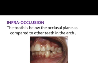
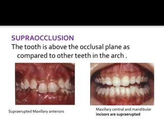
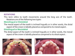
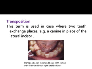
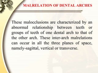
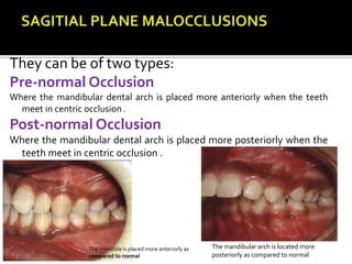
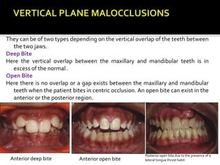
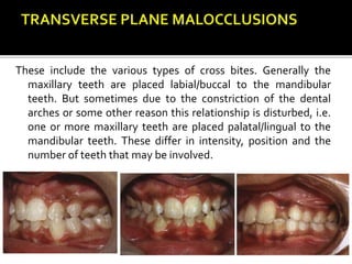
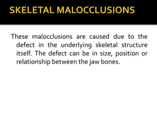
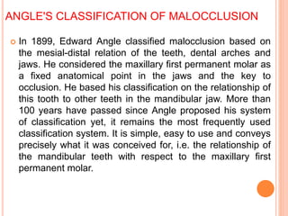
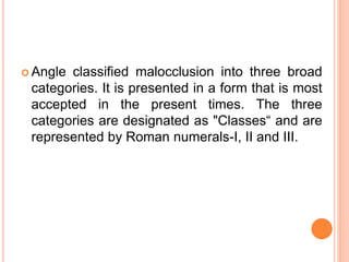
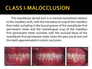
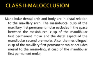
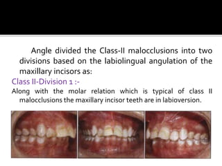
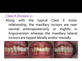
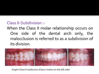
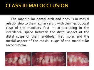
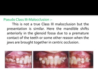
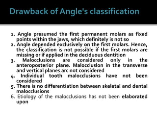
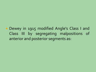
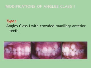
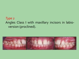
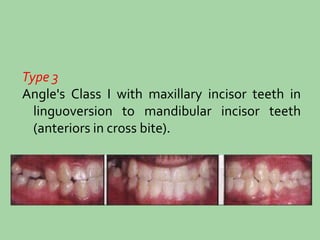
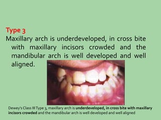
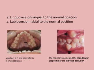
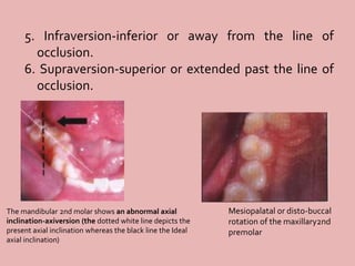
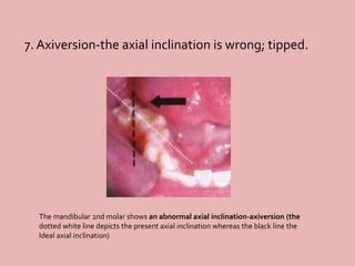
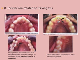
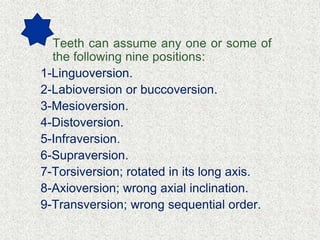
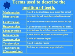
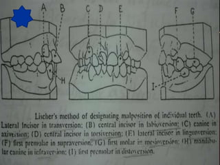
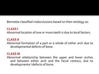
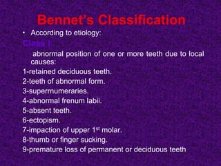
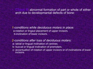
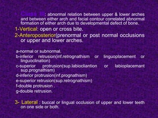
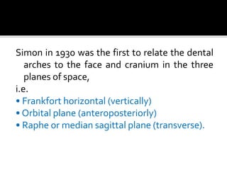
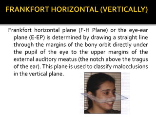
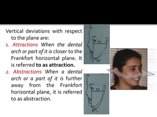
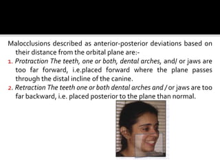
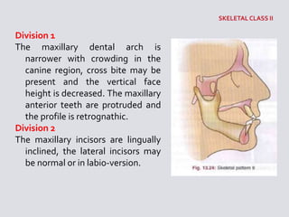
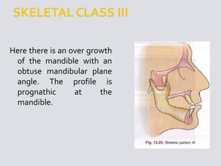
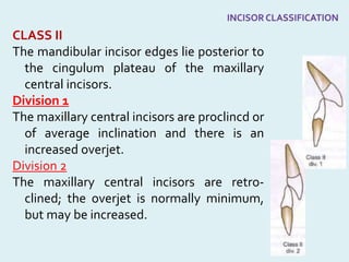
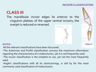
Classification
of
Malocclusion
By; AmmarA. Dae’r
Supervisor: Prof. Maher A. Fouda
 • Introduction
 • Individual tooth malpositions
 • Malrelation of dental arches
 • Skeletal malocclusions
 • Angle's classification of malocclusion
 • Dewey's modification
 • Lischer's modification
 • Bennetle's classification
 • Simon's classification
 • Skeletal classification
 • Ackerman-Profill system of classification
 • Incisor classification
INTRODUCTION
To understand a group of identities it is
advisable to divide them into groups and
subgroups based on certain similarities.
Classification of malocclusion is the description
of dentofacial deviations according to a
common characteristic, or norm. Various
classifications are proposed by different
researchers based on their experiences and
depending upon what they found to be clinically
relevant.
The understanding of these classifications is
essential for the student of orthodontics as
they would be frequently referred to during
communications between consultants and
sometimes, certain identities within a
subgroup will require the same treatment
protocols.
Depending upon which part of the oral and
maxillofacial unit is at fault, malocclusions
can be broadly divided into three types:-
• Individual tooth malpositions.
•Malrelation of the dental arches or
dentoalveolar segments.
• Skeletal malrelationships.
These three can exist individually in a patient or
in combination involving each other,
depending upon where the fault lies-in the
individual dental arch or the dentoalveoJar
segments or the underlying skeletal
structure.
INDIVIDUAL TOOTH MALPOSITIONS
These are malpositions of individual teeth in respect to adjacent
teeth within the same dental arch. Hence, they are also called
intra-arch malocclusions. These can be of the following types:
MESIAL INCLINATION OR TIPPING
The tooth is tilted mesially, i.e. the crown is mesial to the root .
Mesially inclined/tipped central
incisors. The long axis of the teeth
is depicted in black, with the
midline in white dots
DISTAL INCLINATION OR TIPPING
The tooth is tilted distally, i.e. the crown is
distal to the root .
Distally inclined maxillary right lateral
incisor.The long axis is depicted in black,
with the ideal inclination of the tooth
depicted by white dots
LINGUAL INCLINATION OR TIPPING
The tooth is abnormally tilted towards the tongue (or the palate
in the maxillary arch) .
Palatally inclined maxillary left incisor and lingually inclined mandibular second molars
LABIAL/BUCCAL INCLINATION OR TIPPING
The tooth is abnormally inclined towards the
lips/ cheeks .
Labially inclined maxillary right central incisor
INFRA-OCCLUSION
The tooth is below the occlusal plane as
compared to other teeth in the arch .
SUPRAOCCLUSION
The tooth is above the occlusal plane as
compared to other teeth in the arch .
Maxillary central and mandibular
incisors are supraerupted
Supraerupted Maxillary anteriors
ROTATIONS
This term refers to tooth movements around the long axis of the tooth.
Rotations are of the following two types:
Mesiolingual or Distolabial
The mesial aspect of the tooth is inclined lingually or in other words, the distal
aspect of the crown is labially placed as compared to its mesial aspect .
Distolingual or Mesiolabial
The distal aspect of the tooth is inclined lingually or in other words, the mesial
aspect of the crown is labially placed as compared to its distal aspect .
Transposition
This term is used in case where two teeth
exchange places, e.g. a canine in place of the
lateral incisor .
Transposition of the mandibular right canine
with the mandibular right lateral incisor
MALRELATION OF DENTAL ARCHES
These malocclusions are characterized by an
abnormaI relationship between teeth or
groups of teeth of one dental arch to that of
the other arch. These inter-arch malrelations
can occur in all the three planes of space,
namely-sagittal, vertical or transverse.
They can be of two types:
Pre-normal Occlusion
Where the mandibular dental arch is placed more anteriorly when the teeth
meet in centric occlusion .
Post-normal Occlusion
Where the mandibular dental arch is placed more posteriorly when the
teeth meet in centric occlusion .
The mandibular arch is located more
posteriorly as compared to normal
The mandible is placed more anteriorly as
compared to normal
They can be of two types depending on the vertical overlap of the teeth between
the two jaws.
Deep Bite
Here the vertical overlap between the maxillary and mandibular teeth is in
excess of the normal .
Open Bite
Here there is no overlap or a gap exists between the maxillary and mandibular
teeth when the patient bites in centric occlusion. An open bite can exist in the
anterior or the posterior region.
Anterior open biteAnterior deep bite
Posterior open bite due to the presence of a
lateral tongue thrust habit
These include the various types of cross bites. Generally the
maxillary teeth are placed labial/buccal to the mandibular
teeth. But sometimes due to the constriction of the dental
arches or some other reason this relationship is disturbed, i.e.
one or more maxillary teeth are placed palatal/lingual to the
mandibular teeth. These differ in intensity, position and the
number of teeth that may be involved.
These malocclusions are caused due to the
defect in the underlying skeletal structure
itself. The defect can be in size, position or
relationship between the jaw bones.
ANGLE'S CLASSIFICATION OF MALOCCLUSION
 In 1899, Edward Angle classified malocclusion based on
the mesial-distal relation of the teeth, dental arches and
jaws. He considered the maxillary first permanent molar as
a fixed anatomical point in the jaws and the key to
occlusion. He based his classification on the relationship of
this tooth to other teeth in the mandibular jaw. More than
100 years have passed since Angle proposed his system
of classification yet, it remains the most frequently used
classification system. It is simple, easy to use and conveys
precisely what it was conceived for, i.e. the relationship of
the mandibular teeth with respect to the maxillary first
permanent molar.
 Angle classified malocclusion into three broad
categories. It is presented in a form that is most
accepted in the present times. The three
categories are designated as "Classes“ and are
represented by Roman numerals-I, II and III.
The mandibular dental arch is in normal mesiodistal relation
to the maxillary arch, with the mesiobuccal cusp of the maxillary
first molar occluding in the buccal groove of the mandibular first
permanent molar and the mesiolingual cusp of the maxillary
first permanent molar occludes with the occlusal fossa of the
mandibular first permanent molar when the jaws are at rest and
the teeth approximated in centric occlusion.
Mandibular dental arch and body are in distal relation
to the maxillary arch. The mesiobuccal cusp of the
maxillary first permanent molar occludes in the space
between the mesiobuccal cusp of the mandibular
first permanent molar and the distal aspect of the
mandibular second pre-molar. Also, the mesiolingual
cusp of the maxillary first permanent molar occludes
mesial to the mesio-linguaI cusp of the mandibular
first permanent molar.
Angle divided the Class-II malocclusions into two
divisions based on the labiolingual angulation of the
maxillary incisors as:
Class II-Division 1 :-
Along with the molar relation which is typical of class II
malocclusions the maxillary incisor teeth are in labioversion.
Class II-Division 2:-
Along with the typical Class II molar
relationship, the maxillary incisors are near
normal anteroposteriorly or slightly in
linguoversion whereas the maxillary lateral
incisors are tipped labially and/or mesially.
Class II-Subdivision :-
When the Class II molar relationship occurs on
One side of the dental arch only, the
malocclusion is referred to as a subdivision of
its division.
Angle'sClass II subdivision (Class I molars on the left side)
The mandibular dental arch and body is in mesial
relationship to the maxillary arch; with the mesiobuccal
cusp of the maxillary first molar occluding in the
interdental space between the distal aspect of the
distal cusps of the mandibular first molar and the
mesial aspect of the mesial cusps of the mandibular
second molar.
Pseudo Class III-Malocclusion :-
This is not a true Class III malocclusion but the
presentation is similar. Here the mandible shifts
anteriorly in the glenoid fossa due to a premature
contact of the teeth or some other reason when the
jaws are brought together in centric occlusion.
Class III-Subdivision:-
 It is said to exist when the
malocclusion exists unilaterally.
Angle's classification was the first
comprehensive classification of
malocclusion.lt is still the most widely
accepted classification and is used routinely
for day to day communication between
clinicians. With its simplicity, it also had its
inherent drawbacks.
1. Angle presumed the first permanent molars as fixed
points within the jaws, which definitely is not so
2. Angle depended exclusively on the first molars. Hence,
the classification is not possible if the first molars are
missing or if applied in the deciduous dentition
3. Malocclusions are considered only in the
anteroposterior plane. Maloccluslon in the transverse
and vertical planes arc not considered
4. Individual tooth malocclusions have not been
considered
5. There is no differentiation between skeletal and dental
malocclusions
6. Etiology of the malocclusions has not been elaborated
upon
DEWEY'S MODIFICATION
OF ANGLE'S
CLASSIFICATION OF
MALOCCLUSION
 Dewey in 1915 modified Angle's Class I and
Class III by segregating malpositions of
anterior and posterior segments as:
Type 1
Angles Class I with crowded maxillary anterior
teeth.
Type 2
Angles Class I with maxillary incisors in labio-
version (proclined).
Type 3
Angle's Class I with maxillary incisor teeth in
linguoversion to mandibular incisor teeth
(anteriors in cross bite).
Type 4
Molars and/ or premolars are in bucco or
linguo-version, but incisors and canines are in
normal alignment (posteriors in cross bite).
Type 5
Molars are in mesio-version due to early loss of
teeth mesial to them (early loss of deciduous
molars or second premolar).
Dewey's Class IType 5, permanent molar has drifted
mesially due to the early loss of the deciduous 2nd molar
Type 1
Individual arches when viewed individually are
in normal alignment, but when in occlusion
the anteriors are in edge to edge bite.
Dewey's Class III Type 1. individual arches when viewed individually are in
normal alignment, but when in occlusion the anterlors are in edge to edge
bite
Type 2
The mandibular incisors are crowded and
lingual to the maxillary incisors.
Dewey's Class IIIType 2, molars in Angle'sClass III with mandibular
retroclined and/or crowded with maxillary anteriors in labio-version
Type 3
Maxillary arch is underdeveloped, in cross bite
with maxillary incisors crowded and the
mandibular arch is well developed and well
aligned.
Dewey's Class IIIType 3, maxillary arch is underdeveloped, in cross bite with maxillary
incisors crowded and the mandibular arch is well developed and well aligned
LISCHER'S MODIFICATION OF
THE
ANGLE'S CLASSIFICATION OF
MALOCCLUSION
 Lischer in 1933 further modified Angle's
classification by giving substitute names for
Angle's Class I, II and III malocclusions. He also
proposed terms to designate individual tooth
malocclusions.
NEUTRO-OCCLUSION
Neutro-occlusion is the term synonymous with
the Angle's Class I malocclusion.
DISTO-OCCLUSION
Disto-occlusion is synonymous with Angle's
Class II malocclusion.
MESIO·OCCLUSION
Mesio-occlusion is synonymous with Angle's
Class III malocclusion.
Lischers nomenclature for individual tooth
malpositions involved adding the suffix "version" to
a word to indicate the deviation from the normal
position.
1. Mesioversion-mesial to the normal position.
2. Distoversion-distal to the normal position.
Right mandibular 1st
molar is in mesioversion
The right lateral incisor and canine are in distoversion
(black arrow),and the right 1st premolar is rotated
mesio-buccally, i.e. in torsiversion
3. Linguoversion-lingual to the normal position
4. Labioversion-labial to the normal position
Maxillary left 2nd premolar is
in linguocclusion
The maxillary canine and the mandibular
1st premolar are in bucco-occlusion
5. Infraversion-inferior or away from the line of
occlusion.
6. Supraversion-superior or extended past the line of
occlusion.
Mesiopalatal or disto-buccal
rotation of the maxillary2nd
premolar
The mandibular 2nd molar shows an abnormal axial
inclination-axiversion (the dotted white line depicts the
present axial inclination whereas the black line the Ideal
axial inclination)
7. Axiversion-the axial inclination is wrong; tipped.
The mandibular 2nd molar shows an abnormal axial inclination-axiversion (the
dotted white line depicts the present axial inclination whereas the black line the
Ideal axial inclination)
 8.Torsiversion-rotated on its long axis.
The right lateral incisor and canine are in
distoversion (black arrow),and the right 1st
premolar is rotated mesio-buccally, i.e. in
torsiversion
Mesiopalatal or disto-buccal rotation of the
maxillary2nd premolar
 9.Transversion-transposed or changes in the
sequence of position.
Transposition of the mandibular right canine with
the mandibular right lateral incisor
Primary classification
A-Cephalic anomalies:
1-Microcephalus: receding chin
2-Macrocephalus: spacing of the teeth
,mand.prognathism .
3-Osteogenic,neurogenic and tropic
disturbances of the maxilla and
mandible:e.g. leoniasis assea ;hyperostosis of the maxilla
,overgrowth of the mandible,retardation of the dentition ,hemiatrophy of
the face .
B-Dysgnathic anomalies:
suggested by Lischer to denote gross
developmental abnormalities of the teeth,
dental arches, alveolar processes, jaws and
other oral structures.
Dysgnathic anomalies include the following:
1-Macroglossa.
2-Facial clefts.
3-Total or partial agnathia.
4-Tumors of the jaws & diseases of the periodontium.
5-severe changes in the form, structure & relationship of
the teeth and jaws which may be associated with
systemic diseases;e.g.muscular
dystrophy,allergy,endocrine,nutritional disturbances….
C-Eugnathic anomalies:
Suggested by Lischer to denote
anomalies of the teeth alone.
Lischer add the suffix “version” to a
word to indicate the deviation from
normal position
Euganthic anomalies include the following:
1-Disturbance in the degree of tooth
development;impaction,retardation…
2-Position of the tooth or teeth in relation to the
line of occlusion or any of the three palnes of
space.
• Teeth can assume any one or some of
the following nine positions:
1-Linguoversion.
2-Labioversion or buccoversion.
3-Mesioversion.
4-Distoversion.
5-Infraversion.
6-Supraversion.
7-Torsiversion; rotated in its long axis.
8-Axioversion; wrong axial inclination.
9-Transversion; wrong sequential order.
BENNETTE'S CLASSIFICATION OF
MALOCCLUSION
Bennette classified malocclusions based on their etiology as:
CLASS I
Abnormal location of one or more teeth is due to local factors.
CLASS II
Abnormal formation of a part or a whole of either arch due to
developmental defects of bone.
CLASS III
Abnormal relationship between the upper and lower arches
and between either arch and the facial contour, due to
developmenta I defects of bone.
Bennet’s Classification
• According to etiology:
Class I:
abnormal position of one or more teeth due to local
causes:
1-retained deciduous teeth.
2-teeth of abnormal form.
3-superrnumeraries.
4-abnormal frenum labii.
5-absent teeth.
6-ectopism.
7-impaction of upper 1st molar.
8-thumb or finger sucking.
9-premature loss of permanent or deciduous teeth
• Class II: abnormal formation of part or whole of either
arch due to developmantal defects of bone:
1-conditions while deciduous molars in place:
a-rotation or lingaul placement of upper incisors.
b-inclination of lower incisors.
2-conditions after loss of deciduous molars:
a- labial or lingual inclination of canines.
b- buccal or lingual inclination of premolars.
c- accentuation of rotation of upper incisors or of inclinations of lower
incisors.
• Class III: abnormal relation between upper & lower arches
and between either arch and facial contour correlated abnormal
formation of either arch due to developmental defect of bone.
1-Vertical: open or cross bite.
2-Anteroposterior(prenormal or post normal occlusions
or upper and lower arches.
a-normal or subnormal.
b-inferior retrusion(inf.retrognathism or linguoplacement or
linguoclination)
c-superior protrusion(sup.labiocliantion or labioplacemant
sup.prognathism)
d-inferior protrusion(inf.prognathism)
e-superior retrusion(sup.retrognathism)
f-double protrusion .
g-double retrusion.
3- Lateral : buccal or lingual occlusion of upper and lower teeth
on one side or both.
SIMON'S CLASSIFICATION OF
MALOCCLUSION
Simon in 1930 was the first to relate the dental
arches to the face and cranium in the three
planes of space,
i.e.
• Frankfort horizontal (vertically)
• Orbital plane (anteroposteriorly)
• Raphe or median sagittal plane (transverse).
Frankfort horizontal plane (F-H Plane) or the eye-ear
plane (E-EP) is determined by drawing a straight line
through the margins of the bony orbit directly under
the pupil of the eye to the upper margins of the
external auditory meatus (the notch above the tragus
of the ear). This plane is used to classify malocclusions
in the vertical plane.
Vertical deviations with respect
to the plane are:
1. Attractions When the dental
arch or part of it is closer to the
Frankfort horizontal plane. It
is referred to as attraction.
2. Abstractions When a dental
arch or a part of it is further
away from the Frankfort
horizontal plane, it is referred
to as abstraction.
This plane is perpendicular to the eye-ear plane
(Frankfort horizontal plane) at the margin of the bony
orbit directly under the pupil of the eye.
Here it is pertinent to mention the law of the canine.
According to Simon in normal arm relationship, the
orbital plane passes through the distal axial aspect of
the maxillary canine.
Malocclusions described as anterior-posterior deviations based on
their distance from the orbital plane are:-
1. Protraction The teeth, one or both, dental arches, and/ or jaws are
too far forward, i.e.placed forward where the plane passes
through the distal incline of the canine.
2. Retraction The teeth one or both dental arches and / or jaws are too
far backward, i.e. placed posterior to the plane than normal.
The raphe or median sagittal plane is determined by
points approximately 1.5 cm apart on the median
raphe of the palate. The raphe median plane passes
through these two points at right angles to the
Frankfort horizontal plane.
Malocclusions classified according to
transverse deviations from the median
sagittal plane are:
1. Contraction A part or all of the dental
arch is contracted towards the median
sagittal plane.
2. Distraction A part or all of the dental
arch is wider or placed at a distance
which is more than normal.
Deviation of the dental arches in relation to
the orbital plane according to Simon, may
occurs as follows:
1- Both jaws are in normal relation to each other.
2- upper jaw normal, lower jaw distal.
3- upper jaw normal, lower jaw mesial.
4- lower jaw normal, upper jaw mesial.
5- lower jaw normal, upper jaw distal.
6- upper jaw mesial, lower jaw distal.
7- upper jaw distal, lower jaw mesial
SO A classification of malocclusion in which tooth
malpositions are related to three craniofacial
planes: midsagittal, orbital, and Frankfort. Teeth
too close to the midsagittal plane are in
contraction, whereas those too far away are in
distraction. Teeth too anterior to the orbital plane
are in protraction, whereas those too posterior to
the orbital plane are in retraction. Teeth too close
to the Frankfort plane are in attraction, whereas
those too distant are in
distraction.
SKELETAL CLASSIFICATION
Salzmann is 1950 was the first to classify on
occlusion based on the underlying skeletal
structures.
 These malocclusions were
purely dental with the
bones of the face and jaws
being in harmony with one
another and with the rest
of the head. The profile is
orthognathic.
The skeletal Class I was divided further according the dental
malocclusion present as:
Division 1
Local mal-relations of incisors, canine and prernolars.
Division 2
Maxillary incisor protrusion.
Skeletal Class I division 1; local mal relations of incisors, canine and premolars
Skeletal Class I division 2; maxillary incisor protrusion
Division 3
Maxillary incisors in linguoversion.
Division 4
Bimaxillary protrusion.
SkeletalClass I division 3; maxillary anteriors in Iinguo-version
SkeletalClass I division4 rnaloccluslon,bimaxillaryprotrusion
These included malocclusion
with a subnormal distal
mandibular development in
relation to the maxilla.
The skeletal Class II was further
divided into two divisions
based on the features
commonly seen with a
mandible placed in retruded
position.
Division 1
The maxillary dental arch is
narrower with crowding in the
canine region, cross bite may be
present and the vertical face
height is decreased. The maxillary
anterior teeth are protruded and
the profile is retrognathic.
Division 2
The maxillary incisors are lingually
inclined, the lateral incisors may
be normal or in labio-version.
Here there is an over growth
of the mandible with an
obtuse mandibular plane
angle. The profile is
prognathic at the
mandible.
ACKERMAN-PROFITT
SYSTEM OF
CLASSIFICATION
Ackerman and Profitt proposed a very comprehensive system
of classification which divided malocclusions in all the three
planes of space and tended to give an indication towards the
severity of the malocclusion present. The system proposed
by Ackerman-Profitt is based on the set theory, where a set
is defined on the basis of morphologic deviations from the
ideal.
The classification was illustrated using the Venn symbolic
logic diagram. The classification considered nine groups and
five characteristics, and their interrelationships were assessed.
The five characteristics are as follows:-
Venndiagram representing the 5 characters
CHARACTERISTIC 1- ALIGNMENT
CHARACTERISTIC 2- PROFILE
CHARACTERISTIC 3- TRANSVERSE RELATIONSHIPS
CHARACTERISTIC 4- CLASS
CHARACTERISTIC 5- OVERBITE
CHARACTERISTIC 1-ALIGNMENT
Intra arch alignment and symmetry are
assessed as when seen in the occlusal view. A
dental arch is classified as ideal/ crowded /
spaced.
CHARACTERISTIC 2-PROFILE
The profile can be convex/straight/concave.
This also includes the assessment of facial
divergence, i.e. anterior or posterior
divergence.
CHARACTERISTIC 3-TRANSVERSE RELATIONSHIPS
These include the transverse skeletal and dental
relationships. Buccal and palatal cross bites are
noted. These are further subclassified as unilateraI
or bilateral. Distinction is made between skeletal
and dental cross bites.
CHARACTERISTIC 4-CLASS
Here the sagittal relationship of the teeth is assessed
using the Angle classification as Class I/Class II/Class
III. A distinction is made between skeletal and
dental malocclusions.
CHARACTERISTIC 5-OVERBITE
Malocclusions are assessed in the vertical plane. They
are described as anterior open bite/posterior open
bite/anterior deep bite/posterior collapsed bite.
Here again a distinction is made as to whether the
malocclusion is skeletal or dental.
The first characteristic is represented as a square
which contains a larger circle representing the
profile or characteristic 2. This contains three smaller
circles overlapping each other partially, representing
the transverse, sagittal and vertical deviations
respectively. The confluence of these sets form nine
groups, each a combination of certain
characteristics. The ninth group represents the most
complex malocclusion with all possible features.
The 9 groups formed usingt heVenn diagram model
GROUP 1:- Repersented as the outer envelop
or universe.since the degree of alignment &
symmetry are common to all dentitions.
GROUP 2:- the profile is affected by many
malocclusions so it becomes a major set with
in the universe.
GROUP 3-9:- Deviations in three planes of
space are represented by group 3-9 which
includes the overlapping or interlocking
subsets,all with in the profile.
Group 9:- would be more severe with
involvement of criteria from all groups.
ALIGNMENT : both arches crowed
PROFILE : posteriorly
divergent,convex
TYPE : maxillary palatal crossbite,
bilateral skeletal & dental
CLASS : class I,excessive overjet,
class II dental & skeletal
BITE DEPTH : open bite skeletal
 INCISOR CLASSIFICATION
The incisor
classification is
considered simpler and
more relevant than
Angle's classification. It
was adopted by the
British Standards'
Institute in 1983,and is
based upon the
relationship of the lower
incisor edges and the
cingulum plateau of the
maxillary central
incisors.
CLASS I
The mandibular incisor edges occlude with or lie
immediately below the cingulum plateau of the
maxillary central incisors.
CLASS II
The mandibular incisor edges lie posterior to
the cingulum plateau of the maxillary
central incisors.
Division 1
The maxillary central incisors are proclincd or
of average inclination and there is an
increased overjet.
Division 2
The maxillary central incisors are retro-
clined; the overjet is normally minimum,
but may be increased.
CLASS III
The mandibular incisor edges lie anterior to the
cingulum plateau of the upper central incisors; the
overjet is reduced or reversed.
NOTES:-
All the relevant classifications have been discussed.
-The Ackerman and Profitt classification conveys the maximum information
regarding the characteristics of a malocclusion, yet it is not frequently used.
-The incisor classification is the simplest to use, yet not the most frequently
used.
-Angle's classifications with all its shortcomings, is still by far the most
commonly used classification of malocclusion.
Classification by Body-Build
Classification of dental arches according to body type is
advocated by Berger, who used Kretschemer, somatic
types as an adjunct in classifying arch forms. According to
Kretschemer, the human body may be divided
into the following somatic types:
1- Leptosomatic or asthenic:- Long and slender. A tall, thin
person with narrow shoulders, slim arms and hands, the
face is high narrow, the mandible is under-developed and
the bridge of the nose over-developed in length.
2- Pyknic:- short and squat. A person who is
comparatively short, with a short neck and
compact trunk. The face is broad and less high
than the leptosomatic type.
3- Athletic Mascular type;- A person with strongly
developed muscles, broad shoulder, fully
developed chest. The mandible is square and fully
developed.
Note: these types do not always occur in their pure
state and one type may show features of any of
the others.
Etiologic Classification:
1-Osseous:
This category includes problems in abnormal
growth,size,shape or proportion of any of the
bones of the craniofacial complex.When any
bone of the face developed in a perverted,
delayed, advanced or a synchronous,the
aberration may be reflected in an orthodontic
problem.
The term basal bone and apical base used
to describe the areas involved in osseous
dyplasia. The remaining bone, the alveolar
process reacts largely to the needs of dentition
it supports, it can be easily shaped and altered
by tooth movements; the basal bone is less
responsive to the forces of orthodontic
appliances.
2-Muscular: This group includes all problems in
malfunction of the dentofacial musculature,the role of
muscles in etiology of malocclusion includes:
a-Functional: “slide in to occlusion”due to occlusal
interferance.
b-Determinal: sucking habits( e.g.thumb and finger
sucking, etc.)
c-Abnormal pattern of mandibular closure .
d-Incompetant normal reflexes (e.g.lip posture)
e-Abnormal muscular contractions( e.g. tongue
thrusting during swallowing,mouth breathing,etc.)
3-Dental problems: Involve primarily the teeth
and their supporting structures.
This category includes:
a-Malpositions of teeth.
b-Abnormal numbers of teeth.
c-Abnormal size of teeth.
d-Abnormal conformation or texture of teeth.
LOGO

Classification of malocclusion

  • 1.
  • 2.
     • Introduction • Individual tooth malpositions  • Malrelation of dental arches  • Skeletal malocclusions  • Angle's classification of malocclusion  • Dewey's modification  • Lischer's modification  • Bennetle's classification  • Simon's classification  • Skeletal classification  • Ackerman-Profill system of classification  • Incisor classification
  • 3.
    INTRODUCTION To understand agroup of identities it is advisable to divide them into groups and subgroups based on certain similarities. Classification of malocclusion is the description of dentofacial deviations according to a common characteristic, or norm. Various classifications are proposed by different researchers based on their experiences and depending upon what they found to be clinically relevant.
  • 4.
    The understanding ofthese classifications is essential for the student of orthodontics as they would be frequently referred to during communications between consultants and sometimes, certain identities within a subgroup will require the same treatment protocols.
  • 5.
    Depending upon whichpart of the oral and maxillofacial unit is at fault, malocclusions can be broadly divided into three types:- • Individual tooth malpositions. •Malrelation of the dental arches or dentoalveolar segments. • Skeletal malrelationships.
  • 6.
    These three canexist individually in a patient or in combination involving each other, depending upon where the fault lies-in the individual dental arch or the dentoalveoJar segments or the underlying skeletal structure.
  • 7.
    INDIVIDUAL TOOTH MALPOSITIONS Theseare malpositions of individual teeth in respect to adjacent teeth within the same dental arch. Hence, they are also called intra-arch malocclusions. These can be of the following types: MESIAL INCLINATION OR TIPPING The tooth is tilted mesially, i.e. the crown is mesial to the root . Mesially inclined/tipped central incisors. The long axis of the teeth is depicted in black, with the midline in white dots
  • 8.
    DISTAL INCLINATION ORTIPPING The tooth is tilted distally, i.e. the crown is distal to the root . Distally inclined maxillary right lateral incisor.The long axis is depicted in black, with the ideal inclination of the tooth depicted by white dots
  • 9.
    LINGUAL INCLINATION ORTIPPING The tooth is abnormally tilted towards the tongue (or the palate in the maxillary arch) . Palatally inclined maxillary left incisor and lingually inclined mandibular second molars
  • 10.
    LABIAL/BUCCAL INCLINATION ORTIPPING The tooth is abnormally inclined towards the lips/ cheeks . Labially inclined maxillary right central incisor
  • 11.
    INFRA-OCCLUSION The tooth isbelow the occlusal plane as compared to other teeth in the arch .
  • 12.
    SUPRAOCCLUSION The tooth isabove the occlusal plane as compared to other teeth in the arch . Maxillary central and mandibular incisors are supraerupted Supraerupted Maxillary anteriors
  • 13.
    ROTATIONS This term refersto tooth movements around the long axis of the tooth. Rotations are of the following two types: Mesiolingual or Distolabial The mesial aspect of the tooth is inclined lingually or in other words, the distal aspect of the crown is labially placed as compared to its mesial aspect . Distolingual or Mesiolabial The distal aspect of the tooth is inclined lingually or in other words, the mesial aspect of the crown is labially placed as compared to its distal aspect .
  • 14.
    Transposition This term isused in case where two teeth exchange places, e.g. a canine in place of the lateral incisor . Transposition of the mandibular right canine with the mandibular right lateral incisor
  • 15.
    MALRELATION OF DENTALARCHES These malocclusions are characterized by an abnormaI relationship between teeth or groups of teeth of one dental arch to that of the other arch. These inter-arch malrelations can occur in all the three planes of space, namely-sagittal, vertical or transverse.
  • 16.
    They can beof two types: Pre-normal Occlusion Where the mandibular dental arch is placed more anteriorly when the teeth meet in centric occlusion . Post-normal Occlusion Where the mandibular dental arch is placed more posteriorly when the teeth meet in centric occlusion . The mandibular arch is located more posteriorly as compared to normal The mandible is placed more anteriorly as compared to normal
  • 17.
    They can beof two types depending on the vertical overlap of the teeth between the two jaws. Deep Bite Here the vertical overlap between the maxillary and mandibular teeth is in excess of the normal . Open Bite Here there is no overlap or a gap exists between the maxillary and mandibular teeth when the patient bites in centric occlusion. An open bite can exist in the anterior or the posterior region. Anterior open biteAnterior deep bite Posterior open bite due to the presence of a lateral tongue thrust habit
  • 18.
    These include thevarious types of cross bites. Generally the maxillary teeth are placed labial/buccal to the mandibular teeth. But sometimes due to the constriction of the dental arches or some other reason this relationship is disturbed, i.e. one or more maxillary teeth are placed palatal/lingual to the mandibular teeth. These differ in intensity, position and the number of teeth that may be involved.
  • 19.
    These malocclusions arecaused due to the defect in the underlying skeletal structure itself. The defect can be in size, position or relationship between the jaw bones.
  • 20.
    ANGLE'S CLASSIFICATION OFMALOCCLUSION  In 1899, Edward Angle classified malocclusion based on the mesial-distal relation of the teeth, dental arches and jaws. He considered the maxillary first permanent molar as a fixed anatomical point in the jaws and the key to occlusion. He based his classification on the relationship of this tooth to other teeth in the mandibular jaw. More than 100 years have passed since Angle proposed his system of classification yet, it remains the most frequently used classification system. It is simple, easy to use and conveys precisely what it was conceived for, i.e. the relationship of the mandibular teeth with respect to the maxillary first permanent molar.
  • 21.
     Angle classifiedmalocclusion into three broad categories. It is presented in a form that is most accepted in the present times. The three categories are designated as "Classes“ and are represented by Roman numerals-I, II and III.
  • 22.
    The mandibular dentalarch is in normal mesiodistal relation to the maxillary arch, with the mesiobuccal cusp of the maxillary first molar occluding in the buccal groove of the mandibular first permanent molar and the mesiolingual cusp of the maxillary first permanent molar occludes with the occlusal fossa of the mandibular first permanent molar when the jaws are at rest and the teeth approximated in centric occlusion.
  • 23.
    Mandibular dental archand body are in distal relation to the maxillary arch. The mesiobuccal cusp of the maxillary first permanent molar occludes in the space between the mesiobuccal cusp of the mandibular first permanent molar and the distal aspect of the mandibular second pre-molar. Also, the mesiolingual cusp of the maxillary first permanent molar occludes mesial to the mesio-linguaI cusp of the mandibular first permanent molar.
  • 24.
    Angle divided theClass-II malocclusions into two divisions based on the labiolingual angulation of the maxillary incisors as: Class II-Division 1 :- Along with the molar relation which is typical of class II malocclusions the maxillary incisor teeth are in labioversion.
  • 25.
    Class II-Division 2:- Alongwith the typical Class II molar relationship, the maxillary incisors are near normal anteroposteriorly or slightly in linguoversion whereas the maxillary lateral incisors are tipped labially and/or mesially.
  • 26.
    Class II-Subdivision :- Whenthe Class II molar relationship occurs on One side of the dental arch only, the malocclusion is referred to as a subdivision of its division. Angle'sClass II subdivision (Class I molars on the left side)
  • 27.
    The mandibular dentalarch and body is in mesial relationship to the maxillary arch; with the mesiobuccal cusp of the maxillary first molar occluding in the interdental space between the distal aspect of the distal cusps of the mandibular first molar and the mesial aspect of the mesial cusps of the mandibular second molar.
  • 28.
    Pseudo Class III-Malocclusion:- This is not a true Class III malocclusion but the presentation is similar. Here the mandible shifts anteriorly in the glenoid fossa due to a premature contact of the teeth or some other reason when the jaws are brought together in centric occlusion.
  • 29.
    Class III-Subdivision:-  Itis said to exist when the malocclusion exists unilaterally. Angle's classification was the first comprehensive classification of malocclusion.lt is still the most widely accepted classification and is used routinely for day to day communication between clinicians. With its simplicity, it also had its inherent drawbacks.
  • 30.
    1. Angle presumedthe first permanent molars as fixed points within the jaws, which definitely is not so 2. Angle depended exclusively on the first molars. Hence, the classification is not possible if the first molars are missing or if applied in the deciduous dentition 3. Malocclusions are considered only in the anteroposterior plane. Maloccluslon in the transverse and vertical planes arc not considered 4. Individual tooth malocclusions have not been considered 5. There is no differentiation between skeletal and dental malocclusions 6. Etiology of the malocclusions has not been elaborated upon
  • 32.
  • 33.
     Dewey in1915 modified Angle's Class I and Class III by segregating malpositions of anterior and posterior segments as:
  • 34.
    Type 1 Angles ClassI with crowded maxillary anterior teeth.
  • 35.
    Type 2 Angles ClassI with maxillary incisors in labio- version (proclined).
  • 36.
    Type 3 Angle's ClassI with maxillary incisor teeth in linguoversion to mandibular incisor teeth (anteriors in cross bite).
  • 37.
    Type 4 Molars and/or premolars are in bucco or linguo-version, but incisors and canines are in normal alignment (posteriors in cross bite).
  • 38.
    Type 5 Molars arein mesio-version due to early loss of teeth mesial to them (early loss of deciduous molars or second premolar). Dewey's Class IType 5, permanent molar has drifted mesially due to the early loss of the deciduous 2nd molar
  • 39.
    Type 1 Individual archeswhen viewed individually are in normal alignment, but when in occlusion the anteriors are in edge to edge bite. Dewey's Class III Type 1. individual arches when viewed individually are in normal alignment, but when in occlusion the anterlors are in edge to edge bite
  • 40.
    Type 2 The mandibularincisors are crowded and lingual to the maxillary incisors. Dewey's Class IIIType 2, molars in Angle'sClass III with mandibular retroclined and/or crowded with maxillary anteriors in labio-version
  • 41.
    Type 3 Maxillary archis underdeveloped, in cross bite with maxillary incisors crowded and the mandibular arch is well developed and well aligned. Dewey's Class IIIType 3, maxillary arch is underdeveloped, in cross bite with maxillary incisors crowded and the mandibular arch is well developed and well aligned
  • 42.
    LISCHER'S MODIFICATION OF THE ANGLE'SCLASSIFICATION OF MALOCCLUSION
  • 43.
     Lischer in1933 further modified Angle's classification by giving substitute names for Angle's Class I, II and III malocclusions. He also proposed terms to designate individual tooth malocclusions.
  • 44.
    NEUTRO-OCCLUSION Neutro-occlusion is theterm synonymous with the Angle's Class I malocclusion. DISTO-OCCLUSION Disto-occlusion is synonymous with Angle's Class II malocclusion. MESIO·OCCLUSION Mesio-occlusion is synonymous with Angle's Class III malocclusion.
  • 45.
    Lischers nomenclature forindividual tooth malpositions involved adding the suffix "version" to a word to indicate the deviation from the normal position. 1. Mesioversion-mesial to the normal position. 2. Distoversion-distal to the normal position. Right mandibular 1st molar is in mesioversion The right lateral incisor and canine are in distoversion (black arrow),and the right 1st premolar is rotated mesio-buccally, i.e. in torsiversion
  • 46.
    3. Linguoversion-lingual tothe normal position 4. Labioversion-labial to the normal position Maxillary left 2nd premolar is in linguocclusion The maxillary canine and the mandibular 1st premolar are in bucco-occlusion
  • 47.
    5. Infraversion-inferior oraway from the line of occlusion. 6. Supraversion-superior or extended past the line of occlusion. Mesiopalatal or disto-buccal rotation of the maxillary2nd premolar The mandibular 2nd molar shows an abnormal axial inclination-axiversion (the dotted white line depicts the present axial inclination whereas the black line the Ideal axial inclination)
  • 48.
    7. Axiversion-the axialinclination is wrong; tipped. The mandibular 2nd molar shows an abnormal axial inclination-axiversion (the dotted white line depicts the present axial inclination whereas the black line the Ideal axial inclination)
  • 49.
     8.Torsiversion-rotated onits long axis. The right lateral incisor and canine are in distoversion (black arrow),and the right 1st premolar is rotated mesio-buccally, i.e. in torsiversion Mesiopalatal or disto-buccal rotation of the maxillary2nd premolar
  • 50.
     9.Transversion-transposed orchanges in the sequence of position. Transposition of the mandibular right canine with the mandibular right lateral incisor
  • 51.
    Primary classification A-Cephalic anomalies: 1-Microcephalus:receding chin 2-Macrocephalus: spacing of the teeth ,mand.prognathism . 3-Osteogenic,neurogenic and tropic disturbances of the maxilla and mandible:e.g. leoniasis assea ;hyperostosis of the maxilla ,overgrowth of the mandible,retardation of the dentition ,hemiatrophy of the face .
  • 52.
    B-Dysgnathic anomalies: suggested byLischer to denote gross developmental abnormalities of the teeth, dental arches, alveolar processes, jaws and other oral structures. Dysgnathic anomalies include the following: 1-Macroglossa. 2-Facial clefts. 3-Total or partial agnathia. 4-Tumors of the jaws & diseases of the periodontium. 5-severe changes in the form, structure & relationship of the teeth and jaws which may be associated with systemic diseases;e.g.muscular dystrophy,allergy,endocrine,nutritional disturbances….
  • 53.
    C-Eugnathic anomalies: Suggested byLischer to denote anomalies of the teeth alone. Lischer add the suffix “version” to a word to indicate the deviation from normal position Euganthic anomalies include the following: 1-Disturbance in the degree of tooth development;impaction,retardation… 2-Position of the tooth or teeth in relation to the line of occlusion or any of the three palnes of space.
  • 54.
    • Teeth canassume any one or some of the following nine positions: 1-Linguoversion. 2-Labioversion or buccoversion. 3-Mesioversion. 4-Distoversion. 5-Infraversion. 6-Supraversion. 7-Torsiversion; rotated in its long axis. 8-Axioversion; wrong axial inclination. 9-Transversion; wrong sequential order.
  • 57.
  • 58.
    Bennette classified malocclusionsbased on their etiology as: CLASS I Abnormal location of one or more teeth is due to local factors. CLASS II Abnormal formation of a part or a whole of either arch due to developmental defects of bone. CLASS III Abnormal relationship between the upper and lower arches and between either arch and the facial contour, due to developmenta I defects of bone.
  • 59.
    Bennet’s Classification • Accordingto etiology: Class I: abnormal position of one or more teeth due to local causes: 1-retained deciduous teeth. 2-teeth of abnormal form. 3-superrnumeraries. 4-abnormal frenum labii. 5-absent teeth. 6-ectopism. 7-impaction of upper 1st molar. 8-thumb or finger sucking. 9-premature loss of permanent or deciduous teeth
  • 60.
    • Class II:abnormal formation of part or whole of either arch due to developmantal defects of bone: 1-conditions while deciduous molars in place: a-rotation or lingaul placement of upper incisors. b-inclination of lower incisors. 2-conditions after loss of deciduous molars: a- labial or lingual inclination of canines. b- buccal or lingual inclination of premolars. c- accentuation of rotation of upper incisors or of inclinations of lower incisors.
  • 61.
    • Class III:abnormal relation between upper & lower arches and between either arch and facial contour correlated abnormal formation of either arch due to developmental defect of bone. 1-Vertical: open or cross bite. 2-Anteroposterior(prenormal or post normal occlusions or upper and lower arches. a-normal or subnormal. b-inferior retrusion(inf.retrognathism or linguoplacement or linguoclination) c-superior protrusion(sup.labiocliantion or labioplacemant sup.prognathism) d-inferior protrusion(inf.prognathism) e-superior retrusion(sup.retrognathism) f-double protrusion . g-double retrusion. 3- Lateral : buccal or lingual occlusion of upper and lower teeth on one side or both.
  • 62.
  • 63.
    Simon in 1930was the first to relate the dental arches to the face and cranium in the three planes of space, i.e. • Frankfort horizontal (vertically) • Orbital plane (anteroposteriorly) • Raphe or median sagittal plane (transverse).
  • 64.
    Frankfort horizontal plane(F-H Plane) or the eye-ear plane (E-EP) is determined by drawing a straight line through the margins of the bony orbit directly under the pupil of the eye to the upper margins of the external auditory meatus (the notch above the tragus of the ear). This plane is used to classify malocclusions in the vertical plane.
  • 65.
    Vertical deviations withrespect to the plane are: 1. Attractions When the dental arch or part of it is closer to the Frankfort horizontal plane. It is referred to as attraction. 2. Abstractions When a dental arch or a part of it is further away from the Frankfort horizontal plane, it is referred to as abstraction.
  • 66.
    This plane isperpendicular to the eye-ear plane (Frankfort horizontal plane) at the margin of the bony orbit directly under the pupil of the eye. Here it is pertinent to mention the law of the canine. According to Simon in normal arm relationship, the orbital plane passes through the distal axial aspect of the maxillary canine.
  • 67.
    Malocclusions described asanterior-posterior deviations based on their distance from the orbital plane are:- 1. Protraction The teeth, one or both, dental arches, and/ or jaws are too far forward, i.e.placed forward where the plane passes through the distal incline of the canine. 2. Retraction The teeth one or both dental arches and / or jaws are too far backward, i.e. placed posterior to the plane than normal.
  • 69.
    The raphe ormedian sagittal plane is determined by points approximately 1.5 cm apart on the median raphe of the palate. The raphe median plane passes through these two points at right angles to the Frankfort horizontal plane.
  • 70.
    Malocclusions classified accordingto transverse deviations from the median sagittal plane are: 1. Contraction A part or all of the dental arch is contracted towards the median sagittal plane. 2. Distraction A part or all of the dental arch is wider or placed at a distance which is more than normal.
  • 71.
    Deviation of thedental arches in relation to the orbital plane according to Simon, may occurs as follows: 1- Both jaws are in normal relation to each other. 2- upper jaw normal, lower jaw distal. 3- upper jaw normal, lower jaw mesial. 4- lower jaw normal, upper jaw mesial. 5- lower jaw normal, upper jaw distal. 6- upper jaw mesial, lower jaw distal. 7- upper jaw distal, lower jaw mesial
  • 72.
    SO A classificationof malocclusion in which tooth malpositions are related to three craniofacial planes: midsagittal, orbital, and Frankfort. Teeth too close to the midsagittal plane are in contraction, whereas those too far away are in distraction. Teeth too anterior to the orbital plane are in protraction, whereas those too posterior to the orbital plane are in retraction. Teeth too close to the Frankfort plane are in attraction, whereas those too distant are in distraction.
  • 73.
  • 74.
    Salzmann is 1950was the first to classify on occlusion based on the underlying skeletal structures.
  • 75.
     These malocclusionswere purely dental with the bones of the face and jaws being in harmony with one another and with the rest of the head. The profile is orthognathic.
  • 76.
    The skeletal ClassI was divided further according the dental malocclusion present as: Division 1 Local mal-relations of incisors, canine and prernolars. Division 2 Maxillary incisor protrusion. Skeletal Class I division 1; local mal relations of incisors, canine and premolars Skeletal Class I division 2; maxillary incisor protrusion
  • 77.
    Division 3 Maxillary incisorsin linguoversion. Division 4 Bimaxillary protrusion. SkeletalClass I division 3; maxillary anteriors in Iinguo-version SkeletalClass I division4 rnaloccluslon,bimaxillaryprotrusion
  • 78.
    These included malocclusion witha subnormal distal mandibular development in relation to the maxilla. The skeletal Class II was further divided into two divisions based on the features commonly seen with a mandible placed in retruded position.
  • 79.
    Division 1 The maxillarydental arch is narrower with crowding in the canine region, cross bite may be present and the vertical face height is decreased. The maxillary anterior teeth are protruded and the profile is retrognathic. Division 2 The maxillary incisors are lingually inclined, the lateral incisors may be normal or in labio-version.
  • 80.
    Here there isan over growth of the mandible with an obtuse mandibular plane angle. The profile is prognathic at the mandible.
  • 81.
  • 82.
    Ackerman and Profittproposed a very comprehensive system of classification which divided malocclusions in all the three planes of space and tended to give an indication towards the severity of the malocclusion present. The system proposed by Ackerman-Profitt is based on the set theory, where a set is defined on the basis of morphologic deviations from the ideal.
  • 83.
    The classification wasillustrated using the Venn symbolic logic diagram. The classification considered nine groups and five characteristics, and their interrelationships were assessed. The five characteristics are as follows:- Venndiagram representing the 5 characters CHARACTERISTIC 1- ALIGNMENT CHARACTERISTIC 2- PROFILE CHARACTERISTIC 3- TRANSVERSE RELATIONSHIPS CHARACTERISTIC 4- CLASS CHARACTERISTIC 5- OVERBITE
  • 84.
    CHARACTERISTIC 1-ALIGNMENT Intra archalignment and symmetry are assessed as when seen in the occlusal view. A dental arch is classified as ideal/ crowded / spaced.
  • 85.
    CHARACTERISTIC 2-PROFILE The profilecan be convex/straight/concave. This also includes the assessment of facial divergence, i.e. anterior or posterior divergence.
  • 86.
    CHARACTERISTIC 3-TRANSVERSE RELATIONSHIPS Theseinclude the transverse skeletal and dental relationships. Buccal and palatal cross bites are noted. These are further subclassified as unilateraI or bilateral. Distinction is made between skeletal and dental cross bites.
  • 87.
    CHARACTERISTIC 4-CLASS Here thesagittal relationship of the teeth is assessed using the Angle classification as Class I/Class II/Class III. A distinction is made between skeletal and dental malocclusions.
  • 88.
    CHARACTERISTIC 5-OVERBITE Malocclusions areassessed in the vertical plane. They are described as anterior open bite/posterior open bite/anterior deep bite/posterior collapsed bite. Here again a distinction is made as to whether the malocclusion is skeletal or dental.
  • 89.
    The first characteristicis represented as a square which contains a larger circle representing the profile or characteristic 2. This contains three smaller circles overlapping each other partially, representing the transverse, sagittal and vertical deviations respectively. The confluence of these sets form nine groups, each a combination of certain characteristics. The ninth group represents the most complex malocclusion with all possible features. The 9 groups formed usingt heVenn diagram model
  • 92.
    GROUP 1:- Repersentedas the outer envelop or universe.since the degree of alignment & symmetry are common to all dentitions. GROUP 2:- the profile is affected by many malocclusions so it becomes a major set with in the universe. GROUP 3-9:- Deviations in three planes of space are represented by group 3-9 which includes the overlapping or interlocking subsets,all with in the profile.
  • 93.
    Group 9:- wouldbe more severe with involvement of criteria from all groups. ALIGNMENT : both arches crowed PROFILE : posteriorly divergent,convex TYPE : maxillary palatal crossbite, bilateral skeletal & dental CLASS : class I,excessive overjet, class II dental & skeletal BITE DEPTH : open bite skeletal
  • 94.
  • 95.
    The incisor classification is consideredsimpler and more relevant than Angle's classification. It was adopted by the British Standards' Institute in 1983,and is based upon the relationship of the lower incisor edges and the cingulum plateau of the maxillary central incisors.
  • 96.
    CLASS I The mandibularincisor edges occlude with or lie immediately below the cingulum plateau of the maxillary central incisors.
  • 97.
    CLASS II The mandibularincisor edges lie posterior to the cingulum plateau of the maxillary central incisors. Division 1 The maxillary central incisors are proclincd or of average inclination and there is an increased overjet. Division 2 The maxillary central incisors are retro- clined; the overjet is normally minimum, but may be increased.
  • 98.
    CLASS III The mandibularincisor edges lie anterior to the cingulum plateau of the upper central incisors; the overjet is reduced or reversed. NOTES:- All the relevant classifications have been discussed. -The Ackerman and Profitt classification conveys the maximum information regarding the characteristics of a malocclusion, yet it is not frequently used. -The incisor classification is the simplest to use, yet not the most frequently used. -Angle's classifications with all its shortcomings, is still by far the most commonly used classification of malocclusion.
  • 99.
    Classification by Body-Build Classificationof dental arches according to body type is advocated by Berger, who used Kretschemer, somatic types as an adjunct in classifying arch forms. According to Kretschemer, the human body may be divided into the following somatic types: 1- Leptosomatic or asthenic:- Long and slender. A tall, thin person with narrow shoulders, slim arms and hands, the face is high narrow, the mandible is under-developed and the bridge of the nose over-developed in length.
  • 100.
    2- Pyknic:- shortand squat. A person who is comparatively short, with a short neck and compact trunk. The face is broad and less high than the leptosomatic type. 3- Athletic Mascular type;- A person with strongly developed muscles, broad shoulder, fully developed chest. The mandible is square and fully developed. Note: these types do not always occur in their pure state and one type may show features of any of the others.
  • 101.
    Etiologic Classification: 1-Osseous: This categoryincludes problems in abnormal growth,size,shape or proportion of any of the bones of the craniofacial complex.When any bone of the face developed in a perverted, delayed, advanced or a synchronous,the aberration may be reflected in an orthodontic problem.
  • 102.
    The term basalbone and apical base used to describe the areas involved in osseous dyplasia. The remaining bone, the alveolar process reacts largely to the needs of dentition it supports, it can be easily shaped and altered by tooth movements; the basal bone is less responsive to the forces of orthodontic appliances.
  • 103.
    2-Muscular: This groupincludes all problems in malfunction of the dentofacial musculature,the role of muscles in etiology of malocclusion includes: a-Functional: “slide in to occlusion”due to occlusal interferance. b-Determinal: sucking habits( e.g.thumb and finger sucking, etc.) c-Abnormal pattern of mandibular closure . d-Incompetant normal reflexes (e.g.lip posture) e-Abnormal muscular contractions( e.g. tongue thrusting during swallowing,mouth breathing,etc.)
  • 104.
    3-Dental problems: Involveprimarily the teeth and their supporting structures. This category includes: a-Malpositions of teeth. b-Abnormal numbers of teeth. c-Abnormal size of teeth. d-Abnormal conformation or texture of teeth.
  • 105.