This document discusses bracket positioning gauges and techniques for proper bracket placement. It describes:
1. Types of bracket positioning gauges including star shaped and straight rod shaped gauges.
2. Parts of gauges including the holding arm, tooth supporting arm, and slot supporting arm.
3. Correct positioning of gauges which should be perpendicular to the tooth surface to ensure accurate bracket height.
4. Techniques for placing brackets on individual tooth types from different clinician positions to accurately assess bracket alignment.
Introduction by Dr. Yehia under supervision of Dr. Fouda; focus on bracket positioning.
Overview of bracket positioning gauges for vertical accuracy, types of gauges, and importance of gauge positioning. Importance of gauge positioning to avoid bracket height errors (up to 2mm), with techniques for different incisor types.
Technique of bracket placement using wire guidance for accuracy, especially in cases of impacted teeth.
Recommended clinician positions for optimal visibility during bracket placement, including adjustments of the dental unit.
Methods for positioning brackets on upper and lower incisors, including height and position checks.
Bracket placement procedures for upper and lower canines with emphasis on position checks.
Guidelines for positioning brackets on upper and lower bicuspids, with checks for vertical and mesiodistal positions.
Dr Mohammad Yehia
Undersupervision of
Dr Maher Fouda
Mansoura Egypt
Bracket positioning
gauges and
placement
2.
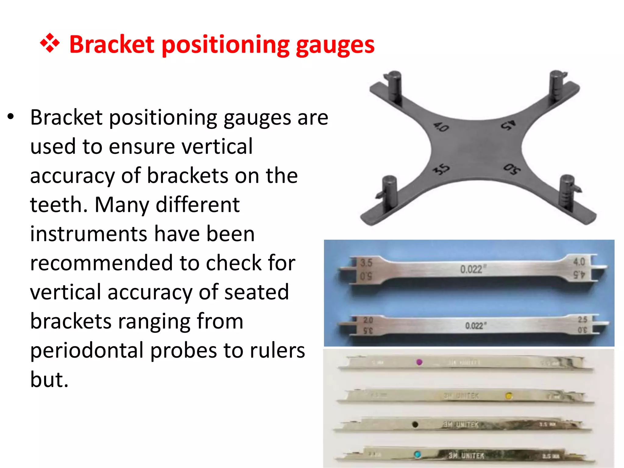
Bracket positioninggauges
• Bracket positioning gauges are
used to ensure vertical
accuracy of brackets on the
teeth. Many different
instruments have been
recommended to check for
vertical accuracy of seated
brackets ranging from
periodontal probes to rulers
but.
3.
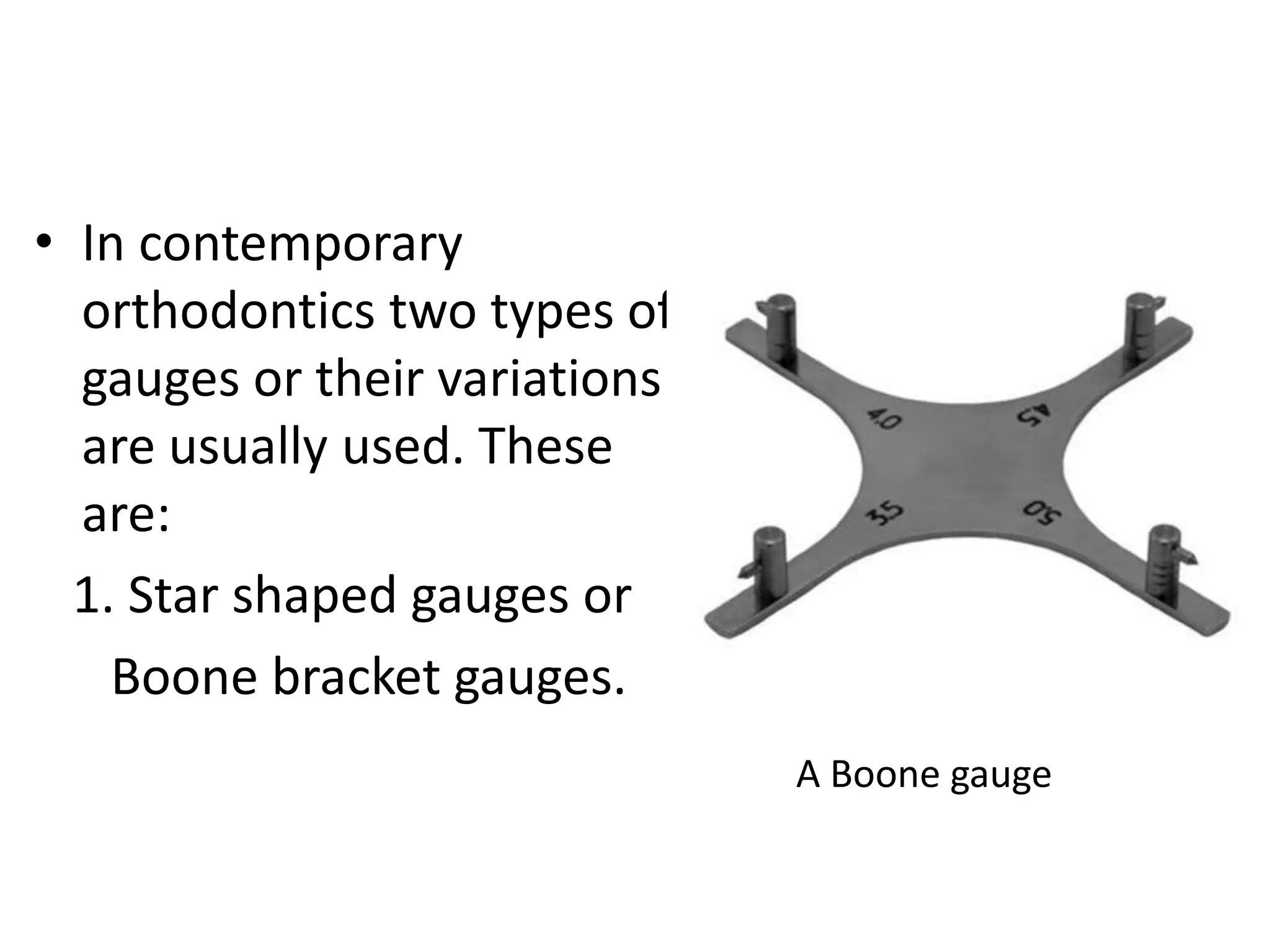
• In contemporary
orthodonticstwo types of
gauges or their variations
are usually used. These
are:
1. Star shaped gauges or
Boone bracket gauges.
A Boone gauge
4.
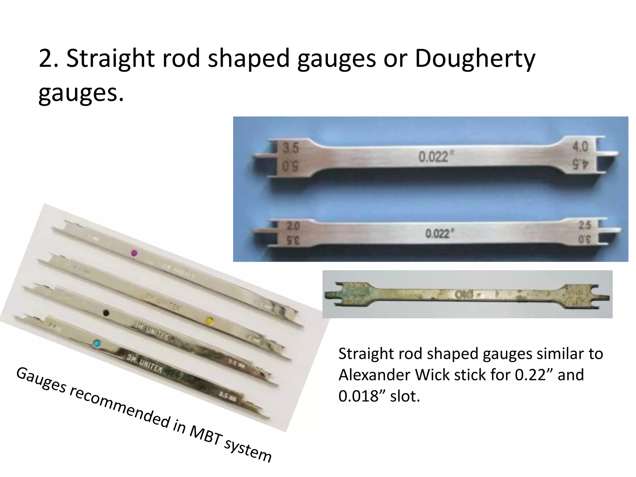
2. Straight rodshaped gauges or Dougherty
gauges.
Straight rod shaped gauges similar to
Alexander Wick stick for 0.22” and
0.018” slot.
5.
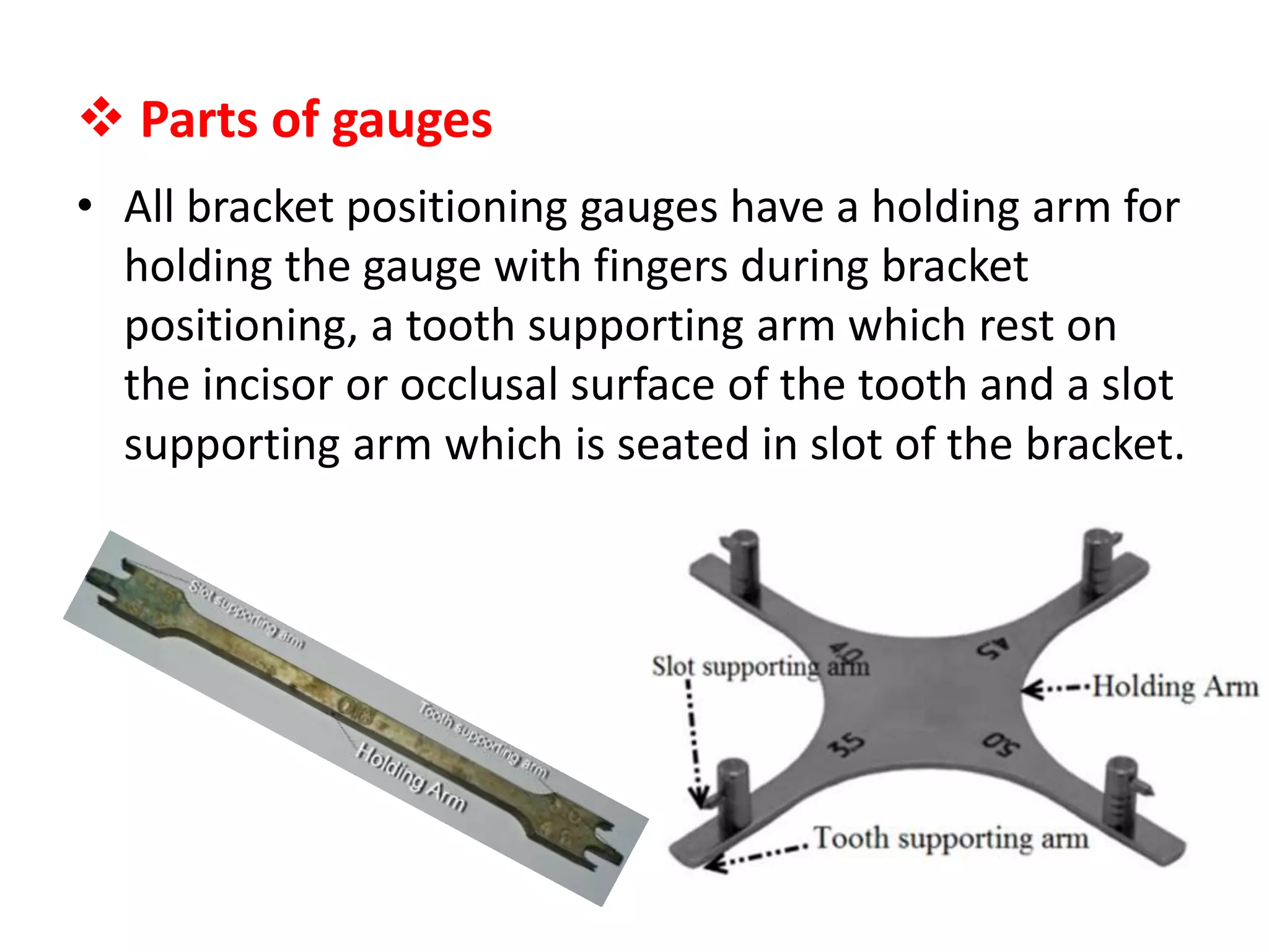
Parts ofgauges
• All bracket positioning gauges have a holding arm for
holding the gauge with fingers during bracket
positioning, a tooth supporting arm which rest on
the incisor or occlusal surface of the tooth and a slot
supporting arm which is seated in slot of the bracket.
6.
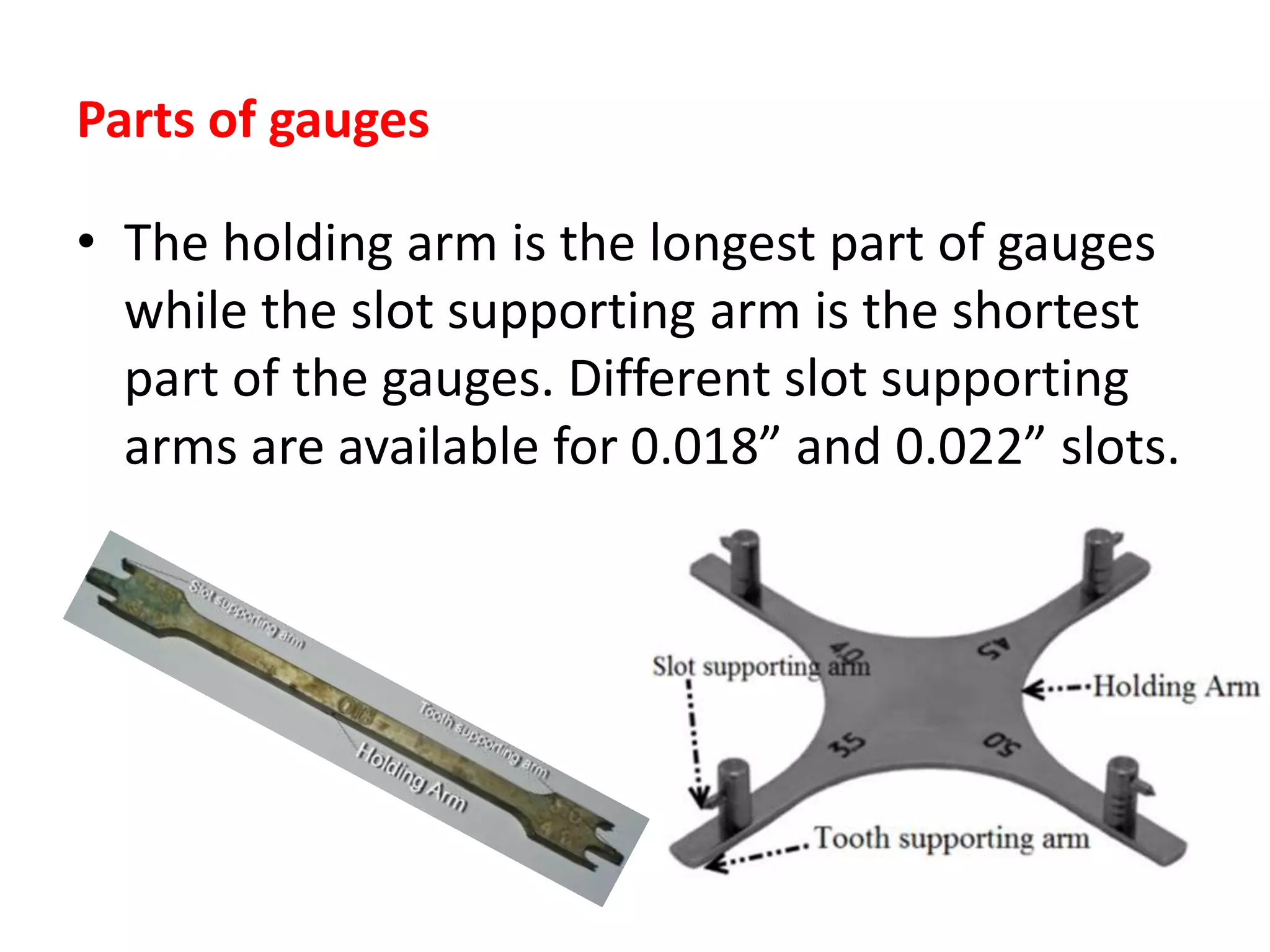
Parts of gauges
•The holding arm is the longest part of gauges
while the slot supporting arm is the shortest
part of the gauges. Different slot supporting
arms are available for 0.018” and 0.022” slots.
7.
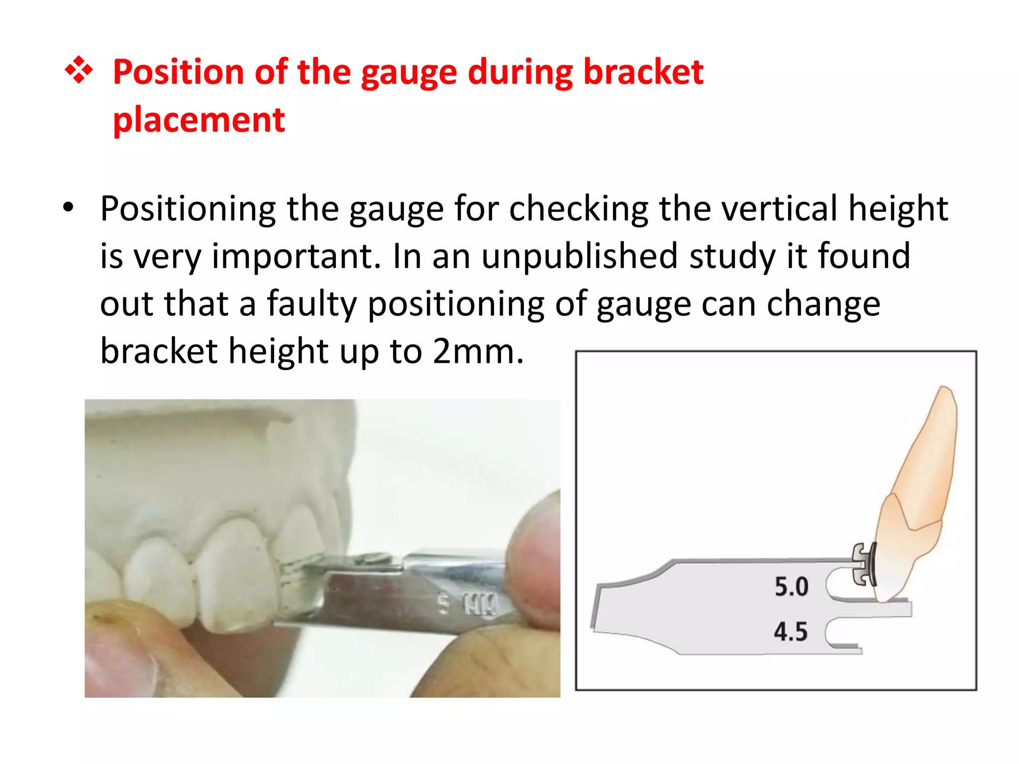
Position ofthe gauge during bracket
placement
• Positioning the gauge for checking the vertical height
is very important. In an unpublished study it found
out that a faulty positioning of gauge can change
bracket height up to 2mm.
8.
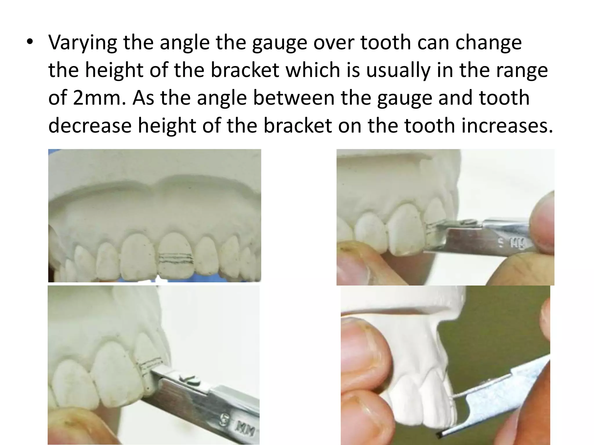
• Varying theangle the gauge over tooth can change
the height of the bracket which is usually in the range
of 2mm. As the angle between the gauge and tooth
decrease height of the bracket on the tooth increases.
9.
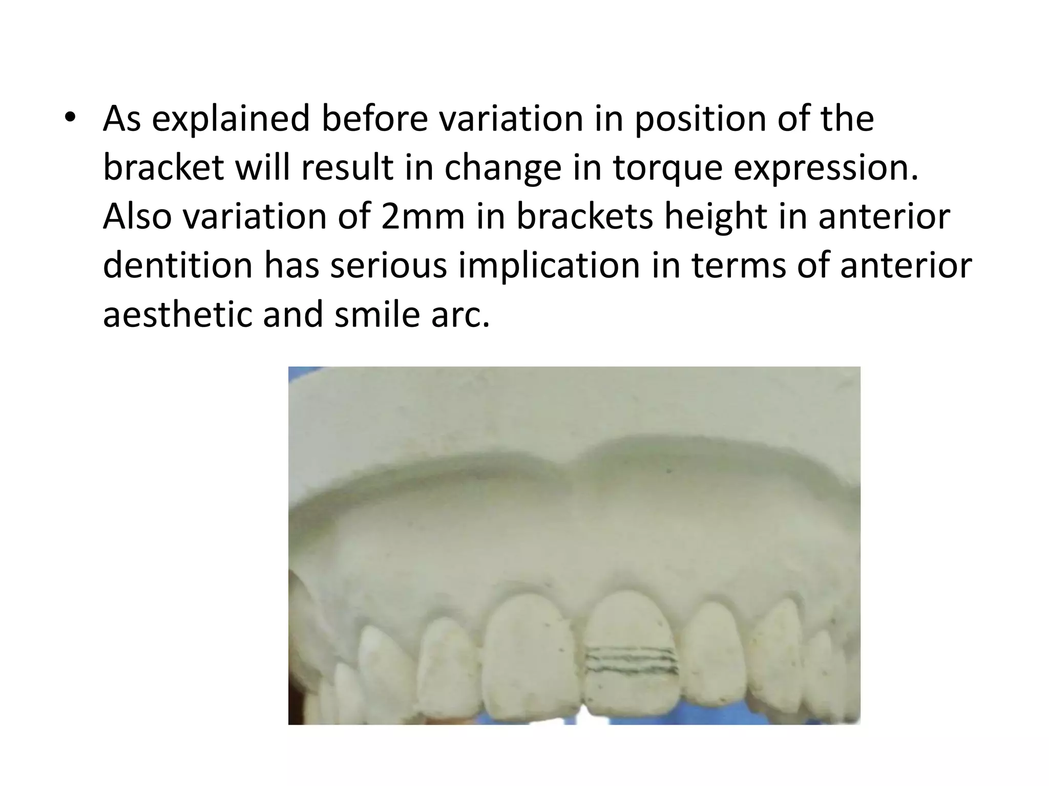
• As explainedbefore variation in position of the
bracket will result in change in torque expression.
Also variation of 2mm in brackets height in anterior
dentition has serious implication in terms of anterior
aesthetic and smile arc.
10.
Position of thegauge during bracket
placement
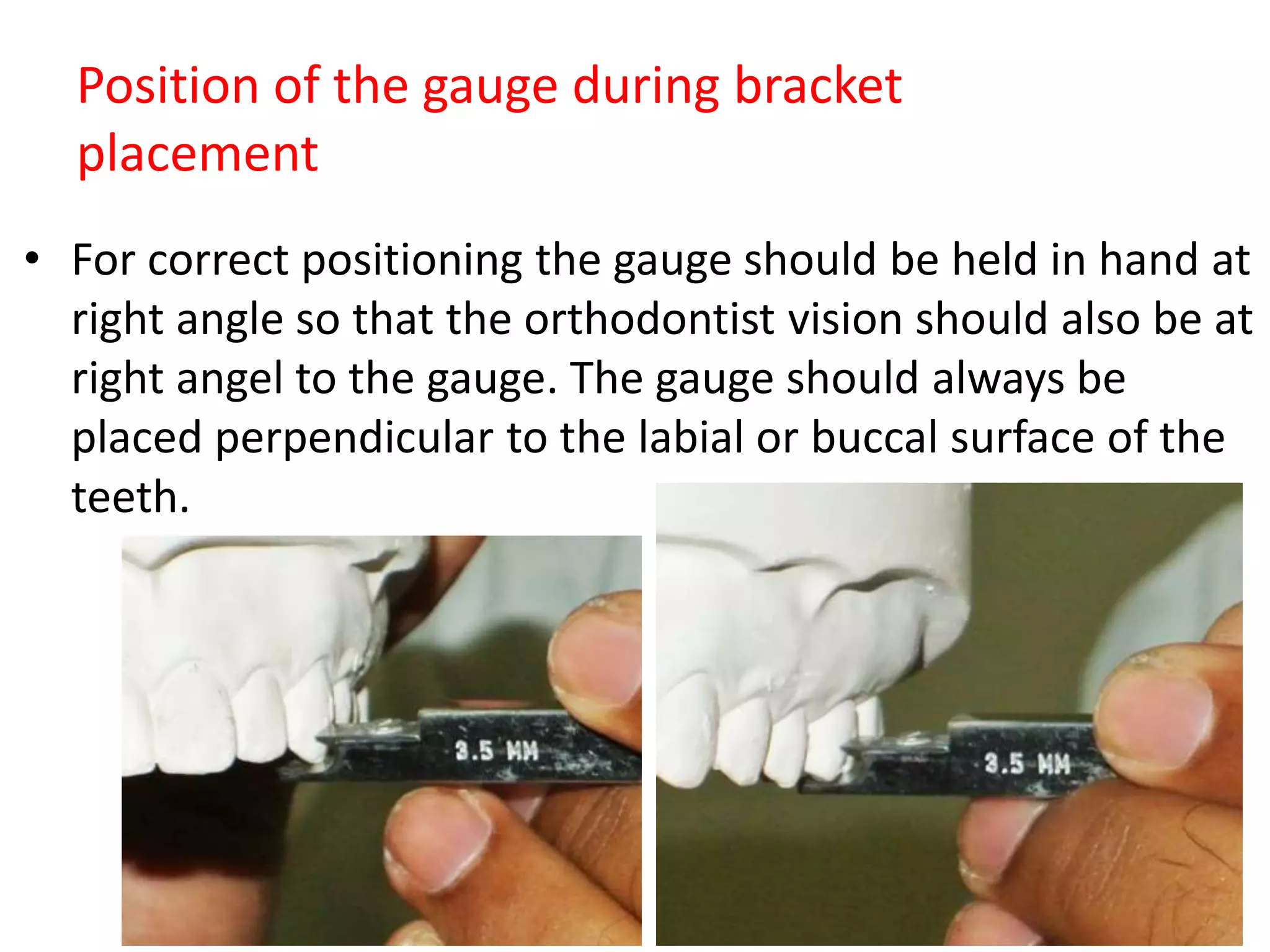
• For correct positioning the gauge should be held in hand at
right angle so that the orthodontist vision should also be at
right angel to the gauge. The gauge should always be
placed perpendicular to the labial or buccal surface of the
teeth.
11.
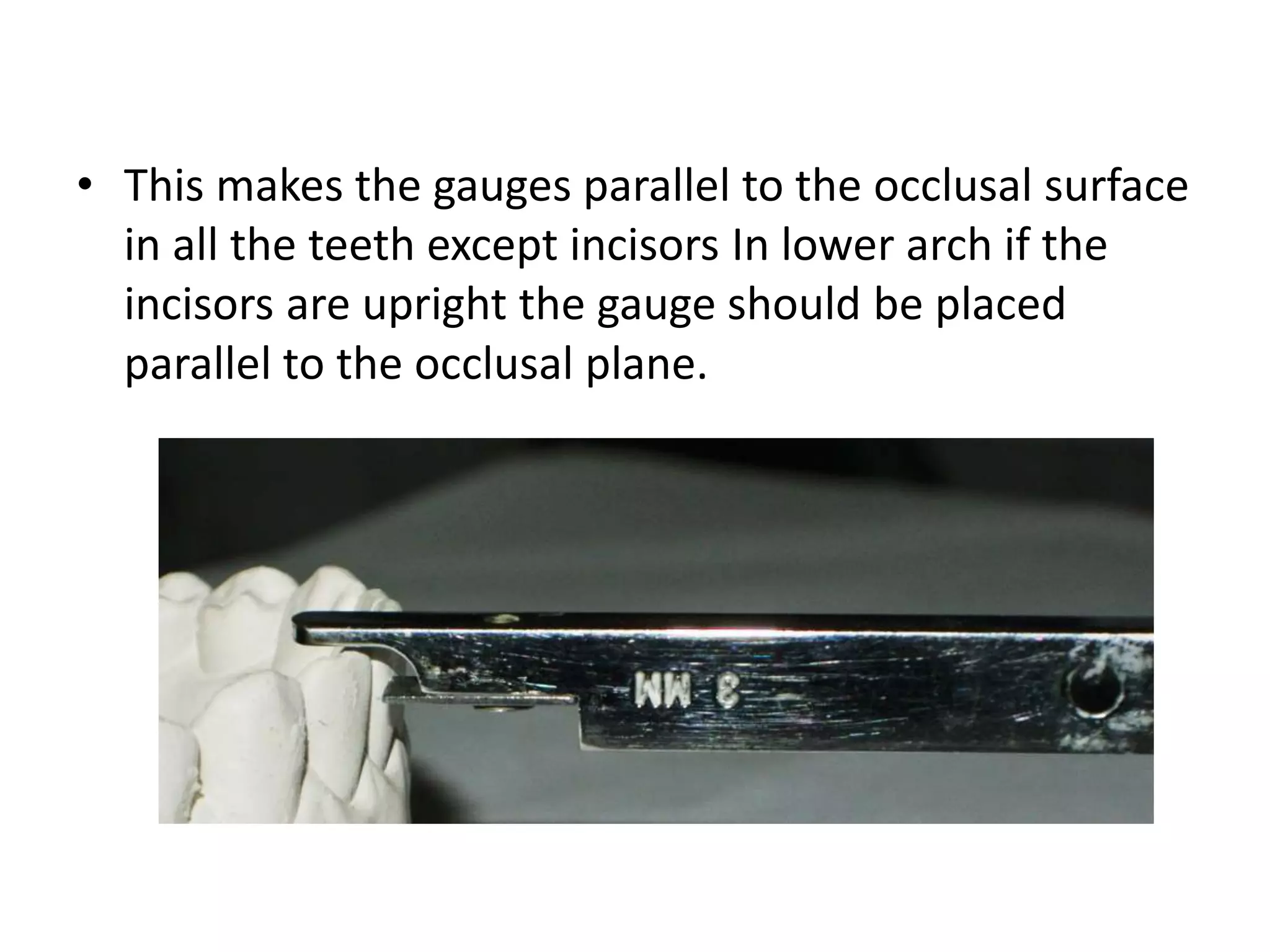
• This makesthe gauges parallel to the occlusal surface
in all the teeth except incisors In lower arch if the
incisors are upright the gauge should be placed
parallel to the occlusal plane.
12.
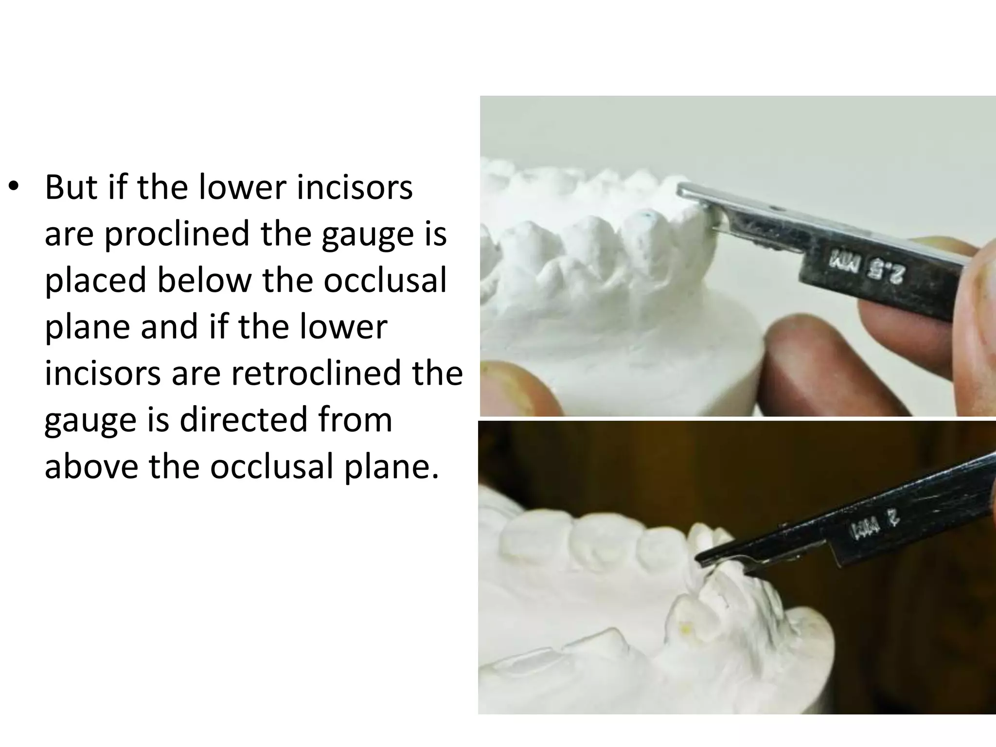
• But ifthe lower incisors
are proclined the gauge is
placed below the occlusal
plane and if the lower
incisors are retroclined the
gauge is directed from
above the occlusal plane.
13.
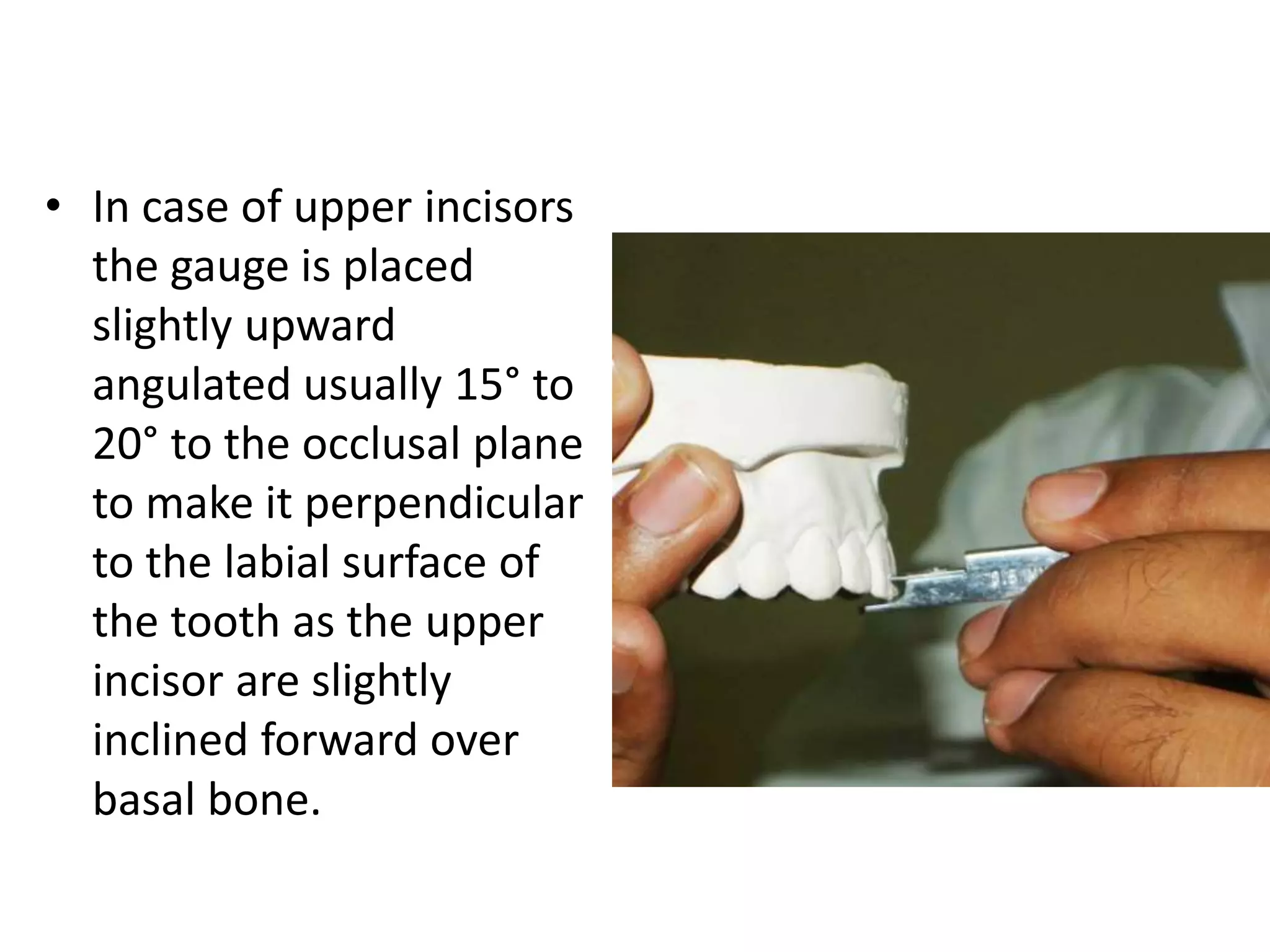
• In caseof upper incisors
the gauge is placed
slightly upward
angulated usually 15° to
20° to the occlusal plane
to make it perpendicular
to the labial surface of
the tooth as the upper
incisor are slightly
inclined forward over
basal bone.
14.
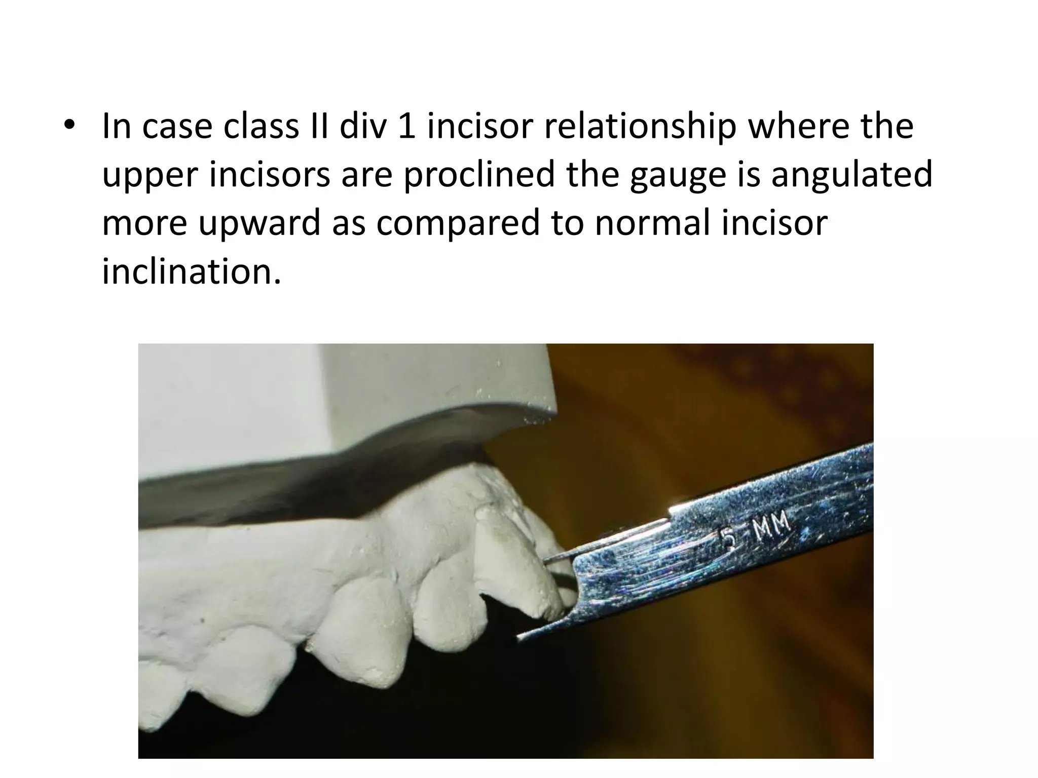
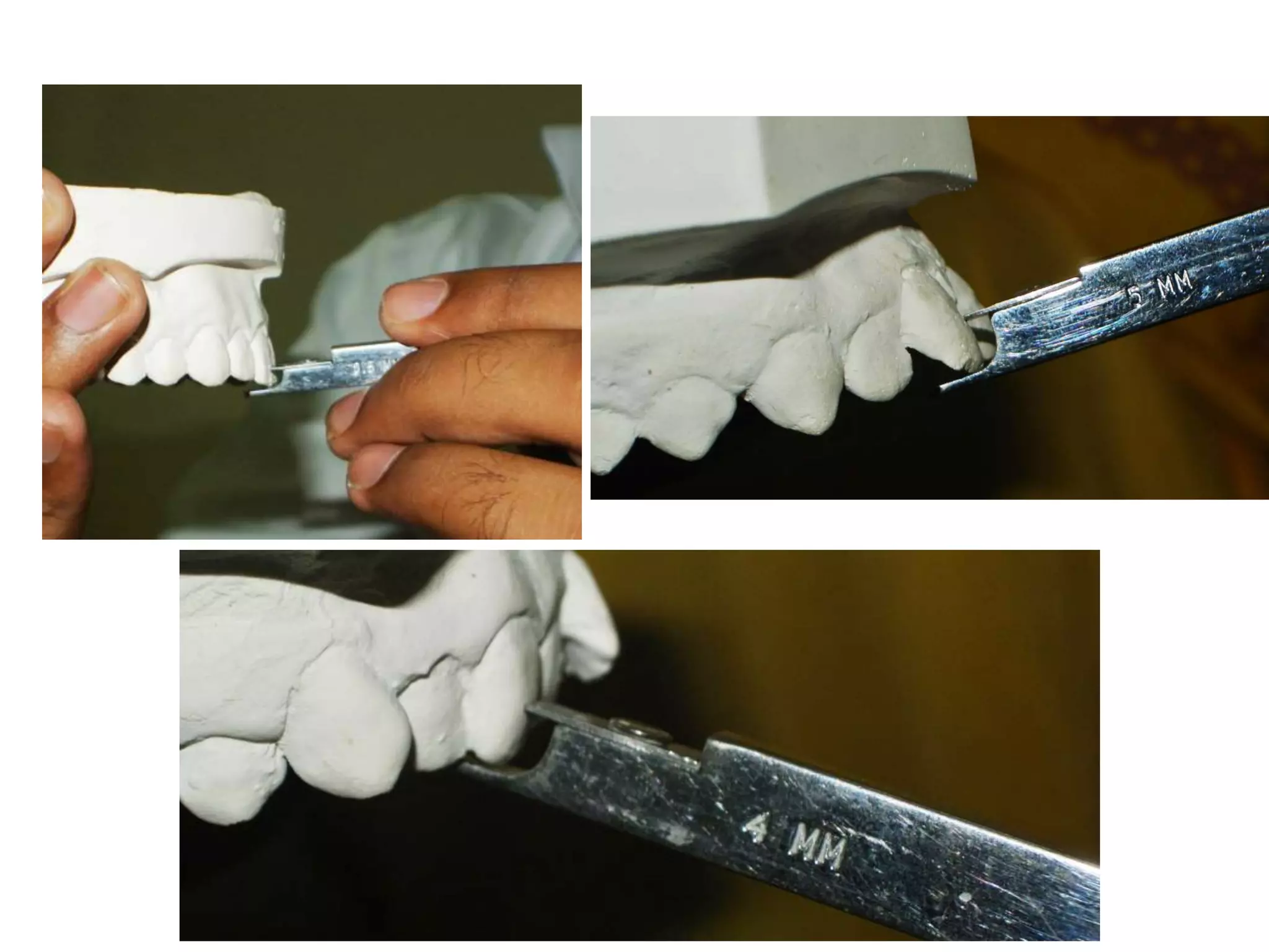
• In caseclass II div 1 incisor relationship where the
upper incisors are proclined the gauge is angulated
more upward as compared to normal incisor
inclination.
15.
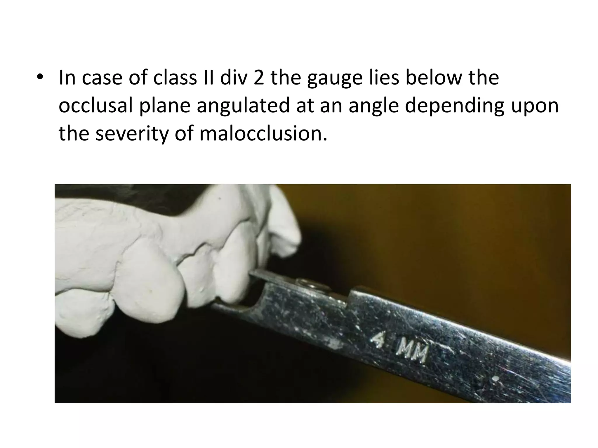
• In caseof class II div 2 the gauge lies below the
occlusal plane angulated at an angle depending upon
the severity of malocclusion.
17.
Bracket placementby wire guidance
• In this technique all the steps of conventional
bonding are done in usual way but before curing the
bracket a heavy wire is passed through the bracket
slot and its bonded neighboring brackets and bands.
The mesiodistal position of the bracket is corrected
manually while axial and vertical positions are guided
by the heavy wire.
18.
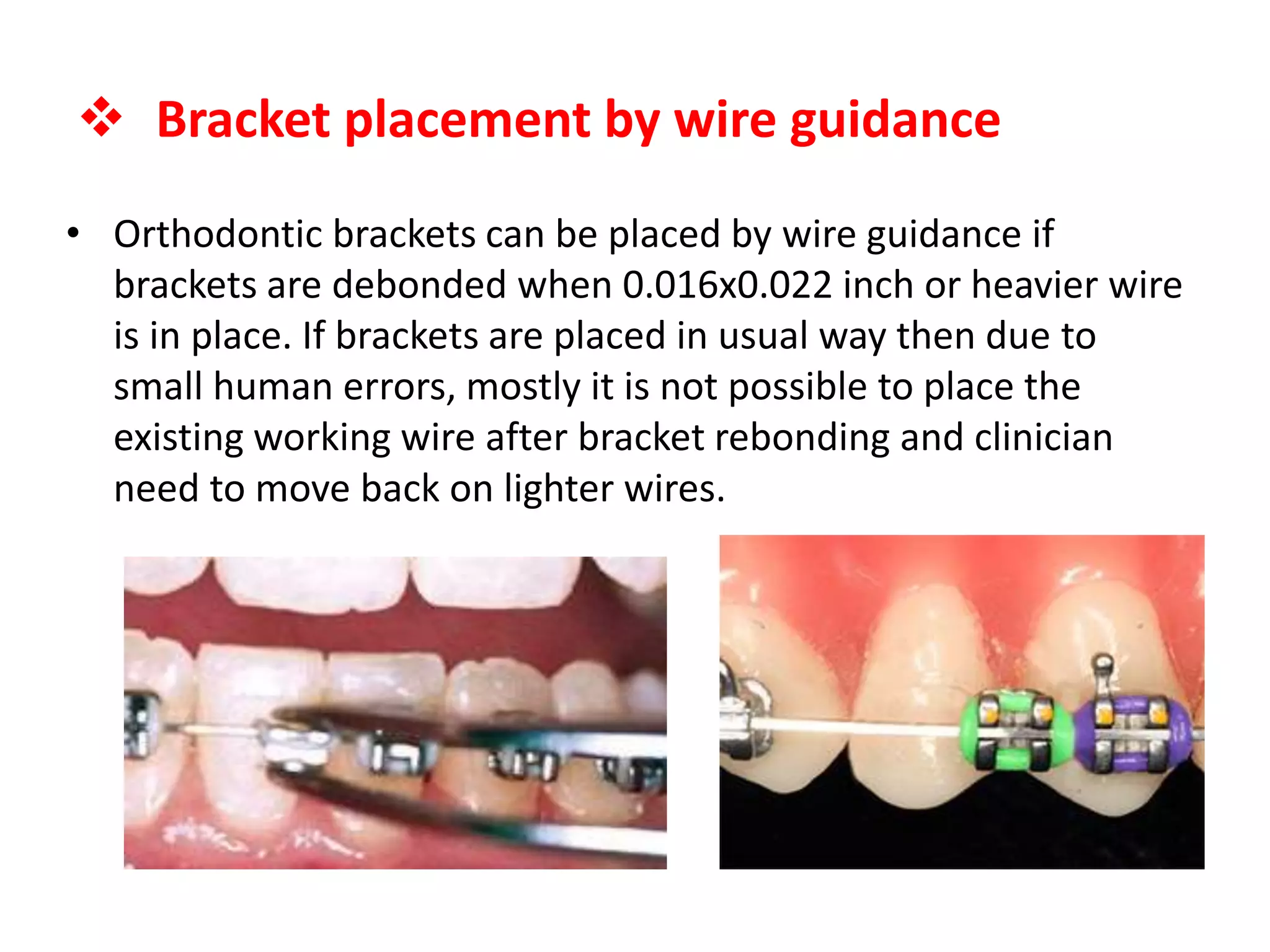
• Orthodontic bracketscan be placed by wire guidance if
brackets are debonded when 0.016x0.022 inch or heavier wire
is in place. If brackets are placed in usual way then due to
small human errors, mostly it is not possible to place the
existing working wire after bracket rebonding and clinician
need to move back on lighter wires.
Bracket placement by wire guidance
19.
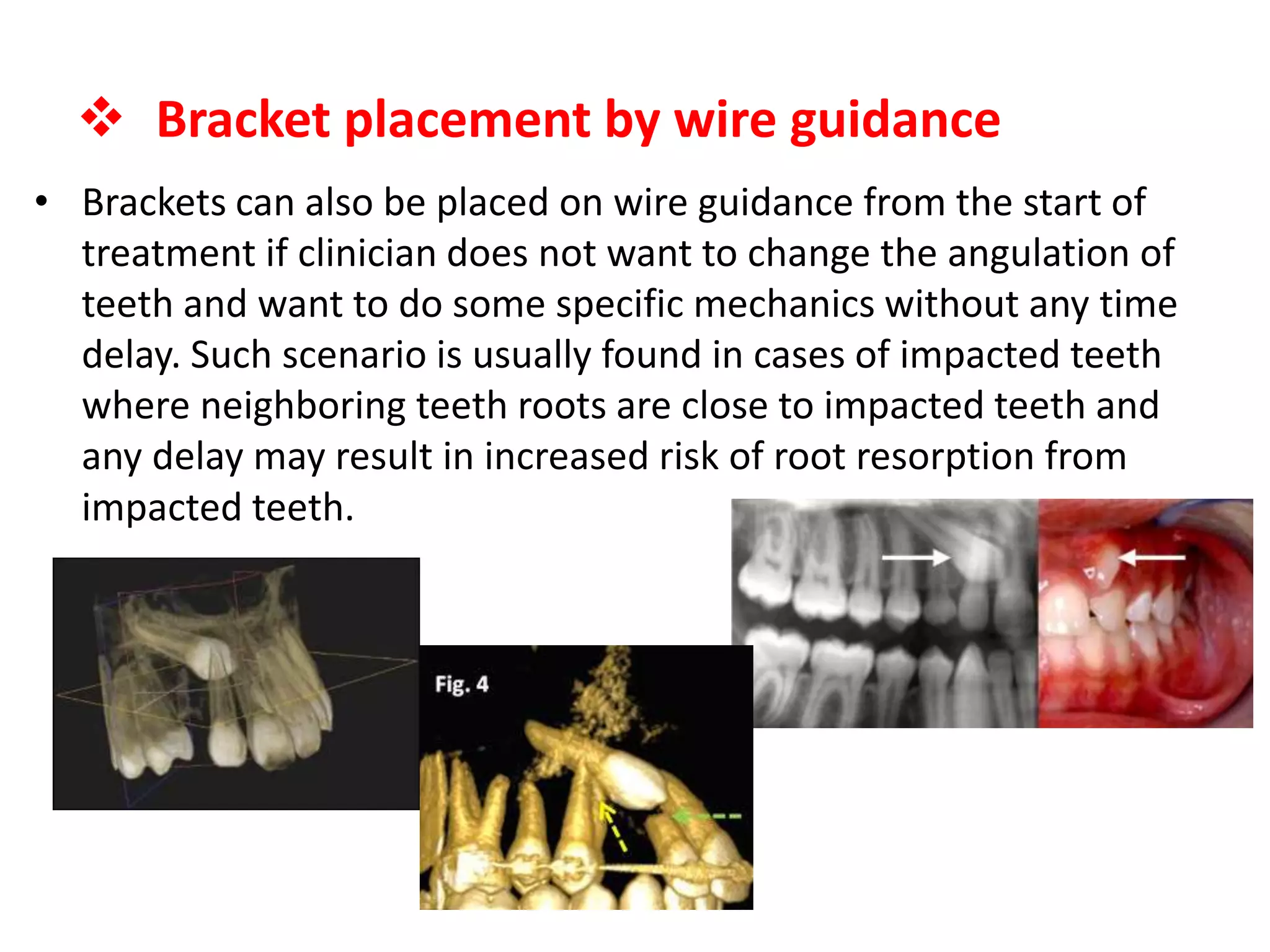
• Brackets canalso be placed on wire guidance from the start of
treatment if clinician does not want to change the angulation of
teeth and want to do some specific mechanics without any time
delay. Such scenario is usually found in cases of impacted teeth
where neighboring teeth roots are close to impacted teeth and
any delay may result in increased risk of root resorption from
impacted teeth.
Bracket placement by wire guidance
20.
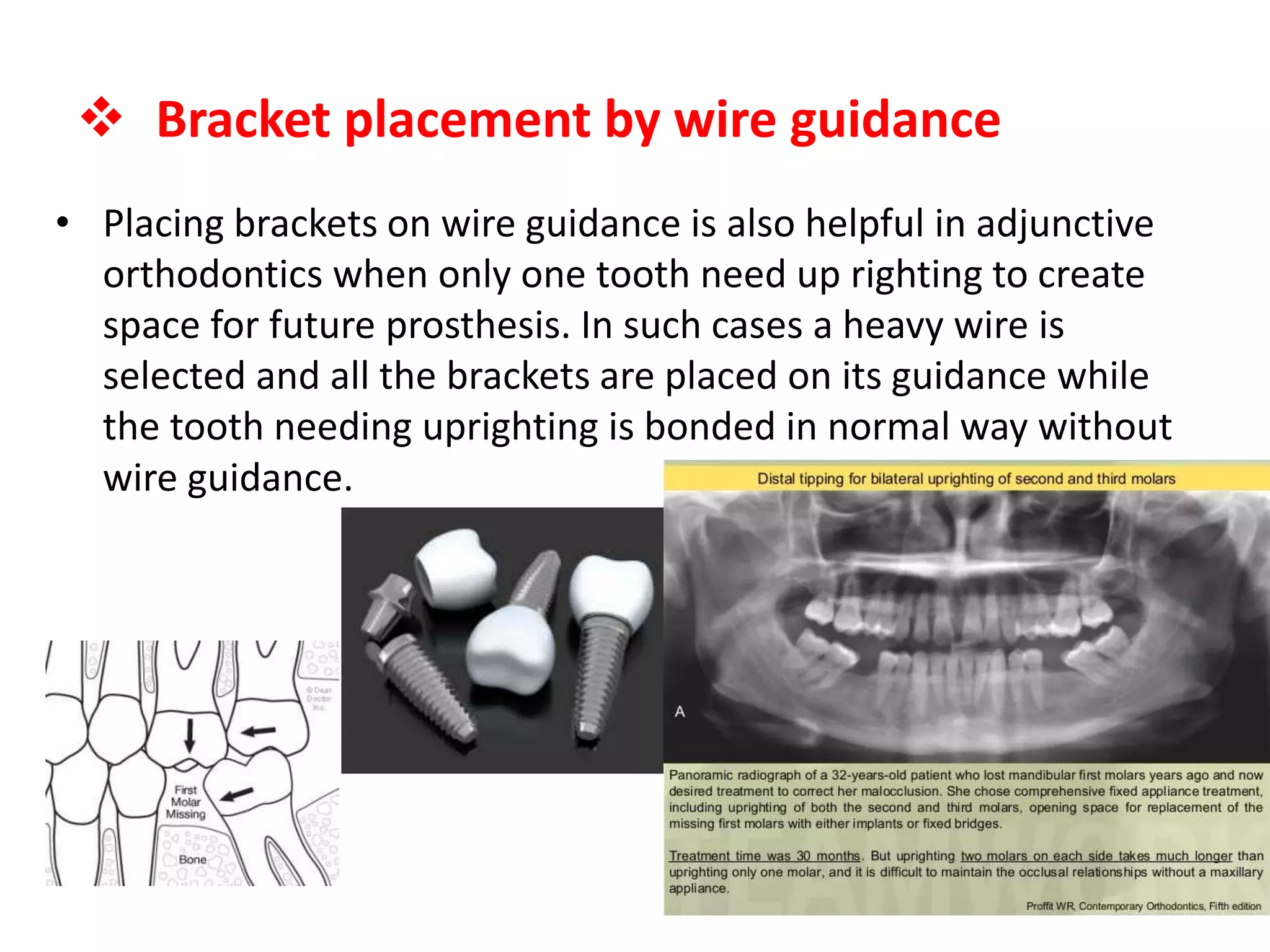
• Placing bracketson wire guidance is also helpful in adjunctive
orthodontics when only one tooth need up righting to create
space for future prosthesis. In such cases a heavy wire is
selected and all the brackets are placed on its guidance while
the tooth needing uprighting is bonded in normal way without
wire guidance.
Bracket placement by wire guidance
21.
Position ofclinician during brackets
placement
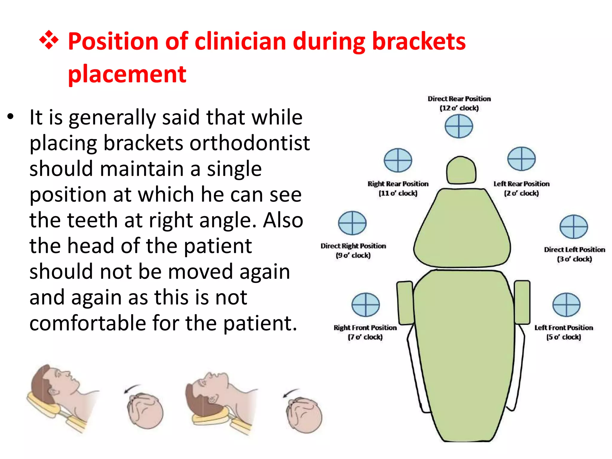
• It is generally said that while
placing brackets orthodontist
should maintain a single
position at which he can see
the teeth at right angle. Also
the head of the patient
should not be moved again
and again as this is not
comfortable for the patient.
22.
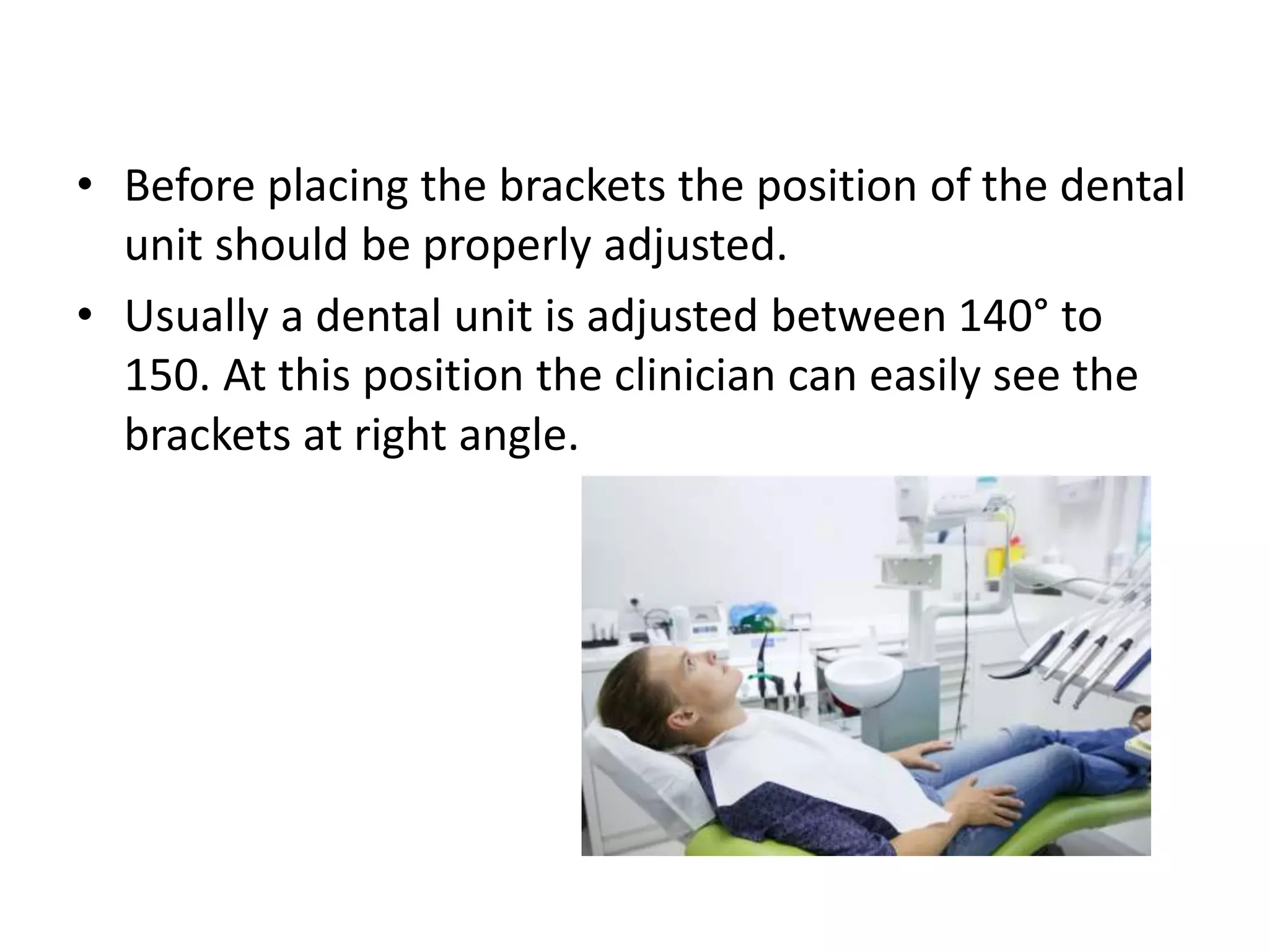
• Before placingthe brackets the position of the dental
unit should be properly adjusted.
• Usually a dental unit is adjusted between 140° to
150. At this position the clinician can easily see the
brackets at right angle.
23.
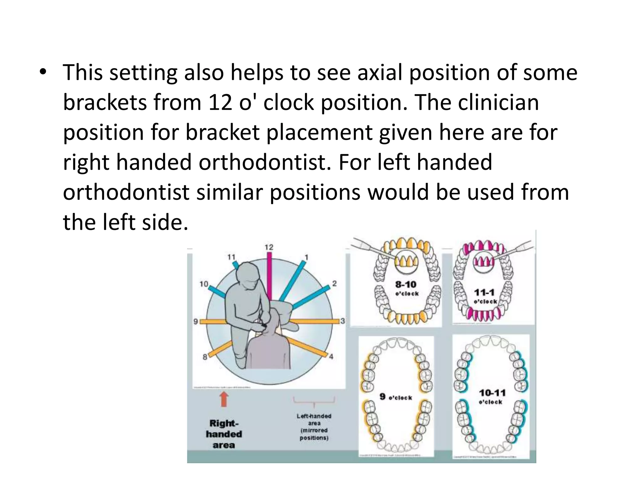
• This settingalso helps to see axial position of some
brackets from 12 o' clock position. The clinician
position for bracket placement given here are for
right handed orthodontist. For left handed
orthodontist similar positions would be used from
the left side.
24.
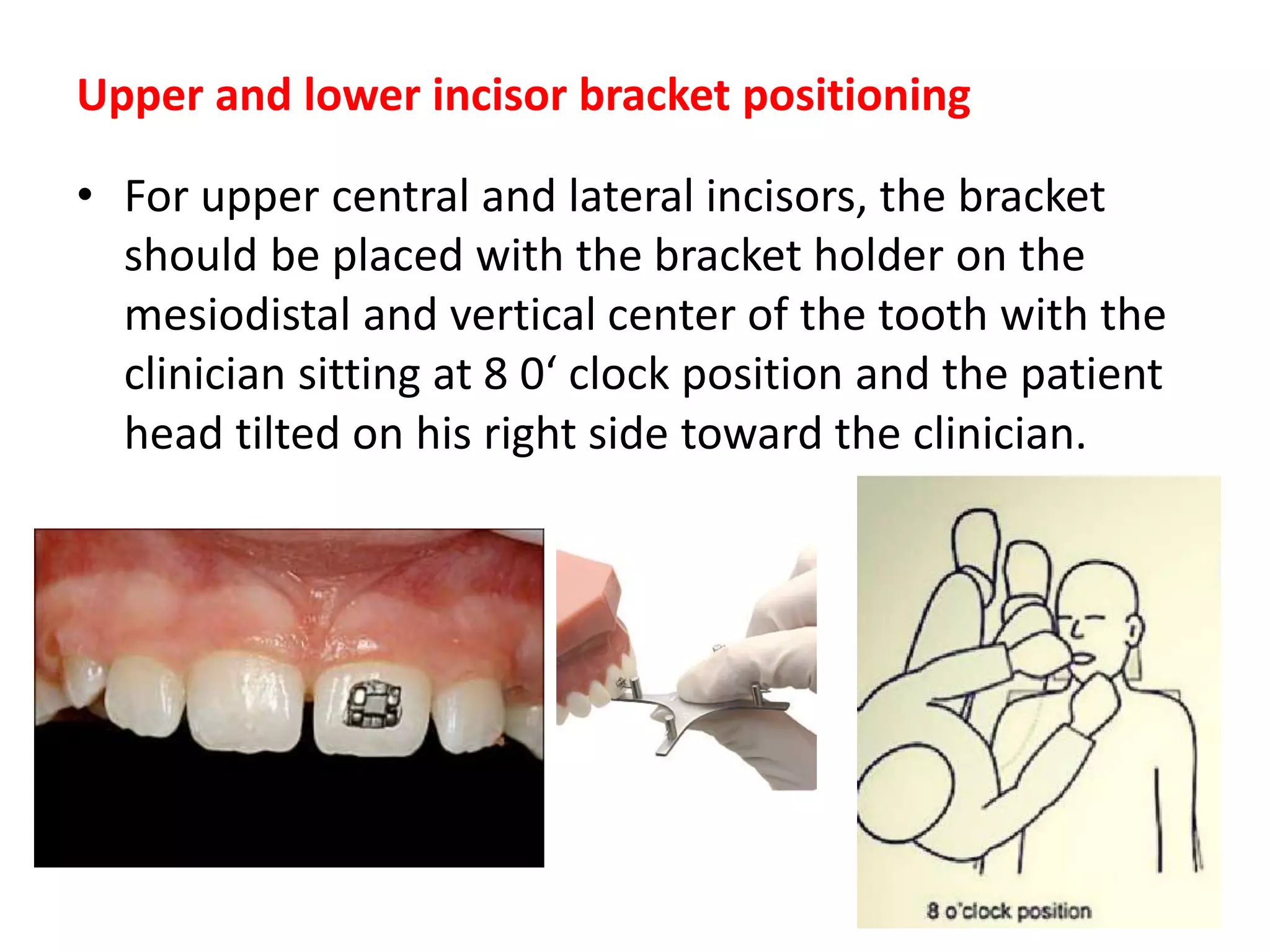
Upper and lowerincisor bracket positioning
• For upper central and lateral incisors, the bracket
should be placed with the bracket holder on the
mesiodistal and vertical center of the tooth with the
clinician sitting at 8 0‘ clock position and the patient
head tilted on his right side toward the clinician.
25.
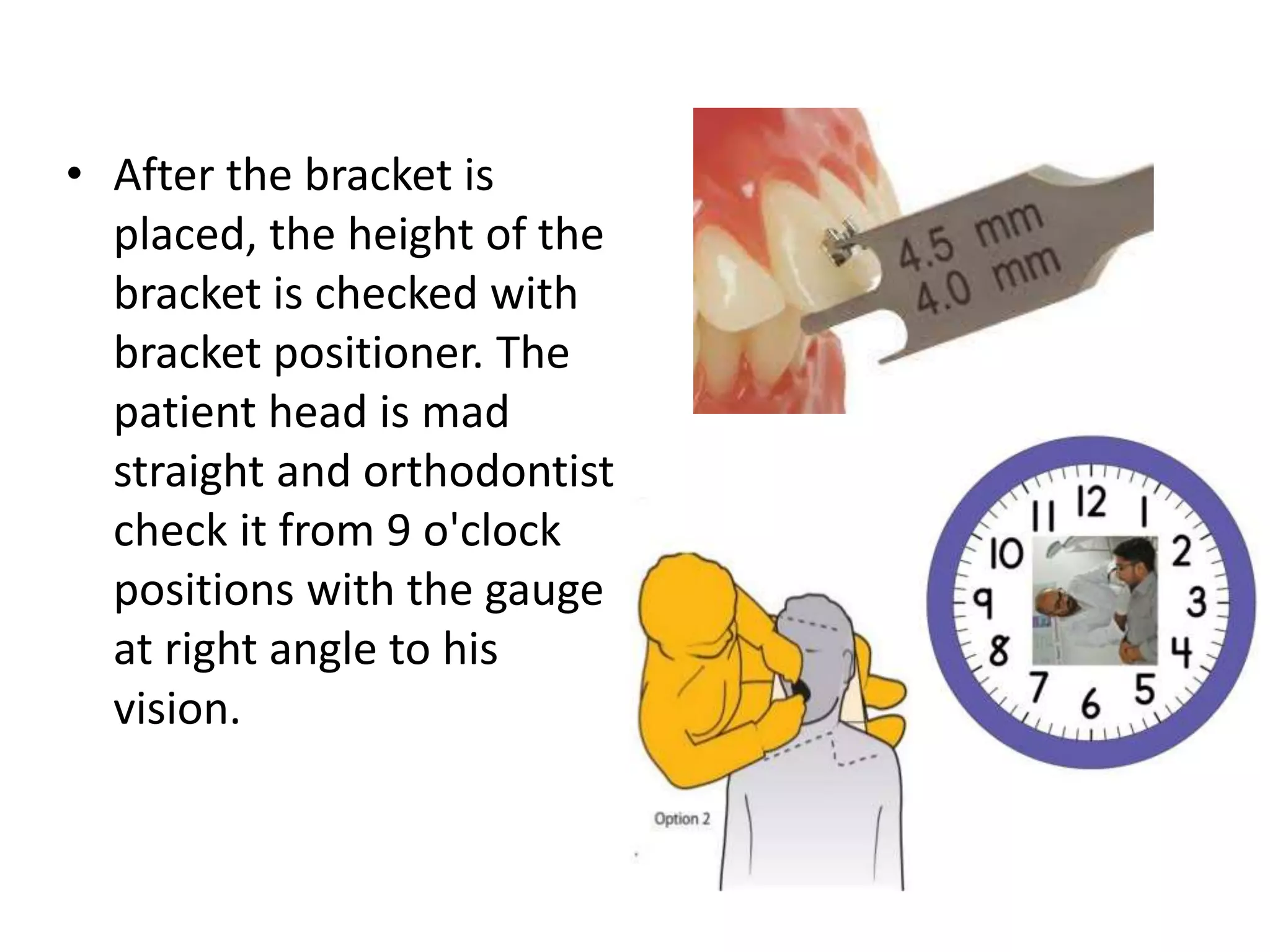
• After thebracket is
placed, the height of the
bracket is checked with
bracket positioner. The
patient head is mad
straight and orthodontist
check it from 9 o'clock
positions with the gauge
at right angle to his
vision.
26.
• To checkthe mesiodistal and axial position of the
bracket the orthodontist moves to 12 o‘ clock
position and place a diagnostic mouth mirror at the
incisor edge to indirectly check the mesiodistal
position of the bracket.
27.
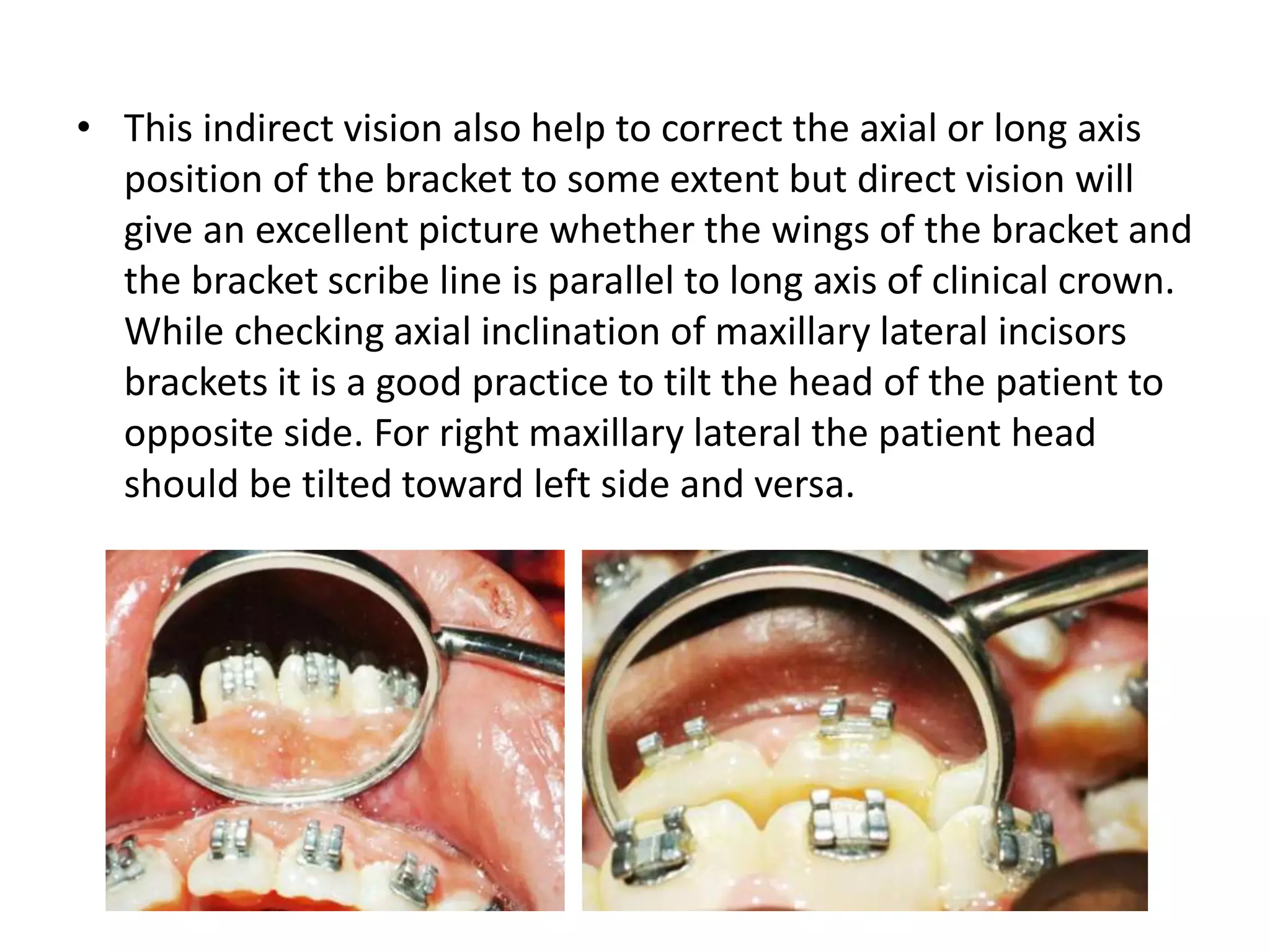
• This indirectvision also help to correct the axial or long axis
position of the bracket to some extent but direct vision will
give an excellent picture whether the wings of the bracket and
the bracket scribe line is parallel to long axis of clinical crown.
While checking axial inclination of maxillary lateral incisors
brackets it is a good practice to tilt the head of the patient to
opposite side. For right maxillary lateral the patient head
should be tilted toward left side and versa.
28.
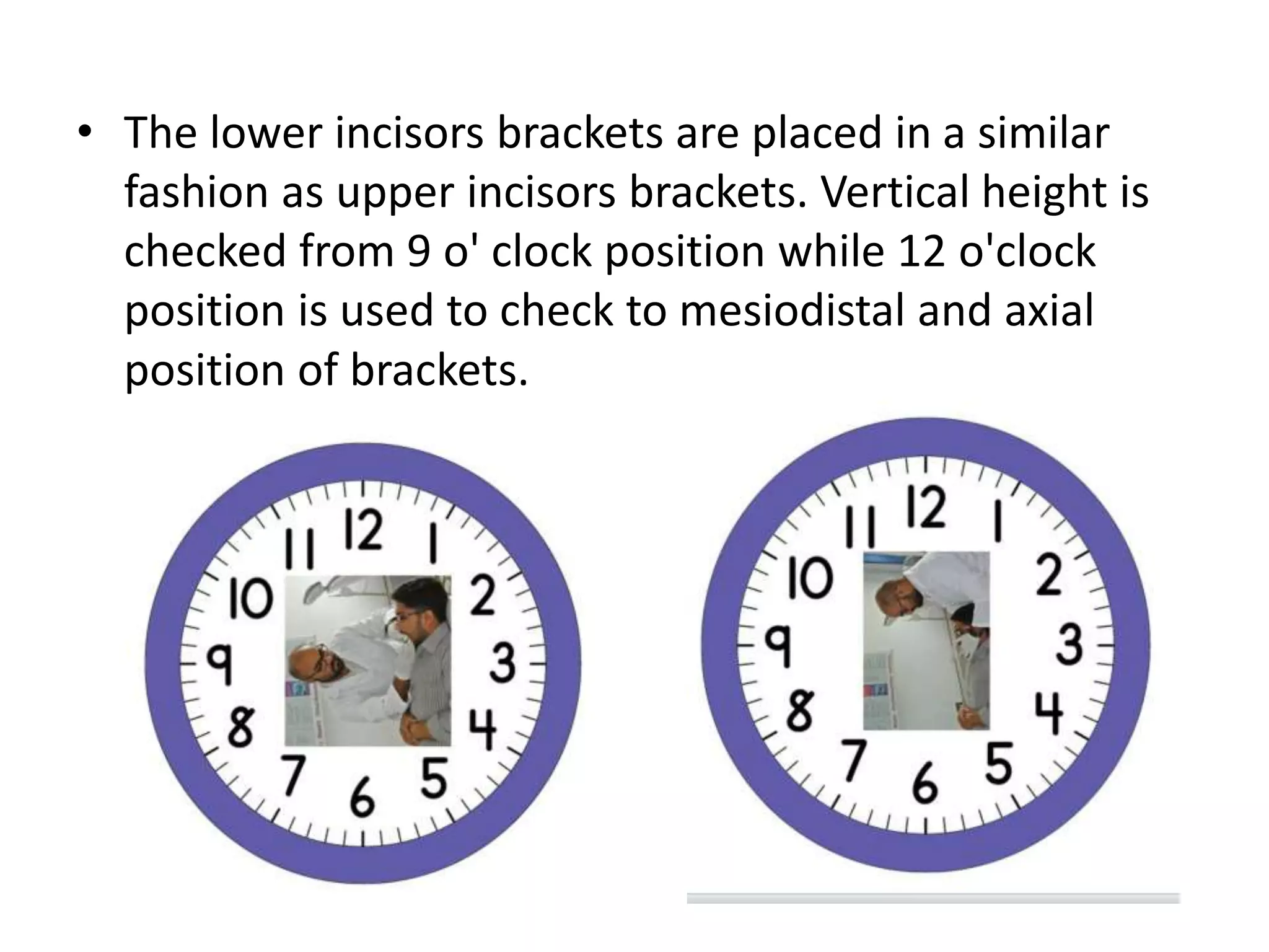
• The lowerincisors brackets are placed in a similar
fashion as upper incisors brackets. Vertical height is
checked from 9 o' clock position while 12 o'clock
position is used to check to mesiodistal and axial
position of brackets.
29.
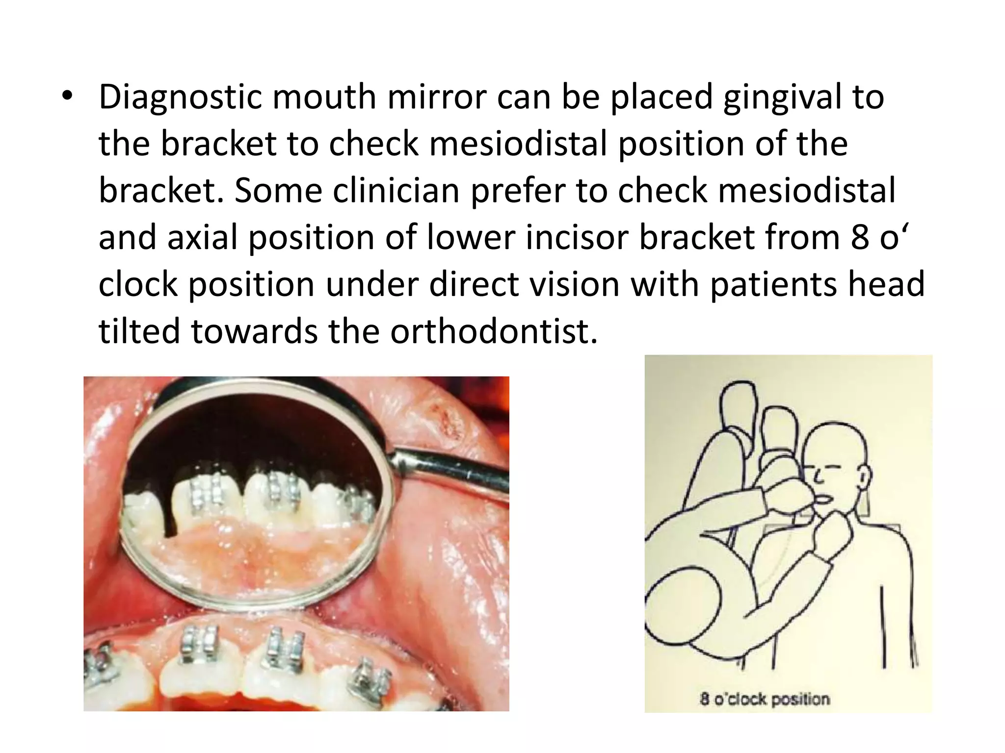
• Diagnostic mouthmirror can be placed gingival to
the bracket to check mesiodistal position of the
bracket. Some clinician prefer to check mesiodistal
and axial position of lower incisor bracket from 8 o‘
clock position under direct vision with patients head
tilted towards the orthodontist.
30.
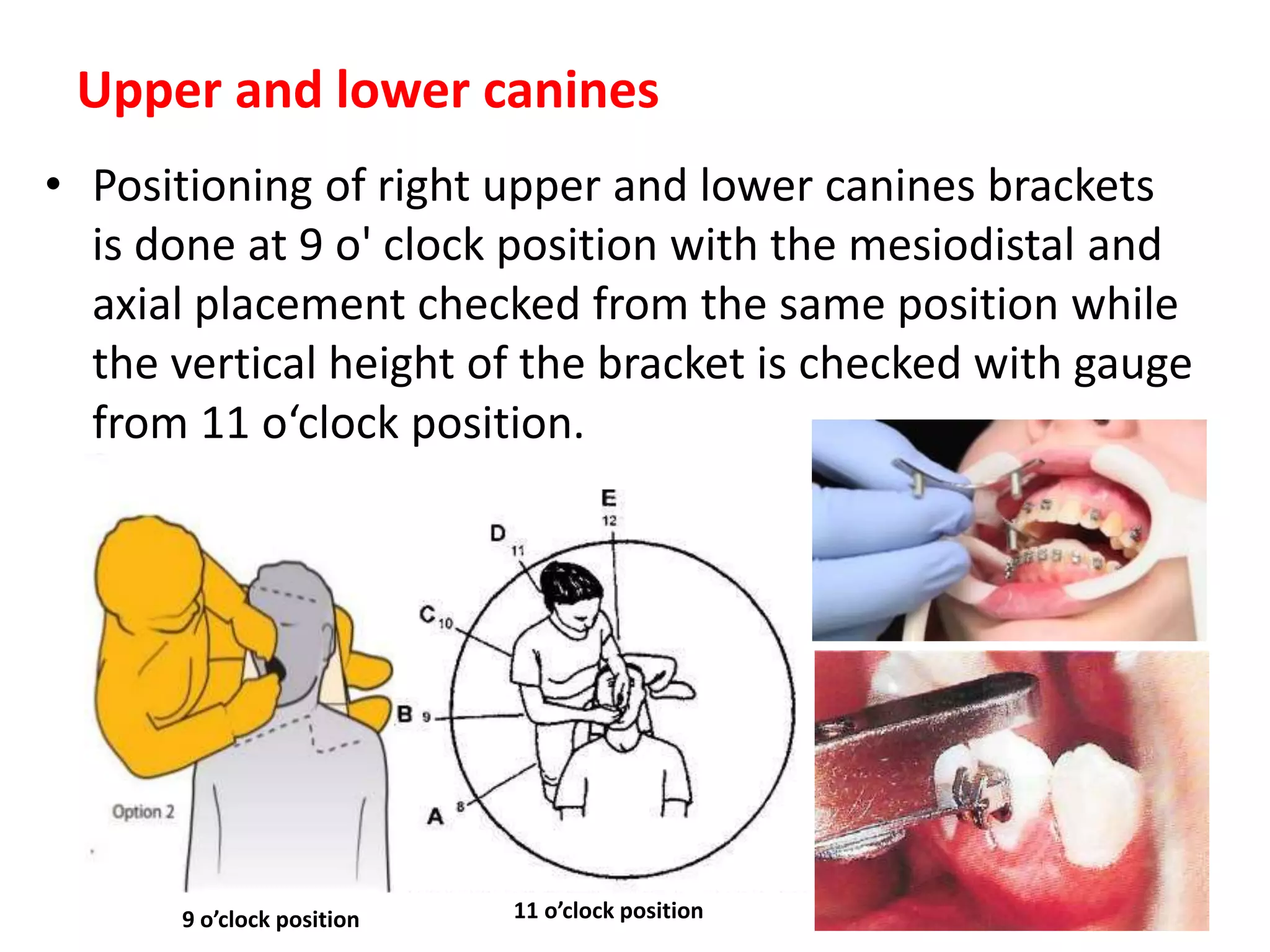
Upper and lowercanines
• Positioning of right upper and lower canines brackets
is done at 9 o' clock position with the mesiodistal and
axial placement checked from the same position while
the vertical height of the bracket is checked with gauge
from 11 o‘clock position.
9 o’clock position 11 o’clock position
31.
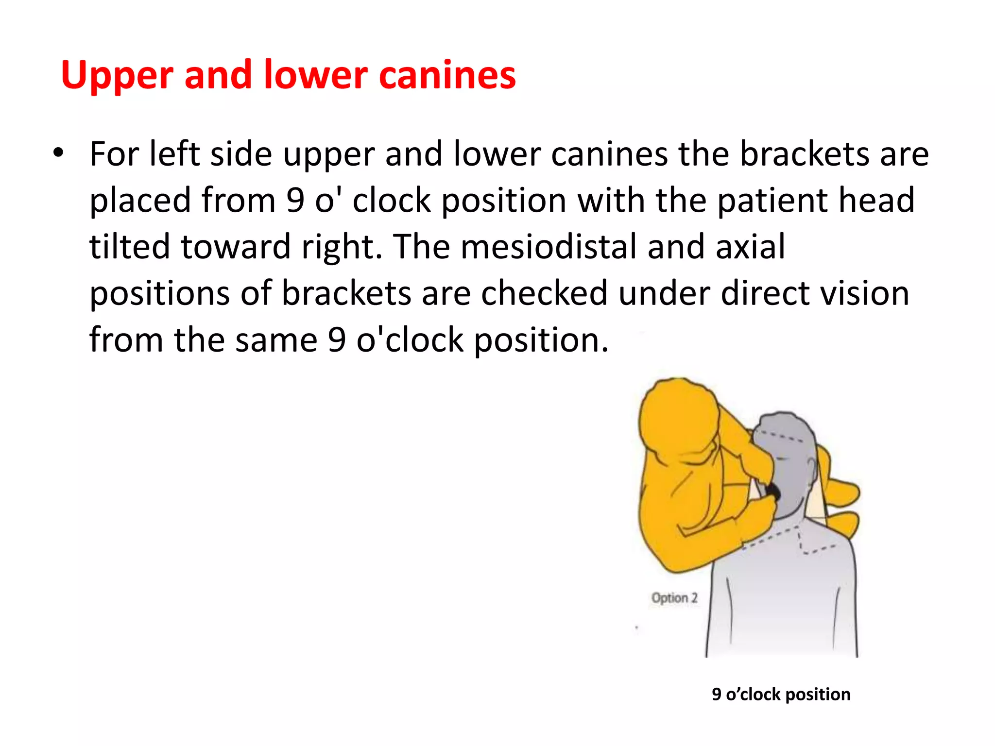
• For leftside upper and lower canines the brackets are
placed from 9 o' clock position with the patient head
tilted toward right. The mesiodistal and axial
positions of brackets are checked under direct vision
from the same 9 o'clock position.
Upper and lower canines
9 o’clock position
32.
Upper and lowerbicuspids
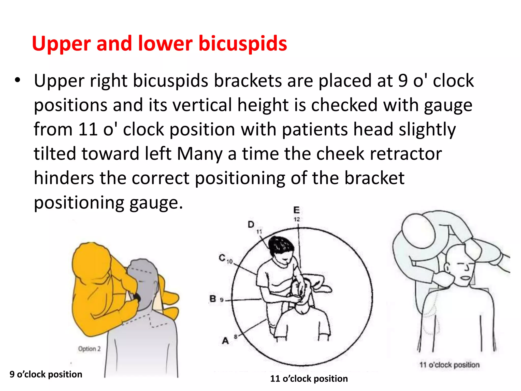
• Upper right bicuspids brackets are placed at 9 o' clock
positions and its vertical height is checked with gauge
from 11 o' clock position with patients head slightly
tilted toward left Many a time the cheek retractor
hinders the correct positioning of the bracket
positioning gauge.
9 o’clock position 11 o’clock position
33.
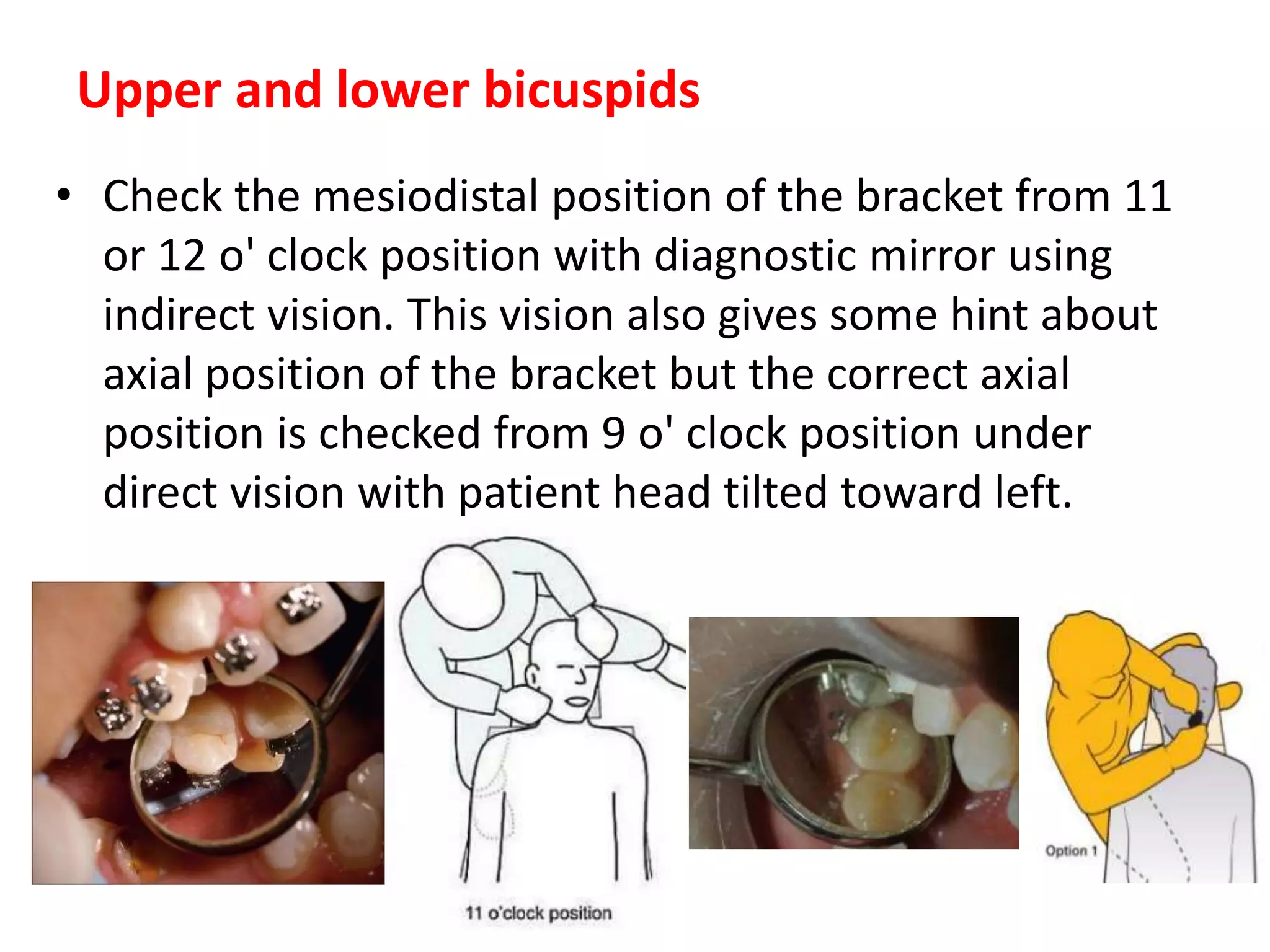
• Check themesiodistal position of the bracket from 11
or 12 o' clock position with diagnostic mirror using
indirect vision. This vision also gives some hint about
axial position of the bracket but the correct axial
position is checked from 9 o' clock position under
direct vision with patient head tilted toward left.
Upper and lower bicuspids
34.
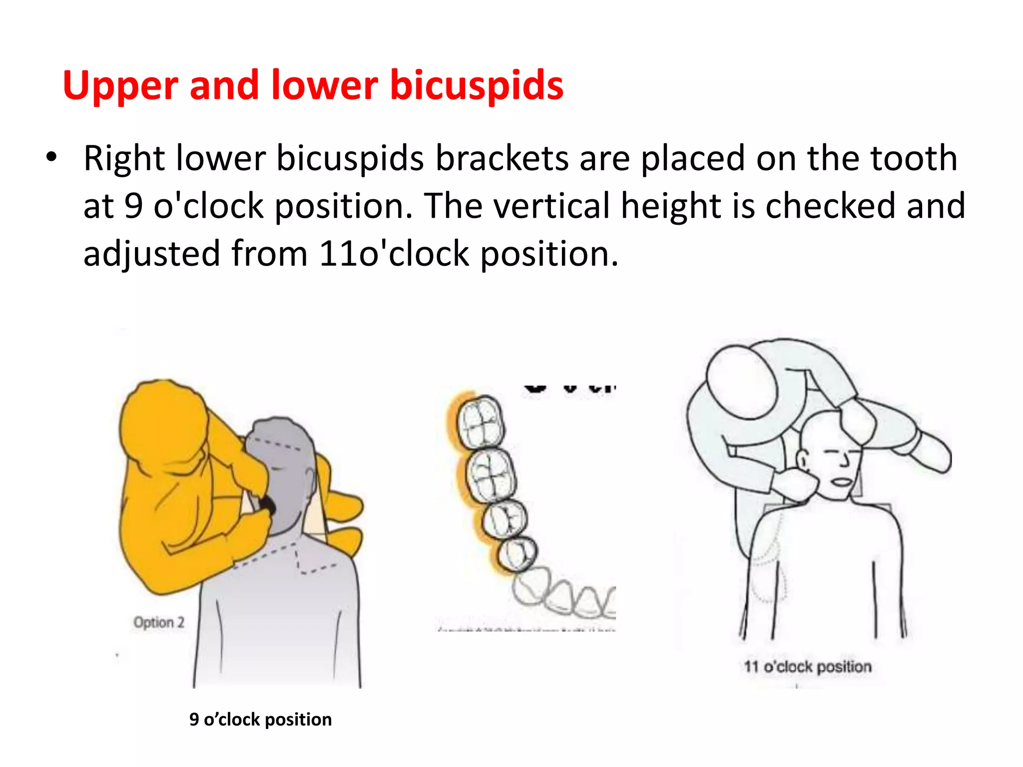
• Right lowerbicuspids brackets are placed on the tooth
at 9 o'clock position. The vertical height is checked and
adjusted from 11o'clock position.
Upper and lower bicuspids
9 o’clock position
35.
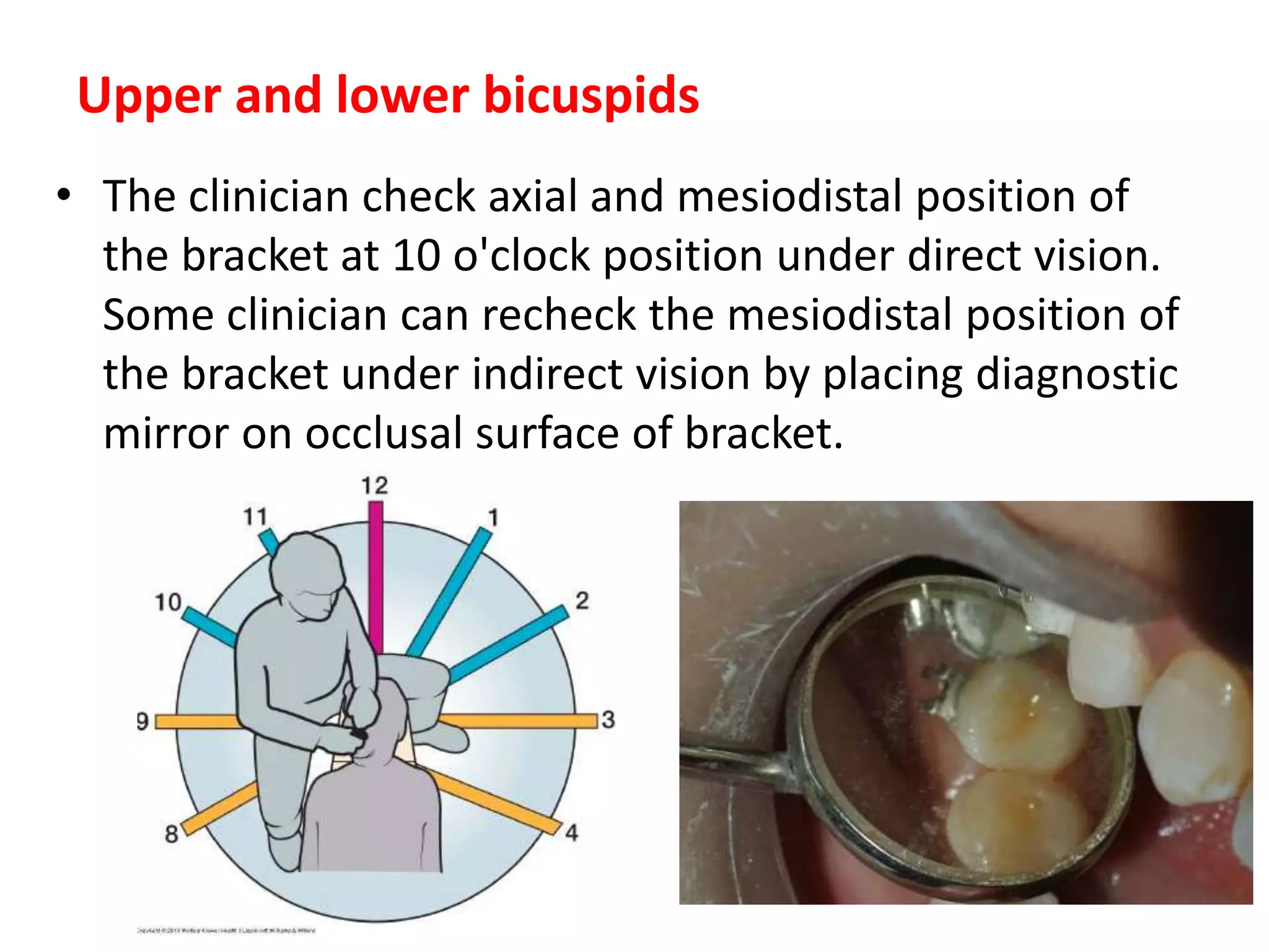
• The cliniciancheck axial and mesiodistal position of
the bracket at 10 o'clock position under direct vision.
Some clinician can recheck the mesiodistal position of
the bracket under indirect vision by placing diagnostic
mirror on occlusal surface of bracket.
Upper and lower bicuspids
36.
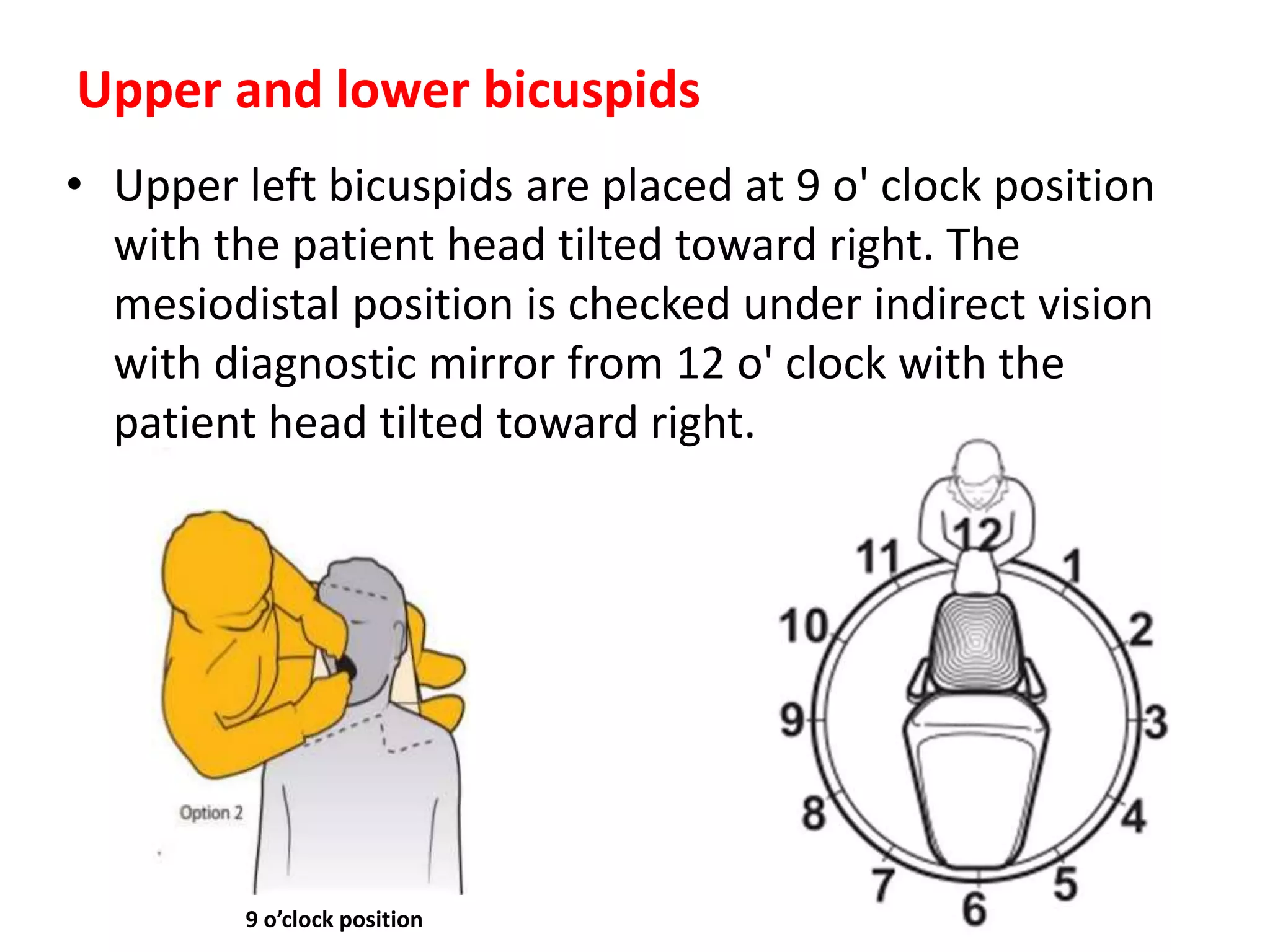
• Upper leftbicuspids are placed at 9 o' clock position
with the patient head tilted toward right. The
mesiodistal position is checked under indirect vision
with diagnostic mirror from 12 o' clock with the
patient head tilted toward right.
Upper and lower bicuspids
9 o’clock position
37.
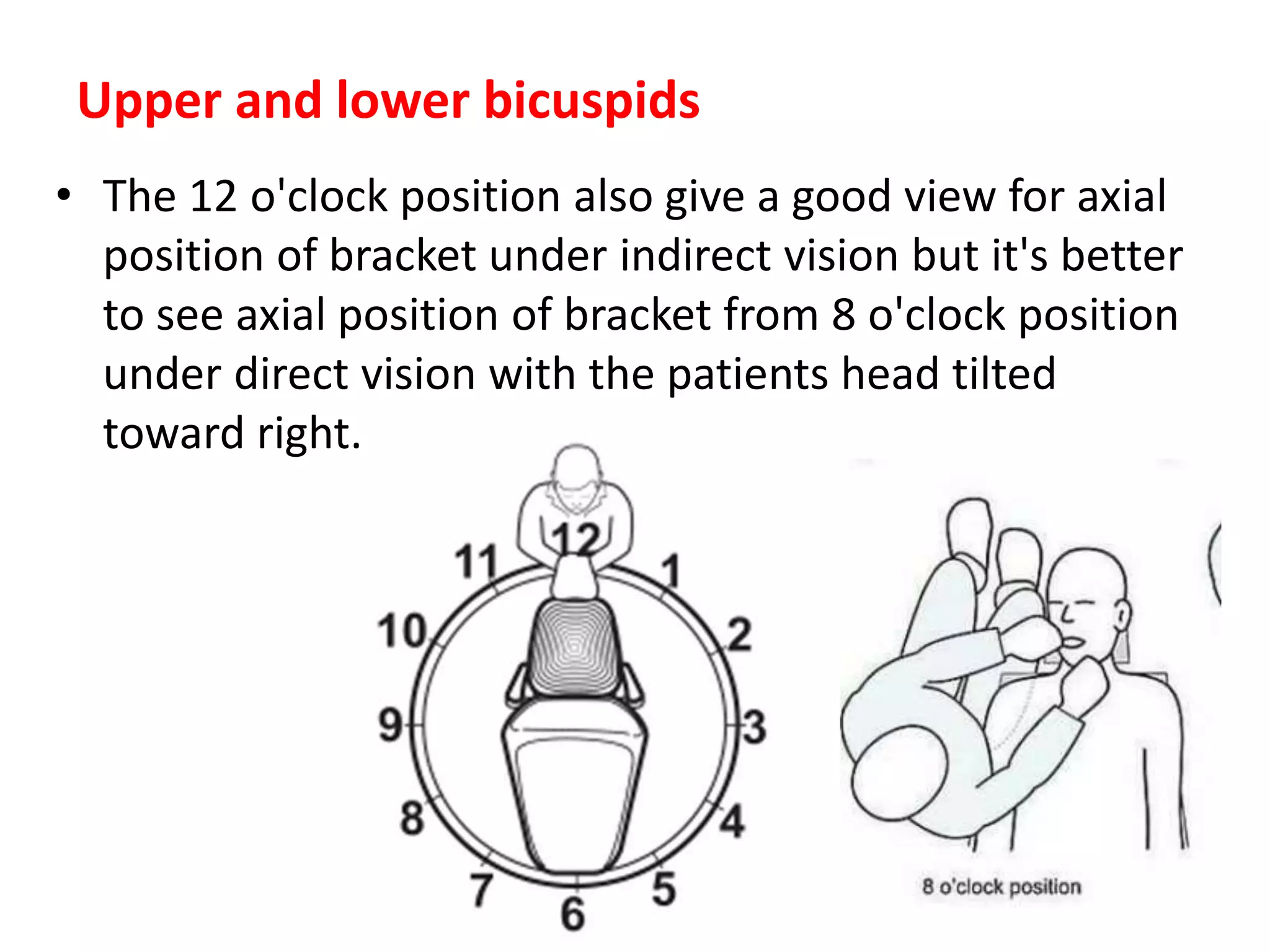
• The 12o'clock position also give a good view for axial
position of bracket under indirect vision but it's better
to see axial position of bracket from 8 o'clock position
under direct vision with the patients head tilted
toward right.
Upper and lower bicuspids
38.
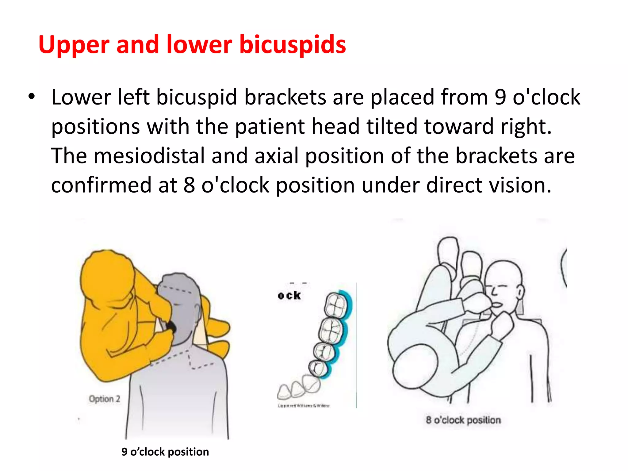
• Lower leftbicuspid brackets are placed from 9 o'clock
positions with the patient head tilted toward right.
The mesiodistal and axial position of the brackets are
confirmed at 8 o'clock position under direct vision.
Upper and lower bicuspids
9 o’clock position