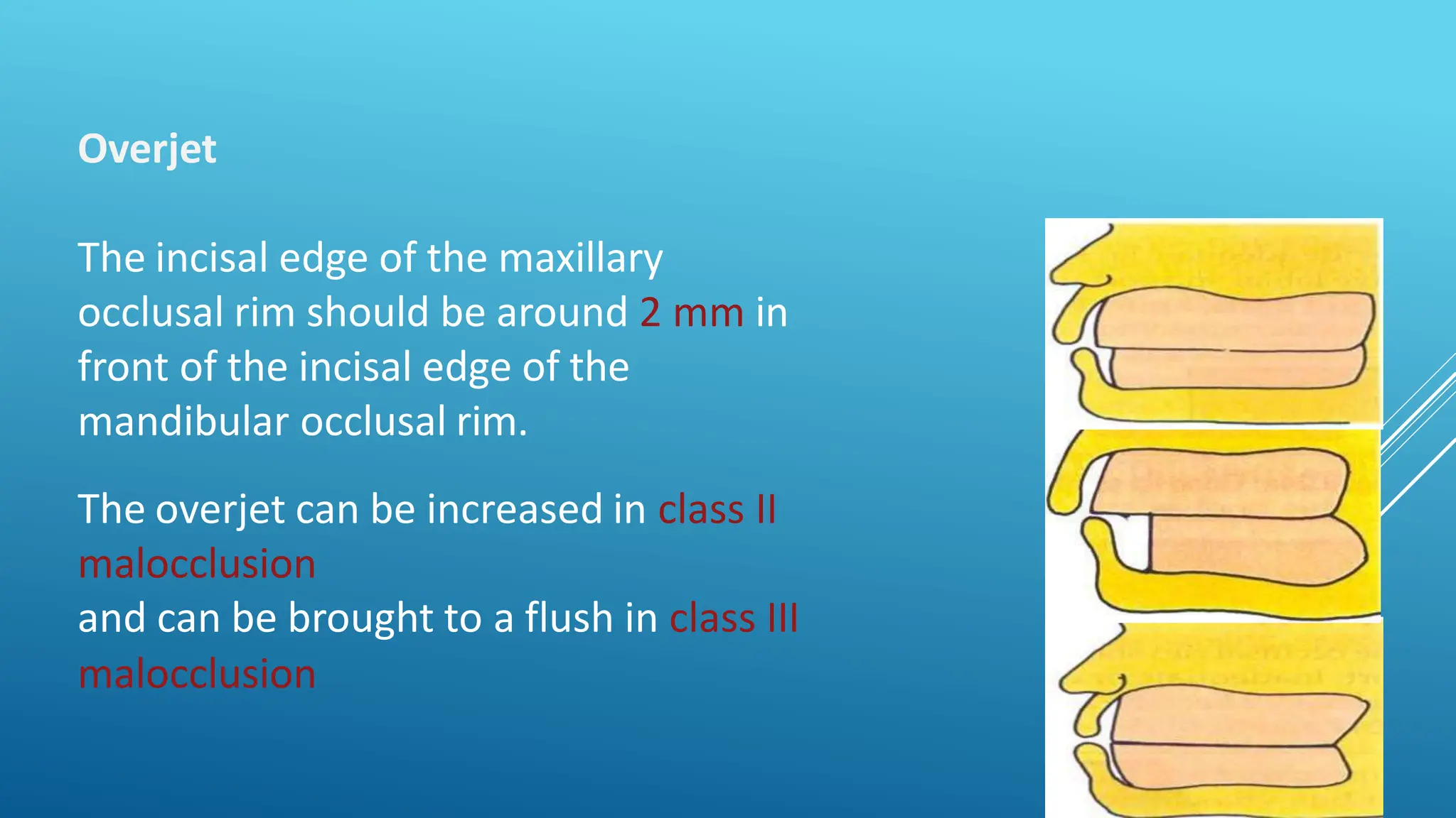
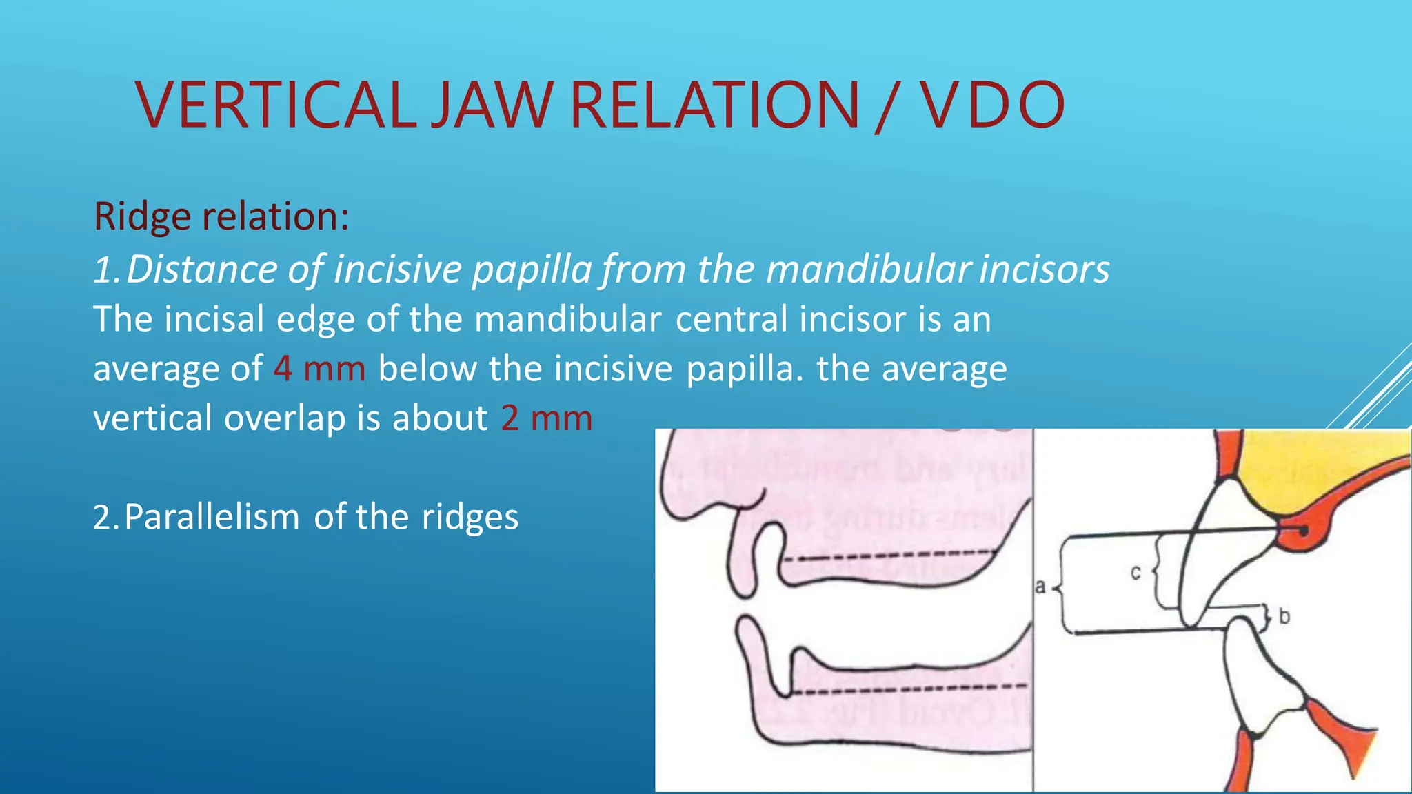
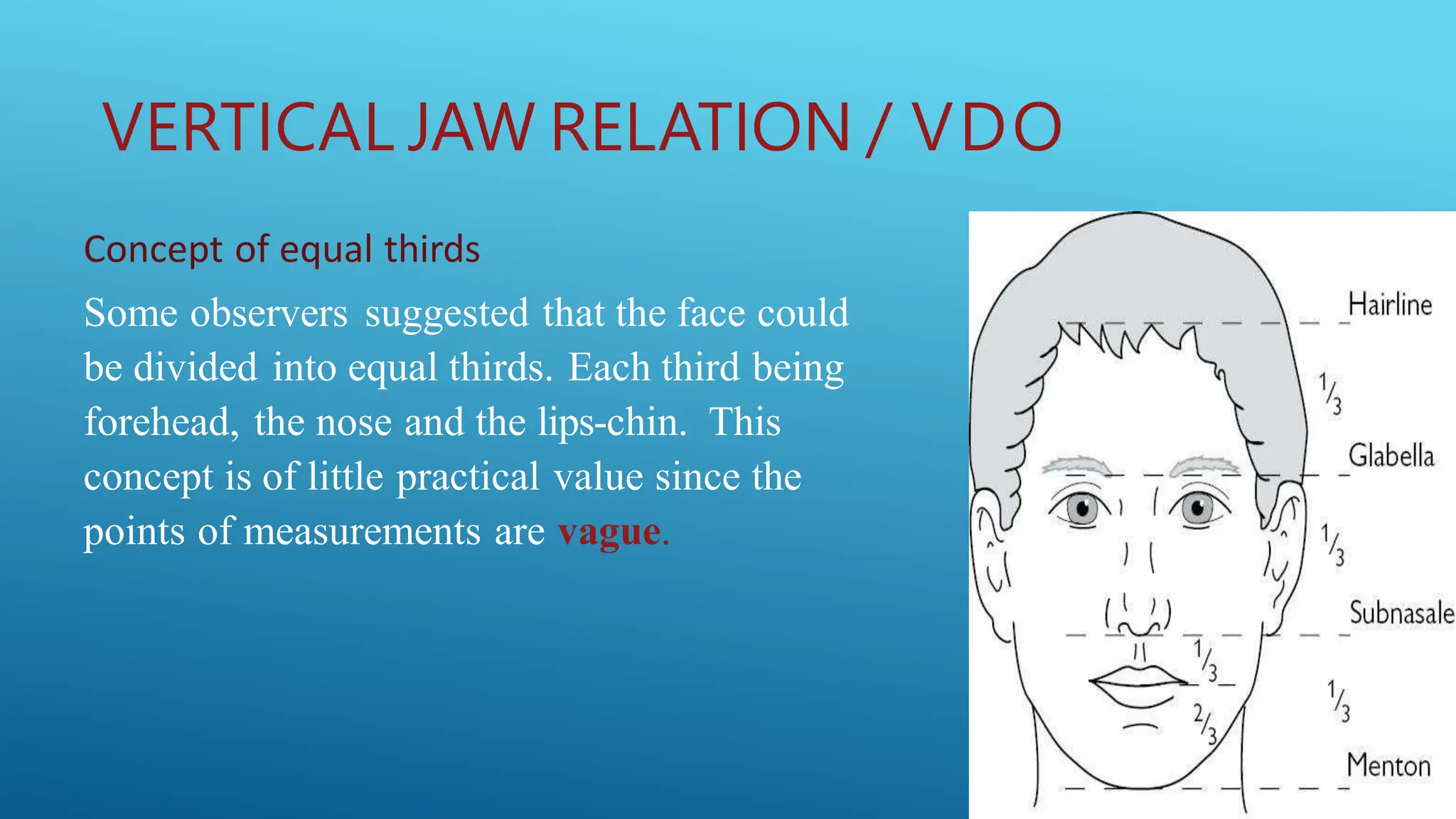
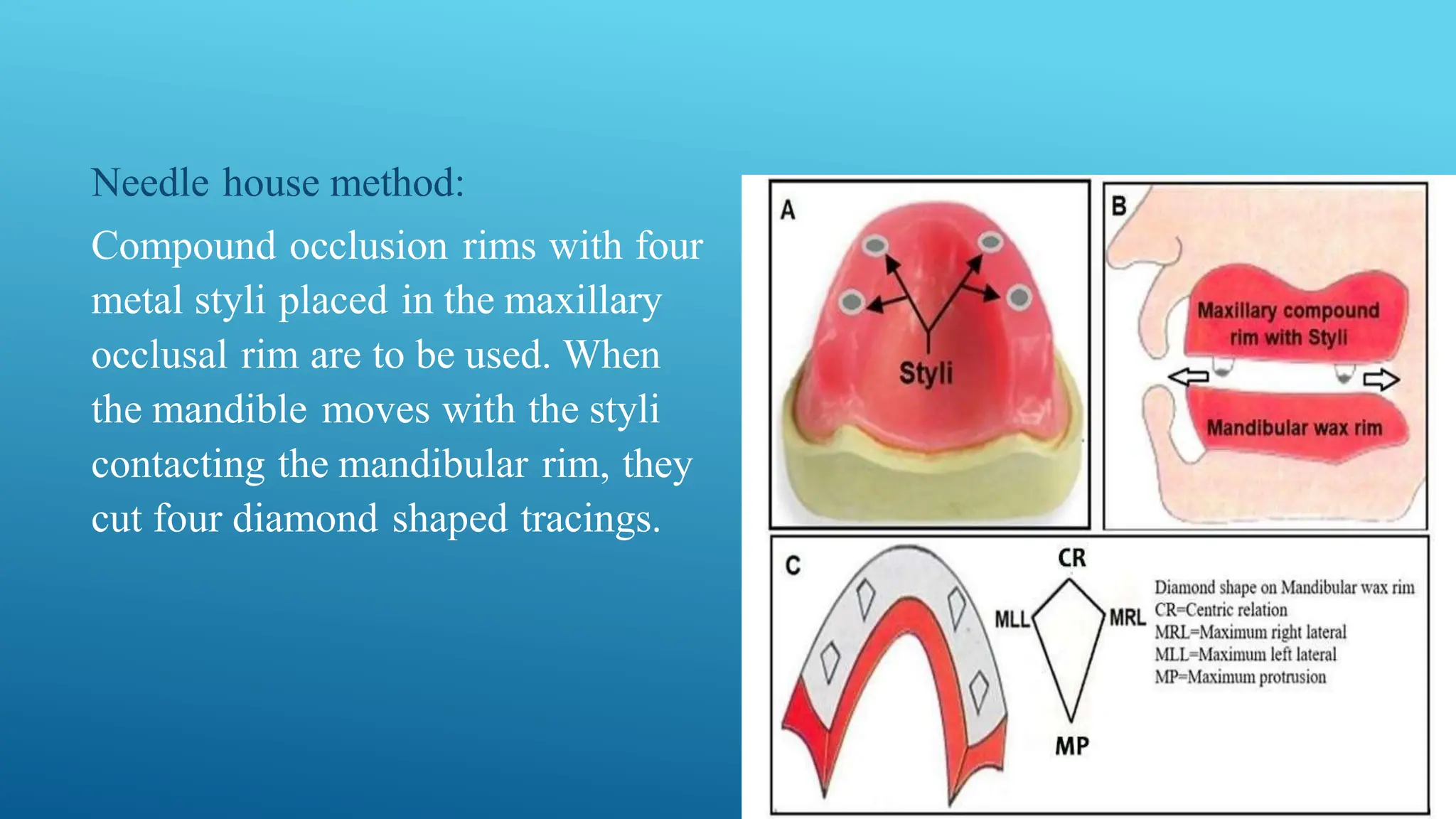
Call Girl Number in Vashi Mumbai📲 9833363713 💞 Full Night Enjoy
jaw relation in complete and partial denture
1. JAW RELATION
FOR COMPLETE AND PARTIAL
DENTURE
Presented BY:
Muamal Fadhil
Supervised BY:
Dr. Ann Akram Nasser
2. Contents
1. Jaw Relation in Complete Denture
a. Orientation Relation
b. Vertical Relation
c. Horizontal Relation
I.Centric relation.
II.Eccentric relation.
a. Protrusive relation.
b. Lateral relation.
2. Jaw Relation in Partial Denture
4. Occlusal Rim /Record Rim
The ideal dimension of maxillary occlusion rim
1. Height
A. Anterior - 24 mm
B. Posterior - 21 mm
2. Width
Anterior - 6 mm
Posterior - 8 mm
5. Occlusal Rim /Record Rim
The ideal dimension of mandibular occlusion rim:
1. Height
A. Anterior - 20 mm
B. Posterior - approximately 18 mm
2. Width
Anterior - 6 mm
Posterior - 8 mm
6. The occlusal rims are inserted into the patient’s mouth and the
following factors are checked:
• Lip support and labial fullness
• Overjet
• Cheek support and buccal fullness
• Level of the occlusal plane
• Orientation of plane of occlusion
7. Lip Support and Labial Fullness
The wax in the flange area is
responsible for the labial fullness.
The amount of wax in the incisal
edge of the occlusal rim
determines the lip support.
8. Overjet
The incisal edge of the maxillary
occlusal rim should be around 2 mm in
front of the incisal edge of the
mandibular occlusal rim.
The overjet can be increased in class II
malocclusion
and can be brought to a flush in class III
malocclusion
9. Cheek Support and Buccal Fullness
The occlusal rims should be
designed to be within the neutral
zone.
10. Level Of The Occlusal Plane
The occlusal plane should be located at the
midpoint of the inter-arch distance.
the length of bite rim anteriorly depended
on length of upper lip.
the length of bite rim Posteriorly it should
be 2/3 the height of the retromolar pad.
11. The occlusal plane can be verified by
using a Fox plane.
The plane of the maxilla is determined
anteriorly by inter-pupillary line and
Posteriorly by the Camper’s line or
the ala-tragus line
12. ORIENTATION OF PLANE OF
OCCLUSION
Orientation jaw relation can be
recorded with a face-bow.
This record gives the angulation of
the maxilla in relation to the base of
the skull.
14. Physiologic Rest Position: It is the postural position of the mandible
when the individual is resting comfortably in an upright position and
the associated muscles are in a state of minimal contractual activity.
Vertical dimension at rest VDR: It is the vertical dimension of face with
jaws in rest position.
Vertical dimension at occlusion VDR:It is the vertical dimension of face
when the teeth or occlusal rims are in contact in centric occlusion:.
Freeway space: It is the difference between the vertical dimension at
rest and vertical dimension at occlusion.
VERTICAL JAW RELATION
15. 1- Niswonger’s method: It is commonly
used today. The patient is seated so
that the ala-tragal line is parallel with
the floor. Two markings are made, one
at the tip of the nose and the other on
the most prominent part of the chin.
VERTICAL JAW RELATION / VDR
16. 2- Tactile sense: Instruct the patient to
open the jaws wide until strain is felt in
the muscles. When this opening
becomes uncomfortable, ask them to
close slowly until the jaws reach a
comfortable, relaxed position. Measure
the distance between the points of
reference.
VERTICAL JAW RELATION / VDR
17. 3- Phonetics: Ask the patient to
repeat pronounce the letter m a
certain number of times, like repeat
the name Emma when the lips touch.
At this time measure between the
two points of reference.
VERTICAL JAW RELATION / VDR
18. 4- Facial expression:
• Skin around the eyes and chin should be
relaxed. It should not be stretched, shiny
or excessively wrinkled.
• The nostrils are relaxed and breathing
should be unobstructed.
• The upper and lower lips should have a
slight contact
VERTICAL JAW RELATION / VDR
19. 5- Willis’s method: The Willis guide is
designed to measure the distance
from the pupils of the eyes to the
corner of the mouth and the distance
from the anterior nasal spine to the
lower border of the mandible. When
these measurements are equal, the
jaws are considered at rest. Its
accuracy is questionable in patients
with facial asymmetry.
VERTICAL JAW RELATION / VDR
20. 6- Electromyographic method (EMG):
By using a special device that
measures the tone of masticatory
muscles, when the tone is at its least,
this means these muscles are in rest
position and the jaws are at rest
position.
VERTICAL JAW RELATION / VDR
21. Mechanical Methods
1. Ridge relation:
a.Distance of incisive papilla
b. Parallelism of the ridges.
2. Measurement of former dentures:
3. Pre-extraction records:
a.Profile photograph
b. Profile radiograph
c. Profile silhouettes
d. Swenson method
e. Articulated casts.
3. Post-extraction methods:
a. Concept of equal thirds
b. phonetic, Silverman’s closest speaking space
c. Boo’s method
VERTICAL JAW RELATION / VDO
Physiological methods
1. Physiologic rest position.
2. Facial expression.
3. Swallowing method.
4. Tactile sense.
22. Ridge relation:
1.Distance of incisive papilla from the mandibularincisors
The incisal edge of the mandibular central incisor is an
average of 4 mm below the incisive papilla. the average
vertical overlap is about 2 mm
2.Parallelism of the ridges
VERTICAL JAW RELATION / VDO
23. Measurement of former dentures: The
patient’s old denture can be used as a
reference for the vertical dimension of
the new denture.
VERTICAL JAW RELATION / VDO
24. Pre-extraction records:
a. Profile photograph
b. Profile radiograph
c. Profile silhouettes
d. Articulated casts
VERTICAL JAW RELATION / VDO
25. Concept of equal thirds
Some observers suggested that the face could
be divided into equal thirds. Each third being
forehead, the nose and the lips-chin. This
concept is of little practical value since the
points of measurements are vague.
VERTICAL JAW RELATION / VDO
26. VERTICAL JAW RELATION / VDO
Silverman’s closest speaking space
It is the minimal amount of interocclusal space between the upper and lower
teeth when sounds like ch, s, and j are pronounced.
There is 1-2 mm clearance between teeth when observed from the profile and
frontal view.
27. VERTICAL JAW RELATION / VDO
Boo’s method
Boo found that there is a point of maximum biting power at occlusion.
28. VERTICAL JAW RELATION / VDO
Physiologic rest position
After the insertion of occlusal rims into the patient’s mouth,
the patient is
asked to swallow and let the jaw relax.
The interocclusal rest space should be 2 to 4 mm.
29. VERTICAL JAW RELATION / VDO
Facial expression
If the vertical dimension is too high the skin of the cheeks
will appear very stretched and the nasolabial fold will be
obliterated, the nasolabial angle will be increased.
Lips the contour and fullness of the lip is affected by the
thickness of the
labial flange. The occlusal rims should be contoured to aid in
lip support.
30. Swallowing method
The position of the mandible at the beginning of the
swallowing act has been used as a guide to the vertical
relation.
VERTICAL JAW RELATION / VDO
31. VERTICAL JAW RELATION / VDO
Tactile sense
The patient’s tactile sense is used as a guide to the
determination of the
oclusal vertical relation.
32. EFFECTS OF INCREASEDVERTICALRELATION
1. Discomfort to the patient
2. Trauma to the mucous membrane by frequent contact of teeth
3. Loss of freeway space, which may lead to:
• Muscular fatigue of any one or group of muscles of mastication.
• Trauma caused by constant pressure on mucous membrane.
• Annoyance from the inability to find comfortable position.
4. Clicking teeth/clattering of teeth.
5. The face has an elongated appearance since at rest the lips are parted and closing them
together will produce an expression of strain.
6. Residual alveolar bone undergoes rapid resorption.
7. Temporomandibular joint pains.
33. 1.Cheek biting: In some cases, there is losing of muscular tone, as a result of reduced vertical height where
the flabby cheek tends to become trapped between the teeth during mastication.
2. Appearance: The general effect of over closure on facial expression is increased with age. There is close
approximation of nose to chin, the soft tissue sag and the lines on the face are deepened. The lips loose
their fullness and the vermillion borders are reduced to approximately a line.
3. Angular cheilitis: A reduced vertical relation results in crease at the corners of the mouth beyond the
vermillion border and the deep fold thus formed becomes bathed in saliva leading to infection and soreness.
4. Pain in TMJ: Trauma in the region of temporomandibular fossa may be attributed to a reduced vertical
relation with symptoms like obscure pains, discomfort, clicking sounds, headaches and neuralgia.
5. Costen’s syndrome: In 1934,Costen listed a number of symptoms that he believed were caused by over
closure of mandible following loss of teeth .The symptoms associated with the syndrome were
impaired hearing, stuffy sensation in the ear,
EFFECTS OF DECREASED VERTICAL RELATION
35. HORIZONTAL JAW RELATION
Methods of Assisting the Patient to Move the Mandible to the Centric Relation:
1. The patient is instructed to let his or her jaw relax, pull it back and close
slowly on the back teeth.
2. The patient is instructed to get the feeling of pushing his upper jaw out and
then close the mouth with back teeth in contact.
3. Assist the patient to protrude and retrude the mandible repeatedly with the
operator holding the finger lightly against the chin.
4. The patient can be instructed to turn the tongue towards the posterior border
of the upper denture base and close the rims together until they meet.
5. Boo’s series of stretch exercises.
36. HORIZONTAL JAW RELATION
Various Methods of Recording Centric Relation
1. Functional (Chew-in) methods
a. Needle house method.
b. Patterson’s method.
c. Meyer’s method.
2. Excursive methods (Graphic method)
3. Tactile or interocclusal check records.
4. Other methods.
a. Heating the surface of one of the rims.
b. Softened wax placed over the occlusal surfaces of the occlusal rim.
c. Soft cones of wax placed on the lower denture trial bases.
37. Needle house method:
Compound occlusion rims with four
metal styli placed in the maxillary
occlusal rim are to be used. When
the mandible moves with the styli
contacting the mandibular rim, they
cut four diamond shaped tracings.
38. Patterson’s method:
A trench is to be made in the mandibular rim
and a mixture of half plaster and half pumice
should be placed in the trench. The
mandibular movements generate
compensating curves. When the paste is
reduced to the predetermined vertical height
of occlusion, the patient is instructed to
retrude the mandible and the occlusal rims
are joined together with metal staples.
40. GRAPHIC
METHOD :
The general concept of this technique is that a pen-like
pointer is attached to one occlusal rim and a recording
plate is placed on the other rim, the plate coated with
carbon or wax on which the needle point can make the
tracing, when the mandiblemoves in horizontalplane,
the pointer draws characteristic patterns on the
recording plate. The characteristic patterns created on
the recording plate is called Arrow Point Tracing, also
known as GothicArch Tracing.
The graphic methodsare either
intraoral or extraoral
41. HORIZONTAL JAW RELATION
Eccentric Relation Record:
It is defined as any relationship of the mandible to the maxilla other than the
centric relation.
It includes protrusive and lateral relations.
The main reason in making an eccentric jaw relation record is to adjust the
horizontal and lateral condylar inclination in the adjustable articulator, and to
establish the balanced occlusion.
The protrusive and left and right lateral movements records are made in
the same manner as for centric relation record and these include:
1. Functional methods.
2. Graphic methods.
3. Inter-occlusal check record method
42. method for establishing occlusal
relationships:
1. Direct Apposition of Casts
2. Interocclusal Records With Posterior
Teeth Remaining
3. Occlusal Relations Using Occlusion
Rims on Record Bases
4. Jaw relation made entirely on
occlusion rims
RELATION IN PARTIAL DENTURE