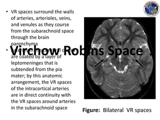
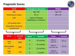
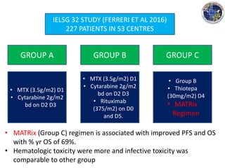
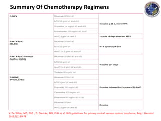
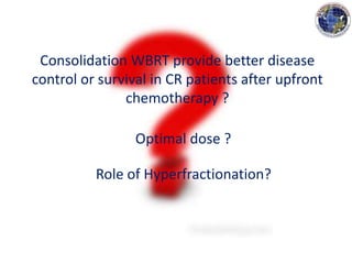
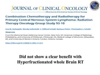
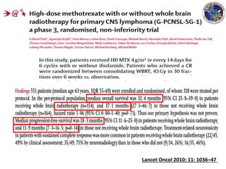
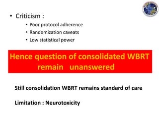
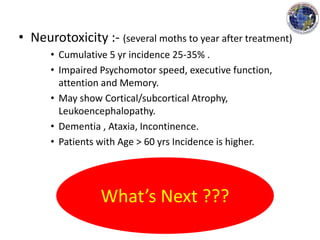
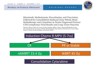
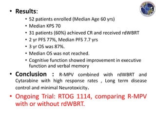
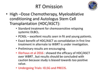
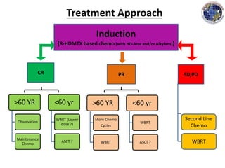
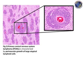
Management of Primary CNS
Lymphoma
Presenter
Dr. Shashank Bansal
JR2- MD Radiotherapy
Dr.B.Borooah Cancer Institute
Moderator
Dr. (Mrs) M Bhattacharyya
Additional Professor , Department of Radiotherapy
Dr.B.Borooah Cancer Institute
OUTLINE
• DEFINITION
• EPIDEMIOLOGY
• PATHOGENSIS AND PATHOLOGY
• PRESENTATION
• DIAGNOSIS
• PROGNOSTICATION
• TREATMENT
DEFINITION
• Primary Central nervous system lymphoma (PSCNL) is a
NHL confined to craniospinal axis (brain
leptomeninges spinal cord or eyes) without evidence of
systemic spread.
• NHL sometimes spares the brain entirely and involves
cranial nerve roots or cauda equina owing to primary
infiltration of the leptomeninges. In rare occasions
there may be predominant infiltration of nerve roots,
nerve plexus or peripheral nerves only – a condition
known as Neurolymphmatosis (NL) .
Epidemiology
• PCNL is rare
• Less than 5% of all brain tumors
• Incidence on the rise owing to increased number of
immunocompromised patients
• Risk is more in patients who have congenital or AIDS,
(3600 times) including organ transplant recipients
• Median age of presentation with:
• HIV :- 31 yrs
• Immunocompetent:- 55 yrs
• Diagnosed in at least 2% of HIV related individuals.
• Considered as AIDS defining illness.
Pathogenesis
• Role of Epstein Barr virus (EBV)
• Detected in 0% to 20% of PCNSLs patients who do not
have compromised immune system
• Nearly in 100% AIDS PCNSL
• Latent membrae protien-1 IHC positive in AIDS PCNSL
compared with AIDS related systemic NHL suggesting a
potentially different pathogenesis.
Pathology
• 90% resemble DLBCL
• 10% are poorly characterized low-grade lymphomas,
Burkitt’s Lymphoma and T-cell Lymphomas.
• The DLBCL type is composed of immunoblasts or
centroblasts.
• Have predilection for blood vessels, resulting in
lymphoid clustering around small cerebral vessels.
• The tumour arises in an extraneural environment with
subsequent localization to the CNS, possibly by virtue
of a specific Neurotropism.
Why it responds different
to therapy ?
REMAINS UNCLEAR
Fig 1: specimen shows large hemorrhagic and necrotic
lesions involving the white matter of bilateral parietal lobes in
a HIV+ve patient.
Fig 2:Primary central nervous system
lymphoma (PCNSL) is characterized
by perivascular growth of large atypical
lymphoid cells
Presentation
• Typically presents with neurological symptoms
rather than systemic B symptoms (< 3mo).
• Altered Mental status
• Seizures
• Headache, Nausea , Vomiting (raised ICT)
• Others (Depending on tumour location)
• PCNSL involving Leptomeninges usually do not
show signs of Leptomeningeal involvement.
• 20% may present with ocular involvement
(blurred vision, floaters, diminished Visual acuity)
Diagnosis
(International PCNL collaborative group guidelines for baseline evaluation)
• Clinical evaluation:-
• Complete medical and Neurological examination
• Dilated eye examination
• Record prognostic factors (Age, PS)
• Serial evaluation of cognitive function.
• Laboratory:
• HIV serology
• CSF cytology, Flow cytometry IgH PCR.
• 24 hr urine collection for Creatinine clearance.
• Central pathology review and IHC.
• Imaging :
• Contrast enhanced cranial MRI
• CT scan – chest, abdomen and pelvis
• Bone marrow aspiration and Biopsy
• Testicular USG in elderly .
Diagnosis
(International PCNL collaborative group guidelines for baseline evaluation)
Abrey LE, et al: Report of an international workshop to standardize baseline evaluation and response
criteria for primary central nervous system lymphoma. J Clin Oncol 23:5034-5043, 2005
• CT scan :- NCCT Iso-Hyperdense on imaging
• MRI :-
• Can detect 10% of lesions missed by CT scans.
• T1- iso-hypointense
• T2- (50%shows Hyperintense), in immunocompetent –
Homogenous enhancement.
• Location on imaging (Mostly seen in these areas):
• Hemispheres, Thalamus and Basal Ganglia,Corpus
Callosum, Ventricular region, Cerebellum [ref].
• HIV : Multifocal (d/d CNS infection).
[ref] Kuker W, et al: Primary central nervous system lymphomas (PCNSL): MRI features at
presentation in 100 patients. J Neurooncol 72:169-177, 2005
Figure: Bilateral VR spaces
• VR spaces surround the walls
of arteries, arterioles, veins,
and venules as they course
from the subarachnoid space
through the brain
parenchyma
• Arteries in the cerebral cortex
are coated by a layer of
leptomeninges that is
subtended from the pia
mater; by this anatomic
arrangement, the VR spaces
of the intracortical arteries
are in direct continuity with
the VR spaces around arteries
in the subarachnoid space
Virchow Robins Space
Fig 3:
(A) Slit-lamp evaluation demonstrating advanced intraocular lymphoma with optic disc
swelling, vasculitis, and subretinal and retinal infiltrates.
(B) Optical coherence tomography demonstrating a nodular hyper-reflective lesion
(arrow) at the retinal pigment epithelium and subretinal space.
(Courtesy Paul Stewart, MD, University of California–San Francisco)
Fig 4:
(A) A T1 axial, postgadolinium image depicts a periventricular contrast-enhancing lesion
with near-uniform contrast enhancement, vasogenic edema and mass effect, in
displacement of the lateral ventricles. Lesional contrast enhancement using MRI is used
for response assessment.
(B) (B) A flair signal abnormality demonstrates the extent of vasogenic edema.
(Courtesy Soonmee Cha, MD, University of California–San Francisco).
Ann Arbor Staging
Do not apply
Prognostication
• International Extranodal Lymphoma Study Group-
Parameters associated with poor prognosis:
• Age (>60yrs) most important
• ECOG performance status >1
• Elevated serum LDH
• High CSF Protein concertation.
• Tumour location within deep regions of brain
(Periventricular, basal ganglia, brainstem etc.)
• Biochemical markers
• BCL-6 (good prognosis)
• P53 and c-Myc (worse prognosis).
Ferreri AJ et al: Prognostic scoring system for primary CNS lymphomas: The International Extranodal
Lymphoma Study Group experience. J Clin Oncol 21:266-272, 2003
IELSG Nottingham/ Barcelona MSKCC
Age Age >60 Age <50
Performance status PS >2 KPS <70
LDH Extent of Disease
(Multifocal or Unifocal )
CSF protein level
Deep Brain structure
Involvement
Score Survival
(2yr)
Low
(0-1)
80%
Intermediate
(2-3)
48%
High
(4-5)
15%
Score Survival
0 55 months
1 41 months
2 32 months
3 1 month
Score Survival
Class 1
(Age<50)
8.5 yr
Class 2
(>50, KPS >70)
3.2 yr
Class 3
(>50, KPS <70)
1.1 yr
Prognostic Scores
Response Assessment
V. De Wilde, MD, PhD , D. Dierickx, MD, PhD et al; BHS guidelines for primary central nervous system lymphoma; Belg J Hematol
2016;7(2):69-78
Management
• Surgery :
– Role limited to establishing diagnosis (stereotactic
biopsy).
– Surgical decompression and shunt placement to
reduce ICT.
• Extremely Radiosensitive and Chemosensistive
tumour
• Role of Steroids
• Should be avoided during evaluation of patient and
before biopsy and CSF examination- false negative
results
• Exert apoptotic effect on Lymphoid cells through
cytoplasmic steroid receptors
• May be started after biopsy is done to control
vasogenic oedema and resultant mass effect.
• Dexamethasone has been associated with Initial CR
(15%) and PR (25%).
• Resistance is common on re-exposure.
• Unclear whether steroids need to be integral part of
any regimen as is true for systemic Lymphomas.
Chemotherapy
• Standard use of systemic NHL treatment
regimens in PCNSL:
• Associated with transient response to brain lesions
• Frequent recurrences
Four prospective trials (one randomized)
All Failed to show an advantage of CHOP/ CHOD plus
WBRT over WBRT alone
CHOP has no role in the treatment of PCNSL
Inability to cross Blood Brain Barrier
• Role Of Methotrexate:
• First recognized when it was discovered that patients
who had systemic NHL that relapsed in the CNS
responded to high dose MTX.
• Ability to penetrate BBB makes it an attractive agent
• Rapid infusion of high dose MTX over 3 hours greatly
raises the drug level in the CSF (Maximum therapeutic
concentration in 4-6 hours after the start of an
infusion), and remains above the minimum therapeutic
concentration in the CSF up to 24 hrs (blood to CSF
ratio 30:1).
• MTX based regimens are the only regimens with a
significant advantage over RT alone.
• Commonly used Methotrexate base
combination modality regimen consists of Five
cycles (10 wks) of pre irradiation.
• IV MTX 2.5 g/m2
• IV Vincristine 1.4 mg/m2
• Oral procarbazine 100mg/m2/d for 7 days
• IT MTX 12 mg
• Dexamethasone taper
• Followed by WBRT 45 Gy and Post- WBRT high dose
Cytarabine 3 g/m2/d.
• Other Combinations:
• IELSG (phase II randomized sudy)
– 2 arms HD MTX + WBRT / HD MTX + Cytarabine + WBRT
– Significant higher CR in the HD MTX + Cytarabine arm
– Significant improved ORR,PFS and a trend towards better OS
were observed in Arm B.
• MTX + Alkylating agent + Rituximab
• Rituximab +MTX + Procarbazine + Vincristine + Low dose
WBRT
• Rituximab + MTX+ Procarbazine+ Vincristine + rdWBRT +
Autologous Stem cell transplant
– ORR 94% and 2 yr PFS 79%.
• No results can be drawn about the effect of each
drug in view of the fact that these trials were
small and single Arm study plus similar results
have been reported with HD MTX.
IELSG 32 STUDY (FERRERI ET AL 2016)
227 PATIENTS IN 53 CENTRES
GROUP A GROUP B GROUP C
• MTX (3.5g/m2) D1
• Cytarabine 2g/m2
bd on D2 D3
• MTX (3.5g/m2) D1
• Cytarabine 2g/m2
bd on D2 D3
• Rituximab
(375/m2) on D0
and D5.
• Group B
• Thiotepa
(30mg/m2) D4
• MATRix
Regimen
• MATRix (Group C) regimen is associated with improved PFS and OS
with % yr OS of 69%.
• Hematologic toxicity were more and infective toxicity was
comparable to other group
Summary Of Chemotherapy Regimens
V. De Wilde, MD, PhD , D. Dierickx, MD, PhD et al; BHS guidelines for primary central nervous system lymphoma; Belg J Hematol
2016;7(2):69-78
Radiotherapy
Historically RT involves whole brain RT
Poor response despite known Radiosensitivity to NHL outside
(RTOG and Princess Margret Hospital report)
Median survival :- 12.2 months to 17 months
Tumour regrowth and uncontrolled disease were common
cause of death
Higher doses causes severe neurotoxicity (dose limitation 40-50Gy)
• Possible reasons of RT failure:
• PCNSL may be confined to the nervous system but it is
potentially disseminated within it at diagnosis.
• Ocular or CSF involvement may be present, even if not
identified on a staging evaluation. These areas could be
potential reservoirs of untreated disease if the patient
receives inly WBRT.
• PCNSL may have a unique biology that accounts for its
worse outcome despite its comparable histological
appearance to most systemic diffuse large B cell
Lymphoma.
Nelson DF et al: NHL of the brain: Can high dose, large volume radiation therapy improve survival?
Report on a prospective trial by the RTOG: RTOG 8315. Int J Radiat Oncol Biol Phys 23:9-17, 1992
Consolidation WBRT provide better disease
control or survival in CR patients after upfront
chemotherapy ?
Optimal dose ?
Role of Hyperfractionation?
Did not show a clear benefit with
Hyperfractionated whole Brain RT
Lancet Oncol 2010; 11: 1036–47
• Criticism :
• Poor protocol adherence
• Randomization caveats
• Low statistical power
Hence question of consolidated WBRT
remain unanswered
Still consolidation WBRT remains standard of care
Limitation : Neurotoxicity
• Neurotoxicity :- (several moths to year after treatment)
• Cumulative 5 yr incidence 25-35% .
• Impaired Psychomotor speed, executive function,
attention and Memory.
• May show Cortical/subcortical Atrophy,
Leukoencephalopathy.
• Dementia , Ataxia, Incontinence.
• Patients with Age > 60 yrs Incidence is higher.
What’s Next ???
RT Dose Reduction
Induction Chemo R-MPV (5-7cy)
CR
rdWBRT 23.4 Gy
PR or Stable
WBRT 45 Gy
Consolidation Cytarabine
• Results:
• 52 patients enrolled (Median Age 60 yrs)
• Median KPS 70
• 31 patients (60%) achieved CR and received rdWBRT
• 2 yr PFS 77%, Median PFS 7.7 yrs
• 3 yr OS was 87%.
• Median OS was not reached.
• Cognitive function showed improvement in executive
function and verbal memory
• Conclusion : R-MPV combined with rdWBRT and
Cytarabine with high response rates , Long term disease
control and minimal Neurotoxicity.
• Ongoing Trial: RTOG 1114, comparing R-MPV
with or without rdWBRT.
RT Omission
• High –Dose Chemotherapy, Myeloablative
conditioning and Autologus Stem Cell
Transplantation (HDC/ASCT):
• Standard treatment for chemosensitive relapsing
systemic DLBCL.
• PCNSL – excellent results seen in fit and young patients.
• Exact benefit of HDC/ASCT as consolidation in first line
treatment in alternate to WBRT is under invetigation.
• Preliminary results are encouraging.
• Illerhaus et al 2016 : showed the efficacy of HDC/ASCT
over WBRT , but results should be concluded with
caution because study is biased towards too positive
value.
• Undergoing Trials: IELSG and PRECIS.
Induction
{R-HDMTX based chemo (with HD-Arac and/or Alkylans)}
CR PR SD,PD
>60 YR
Observation
Maintenance
Chemo
<60 yr
WBRT (Lower
dose ?)
ASCT ?
>60 YR
More Chemo
Cycles
WBRT
<60 yr
WBRT
ASCT ?
Second Line
Chemo
WBRT
Treatment Approach
Conclusion
• Rare form of Extra nodal NHL and its typically a DLBCL
that is confined to the nervous system and eyes.
• The diagnosis of PCNSL is supported by CT and MRI
studies as well as CSF testing, but is ultimately
confirmed on the basis of stereotatic biopsy.
• Current treatment regimens are achieving log term
remissions though in a small fractions of patients .
• The optimal role and timing of WBRT in the
management of newly diagnosed PCNSL patients has
yet to be established.
• Minimizing the risk of neurotoxicity by deferring WBRT
in Patients older than age 60 is an important objective.
THANK YOU

Primary cns lymphoma ppt

  • 1.
    Management of PrimaryCNS Lymphoma Presenter Dr. Shashank Bansal JR2- MD Radiotherapy Dr.B.Borooah Cancer Institute Moderator Dr. (Mrs) M Bhattacharyya Additional Professor , Department of Radiotherapy Dr.B.Borooah Cancer Institute
  • 2.
    OUTLINE • DEFINITION • EPIDEMIOLOGY •PATHOGENSIS AND PATHOLOGY • PRESENTATION • DIAGNOSIS • PROGNOSTICATION • TREATMENT
  • 3.
    DEFINITION • Primary Centralnervous system lymphoma (PSCNL) is a NHL confined to craniospinal axis (brain leptomeninges spinal cord or eyes) without evidence of systemic spread. • NHL sometimes spares the brain entirely and involves cranial nerve roots or cauda equina owing to primary infiltration of the leptomeninges. In rare occasions there may be predominant infiltration of nerve roots, nerve plexus or peripheral nerves only – a condition known as Neurolymphmatosis (NL) .
  • 4.
    Epidemiology • PCNL israre • Less than 5% of all brain tumors • Incidence on the rise owing to increased number of immunocompromised patients • Risk is more in patients who have congenital or AIDS, (3600 times) including organ transplant recipients • Median age of presentation with: • HIV :- 31 yrs • Immunocompetent:- 55 yrs • Diagnosed in at least 2% of HIV related individuals. • Considered as AIDS defining illness.
  • 5.
    Pathogenesis • Role ofEpstein Barr virus (EBV) • Detected in 0% to 20% of PCNSLs patients who do not have compromised immune system • Nearly in 100% AIDS PCNSL • Latent membrae protien-1 IHC positive in AIDS PCNSL compared with AIDS related systemic NHL suggesting a potentially different pathogenesis.
  • 6.
    Pathology • 90% resembleDLBCL • 10% are poorly characterized low-grade lymphomas, Burkitt’s Lymphoma and T-cell Lymphomas. • The DLBCL type is composed of immunoblasts or centroblasts. • Have predilection for blood vessels, resulting in lymphoid clustering around small cerebral vessels. • The tumour arises in an extraneural environment with subsequent localization to the CNS, possibly by virtue of a specific Neurotropism. Why it responds different to therapy ? REMAINS UNCLEAR
  • 7.
    Fig 1: specimenshows large hemorrhagic and necrotic lesions involving the white matter of bilateral parietal lobes in a HIV+ve patient.
  • 8.
    Fig 2:Primary centralnervous system lymphoma (PCNSL) is characterized by perivascular growth of large atypical lymphoid cells
  • 9.
    Presentation • Typically presentswith neurological symptoms rather than systemic B symptoms (< 3mo). • Altered Mental status • Seizures • Headache, Nausea , Vomiting (raised ICT) • Others (Depending on tumour location) • PCNSL involving Leptomeninges usually do not show signs of Leptomeningeal involvement. • 20% may present with ocular involvement (blurred vision, floaters, diminished Visual acuity)
  • 10.
    Diagnosis (International PCNL collaborativegroup guidelines for baseline evaluation) • Clinical evaluation:- • Complete medical and Neurological examination • Dilated eye examination • Record prognostic factors (Age, PS) • Serial evaluation of cognitive function. • Laboratory: • HIV serology • CSF cytology, Flow cytometry IgH PCR. • 24 hr urine collection for Creatinine clearance. • Central pathology review and IHC.
  • 11.
    • Imaging : •Contrast enhanced cranial MRI • CT scan – chest, abdomen and pelvis • Bone marrow aspiration and Biopsy • Testicular USG in elderly . Diagnosis (International PCNL collaborative group guidelines for baseline evaluation) Abrey LE, et al: Report of an international workshop to standardize baseline evaluation and response criteria for primary central nervous system lymphoma. J Clin Oncol 23:5034-5043, 2005
  • 12.
    • CT scan:- NCCT Iso-Hyperdense on imaging • MRI :- • Can detect 10% of lesions missed by CT scans. • T1- iso-hypointense • T2- (50%shows Hyperintense), in immunocompetent – Homogenous enhancement. • Location on imaging (Mostly seen in these areas): • Hemispheres, Thalamus and Basal Ganglia,Corpus Callosum, Ventricular region, Cerebellum [ref]. • HIV : Multifocal (d/d CNS infection). [ref] Kuker W, et al: Primary central nervous system lymphomas (PCNSL): MRI features at presentation in 100 patients. J Neurooncol 72:169-177, 2005
  • 13.
    Figure: Bilateral VRspaces • VR spaces surround the walls of arteries, arterioles, veins, and venules as they course from the subarachnoid space through the brain parenchyma • Arteries in the cerebral cortex are coated by a layer of leptomeninges that is subtended from the pia mater; by this anatomic arrangement, the VR spaces of the intracortical arteries are in direct continuity with the VR spaces around arteries in the subarachnoid space Virchow Robins Space
  • 14.
    Fig 3: (A) Slit-lampevaluation demonstrating advanced intraocular lymphoma with optic disc swelling, vasculitis, and subretinal and retinal infiltrates. (B) Optical coherence tomography demonstrating a nodular hyper-reflective lesion (arrow) at the retinal pigment epithelium and subretinal space. (Courtesy Paul Stewart, MD, University of California–San Francisco)
  • 15.
    Fig 4: (A) AT1 axial, postgadolinium image depicts a periventricular contrast-enhancing lesion with near-uniform contrast enhancement, vasogenic edema and mass effect, in displacement of the lateral ventricles. Lesional contrast enhancement using MRI is used for response assessment. (B) (B) A flair signal abnormality demonstrates the extent of vasogenic edema. (Courtesy Soonmee Cha, MD, University of California–San Francisco).
  • 16.
  • 17.
    Prognostication • International ExtranodalLymphoma Study Group- Parameters associated with poor prognosis: • Age (>60yrs) most important • ECOG performance status >1 • Elevated serum LDH • High CSF Protein concertation. • Tumour location within deep regions of brain (Periventricular, basal ganglia, brainstem etc.) • Biochemical markers • BCL-6 (good prognosis) • P53 and c-Myc (worse prognosis). Ferreri AJ et al: Prognostic scoring system for primary CNS lymphomas: The International Extranodal Lymphoma Study Group experience. J Clin Oncol 21:266-272, 2003
  • 18.
    IELSG Nottingham/ BarcelonaMSKCC Age Age >60 Age <50 Performance status PS >2 KPS <70 LDH Extent of Disease (Multifocal or Unifocal ) CSF protein level Deep Brain structure Involvement Score Survival (2yr) Low (0-1) 80% Intermediate (2-3) 48% High (4-5) 15% Score Survival 0 55 months 1 41 months 2 32 months 3 1 month Score Survival Class 1 (Age<50) 8.5 yr Class 2 (>50, KPS >70) 3.2 yr Class 3 (>50, KPS <70) 1.1 yr Prognostic Scores
  • 19.
    Response Assessment V. DeWilde, MD, PhD , D. Dierickx, MD, PhD et al; BHS guidelines for primary central nervous system lymphoma; Belg J Hematol 2016;7(2):69-78
  • 20.
    Management • Surgery : –Role limited to establishing diagnosis (stereotactic biopsy). – Surgical decompression and shunt placement to reduce ICT. • Extremely Radiosensitive and Chemosensistive tumour
  • 21.
    • Role ofSteroids • Should be avoided during evaluation of patient and before biopsy and CSF examination- false negative results • Exert apoptotic effect on Lymphoid cells through cytoplasmic steroid receptors • May be started after biopsy is done to control vasogenic oedema and resultant mass effect. • Dexamethasone has been associated with Initial CR (15%) and PR (25%). • Resistance is common on re-exposure. • Unclear whether steroids need to be integral part of any regimen as is true for systemic Lymphomas.
  • 22.
    Chemotherapy • Standard useof systemic NHL treatment regimens in PCNSL: • Associated with transient response to brain lesions • Frequent recurrences Four prospective trials (one randomized) All Failed to show an advantage of CHOP/ CHOD plus WBRT over WBRT alone CHOP has no role in the treatment of PCNSL Inability to cross Blood Brain Barrier
  • 23.
    • Role OfMethotrexate: • First recognized when it was discovered that patients who had systemic NHL that relapsed in the CNS responded to high dose MTX. • Ability to penetrate BBB makes it an attractive agent • Rapid infusion of high dose MTX over 3 hours greatly raises the drug level in the CSF (Maximum therapeutic concentration in 4-6 hours after the start of an infusion), and remains above the minimum therapeutic concentration in the CSF up to 24 hrs (blood to CSF ratio 30:1). • MTX based regimens are the only regimens with a significant advantage over RT alone.
  • 24.
    • Commonly usedMethotrexate base combination modality regimen consists of Five cycles (10 wks) of pre irradiation. • IV MTX 2.5 g/m2 • IV Vincristine 1.4 mg/m2 • Oral procarbazine 100mg/m2/d for 7 days • IT MTX 12 mg • Dexamethasone taper • Followed by WBRT 45 Gy and Post- WBRT high dose Cytarabine 3 g/m2/d.
  • 25.
    • Other Combinations: •IELSG (phase II randomized sudy) – 2 arms HD MTX + WBRT / HD MTX + Cytarabine + WBRT – Significant higher CR in the HD MTX + Cytarabine arm – Significant improved ORR,PFS and a trend towards better OS were observed in Arm B. • MTX + Alkylating agent + Rituximab • Rituximab +MTX + Procarbazine + Vincristine + Low dose WBRT • Rituximab + MTX+ Procarbazine+ Vincristine + rdWBRT + Autologous Stem cell transplant – ORR 94% and 2 yr PFS 79%. • No results can be drawn about the effect of each drug in view of the fact that these trials were small and single Arm study plus similar results have been reported with HD MTX.
  • 26.
    IELSG 32 STUDY(FERRERI ET AL 2016) 227 PATIENTS IN 53 CENTRES GROUP A GROUP B GROUP C • MTX (3.5g/m2) D1 • Cytarabine 2g/m2 bd on D2 D3 • MTX (3.5g/m2) D1 • Cytarabine 2g/m2 bd on D2 D3 • Rituximab (375/m2) on D0 and D5. • Group B • Thiotepa (30mg/m2) D4 • MATRix Regimen • MATRix (Group C) regimen is associated with improved PFS and OS with % yr OS of 69%. • Hematologic toxicity were more and infective toxicity was comparable to other group
  • 27.
    Summary Of ChemotherapyRegimens V. De Wilde, MD, PhD , D. Dierickx, MD, PhD et al; BHS guidelines for primary central nervous system lymphoma; Belg J Hematol 2016;7(2):69-78
  • 28.
    Radiotherapy Historically RT involveswhole brain RT Poor response despite known Radiosensitivity to NHL outside (RTOG and Princess Margret Hospital report) Median survival :- 12.2 months to 17 months Tumour regrowth and uncontrolled disease were common cause of death Higher doses causes severe neurotoxicity (dose limitation 40-50Gy)
  • 29.
    • Possible reasonsof RT failure: • PCNSL may be confined to the nervous system but it is potentially disseminated within it at diagnosis. • Ocular or CSF involvement may be present, even if not identified on a staging evaluation. These areas could be potential reservoirs of untreated disease if the patient receives inly WBRT. • PCNSL may have a unique biology that accounts for its worse outcome despite its comparable histological appearance to most systemic diffuse large B cell Lymphoma. Nelson DF et al: NHL of the brain: Can high dose, large volume radiation therapy improve survival? Report on a prospective trial by the RTOG: RTOG 8315. Int J Radiat Oncol Biol Phys 23:9-17, 1992
  • 30.
    Consolidation WBRT providebetter disease control or survival in CR patients after upfront chemotherapy ? Optimal dose ? Role of Hyperfractionation?
  • 31.
    Did not showa clear benefit with Hyperfractionated whole Brain RT
  • 32.
    Lancet Oncol 2010;11: 1036–47
  • 33.
    • Criticism : •Poor protocol adherence • Randomization caveats • Low statistical power Hence question of consolidated WBRT remain unanswered Still consolidation WBRT remains standard of care Limitation : Neurotoxicity
  • 34.
    • Neurotoxicity :-(several moths to year after treatment) • Cumulative 5 yr incidence 25-35% . • Impaired Psychomotor speed, executive function, attention and Memory. • May show Cortical/subcortical Atrophy, Leukoencephalopathy. • Dementia , Ataxia, Incontinence. • Patients with Age > 60 yrs Incidence is higher. What’s Next ???
  • 35.
  • 36.
    Induction Chemo R-MPV(5-7cy) CR rdWBRT 23.4 Gy PR or Stable WBRT 45 Gy Consolidation Cytarabine
  • 37.
    • Results: • 52patients enrolled (Median Age 60 yrs) • Median KPS 70 • 31 patients (60%) achieved CR and received rdWBRT • 2 yr PFS 77%, Median PFS 7.7 yrs • 3 yr OS was 87%. • Median OS was not reached. • Cognitive function showed improvement in executive function and verbal memory • Conclusion : R-MPV combined with rdWBRT and Cytarabine with high response rates , Long term disease control and minimal Neurotoxicity. • Ongoing Trial: RTOG 1114, comparing R-MPV with or without rdWBRT.
  • 38.
    RT Omission • High–Dose Chemotherapy, Myeloablative conditioning and Autologus Stem Cell Transplantation (HDC/ASCT): • Standard treatment for chemosensitive relapsing systemic DLBCL. • PCNSL – excellent results seen in fit and young patients. • Exact benefit of HDC/ASCT as consolidation in first line treatment in alternate to WBRT is under invetigation. • Preliminary results are encouraging. • Illerhaus et al 2016 : showed the efficacy of HDC/ASCT over WBRT , but results should be concluded with caution because study is biased towards too positive value. • Undergoing Trials: IELSG and PRECIS.
  • 39.
    Induction {R-HDMTX based chemo(with HD-Arac and/or Alkylans)} CR PR SD,PD >60 YR Observation Maintenance Chemo <60 yr WBRT (Lower dose ?) ASCT ? >60 YR More Chemo Cycles WBRT <60 yr WBRT ASCT ? Second Line Chemo WBRT Treatment Approach
  • 40.
    Conclusion • Rare formof Extra nodal NHL and its typically a DLBCL that is confined to the nervous system and eyes. • The diagnosis of PCNSL is supported by CT and MRI studies as well as CSF testing, but is ultimately confirmed on the basis of stereotatic biopsy. • Current treatment regimens are achieving log term remissions though in a small fractions of patients . • The optimal role and timing of WBRT in the management of newly diagnosed PCNSL patients has yet to be established. • Minimizing the risk of neurotoxicity by deferring WBRT in Patients older than age 60 is an important objective.
  • 41.