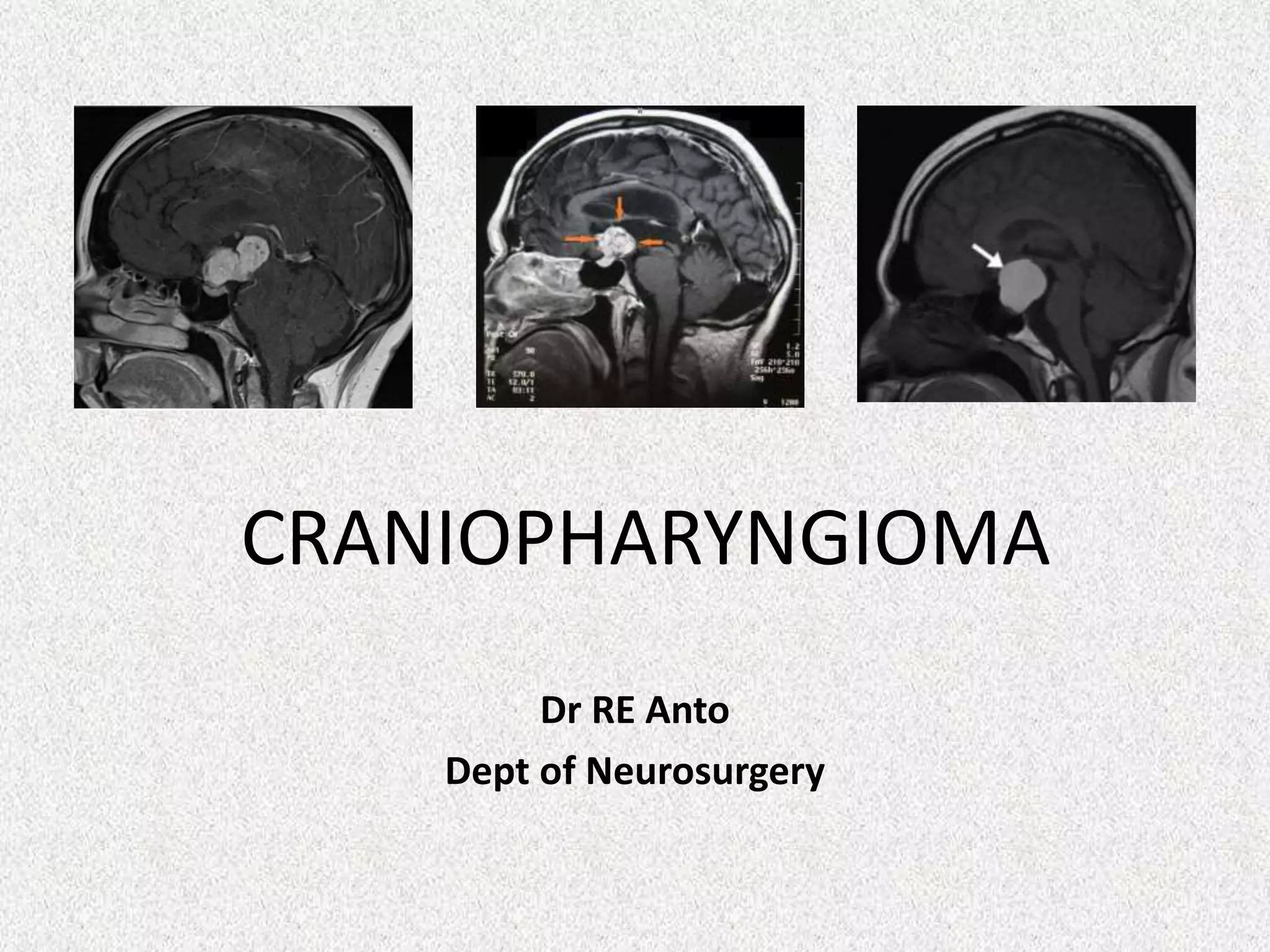
CRANIOPHARYNGIOMA
Dr RE Anto
Dept of Neurosurgery
• “The most formidable of all intracranial
tumours”
• “The kaleidoscopic tumours, solid and
cystic....” whose management is “one of the
most baffling problems to the neurosurgeon”
-Harvey Cushing (1936)
Craniopharyngioma
-Benign intracranial tumour
-Intimate relationship with critical structures
• Visual apparatus
• Pituitary
• Hypothalamus
• Limbic system
-Devestating clinical and psychosocial consequences
PATHOPHYSIOLOGY
Two hypotheses
• Embryogenic
• Transformation of the squamous cell epithelium along the
path of the craniopharyngeal duct
• Metaplastic
• Metaplasia of adenohypophyseal cells in pituitary stalk or
gland “Rathke’s Pouch”
Defect in Wnt signaling pathway reactivation
• β-Catenin gene mutations effecting exon 3 suggesting nuclear
β-Catenin accumulation
EPIDEMIOLOGY
• 3% of all intracranial tumours
• >50% suprasellar tumours in childhood
• Most common nonglial tumour of childhood
• Bimodal distribution
– Children (5-15 years)
– Late adulthood (45-60 years)
• Slight male predominance (55%)
PATHOLOGY
Adamantinomatous (WHO Grade 1)
• More common variant
• Epithelium of tooth
primordia
• Cysts in these tumours
contain “motor oil” liquid
• Associated with CTNNB1
mutation
Papillary (WHO Grade 1)
• Adults
• Predominantly solid tumours
• Cyst – fluid less oily and dark
• Derived from buccal mucosa
primordia
• Associated with BRAF V600E
mutation
Mixed
• 15% of craniopharyngiomas
SUPRASELLAR
(most common)
INTRAVENTRICULAR
(rare)
INTRASELLAR
(less common)
Hoffman et al
• Anterior pituitary
deficiency (>85%)
Paediatric
• Visual dysfunction
(>85%)
Adult
• Diabetes insipidus
• HCP
• Cognitive dys
• Memory impx
Both
Hypothalamic syndrome
• Obesity
• Sexual dysfunction
• Sleep/Wake cycle
• Pituitary failure
• Temperature dysregulation
• Behavioral (Depression/Aggression)
• Social / Emotional withdrawal
NEUROIMAGING
Various imaging modalities
• Skull Xray/CT/MR sequences
• 60-80%: calcifications in the suprasellar region
• 75%: one or more cysts
CT
• Better at evaluating the calcifications
• The skull base (enlargement of the sella turcica or erosion of the dorsum
sella)
MR
• Better evaluation of the relationships of tumour to surrounding
structures
MRI
• Cysts are uniformly hyperintense on T2
• Heterogenous on T1
MR Angiography
• Relationship between the tumour and the surrounding
vessels and the circle of Willis
MR Spectroscopy
• Shows a dominant peak consistent with lactate or lipids
• 7 year old boy with
2/12 hx of visual
problems and
headaches.
CT scan
T1WI Sagittal T2WI Sagittal
T1WI C+ Sagittal T1WI C+ Coronal
Perfusion MR MR Spectroscopy
Autopsy
Role of surgery
Maximal safe resection
• Transcranial vs Endoscopic
endonasal
• Gross total vs Subtotal
Management of cysts
• Drainage vs Ommaya reservoir
Management of hydrocephalus
• Shunt placement
Transcranial approaches
• Subfrontal
• Pterional
• Orbitofrontal
• Lamina terminalis
• Transcallosal
• Transcortical
Endoscopic Endonasal Approach
Intracystic therapy
• Reserved for tumours with one big dominant cyst
• Delay time to defintive surgery or radiation
• Requires surgery for placement and CT “leak test”
• Radiation - P32 (one time)
• 79.5-96% regression of cysts
• Chemotherapy - Bleomycin (3 week course)
• 50% of cyst regression
• Chemotherapy - Interferon-α (4 week course)
• 78% of cysts shrunk by 50% or more
• 2 x Ommaya reservoir
insertions in 2020 at
Polokwane Hospital
• A series of 17 children, intracystic bleomycin was
well tolerated, with five complete remissions and a
median progression-free interval of 1.8 years
• A series of 47 patients, given a radiation dose of 250 Gy,
The overall response rate was 78% and the mean
survival was 10 years.
• The survival rate at 1, 3, 5, and 10 years after p32
therapy was 91%, 77%, 73%, and 52%, respectively.
Radiation
LINAC
• Linear Accelarator (external beam RT)
• TruBeam
• CyberKnife
Stereotactic radiosurgery
• GammaKnife
Proton Beam
• Uses charged particles rather than photons to deliver high
doses of radiation to the target volume while limiting the
"scatter" dose received by surrounding tissues
Radiation therapy
• 54 Gy in 30 fractions
• 6 weeks of treatment
• Adverse events
• Cyst enlargement
• HCP
• Pituitary failure
(20-60% at 5 years)
• Cognitive decline
• Total dose temporal lobes
• Radiation necrosis
Evidence favouring radiation
• Study of 122 patients, looked at those treated
with GTR, STR and STR + RT, and the primary
endpoints were PFS and OS.
UCSF database
Progression Free Survival
Timing of radiation
• Controversial
• Immediate post-op (adjunctive) vs recurrence
(salvage)
• Another series noted control rate did not depend
on timing of RT
– DI and Visual impx (delayed > immediate)
• General recommendation is immediate
adjunctive therapy
– Cautious in children (delay) because of the greater
sensitivity to carcinogenic and adverse neurocognitive
effects of RT
SUMMARY
Surgery
• Maximum safe resection is 1st line Rx
• Cyst may require Ommaya reservoir
• HCP may require shunt
Radiation
• No proven difference between GK, CK, LINAC
• SBT + RT = GTR with low morbidity
• Radiation may be delayed until recurrence vs immediately post op.
Chemotherapy
• Intracystic interferon-α, bleomycin or p32
Medical therapy
• Hormone replacement
FUTURE DIRECTION
• Targeted molecular therapies
• Genetic studies
– CTNNB1 (B-catennin) : Adamantinomatous
– BRAF : Papillary
• Inhibitors of BRAF Proto-oncogene
– Dabrafenib/Vemurafanib
• MEK inhibitors
– Trametinib
• Multicenter phase-2 clinical trials at the
National Cancer Institute are currently
underway evaluating BRAF/MEK inhibition
in the treatment of craniopharyngioma
CONCLUSION
• Regardless of the therapeutic
strategies that are utilized, it is
evident that craniopharyngiomas
continue to present a distinct
challenge that still needs to be
overcome.
• Quality of life is a key consideration
in this disease, and long-term follow
up, involving a multidisciplinary
team, is a necessary element of care
of these patients.

Craniopharyngioma

  • 1.
  • 2.
    • “The mostformidable of all intracranial tumours” • “The kaleidoscopic tumours, solid and cystic....” whose management is “one of the most baffling problems to the neurosurgeon” -Harvey Cushing (1936)
  • 3.
    Craniopharyngioma -Benign intracranial tumour -Intimaterelationship with critical structures • Visual apparatus • Pituitary • Hypothalamus • Limbic system -Devestating clinical and psychosocial consequences
  • 6.
    PATHOPHYSIOLOGY Two hypotheses • Embryogenic •Transformation of the squamous cell epithelium along the path of the craniopharyngeal duct • Metaplastic • Metaplasia of adenohypophyseal cells in pituitary stalk or gland “Rathke’s Pouch” Defect in Wnt signaling pathway reactivation • β-Catenin gene mutations effecting exon 3 suggesting nuclear β-Catenin accumulation
  • 8.
    EPIDEMIOLOGY • 3% ofall intracranial tumours • >50% suprasellar tumours in childhood • Most common nonglial tumour of childhood • Bimodal distribution – Children (5-15 years) – Late adulthood (45-60 years) • Slight male predominance (55%)
  • 9.
    PATHOLOGY Adamantinomatous (WHO Grade1) • More common variant • Epithelium of tooth primordia • Cysts in these tumours contain “motor oil” liquid • Associated with CTNNB1 mutation
  • 11.
    Papillary (WHO Grade1) • Adults • Predominantly solid tumours • Cyst – fluid less oily and dark • Derived from buccal mucosa primordia • Associated with BRAF V600E mutation Mixed • 15% of craniopharyngiomas
  • 12.
  • 13.
    • Anterior pituitary deficiency(>85%) Paediatric • Visual dysfunction (>85%) Adult • Diabetes insipidus • HCP • Cognitive dys • Memory impx Both
  • 14.
    Hypothalamic syndrome • Obesity •Sexual dysfunction • Sleep/Wake cycle • Pituitary failure • Temperature dysregulation • Behavioral (Depression/Aggression) • Social / Emotional withdrawal
  • 15.
    NEUROIMAGING Various imaging modalities •Skull Xray/CT/MR sequences • 60-80%: calcifications in the suprasellar region • 75%: one or more cysts CT • Better at evaluating the calcifications • The skull base (enlargement of the sella turcica or erosion of the dorsum sella) MR • Better evaluation of the relationships of tumour to surrounding structures
  • 16.
    MRI • Cysts areuniformly hyperintense on T2 • Heterogenous on T1 MR Angiography • Relationship between the tumour and the surrounding vessels and the circle of Willis MR Spectroscopy • Shows a dominant peak consistent with lactate or lipids
  • 17.
    • 7 yearold boy with 2/12 hx of visual problems and headaches. CT scan
  • 18.
  • 19.
    T1WI C+ SagittalT1WI C+ Coronal
  • 20.
    Perfusion MR MRSpectroscopy
  • 21.
  • 22.
    Role of surgery Maximalsafe resection • Transcranial vs Endoscopic endonasal • Gross total vs Subtotal Management of cysts • Drainage vs Ommaya reservoir Management of hydrocephalus • Shunt placement
  • 23.
    Transcranial approaches • Subfrontal •Pterional • Orbitofrontal • Lamina terminalis • Transcallosal • Transcortical
  • 25.
  • 27.
    Intracystic therapy • Reservedfor tumours with one big dominant cyst • Delay time to defintive surgery or radiation • Requires surgery for placement and CT “leak test” • Radiation - P32 (one time) • 79.5-96% regression of cysts • Chemotherapy - Bleomycin (3 week course) • 50% of cyst regression • Chemotherapy - Interferon-α (4 week course) • 78% of cysts shrunk by 50% or more
  • 28.
    • 2 xOmmaya reservoir insertions in 2020 at Polokwane Hospital
  • 29.
    • A seriesof 17 children, intracystic bleomycin was well tolerated, with five complete remissions and a median progression-free interval of 1.8 years
  • 30.
    • A seriesof 47 patients, given a radiation dose of 250 Gy, The overall response rate was 78% and the mean survival was 10 years. • The survival rate at 1, 3, 5, and 10 years after p32 therapy was 91%, 77%, 73%, and 52%, respectively.
  • 31.
    Radiation LINAC • Linear Accelarator(external beam RT) • TruBeam • CyberKnife Stereotactic radiosurgery • GammaKnife Proton Beam • Uses charged particles rather than photons to deliver high doses of radiation to the target volume while limiting the "scatter" dose received by surrounding tissues
  • 32.
    Radiation therapy • 54Gy in 30 fractions • 6 weeks of treatment • Adverse events • Cyst enlargement • HCP • Pituitary failure (20-60% at 5 years) • Cognitive decline • Total dose temporal lobes • Radiation necrosis
  • 33.
    Evidence favouring radiation •Study of 122 patients, looked at those treated with GTR, STR and STR + RT, and the primary endpoints were PFS and OS.
  • 34.
  • 35.
  • 36.
    Timing of radiation •Controversial • Immediate post-op (adjunctive) vs recurrence (salvage) • Another series noted control rate did not depend on timing of RT – DI and Visual impx (delayed > immediate) • General recommendation is immediate adjunctive therapy – Cautious in children (delay) because of the greater sensitivity to carcinogenic and adverse neurocognitive effects of RT
  • 37.
    SUMMARY Surgery • Maximum saferesection is 1st line Rx • Cyst may require Ommaya reservoir • HCP may require shunt Radiation • No proven difference between GK, CK, LINAC • SBT + RT = GTR with low morbidity • Radiation may be delayed until recurrence vs immediately post op. Chemotherapy • Intracystic interferon-α, bleomycin or p32 Medical therapy • Hormone replacement
  • 38.
    FUTURE DIRECTION • Targetedmolecular therapies • Genetic studies – CTNNB1 (B-catennin) : Adamantinomatous – BRAF : Papillary • Inhibitors of BRAF Proto-oncogene – Dabrafenib/Vemurafanib • MEK inhibitors – Trametinib • Multicenter phase-2 clinical trials at the National Cancer Institute are currently underway evaluating BRAF/MEK inhibition in the treatment of craniopharyngioma
  • 39.
    CONCLUSION • Regardless ofthe therapeutic strategies that are utilized, it is evident that craniopharyngiomas continue to present a distinct challenge that still needs to be overcome. • Quality of life is a key consideration in this disease, and long-term follow up, involving a multidisciplinary team, is a necessary element of care of these patients.