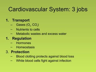
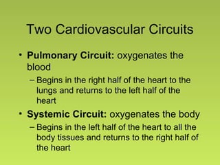
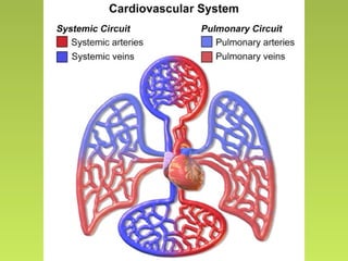
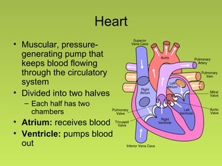
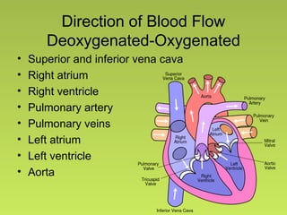
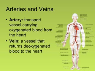
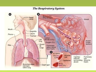
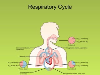
Cardiovascular and Respiratory
Lab
Bio 102
Cardiovascular System: 3 jobs
1. Transport
– Gases (O2, CO2)
– Nutrients to cells
– Metabolic wastes and excess water
1. Regulation
– Hormones
– Homeostasis
3. Protection
– Blood clotting protects against blood loss
– White blood cells fight against infection
Two Cardiovascular Circuits
• Pulmonary Circuit: oxygenates the
blood
– Begins in the right half of the heart to the
lungs and returns to the left half of the
heart
• Systemic Circuit: oxygenates the body
– Begins in the left half of the heart to all the
body tissues and returns to the right half of
the heart
Heart
• Muscular, pressure-
generating pump that
keeps blood flowing
through the circulatory
system
• Divided into two halves
– Each half has two
chambers
• Atrium: receives blood
• Ventricle: pumps blood
out
Direction of Blood Flow
Deoxygenated-Oxygenated
• Superior and inferior vena cava
• Right atrium
• Right ventricle
• Pulmonary artery
• Pulmonary veins
• Left atrium
• Left ventricle
• Aorta
Arteries and Veins
• Artery: transport
vessel carrying
oxygenated blood from
the heart
• Vein: a vessel that
returns deoxygenated
blood to the heart
Respiration
• Physiological process by which oxygen
moves into the internal environment and
carbon dioxide moves out
• Oxygen is needed for aerobic respiration
• Carbon dioxide is produced as a bi-
product of aerobic respiration
• Gases enter and leave an animal by
crossing a respiratory surface
–Thin layer of constantly moist epithelium
Human Respiratory System
Functions
• Gas exchange
• Aid in sense of smell, speech
• Helps blood return to the heart
• Helps body get rid of heat and water
• Helps maintain internal pH
Respiratory Structures
Respiratory Cycle
Respiratory Volumes
• Spirometer: instrument used to measure
different volumes of air
– Developed over 100 years ago
• Vital Capacity: maximal amount of air
exhaled steadily from full inspiration to
maximal expiration
• Forced vital capacity: The total amount
of air that you blow out in one breath
Respiratory Volumes 2
• Forced expiratory volume in one
second: The amount of air you can blow
out in one second.
• Forced expiratory ratio: Percentage of
the FVC expelled in the first second of a
forced expiration
• Peak expiratory flow: Peak expiratory
flow in liters per minute
Today’s lab:
• View the models to identify the major
structures involved in circulation and
respiration
• Use the spirometer to measure your
respiratory volumes
• Investigate how various activities affect
your heart rate and your breathing rate
– Develop and implement your own
experiments

Circulatory Respiratory Lab

  • 1.
  • 2.
    Cardiovascular System: 3jobs 1. Transport – Gases (O2, CO2) – Nutrients to cells – Metabolic wastes and excess water 1. Regulation – Hormones – Homeostasis 3. Protection – Blood clotting protects against blood loss – White blood cells fight against infection
  • 3.
    Two Cardiovascular Circuits •Pulmonary Circuit: oxygenates the blood – Begins in the right half of the heart to the lungs and returns to the left half of the heart • Systemic Circuit: oxygenates the body – Begins in the left half of the heart to all the body tissues and returns to the right half of the heart
  • 5.
    Heart • Muscular, pressure- generatingpump that keeps blood flowing through the circulatory system • Divided into two halves – Each half has two chambers • Atrium: receives blood • Ventricle: pumps blood out
  • 6.
    Direction of BloodFlow Deoxygenated-Oxygenated • Superior and inferior vena cava • Right atrium • Right ventricle • Pulmonary artery • Pulmonary veins • Left atrium • Left ventricle • Aorta
  • 7.
    Arteries and Veins •Artery: transport vessel carrying oxygenated blood from the heart • Vein: a vessel that returns deoxygenated blood to the heart
  • 8.
    Respiration • Physiological processby which oxygen moves into the internal environment and carbon dioxide moves out • Oxygen is needed for aerobic respiration • Carbon dioxide is produced as a bi- product of aerobic respiration • Gases enter and leave an animal by crossing a respiratory surface –Thin layer of constantly moist epithelium
  • 9.
    Human Respiratory System Functions •Gas exchange • Aid in sense of smell, speech • Helps blood return to the heart • Helps body get rid of heat and water • Helps maintain internal pH
  • 11.
  • 12.
  • 13.
    Respiratory Volumes • Spirometer:instrument used to measure different volumes of air – Developed over 100 years ago • Vital Capacity: maximal amount of air exhaled steadily from full inspiration to maximal expiration • Forced vital capacity: The total amount of air that you blow out in one breath
  • 14.
    Respiratory Volumes 2 •Forced expiratory volume in one second: The amount of air you can blow out in one second. • Forced expiratory ratio: Percentage of the FVC expelled in the first second of a forced expiration • Peak expiratory flow: Peak expiratory flow in liters per minute
  • 15.
    Today’s lab: • Viewthe models to identify the major structures involved in circulation and respiration • Use the spirometer to measure your respiratory volumes • Investigate how various activities affect your heart rate and your breathing rate – Develop and implement your own experiments

Editor's Notes

  • #5 Cardiovascular System. Authored by: Blausen Medical Communications. Located at: https://commons.wikimedia.org/wiki/File:Blausen_0168_CardiovascularSystem.png. License: CC BY
  • #6 Diagram of the Human Heart. Authored by: Wapcaplet. Located at: https://commons.wikimedia.org/wiki/File:Diagram_of_the_human_heart_(cropped).svg. License: CC BY-SA
  • #7 Diagram of the Human Heart. Authored by: Wapcaplet. Located at: https://commons.wikimedia.org/wiki/File:Diagram_of_the_human_heart_(cropped).svg. License: CC BY-SA
  • #8 Arterial System. Authored by: LadyofHats. Located at: https://commons.wikimedia.org/wiki/File:Arterial_System_en.svg. License: Public domain
  • #11 The Human Respiratory System. Provided by: National Institutes of Health. Located at: http://www.nhlbi.nih.gov/health/health-topics/topics/hlw/system. License: Public domain
  • #12 Head and Neck Overview. Provided by: National Cancer Institute. Located at: http://training.seer.cancer.gov/head-neck/anatomy/overview.html. License: Public domain Alveoli diagram. Authored by: Pdefer. Located at: https://commons.wikimedia.org/wiki/File:Alveoli_diagram.png. License: CC BY-SA Illu bronchi lungs. Provided by: National Cancer Institute. Located at: https://commons.wikimedia.org/wiki/File:Illu_bronchi_lungs.jpg. License: Public domain
  • #13 Partial Pressures. Provided by: Boundless. Located at: https://www.boundless.com/biology/textbooks/boundless-biology-textbook/the-respiratory-system-39/gas-exchange-across-respiratory-surfaces-220/gas-exchange-across-the-alveoli-836-12081/. License: CC BY-SA