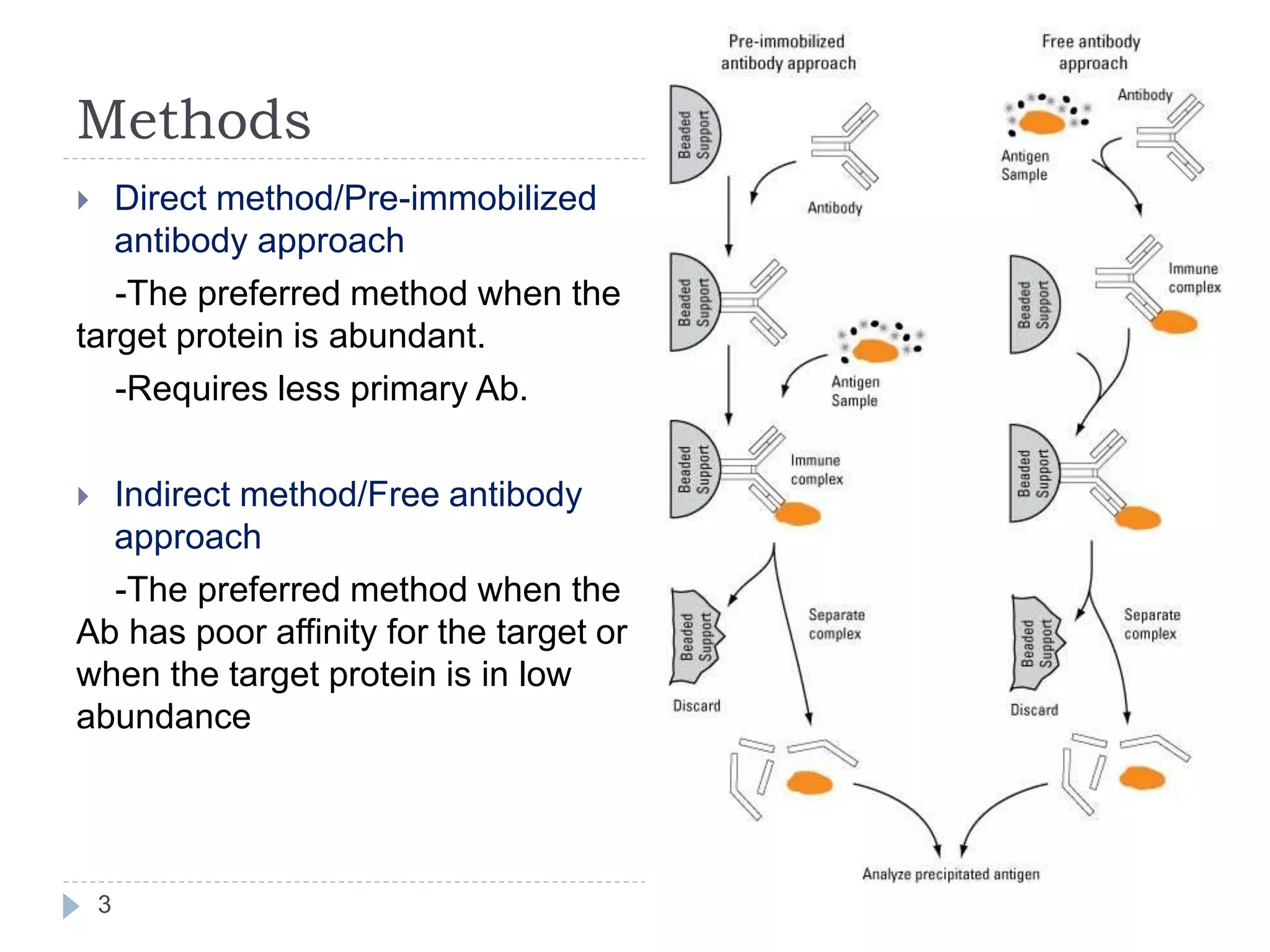
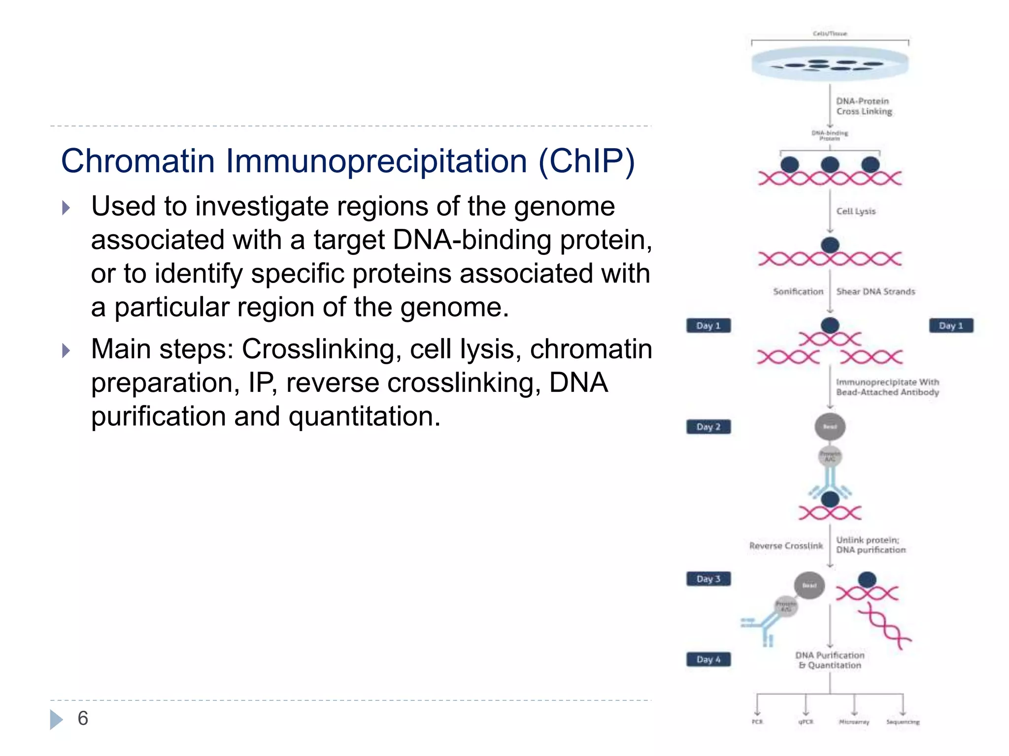
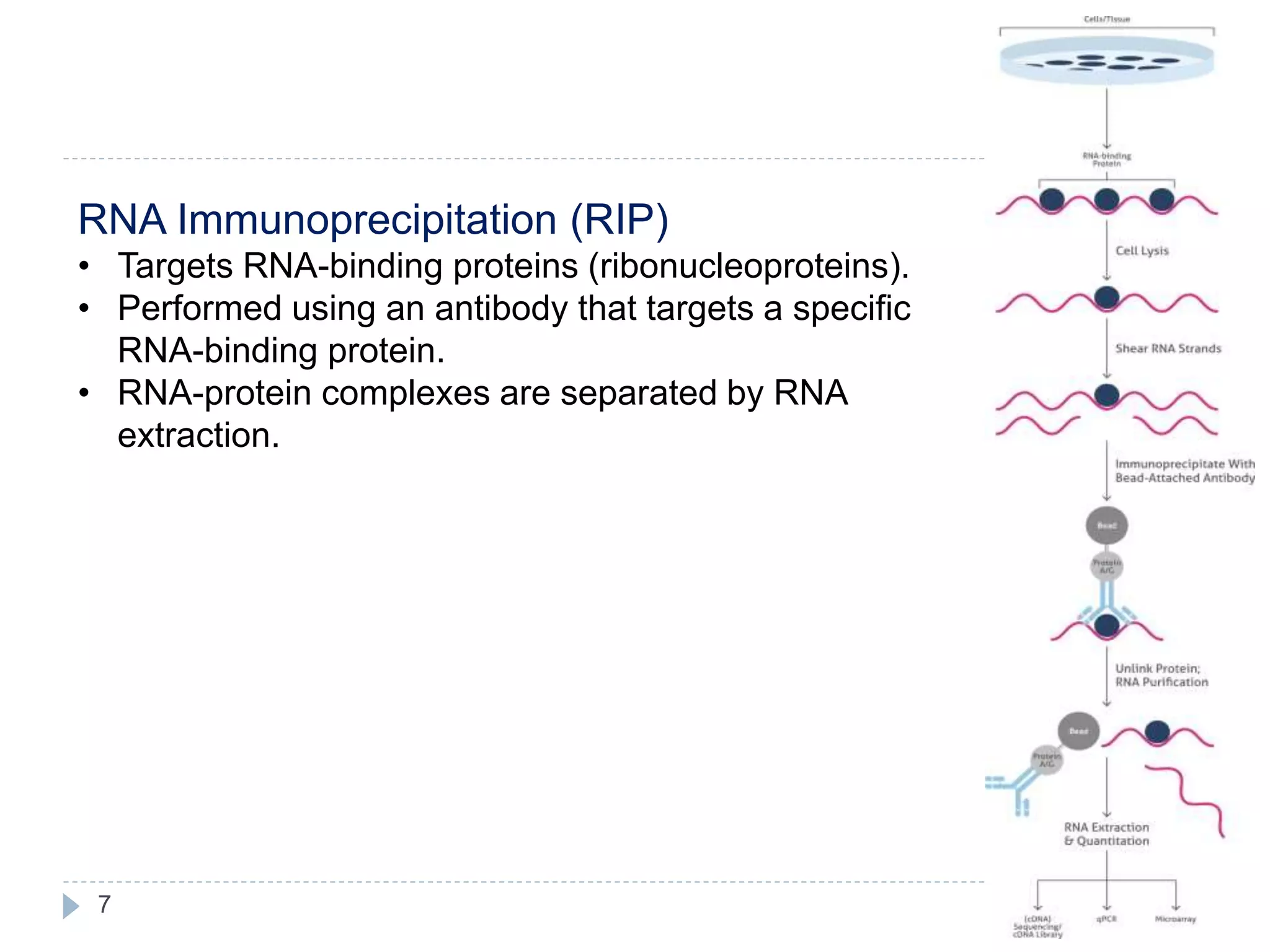
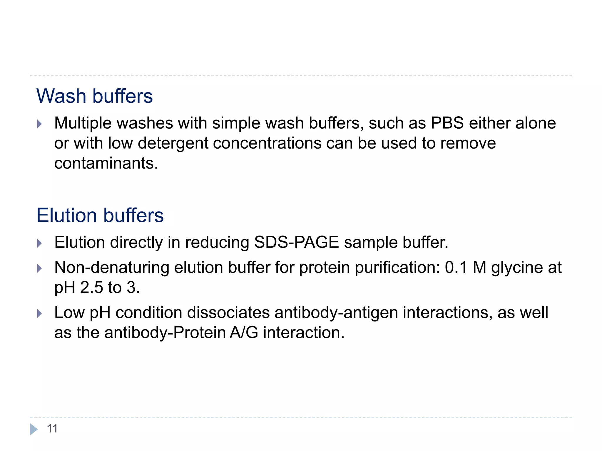
Immunoprecipitation
Aishwarya Babu
1
Immunoprecipitation
2
• Technique of precipitating protein antigens out of a
solution using a specific antibody which is
immobilized to a solid support.
• Widely used method for protein isolation from
complex samples such as cell lysates, serum and
tissue homogenates.
• Applications:
-to measure the molecular weight of the given
protein
-to determine post translational modifications
-to analyze the expression level of a protein of
interest and
-in the study of protein-protein as well as protein-
nucleic acid interactions.
Methods
3
 Direct method/Pre-immobilized
antibody approach
-The preferred method when the
target protein is abundant.
-Requires less primary Ab.
 Indirect method/Free antibody
approach
-The preferred method when the
Ab has poor affinity for the target or
when the target protein is in low
abundance
Magnetic beads vs. agarose resin for
immunoprecipitation
4
 Magnetic beads provide the
best balance of capacity/yield,
reproducibility, purity, and cost
savings for routine small-scale
isolation of specific proteins
and protein complexes.
 Agarose is most appropriate
for large-scale IP reactions
when sufficient antibody is
plentiful at a low cost and
where the goal is to purify a
sufficient amount of target
protein for multiple
downstream assays.
Types of immunoprecipitation
5
Co-immunoprecipitation
 Used to analyze protein–protein interactions.
 Main purpose of Co-IP is the identification of
interaction partners (other proteins, ligands, co-
factors, or signaling molecules) to the protein of
interest.
6
Chromatin Immunoprecipitation (ChIP)
 Used to investigate regions of the genome
associated with a target DNA-binding protein,
or to identify specific proteins associated with
a particular region of the genome.
 Main steps: Crosslinking, cell lysis, chromatin
preparation, IP, reverse crosslinking, DNA
purification and quantitation.
7
RNA Immunoprecipitation (RIP)
• Targets RNA-binding proteins (ribonucleoproteins).
• Performed using an antibody that targets a specific
RNA-binding protein.
• RNA-protein complexes are separated by RNA
extraction.
8
Tagged protein IP
 Proteins can be tagged with an epitope to which a high affinity
antibody is available and expressed in the cell of interest.
 Tags can be either short peptide sequences or fluorescent proteins,
including: FLAG, c-Myc, Hemagglutinin (HA), Green fluorescent
protein (GFP).
Limitation:
 Overexpressed tagged protein is immunoprecipitated
 Tagging the protein may interfere with protein function.
Antibody immobilization
9
1. Antibody-Binding
proteins
2. Covalent antibody
immobilization
3. Crosslinking antibody
immobiliaztion
IP Buffers
10
Lysis buffers
 Stabilizes native protein conformation,
 Inhibits enzymatic activity,
 Maximizes the release of proteins from the cells or tissue.
 Non-denaturing buffers: NP-40 or Triton X-100
 Denaturing buffers: radio-immunoprecipitation assay (RIPA)
 Contains NaCl and Tris-HCl and have a slightly basic pH (7.4 to 8).
 Proteasomal inhibitors: PMSF, aprotinin and leupeptin
11
Wash buffers
 Multiple washes with simple wash buffers, such as PBS either alone
or with low detergent concentrations can be used to remove
contaminants.
Elution buffers
 Elution directly in reducing SDS-PAGE sample buffer.
 Non-denaturing elution buffer for protein purification: 0.1 M glycine at
pH 2.5 to 3.
 Low pH condition dissociates antibody-antigen interactions, as well
as the antibody-Protein A/G interaction.
12
Thank you

IMMUNOPRECIPITATION

  • 1.
  • 2.
    Immunoprecipitation 2 • Technique ofprecipitating protein antigens out of a solution using a specific antibody which is immobilized to a solid support. • Widely used method for protein isolation from complex samples such as cell lysates, serum and tissue homogenates. • Applications: -to measure the molecular weight of the given protein -to determine post translational modifications -to analyze the expression level of a protein of interest and -in the study of protein-protein as well as protein- nucleic acid interactions.
  • 3.
    Methods 3  Direct method/Pre-immobilized antibodyapproach -The preferred method when the target protein is abundant. -Requires less primary Ab.  Indirect method/Free antibody approach -The preferred method when the Ab has poor affinity for the target or when the target protein is in low abundance
  • 4.
    Magnetic beads vs.agarose resin for immunoprecipitation 4  Magnetic beads provide the best balance of capacity/yield, reproducibility, purity, and cost savings for routine small-scale isolation of specific proteins and protein complexes.  Agarose is most appropriate for large-scale IP reactions when sufficient antibody is plentiful at a low cost and where the goal is to purify a sufficient amount of target protein for multiple downstream assays.
  • 5.
    Types of immunoprecipitation 5 Co-immunoprecipitation Used to analyze protein–protein interactions.  Main purpose of Co-IP is the identification of interaction partners (other proteins, ligands, co- factors, or signaling molecules) to the protein of interest.
  • 6.
    6 Chromatin Immunoprecipitation (ChIP) Used to investigate regions of the genome associated with a target DNA-binding protein, or to identify specific proteins associated with a particular region of the genome.  Main steps: Crosslinking, cell lysis, chromatin preparation, IP, reverse crosslinking, DNA purification and quantitation.
  • 7.
    7 RNA Immunoprecipitation (RIP) •Targets RNA-binding proteins (ribonucleoproteins). • Performed using an antibody that targets a specific RNA-binding protein. • RNA-protein complexes are separated by RNA extraction.
  • 8.
    8 Tagged protein IP Proteins can be tagged with an epitope to which a high affinity antibody is available and expressed in the cell of interest.  Tags can be either short peptide sequences or fluorescent proteins, including: FLAG, c-Myc, Hemagglutinin (HA), Green fluorescent protein (GFP). Limitation:  Overexpressed tagged protein is immunoprecipitated  Tagging the protein may interfere with protein function.
  • 9.
    Antibody immobilization 9 1. Antibody-Binding proteins 2.Covalent antibody immobilization 3. Crosslinking antibody immobiliaztion
  • 10.
    IP Buffers 10 Lysis buffers Stabilizes native protein conformation,  Inhibits enzymatic activity,  Maximizes the release of proteins from the cells or tissue.  Non-denaturing buffers: NP-40 or Triton X-100  Denaturing buffers: radio-immunoprecipitation assay (RIPA)  Contains NaCl and Tris-HCl and have a slightly basic pH (7.4 to 8).  Proteasomal inhibitors: PMSF, aprotinin and leupeptin
  • 11.
    11 Wash buffers  Multiplewashes with simple wash buffers, such as PBS either alone or with low detergent concentrations can be used to remove contaminants. Elution buffers  Elution directly in reducing SDS-PAGE sample buffer.  Non-denaturing elution buffer for protein purification: 0.1 M glycine at pH 2.5 to 3.  Low pH condition dissociates antibody-antigen interactions, as well as the antibody-Protein A/G interaction.
  • 12.