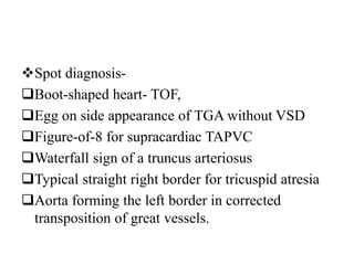
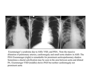
This document provides guidance on evaluating chest x-rays for congenital heart disease. It outlines key aspects to examine such as cardiac size and position, pulmonary vasculature, and abnormalities in specific heart structures. Common errors made in interpretation are also discussed, such as misdiagnosing over- or under-penetration of images. Example x-rays of various congenital heart conditions are provided to demonstrate typical features, including tetralogy of Fallot, transposition of the great vessels, and total anomalous pulmonary venous return.
AV nodal reentrant tachycardia (AVNRT), or atrioventricular nodal reentrant tachycardia, is a type of tachycardia (fast rhythm) of the heart. It is a type of supraventricular tachycardia (SVT), meaning that it originates from a location within the heart above the bundle of His. AV nodal reentrant tachycardia is the most common regular supraventricular tachycardia. It is more common in women than men (approximately 75% of cases occur in females). The main symptom is palpitations. Treatment may be with specific physical maneuvers, medication, or, rarely, synchronized cardioversion. Frequent attacks may require radiofrequency ablation, in which the abnormally conducting tissue in the heart is destroyed.
AVNRT occurs when a reentry circuit forms within or just next to the atrioventricular node. The circuit usually involves two anatomical pathways: the fast pathway and the slow pathway, which are both in the right atrium. The slow pathway (which is usually targeted for ablation) is located inferior and slightly posterior to the AV node, often following the anterior margin of the coronary sinus. The fast pathway is usually located just superior and posterior to the AV node. These pathways are formed from tissue that behaves very much like the AV node, and some authors regard them as part of the AV node.
The fast and slow pathways should not be confused with the accessory pathways that give rise to Wolff-Parkinson-White syndrome (WPW syndrome) or atrioventricular reciprocating tachycardia (AVRT). In AVNRT, the fast and slow pathways are located within the right atrium close to or within the AV node and exhibit electrophysiologic properties similar to AV nodal tissue. Accessory pathways that give rise to WPW syndrome and AVRT are located in the atrioventricular valvular rings. They provide a direct connection between the atria and ventricles, and have electrophysiologic properties similar to ventricular myocardium.
preop TEE assessment of atrial septal defect is very important for making decision for device closure, properly assessed adequate rims of ASD will reduce risk of device embolization to almost nil.
Today, in addition to measurement of left ventricular ejection fraction, the simple 12-lead surface ECG remains the only evidence-based means of identifying patients who may obtain the substantial benefits of CRT
Speckle tracking echocardiography (STE) is an echocardiographic imaging technique that analyzes the motion of tissues in the heart by using the naturally occurring speckle pattern in the myocardium or blood when imaged by ultrasound.
This file was made while my course of studying pediatrics at college,intednded to make the cardiology lessons more organized and easier to study and memorize. And I do hope it will be useful to the other medical students who read it.
Report Back from SGO 2024: What’s the Latest in Cervical Cancer?bkling
Are you curious about what’s new in cervical cancer research or unsure what the findings mean? Join Dr. Emily Ko, a gynecologic oncologist at Penn Medicine, to learn about the latest updates from the Society of Gynecologic Oncology (SGO) 2024 Annual Meeting on Women’s Cancer. Dr. Ko will discuss what the research presented at the conference means for you and answer your questions about the new developments.
New Directions in Targeted Therapeutic Approaches for Older Adults With Mantl...i3 Health
i3 Health is pleased to make the speaker slides from this activity available for use as a non-accredited self-study or teaching resource.
This slide deck presented by Dr. Kami Maddocks, Professor-Clinical in the Division of Hematology and
Associate Division Director for Ambulatory Operations
The Ohio State University Comprehensive Cancer Center, will provide insight into new directions in targeted therapeutic approaches for older adults with mantle cell lymphoma.
STATEMENT OF NEED
Mantle cell lymphoma (MCL) is a rare, aggressive B-cell non-Hodgkin lymphoma (NHL) accounting for 5% to 7% of all lymphomas. Its prognosis ranges from indolent disease that does not require treatment for years to very aggressive disease, which is associated with poor survival (Silkenstedt et al, 2021). Typically, MCL is diagnosed at advanced stage and in older patients who cannot tolerate intensive therapy (NCCN, 2022). Although recent advances have slightly increased remission rates, recurrence and relapse remain very common, leading to a median overall survival between 3 and 6 years (LLS, 2021). Though there are several effective options, progress is still needed towards establishing an accepted frontline approach for MCL (Castellino et al, 2022). Treatment selection and management of MCL are complicated by the heterogeneity of prognosis, advanced age and comorbidities of patients, and lack of an established standard approach for treatment, making it vital that clinicians be familiar with the latest research and advances in this area. In this activity chaired by Michael Wang, MD, Professor in the Department of Lymphoma & Myeloma at MD Anderson Cancer Center, expert faculty will discuss prognostic factors informing treatment, the promising results of recent trials in new therapeutic approaches, and the implications of treatment resistance in therapeutic selection for MCL.
Target Audience
Hematology/oncology fellows, attending faculty, and other health care professionals involved in the treatment of patients with mantle cell lymphoma (MCL).
Learning Objectives
1.) Identify clinical and biological prognostic factors that can guide treatment decision making for older adults with MCL
2.) Evaluate emerging data on targeted therapeutic approaches for treatment-naive and relapsed/refractory MCL and their applicability to older adults
3.) Assess mechanisms of resistance to targeted therapies for MCL and their implications for treatment selection
Title: Sense of Smell
Presenter: Dr. Faiza, Assistant Professor of Physiology
Qualifications:
MBBS (Best Graduate, AIMC Lahore)
FCPS Physiology
ICMT, CHPE, DHPE (STMU)
MPH (GC University, Faisalabad)
MBA (Virtual University of Pakistan)
Learning Objectives:
Describe the primary categories of smells and the concept of odor blindness.
Explain the structure and location of the olfactory membrane and mucosa, including the types and roles of cells involved in olfaction.
Describe the pathway and mechanisms of olfactory signal transmission from the olfactory receptors to the brain.
Illustrate the biochemical cascade triggered by odorant binding to olfactory receptors, including the role of G-proteins and second messengers in generating an action potential.
Identify different types of olfactory disorders such as anosmia, hyposmia, hyperosmia, and dysosmia, including their potential causes.
Key Topics:
Olfactory Genes:
3% of the human genome accounts for olfactory genes.
400 genes for odorant receptors.
Olfactory Membrane:
Located in the superior part of the nasal cavity.
Medially: Folds downward along the superior septum.
Laterally: Folds over the superior turbinate and upper surface of the middle turbinate.
Total surface area: 5-10 square centimeters.
Olfactory Mucosa:
Olfactory Cells: Bipolar nerve cells derived from the CNS (100 million), with 4-25 olfactory cilia per cell.
Sustentacular Cells: Produce mucus and maintain ionic and molecular environment.
Basal Cells: Replace worn-out olfactory cells with an average lifespan of 1-2 months.
Bowman’s Gland: Secretes mucus.
Stimulation of Olfactory Cells:
Odorant dissolves in mucus and attaches to receptors on olfactory cilia.
Involves a cascade effect through G-proteins and second messengers, leading to depolarization and action potential generation in the olfactory nerve.
Quality of a Good Odorant:
Small (3-20 Carbon atoms), volatile, water-soluble, and lipid-soluble.
Facilitated by odorant-binding proteins in mucus.
Membrane Potential and Action Potential:
Resting membrane potential: -55mV.
Action potential frequency in the olfactory nerve increases with odorant strength.
Adaptation Towards the Sense of Smell:
Rapid adaptation within the first second, with further slow adaptation.
Psychological adaptation greater than receptor adaptation, involving feedback inhibition from the central nervous system.
Primary Sensations of Smell:
Camphoraceous, Musky, Floral, Pepperminty, Ethereal, Pungent, Putrid.
Odor Detection Threshold:
Examples: Hydrogen sulfide (0.0005 ppm), Methyl-mercaptan (0.002 ppm).
Some toxic substances are odorless at lethal concentrations.
Characteristics of Smell:
Odor blindness for single substances due to lack of appropriate receptor protein.
Behavioral and emotional influences of smell.
Transmission of Olfactory Signals:
From olfactory cells to glomeruli in the olfactory bulb, involving lateral inhibition.
Primitive, less old, and new olfactory systems with different path
Ethanol (CH3CH2OH), or beverage alcohol, is a two-carbon alcohol
that is rapidly distributed in the body and brain. Ethanol alters many
neurochemical systems and has rewarding and addictive properties. It
is the oldest recreational drug and likely contributes to more morbidity,
mortality, and public health costs than all illicit drugs combined. The
5th edition of the Diagnostic and Statistical Manual of Mental Disorders
(DSM-5) integrates alcohol abuse and alcohol dependence into a single
disorder called alcohol use disorder (AUD), with mild, moderate,
and severe subclassifications (American Psychiatric Association, 2013).
In the DSM-5, all types of substance abuse and dependence have been
combined into a single substance use disorder (SUD) on a continuum
from mild to severe. A diagnosis of AUD requires that at least two of
the 11 DSM-5 behaviors be present within a 12-month period (mild
AUD: 2–3 criteria; moderate AUD: 4–5 criteria; severe AUD: 6–11 criteria).
The four main behavioral effects of AUD are impaired control over
drinking, negative social consequences, risky use, and altered physiological
effects (tolerance, withdrawal). This chapter presents an overview
of the prevalence and harmful consequences of AUD in the U.S.,
the systemic nature of the disease, neurocircuitry and stages of AUD,
comorbidities, fetal alcohol spectrum disorders, genetic risk factors, and
pharmacotherapies for AUD.
Title: Sense of Taste
Presenter: Dr. Faiza, Assistant Professor of Physiology
Qualifications:
MBBS (Best Graduate, AIMC Lahore)
FCPS Physiology
ICMT, CHPE, DHPE (STMU)
MPH (GC University, Faisalabad)
MBA (Virtual University of Pakistan)
Learning Objectives:
Describe the structure and function of taste buds.
Describe the relationship between the taste threshold and taste index of common substances.
Explain the chemical basis and signal transduction of taste perception for each type of primary taste sensation.
Recognize different abnormalities of taste perception and their causes.
Key Topics:
Significance of Taste Sensation:
Differentiation between pleasant and harmful food
Influence on behavior
Selection of food based on metabolic needs
Receptors of Taste:
Taste buds on the tongue
Influence of sense of smell, texture of food, and pain stimulation (e.g., by pepper)
Primary and Secondary Taste Sensations:
Primary taste sensations: Sweet, Sour, Salty, Bitter, Umami
Chemical basis and signal transduction mechanisms for each taste
Taste Threshold and Index:
Taste threshold values for Sweet (sucrose), Salty (NaCl), Sour (HCl), and Bitter (Quinine)
Taste index relationship: Inversely proportional to taste threshold
Taste Blindness:
Inability to taste certain substances, particularly thiourea compounds
Example: Phenylthiocarbamide
Structure and Function of Taste Buds:
Composition: Epithelial cells, Sustentacular/Supporting cells, Taste cells, Basal cells
Features: Taste pores, Taste hairs/microvilli, and Taste nerve fibers
Location of Taste Buds:
Found in papillae of the tongue (Fungiform, Circumvallate, Foliate)
Also present on the palate, tonsillar pillars, epiglottis, and proximal esophagus
Mechanism of Taste Stimulation:
Interaction of taste substances with receptors on microvilli
Signal transduction pathways for Umami, Sweet, Bitter, Sour, and Salty tastes
Taste Sensitivity and Adaptation:
Decrease in sensitivity with age
Rapid adaptation of taste sensation
Role of Saliva in Taste:
Dissolution of tastants to reach receptors
Washing away the stimulus
Taste Preferences and Aversions:
Mechanisms behind taste preference and aversion
Influence of receptors and neural pathways
Impact of Sensory Nerve Damage:
Degeneration of taste buds if the sensory nerve fiber is cut
Abnormalities of Taste Detection:
Conditions: Ageusia, Hypogeusia, Dysgeusia (parageusia)
Causes: Nerve damage, neurological disorders, infections, poor oral hygiene, adverse drug effects, deficiencies, aging, tobacco use, altered neurotransmitter levels
Neurotransmitters and Taste Threshold:
Effects of serotonin (5-HT) and norepinephrine (NE) on taste sensitivity
Supertasters:
25% of the population with heightened sensitivity to taste, especially bitterness
Increased number of fungiform papillae
Prix Galien International 2024 Forum ProgramLevi Shapiro
June 20, 2024, Prix Galien International and Jerusalem Ethics Forum in ROME. Detailed agenda including panels:
- ADVANCES IN CARDIOLOGY: A NEW PARADIGM IS COMING
- WOMEN’S HEALTH: FERTILITY PRESERVATION
- WHAT’S NEW IN THE TREATMENT OF INFECTIOUS,
ONCOLOGICAL AND INFLAMMATORY SKIN DISEASES?
- ARTIFICIAL INTELLIGENCE AND ETHICS
- GENE THERAPY
- BEYOND BORDERS: GLOBAL INITIATIVES FOR DEMOCRATIZING LIFE SCIENCE TECHNOLOGIES AND PROMOTING ACCESS TO HEALTHCARE
- ETHICAL CHALLENGES IN LIFE SCIENCES
- Prix Galien International Awards Ceremony
MANAGEMENT OF ATRIOVENTRICULAR CONDUCTION BLOCK.pdfJim Jacob Roy
Cardiac conduction defects can occur due to various causes.
Atrioventricular conduction blocks ( AV blocks ) are classified into 3 types.
This document describes the acute management of AV block.
These simplified slides by Dr. Sidra Arshad present an overview of the non-respiratory functions of the respiratory tract.
Learning objectives:
1. Enlist the non-respiratory functions of the respiratory tract
2. Briefly explain how these functions are carried out
3. Discuss the significance of dead space
4. Differentiate between minute ventilation and alveolar ventilation
5. Describe the cough and sneeze reflexes
Study Resources:
1. Chapter 39, Guyton and Hall Textbook of Medical Physiology, 14th edition
2. Chapter 34, Ganong’s Review of Medical Physiology, 26th edition
3. Chapter 17, Human Physiology by Lauralee Sherwood, 9th edition
4. Non-respiratory functions of the lungs https://academic.oup.com/bjaed/article/13/3/98/278874
- Video recording of this lecture in English language: https://youtu.be/lK81BzxMqdo
- Video recording of this lecture in Arabic language: https://youtu.be/Ve4P0COk9OI
- Link to download the book free: https://nephrotube.blogspot.com/p/nephrotube-nephrology-books.html
- Link to NephroTube website: www.NephroTube.com
- Link to NephroTube social media accounts: https://nephrotube.blogspot.com/p/join-nephrotube-on-social-media.html
3. Reading of chest X-rays in CHD
Is there cardiomegaly?
Cardiothoracic ratio >60% in neonates
> 55% in infants
> 50% in older children/adult
4. Is the situs normal?
The situs as defined by-
Stomach bubble
Liver shadow
Bronchial morphology of more horizontal and
short right bronchus
5. Situs inversus totalis and dextrocardia- rarely
has heart disease
Mesocardia -commonly has c TGA
Left-sided heart in situs inversus- has complex
heart disease.
Apex -dome of diaphragm is lower on the side
of apex.
6. Is there a particular chamber enlargement ?
RA enlargement-
Vertical height compared to height from aortic
arch to SVC-RA junction
lateral 3 spaces criteria
The RV type- upturned apex e.g TOF
LV type of apex -tricuspid atresia
7.
8. Is the cardiac silhouette normal?
L-posed aorta
Enlarged thymus
9. What is the pulmonary blood flow—
increased, decreased or normal?
Increased pulmonary blood flow - more than 5
end-on vessels in the lungs, or more than 3 in
one lung.
The end-on vessels should be more than twice
the size of an accompanying bronchus.
More than 6 vessels could be traced in the
periphery in increased flow states.
10. Decreased flow - when the vessels are thin and
small.
Less than 3 vessels are seen in the periphery
Pulmonary conus and main arteries are not
prominent.
11. Is there PAH? How severe?
In adults NORMAL descending branch of right
pulmonary artery-
9–14 mm in females.
10–15 mm in males.
In children, the artery size larger than the trachea
indicates enlargement.
Peripheral pruning—that is sudden taper of PA
branches as it travels to periphery indicates PAH.
Pruning is seen in functional as well as organic PAH.
12. Is there PVH ? How severe?
PVH is present when the PAWP >12 mm Hg.
Equalization
Cephalization
Perihilar haze
Interstitial edema indicate progressively
increasing PVH.
o Kerley lines generally indicate chronic severe
PVH
13. Is there aortic arch abnormality?
The aortic arch is identified by the impression on
trachea.
A right aortic arch is commonly associated with
VSD.
A right arch and increased blood flow is typically
seen in truncus arteriosus.
An inconspicuous aortic arch could mean double
arch.
Coarctation with arch hypoplasia.
15. Is there asymmetry of findings, e.g.
Decreased vasculature on one side?
This could indicate ipsilateral pulmonary artery
stenosis.
Pulmonary embolism.
Anomalous pulmonary venous drainage
Lung disease.
Is there rib notching? Signs of previous
surgery like rib regeneration?
16. Are there serial changes?
The changes of pulmonary plethora diminish
but in Eisenmenger’s syndrome
17. Spot diagnosis-
Boot-shaped heart- TOF,
Egg on side appearance of TGA without VSD
Figure-of-8 for supracardiac TAPVC
Waterfall sign of a truncus arteriosus
Typical straight right border for tricuspid atresia
Aorta forming the left border in corrected
transposition of great vessels.
18. COMMON ERRORS
Following are the common errors seen in day-
to-day clinical practice:
Spurious cardiomegaly due to expiratory film.
Wrong assessment of lung vasculature due to
over- and underpenetration (underpenetration
increases lung vasculature and vice versa)
Rotation of film leading to wrong
interpretation of cardiac silhouette or hilum.
19. Missing a spinal deformity causing
cardiomegaly, or altered silhouette.
Over-relying on patterns without taking into
account the sensitivity and specificity of a
finding- e.g RA dilatation, type of apex (RV or
LV), egg-on-side pattern for TGA.
20. Misunderstanding PVH for pulmonary plethora
can lead to misclassification of the disease.
Wrong reading of hilar shadows is frequent cause
of errors.
Many a times, dilated PAs have been interpreted
as lymph nodes and patients prescribed
antitubercular treatment.
Not paying enough attention to lungs e.g
infiltration, or military shadows missed or over-
read.
21. Misinterpretation of normal but infrequent
seen structures such as the azygous lobe,
diaphragmatic humps, atypical thymus
shadow, etc.
22. TOF with absent pulmonary valve -Dilated main pulmonary artery
The pulmonary vasculature is not markedly decreased and may be
normal or increased
23. TOF - markedly decreased lung vasculature
and pulmonary bay and right aortic arch
24. TOF and endocarditis. Note the infiltration in the lungs due
to septic emboli from the right-sided vegetations. The boot-
shaped heart and decreased pulmonary blood flow is seen
25. Tricuspid atresia with decreased pulmonary blood flow.
LV type of apex. note the left SVC.
26. Corrected transposition of great vessels. Note the mesocardia, and shadow
of L-posed aorta. Pulmonary artery shadow is not distinctly seen and is
behind the aortic shadow.
27. Note the figure-of-8 shadow and increased pulmonary blood flow from
supracardiac TAPVC that is not obstructed (A thymic shadow can sometime
mimic this, but then the pulmonary blood flow will be normal)
29. Truncus arteriosus. Note cardiomegaly and increased
pulmonary blood flow. The main pulmonary artery is not in
normal place. Rt PA origin seems high. A right arch would
have made the diagnosis easy but is not present
30. Two examples of transposition with intect septum. Note the variation in TGA
X-rays.
31. Marked cardiomegaly in newborn has few differential diagnoses, Ebstein’s
anomaly in this case
33. Diffuse cardiomegaly and overpenetrated
film. No comments possible regarding lung
vasculature
Hyperinflated lung in double aortic arch
34. TOF and cardiomegaly due to severe anemia. Cardiomegaly does not
occur in all lesions of “TOF physiology” unless complicated by other
things
35. Typical ground-glass appearance in a neonate from obstructed infracardiac
TAPVC. The angiogram shows the descending vertical vein going below the
diaphragm
36. Increased vasculature in VSD
PAH, peripheral pruning, dilated right
descending PA, and prominent main
PA.
37. Eisenmenger’s syndrome due to ASD, VSD, and PDA . Note the massive
dilatation of pulmonary arteries, cardiomegaly and small aorta shadow in ASD. The
PDA Eisenmenger (right) is remarkable for prominent aorticopulmonary shadow.
Sometimes a ductal calcification may be seen in the area between aorta and dilated
PA. Eisenmenger VSD (middle) shows PAH but neither cardiomegaly nor
prominent aorta
Right atrial enlargement is diagnosed when A > B (the horizontal lines are drawn at the top of aortic arch) at superior vena cava-right atrium (SVC-RA) junction, and lower limit of heart at right border
The right pulmonary artery (RPA) is also commonly dilated, but not seen here.
Localizing cardiac prosthetic valves can be difficult. There are a number of strategies that can be employed to aid in characterizing the type of prosthetic valve. The best strategy involves assessing the location of the valve and then determining the orientation and direction of flow. Additional aids which serve as adjunts are the statistical nature of valve replacement and including patient history.The location of the cardiac valves is best determined on the lateral radiograph. A line is drawn on the lateral radiograph from the carina to the cardiac apex. The pulmonic and aortic valves generally sit above this line and the tricuspid and mitral valves sit below this line. Keep in mind that sometimes the aortic root can be inferiorly displaced which will shift the aortic valve below this line.
A second technique to further localize prosthetic valves involves drawing a second line which is perpendicular to the patient's upright position which bisects the cardiac silouette. The aortic valve projects in the upper quadrant, the mitral valve in the lower quadrant and the tricuspid valve in the anterior quadrant. The pulmonary valve projects in the superior portion of the posterior quadrant which is illustrated in the below images:
An alternative method, which is less reproducible, involves the frontal radiograph. On the frontal chest radiograph (either the AP or PA view) cardiac valvular prostheses can be localized by drawing a longitudinal line through the mid sternal body. Use this line to bisect the sternum in the sagittal plane and then draw a perpendicular line dividing the heart horizontally. The aortic valve should overlie the intersection of these two lines. The mitral valve will lie in the lower left quadrant (the patient’s left). The tricuspid valve would lie in the lower right corner (the patient's right) and the pulmonic valve will lie in the upper left corner (the patient's left). A word of warning pertains to the above strategies. Patients with cardiac valves often have chamber enlargement and cardiac rotation which can displace the positions of the valves as well as create difficulty when drawing lines through the cardiac silhouette. These rules are meant as a guideline to better localize cardiac valves although they do not always work.
One additional pearl in determining the location of cardiac valves includes localizing the direction of flow. Some bioprosthetic valves have components that determine the direction of flow which helps localize the valve prosthesis. If the direction of flow is from inferior to superior and then an aortic valve is likely. If the direction of flow is from superior to inferior in the left chest then the valve is likely a mitral valve.