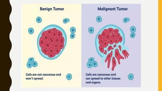
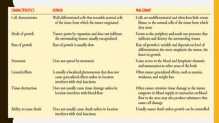
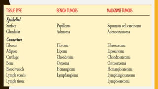
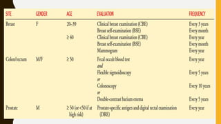
ONCOLOGICAL
DISORDERS
INTRODUCTION
B Y,
M S . E K TA S PAT E L ,
A S S I S TA N T P R O F E S S O R
MEANING
A serious disease that is caused when cells in
the body grow in a way that is uncontrolled and
not normal.
A disease caused by an uncontrolled division of abnormal
cells in a part of the body.
• Cancer can involve any tissue of the body and have many
different forms in each body area.
• Most cancers are named for the type of cell or organ in
which they start.
DEFINITION:
• An abnormal growth of cells which tend to proliferate in an
uncontrolled way and, in some cases, to metastasize
(spread).
• A term for diseases in which abnormal cells divide without
control and can invade nearby tissues.
BENIGN / MALIGNANT
C H A R A C T E R I S T I C S O F
B E N I G N A N D M A L I G N A N T
C A N C E R
CHARACTERISTICS OF BENIGN TUMORS
• Cells tend not to spread
• Most grow slowly
• Do not invade nearby tissue
• Do not metastasize (spread) to other parts of the body
• Tend to have clear boundaries
• Under a pathologist's microscope, shape, chromosomes,
and DNA of cells appear normal
• Do not secrete hormones or other substances (an
exception: pheochromocytomas of the adrenal gland)
• May not require treatment if not health threatening
• Unlikely to recure if removed or require further treatment
such as radiation or chemotherapy
CHARACTERISTICS OF MALIGNANT TUMORS
• Cells can spread
• Usually grow fairly rapidly
• Often invade basal membrane that surrounds nearby
healthy tissue
• Can spread via bloodstream or lymphatic system.
• May recur after removal, sometimes in areas other the original
site
• Cells have abnormal chromosomes and DNA characterized by
large, dark nuclei; may have abnormal shape
• Can secrete substances that cause fatigue and weight loss
(paraneoplastic syndrome)
• May require aggressive treatment, including surgery, radiation,
chemotherapy, and immunotherapy medications
T U M O R S A N D T I S S U E T Y P E S
A M E R I C A N C A N C E R S O C I E T Y
R E C O M M E N D AT I O N S F O R
E A R LY D E T E C T I O N O F
C A N C E R I N A S Y M P T O M AT I C ,
A V E R A G E - R I S K P E O P L E
IMAGING TESTS USED TO DETECT CANCER
• TEST : Tumor marker identification
• DESCRIPTION : Analysis of substances found in the blood
or other body fluids that are made by the tumor or by the
body in response to the tumor
• DIAGNOSTIC USES : Breast, colon, lungs, ovaries, testes,
protest
• TEST : Magnetic resonance imaging (MRI)
• DESCRIPTION : Use of magnetic fields and radiofrequency
signals to create sectioned images of various body
structures
• DIAGNOSTIC USES : Neurologic, pelvic, abdominal, thoracic
cancers
• TEST : Magnetic resonance imaging (MRI)
• DESCRIPTION : Use of magnetic fields and radiofrequency
signals to create sectioned images of various body
structures
• DIAGNOSTIC USES : Neurologic, pelvic, abdominal, thoracic
cancers
• TEST : Computed tomography (CT scan)
• DESCRIPTION : Use of narrow beam x-ray to scan
successive layers of tissue for a cross-sectional view
• DIAGNOSTIC USES : Neurologic, pelvic, skeletal, abdominal,
thoracic cancers
• TEST : Fluoroscopy
• DESCRIPTION : Use of x-rays that identify contrasts in body
tissue densities; may involve the use of contrast agents
• DIAGNOSTIC USES : Skeletal, lung, gastrointestinal cancers
• TEST : Ultrasonography (ultrasound)
• DESCRIPTION : High-frequency sound waves echoing off body
tissues are converted electronically into images; used to assess
tissues deep within the body
• DIAGNOSTIC USES : Abdominal and pelvic cancers
• TEST : Endoscopy
• DESCRIPTION : Direct visualization of a body cavity or
passageway by insertion of an endoscope into a body cavity or
opening; allows tissue biopsy, fluid aspiration and excision of
small tumors; both diagnostic and therapeutic
• DIAGNOSTIC USES : Bronchial, gastrointestinal cancers
• TEST : Nuclear medicine imaging
• DESCRIPTION : Uses intravenous injection or ingestion of
radioisotope substances followed by imaging of tissues that
have concentrated the radioisotopes
• DIAGNOSTIC USES : Bone, liver, kidney, spleen, brain, thyroid
cancers
• TEST : Positron emission tomography (PET scan)
• DESCRIPTION : Computed cross-sectional images of increased
concentration of radioisotopes in malignant cells provide
information about biologic activity of malignant cells; help
distinguish between benign and malignant processes and
responses to treatment
• DIAGNOSTIC USES : Lung, colon, liver, pancreatic, breast,
esophagus cancers; Hodgkin’s and non-Hodgkin’s lymphoma
and melanoma
• TEST : Radioimmunoconjugates (Radio-immuno-conjugates)
• DESCRIPTION : Monoclonal antibodies are labeled with a
radioisotope and injected intravenously into the patient; the
antibodies that aggregate at the tumor site are visualized with
scanners
• DIAGNOSTIC USES : Colorectal, breast, ovarian, head and neck
cancers; lymphoma and melanoma
WARNING SIGNS OF
CANCER
C
A
U
T
I
O
N
• Change in bowel or bladder habits
• A sore that does not heal
• Unusual bleeding or discharge
• Thickening or lump in the breast or elsewhere
• Indigestion or difficulty in swallowing
• Obvious change in a wart or mole
• Nagging cough or hoarseness
S

Cancer introduction

  • 1.
  • 2.
    INTRODUCTION B Y, M S. E K TA S PAT E L , A S S I S TA N T P R O F E S S O R
  • 3.
    MEANING A serious diseasethat is caused when cells in the body grow in a way that is uncontrolled and not normal. A disease caused by an uncontrolled division of abnormal cells in a part of the body.
  • 4.
    • Cancer caninvolve any tissue of the body and have many different forms in each body area. • Most cancers are named for the type of cell or organ in which they start.
  • 5.
    DEFINITION: • An abnormalgrowth of cells which tend to proliferate in an uncontrolled way and, in some cases, to metastasize (spread). • A term for diseases in which abnormal cells divide without control and can invade nearby tissues.
  • 6.
  • 8.
    C H AR A C T E R I S T I C S O F B E N I G N A N D M A L I G N A N T C A N C E R
  • 9.
    CHARACTERISTICS OF BENIGNTUMORS • Cells tend not to spread • Most grow slowly • Do not invade nearby tissue • Do not metastasize (spread) to other parts of the body • Tend to have clear boundaries
  • 10.
    • Under apathologist's microscope, shape, chromosomes, and DNA of cells appear normal • Do not secrete hormones or other substances (an exception: pheochromocytomas of the adrenal gland) • May not require treatment if not health threatening • Unlikely to recure if removed or require further treatment such as radiation or chemotherapy
  • 11.
    CHARACTERISTICS OF MALIGNANTTUMORS • Cells can spread • Usually grow fairly rapidly • Often invade basal membrane that surrounds nearby healthy tissue • Can spread via bloodstream or lymphatic system.
  • 12.
    • May recurafter removal, sometimes in areas other the original site • Cells have abnormal chromosomes and DNA characterized by large, dark nuclei; may have abnormal shape • Can secrete substances that cause fatigue and weight loss (paraneoplastic syndrome) • May require aggressive treatment, including surgery, radiation, chemotherapy, and immunotherapy medications
  • 14.
    T U MO R S A N D T I S S U E T Y P E S
  • 18.
    A M ER I C A N C A N C E R S O C I E T Y R E C O M M E N D AT I O N S F O R E A R LY D E T E C T I O N O F C A N C E R I N A S Y M P T O M AT I C , A V E R A G E - R I S K P E O P L E
  • 21.
    IMAGING TESTS USEDTO DETECT CANCER • TEST : Tumor marker identification • DESCRIPTION : Analysis of substances found in the blood or other body fluids that are made by the tumor or by the body in response to the tumor • DIAGNOSTIC USES : Breast, colon, lungs, ovaries, testes, protest
  • 22.
    • TEST :Magnetic resonance imaging (MRI) • DESCRIPTION : Use of magnetic fields and radiofrequency signals to create sectioned images of various body structures • DIAGNOSTIC USES : Neurologic, pelvic, abdominal, thoracic cancers • TEST : Magnetic resonance imaging (MRI) • DESCRIPTION : Use of magnetic fields and radiofrequency signals to create sectioned images of various body structures • DIAGNOSTIC USES : Neurologic, pelvic, abdominal, thoracic cancers
  • 24.
    • TEST :Computed tomography (CT scan) • DESCRIPTION : Use of narrow beam x-ray to scan successive layers of tissue for a cross-sectional view • DIAGNOSTIC USES : Neurologic, pelvic, skeletal, abdominal, thoracic cancers
  • 26.
    • TEST :Fluoroscopy • DESCRIPTION : Use of x-rays that identify contrasts in body tissue densities; may involve the use of contrast agents • DIAGNOSTIC USES : Skeletal, lung, gastrointestinal cancers
  • 28.
    • TEST :Ultrasonography (ultrasound) • DESCRIPTION : High-frequency sound waves echoing off body tissues are converted electronically into images; used to assess tissues deep within the body • DIAGNOSTIC USES : Abdominal and pelvic cancers
  • 29.
    • TEST :Endoscopy • DESCRIPTION : Direct visualization of a body cavity or passageway by insertion of an endoscope into a body cavity or opening; allows tissue biopsy, fluid aspiration and excision of small tumors; both diagnostic and therapeutic • DIAGNOSTIC USES : Bronchial, gastrointestinal cancers
  • 31.
    • TEST :Nuclear medicine imaging • DESCRIPTION : Uses intravenous injection or ingestion of radioisotope substances followed by imaging of tissues that have concentrated the radioisotopes • DIAGNOSTIC USES : Bone, liver, kidney, spleen, brain, thyroid cancers
  • 33.
    • TEST :Positron emission tomography (PET scan) • DESCRIPTION : Computed cross-sectional images of increased concentration of radioisotopes in malignant cells provide information about biologic activity of malignant cells; help distinguish between benign and malignant processes and responses to treatment • DIAGNOSTIC USES : Lung, colon, liver, pancreatic, breast, esophagus cancers; Hodgkin’s and non-Hodgkin’s lymphoma and melanoma
  • 35.
    • TEST :Radioimmunoconjugates (Radio-immuno-conjugates) • DESCRIPTION : Monoclonal antibodies are labeled with a radioisotope and injected intravenously into the patient; the antibodies that aggregate at the tumor site are visualized with scanners • DIAGNOSTIC USES : Colorectal, breast, ovarian, head and neck cancers; lymphoma and melanoma
  • 36.
  • 37.
  • 38.
    • Change inbowel or bladder habits • A sore that does not heal • Unusual bleeding or discharge • Thickening or lump in the breast or elsewhere • Indigestion or difficulty in swallowing • Obvious change in a wart or mole • Nagging cough or hoarseness
  • 39.