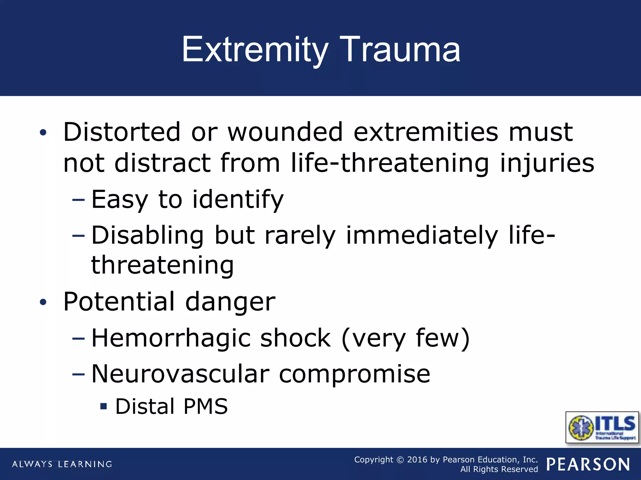
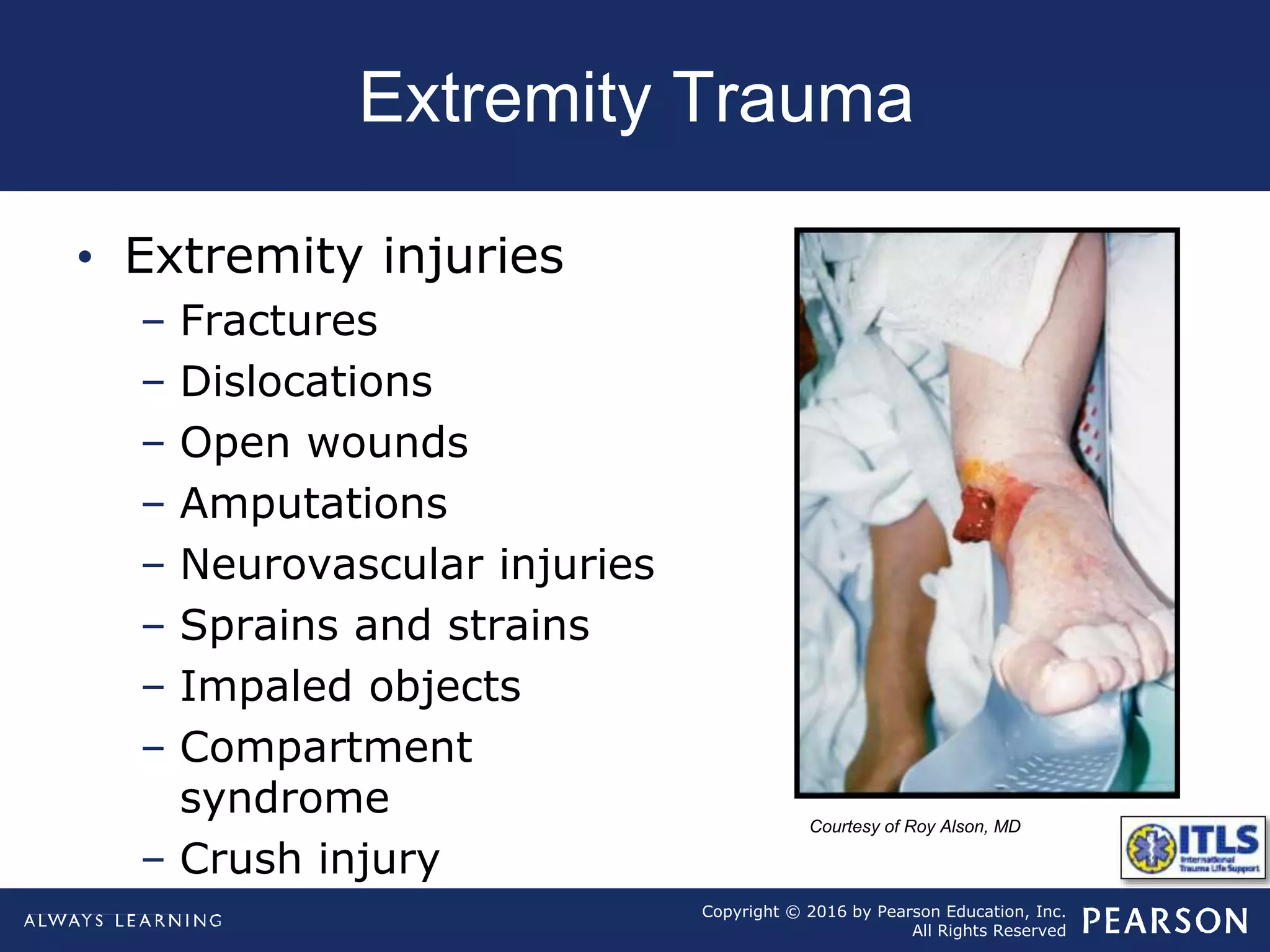
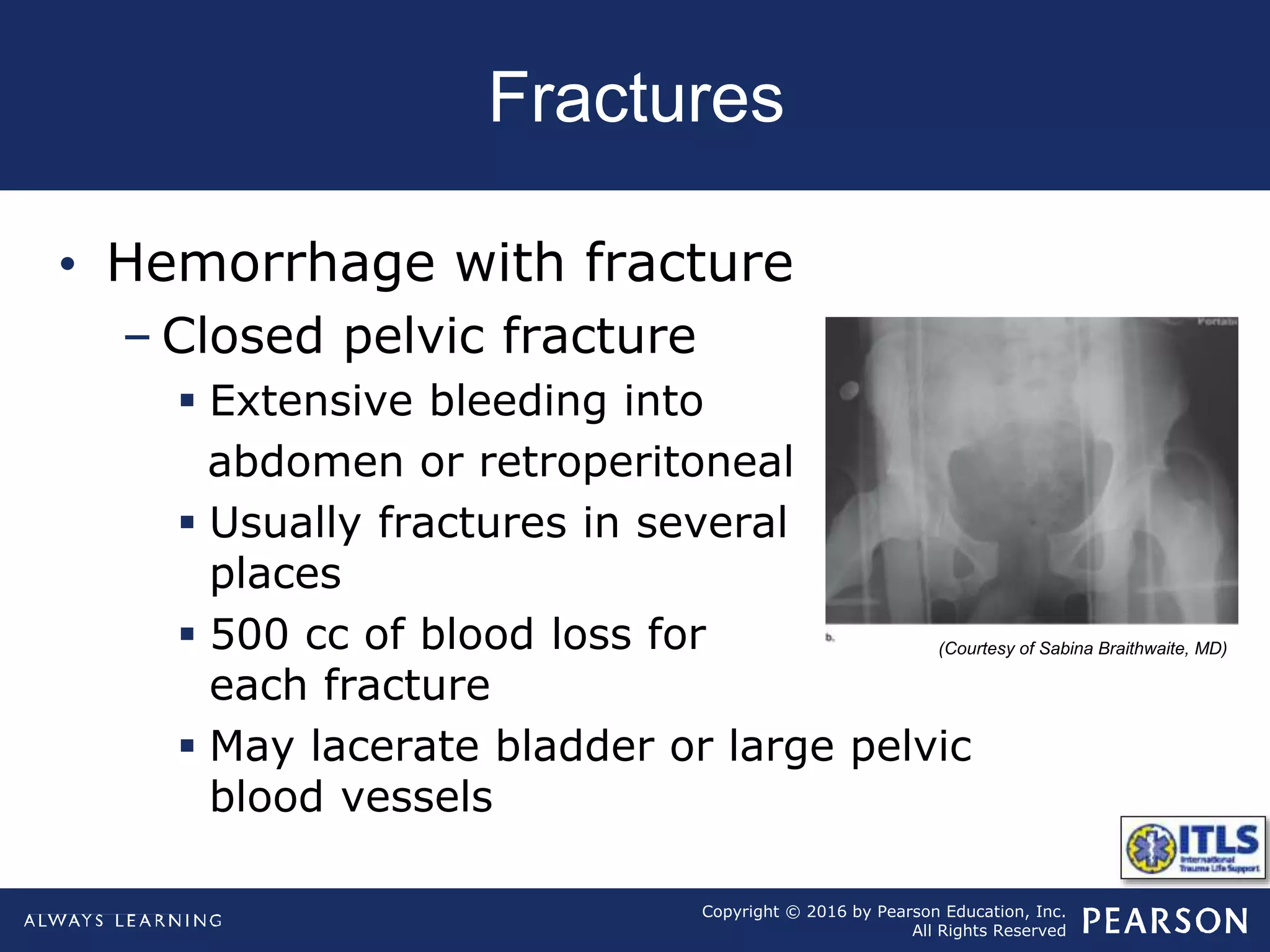
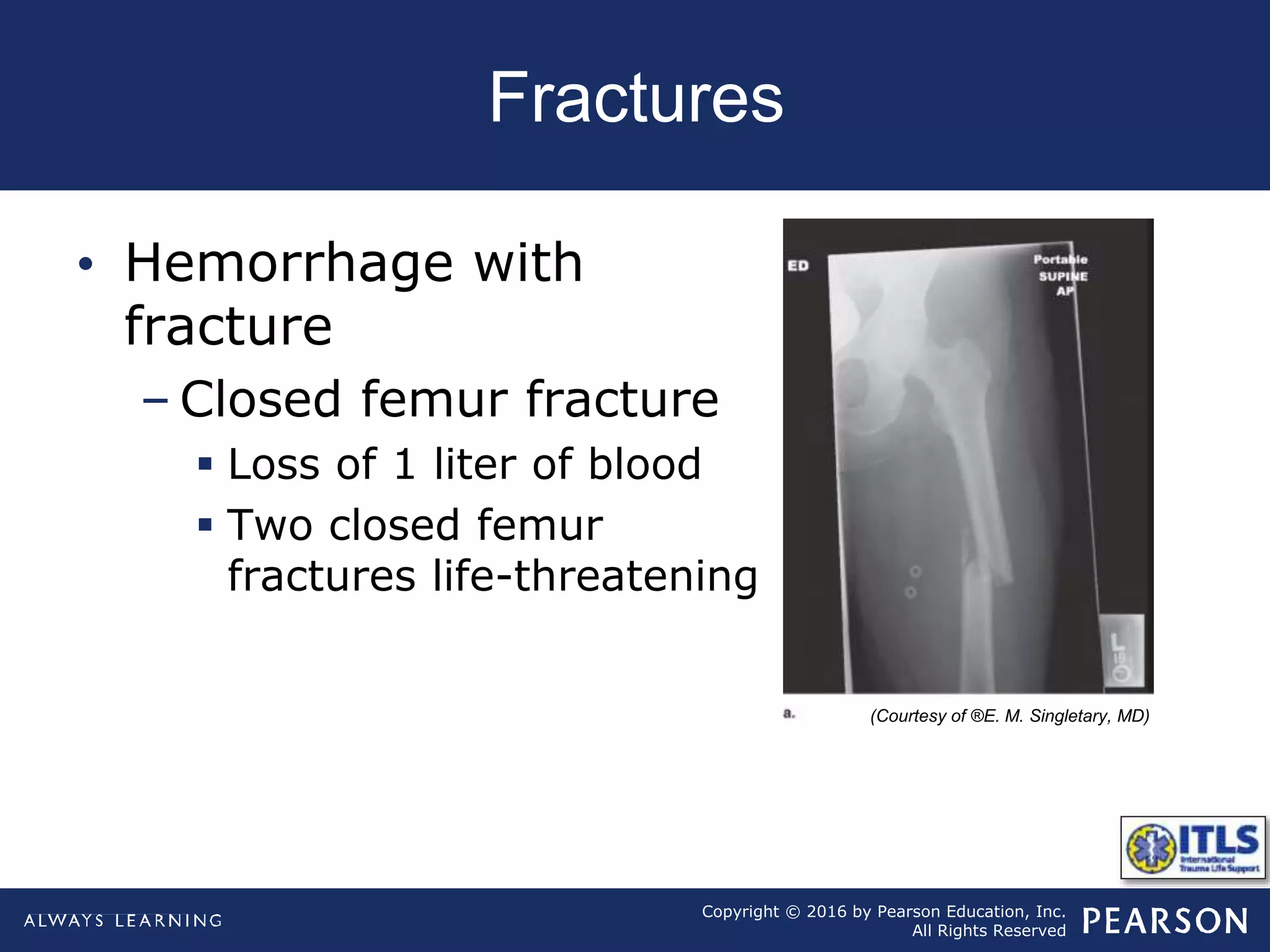
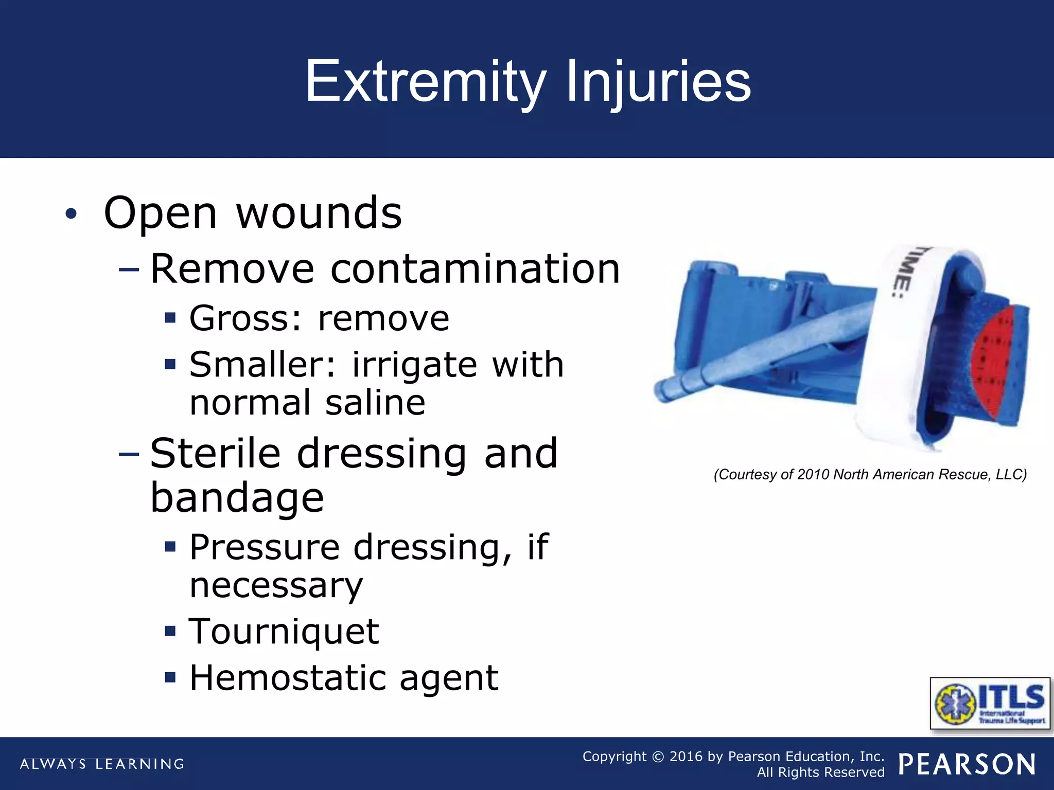
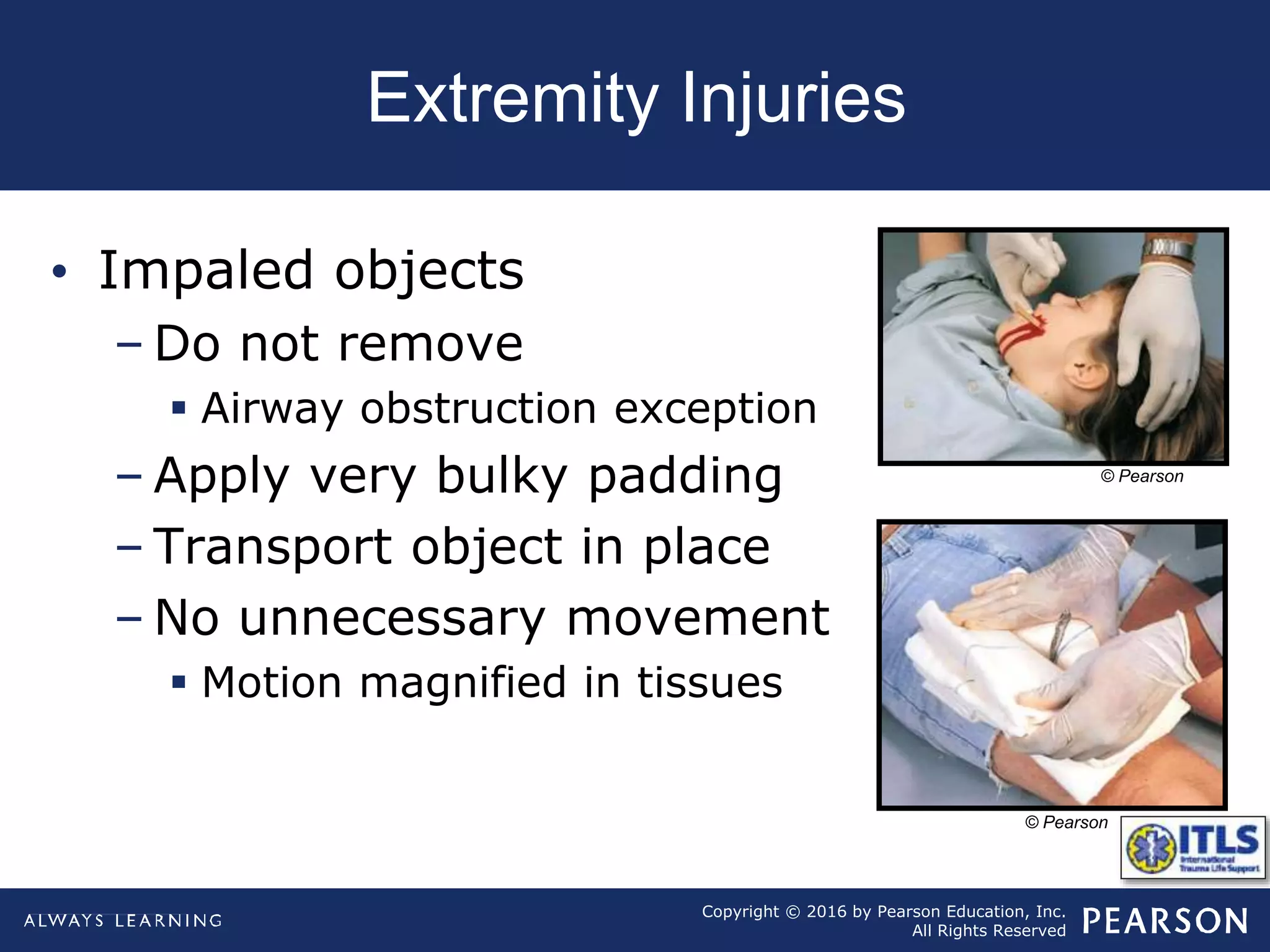
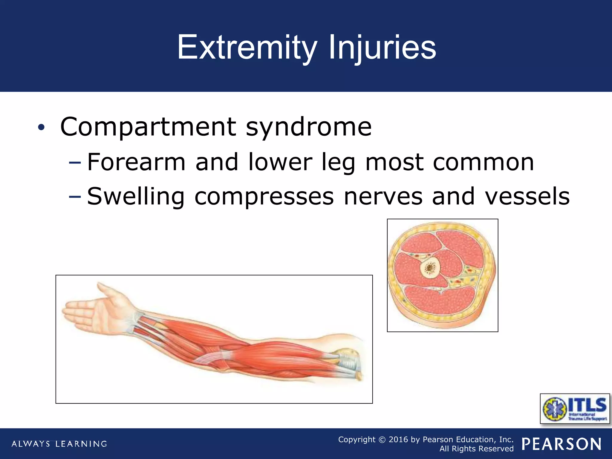
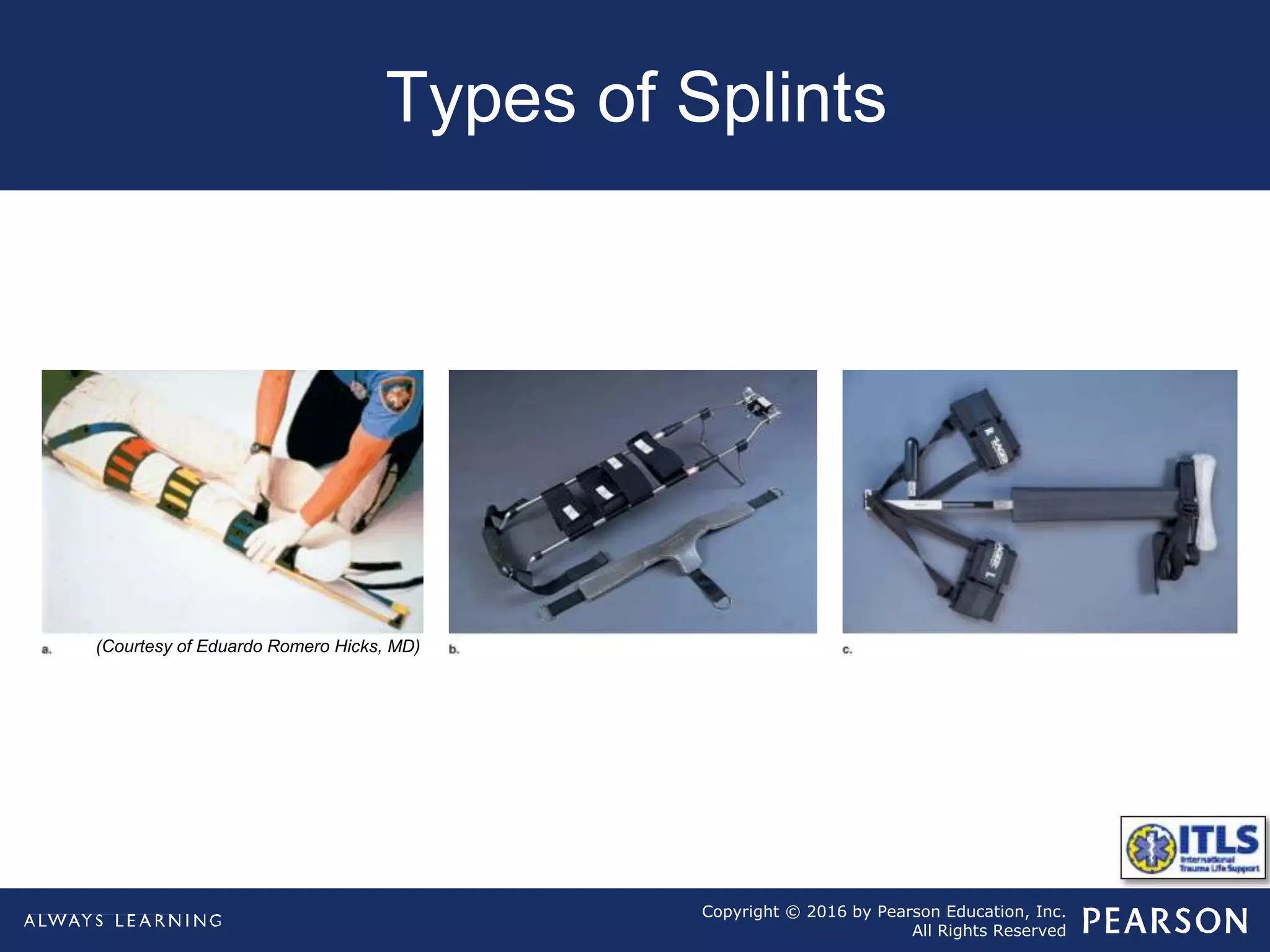
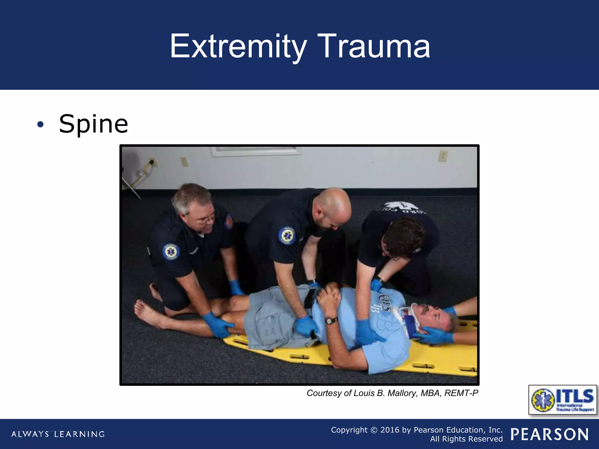
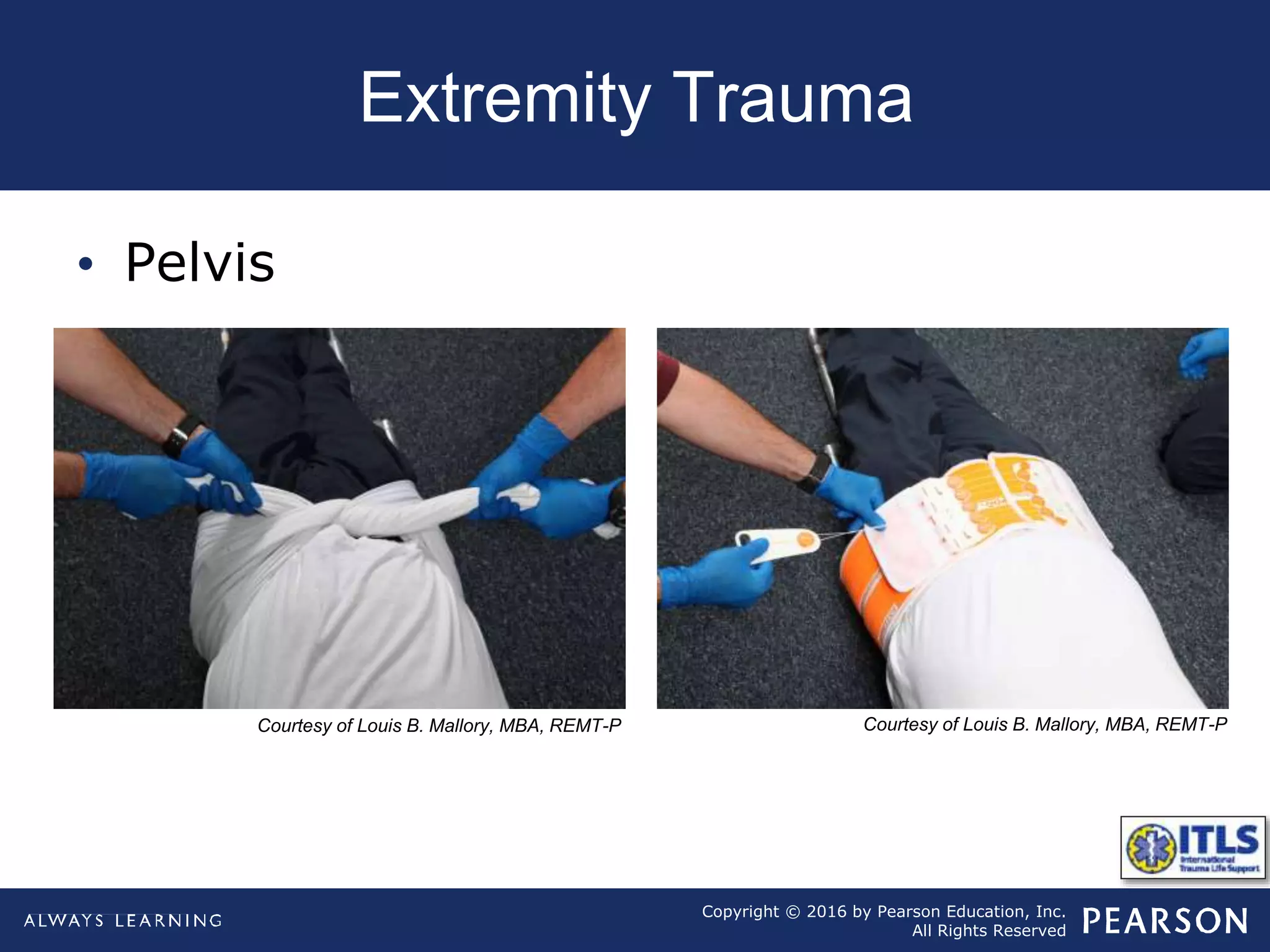
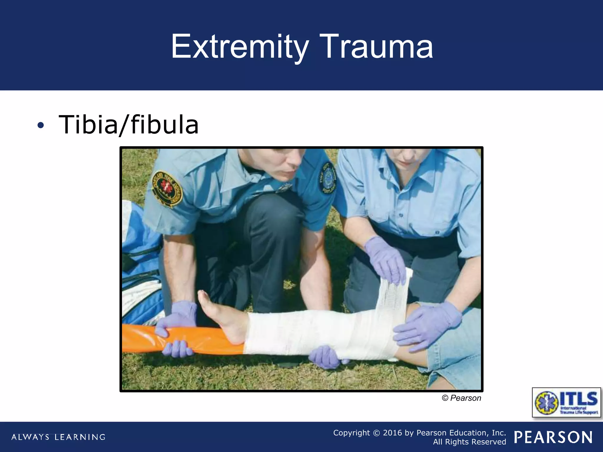
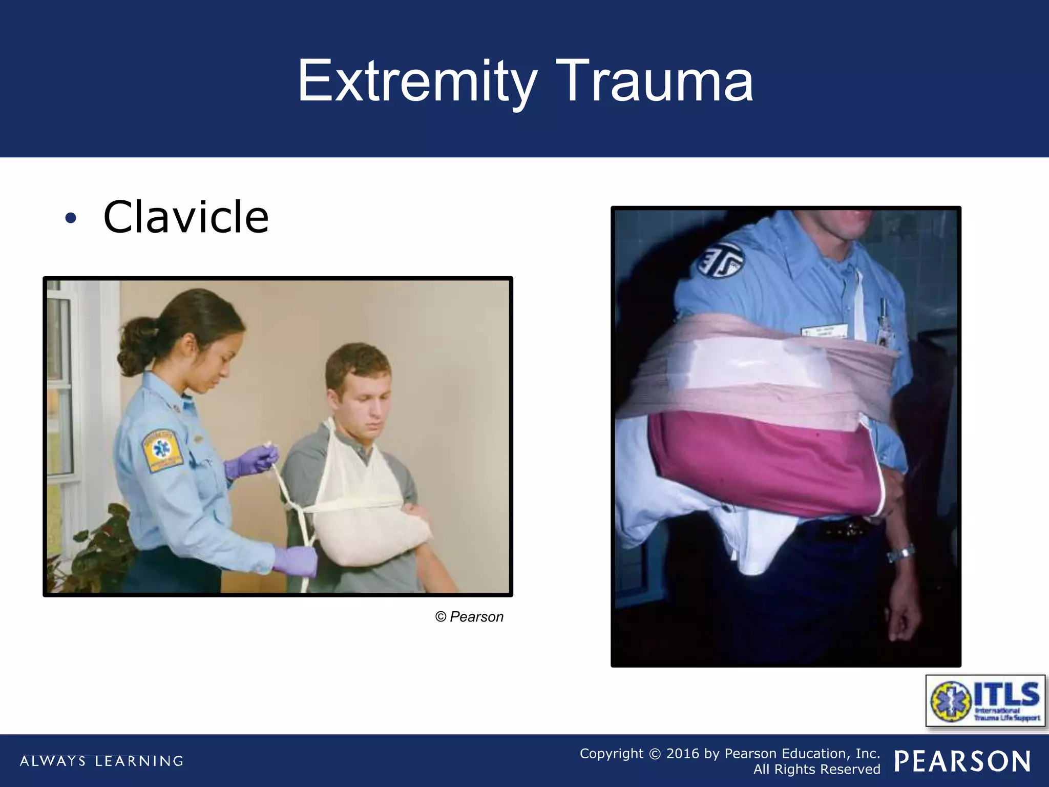
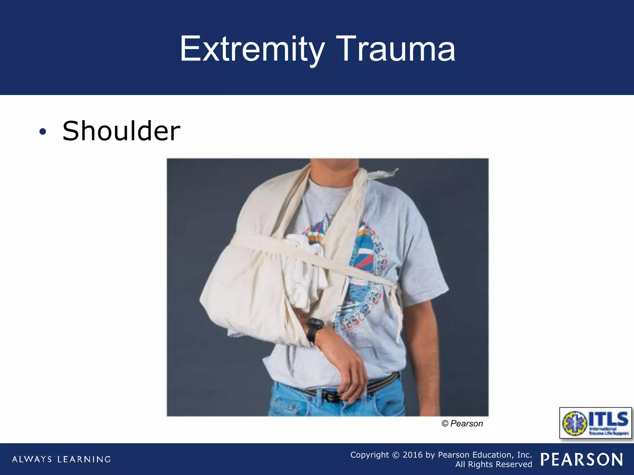
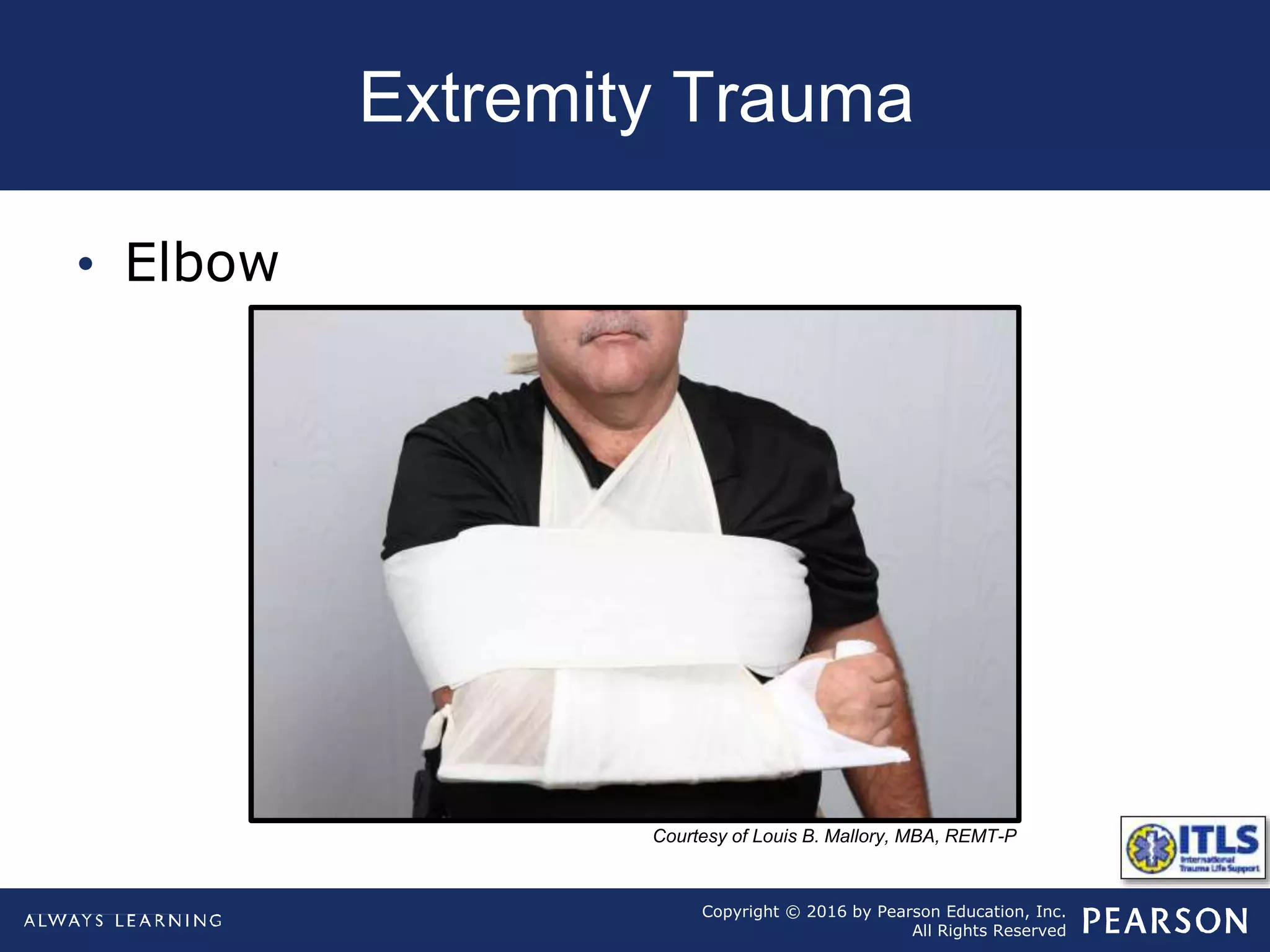
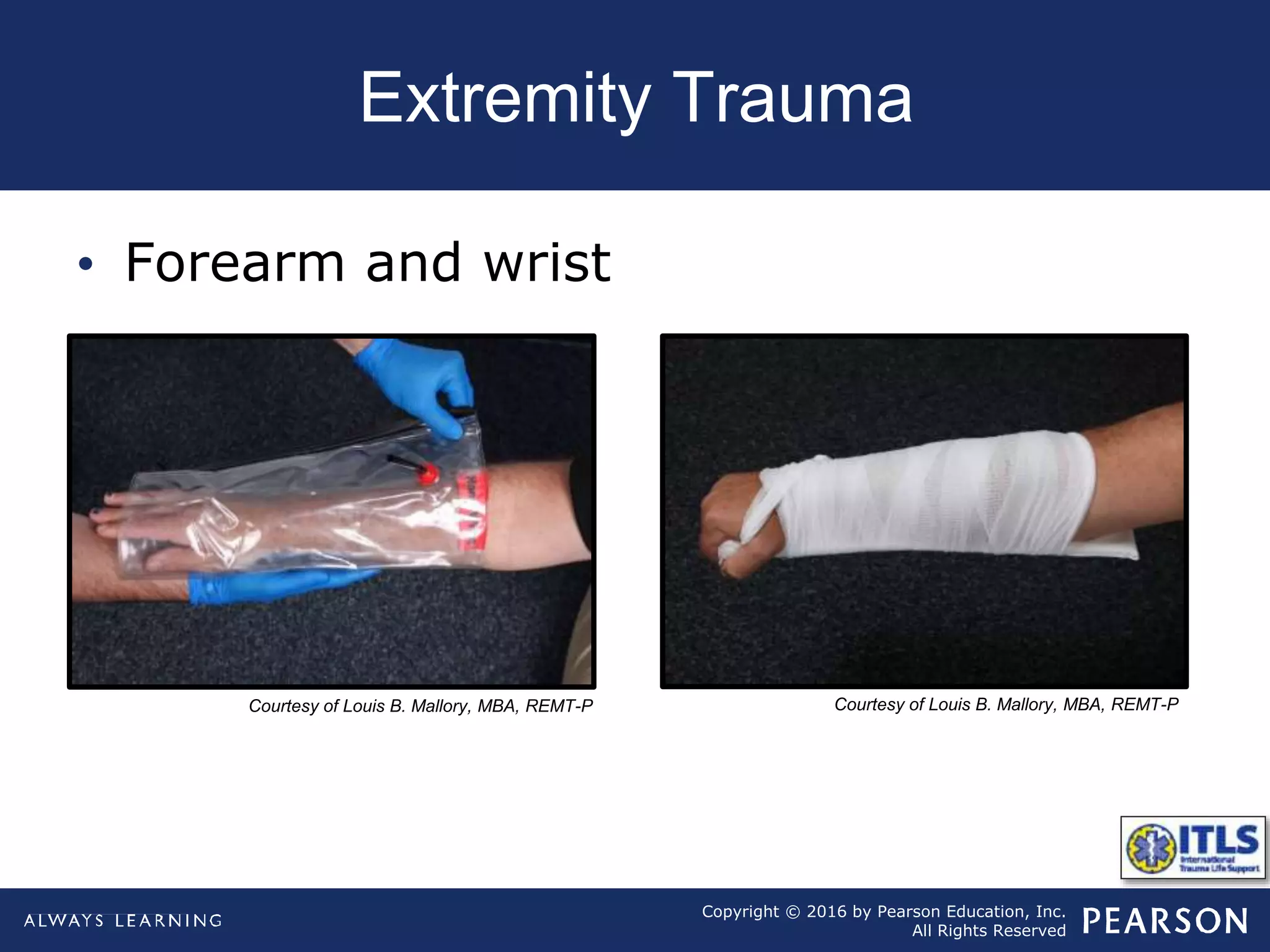
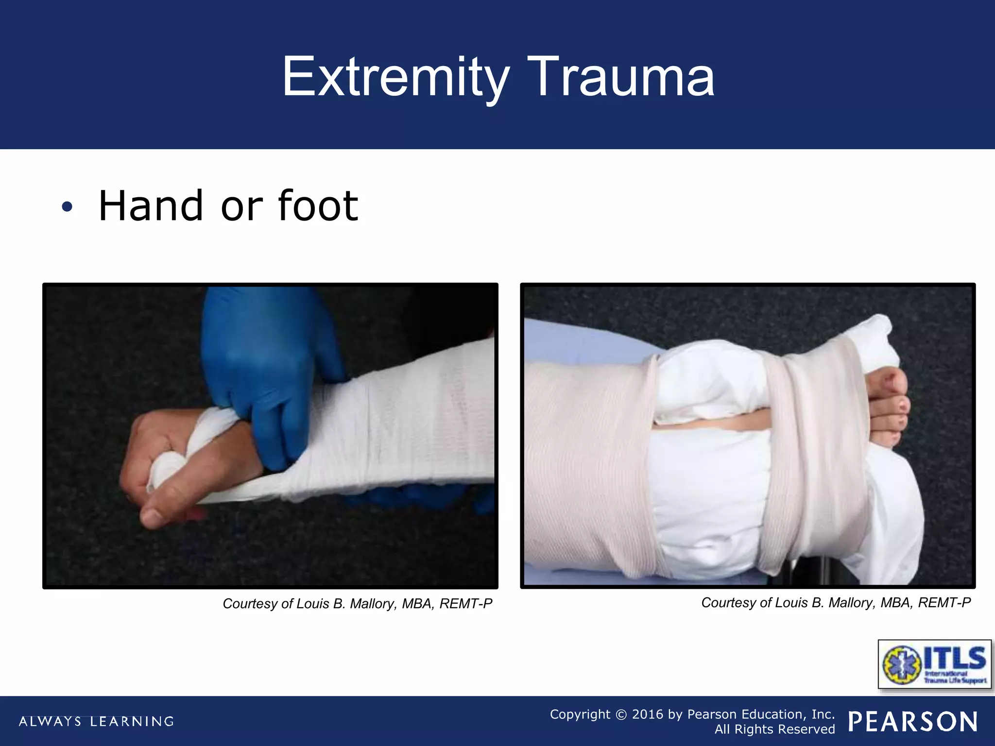
The document discusses extremity trauma, including fractures, dislocations, open wounds, amputations, and other injuries. It emphasizes that extremity injuries are generally not life-threatening, but can cause disability or complications like hemorrhage or compartment syndrome. The key priorities are to address any life-threatening injuries first, check neurovascular status, control bleeding, clean and dress wounds, splint suspected fractures to prevent further injury, and watch for signs of compartment syndrome.
Assessment and management of shock in acute trauma setting based on ATLS recommendations .Lecture given in Trauma update at Perintalmanna on19th August 2014.
Prehospital care in trauma is as important as in hospital care. The presentation addresses simple and basic approach to care a polytrauma victim in platinum 10 minutes based on BTLS.
Trauma is a global problem and continues to be a leading cause of disability and death.
Approximately 25% to 30% of deaths caused by trauma can be prevented when a systematic and organized approach is used.
The main goal of the initial assessment
Recognize the patient who does have life-threatening injuries
Establish treatment priorities, and
Manage them aggressively
CHEST INJURY- BLUNT/ Trauma Surgery
Dear viewers,
Greetings from “Surgical Educator”
Today I have uploaded a video on CHEST INJURY- BLUNT- an important topic in trauma. Even the blunt chest trauma can turn into penetrating one because of jagged edges of the broken ribs. I haven’t talked elaborately but have included the essential minimum an undergraduate medical student should know. I have talked about pathophysiology, clinical approach, symptoms, signs, investigations, different individual types of Chest injuries and management of all the varieties of Chest injuries. My aim is, after watching this video all of you should be able to arrive at a correct working diagnosis of the type of chest injury and should also be able to institute immediate lifesaving treatment to the patients if there is a need. You can watch the video in the following links:
surgicaleducator.blogspot.com
youtube.com/c/surgicaleducator
Thank you for watching the video.
Successful management of Polytrauma must achieve the following goals, 1- Keep someone alive that would be dead without you 2- Prioritize treatment to prevent killing someone 3- Treat extremity injuries to return the patient to a functional life. The Priorities are 1- Life threatening, 2- Limb threatening, 3- Function threatening. The question about the best strategy in the management Polytrauma and the choice between an Early Total Care (ETC) vs. Damage Control Orthopedics (DCO) will be answered in this presentation.
extremity vascular injury, arterial injury, causes of arterial injury, mechanisms of arterial injury, investigations for arterial injury, treatment of arterial injury, , extremity vascular injuryfor medical students
Agnesian HealthCare Know & Go Showcase: Orthopedic ServicesAgnesian HealthCare
The doctors from the Agnesian HealthCare Center for Bone & Joint Health discuss what is going on in the world of orthopedics, including the most common diagnoses and the treatments offered to the patients.
management priorities in high energy trauma
Define the terms of fracture, dislocation and Subluxation
Identify the clinical and radiological pictures of fractures
Classify the different types of fractures
general principles of fracture management
Principles of open fracture management
Trauma Outpatient Center is a comprehensive facility dedicated to addressing mental health challenges and providing medication-assisted treatment. We offer a diverse range of services aimed at assisting individuals in overcoming addiction, mental health disorders, and related obstacles. Our team consists of seasoned professionals who are both experienced and compassionate, committed to delivering the highest standard of care to our clients. By utilizing evidence-based treatment methods, we strive to help our clients achieve their goals and lead healthier, more fulfilling lives.
Our mission is to provide a safe and supportive environment where our clients can receive the highest quality of care. We are dedicated to assisting our clients in reaching their objectives and improving their overall well-being. We prioritize our clients' needs and individualize treatment plans to ensure they receive tailored care. Our approach is rooted in evidence-based practices proven effective in treating addiction and mental health disorders.
This document is designed as an introductory to medical students,nursing students,midwives or other healthcare trainees to improve their understanding about how health system in Sri Lanka cares children health.
Navigating Challenges: Mental Health, Legislation, and the Prison System in B...Guillermo Rivera
This conference will delve into the intricate intersections between mental health, legal frameworks, and the prison system in Bolivia. It aims to provide a comprehensive overview of the current challenges faced by mental health professionals working within the legislative and correctional landscapes. Topics of discussion will include the prevalence and impact of mental health issues among the incarcerated population, the effectiveness of existing mental health policies and legislation, and potential reforms to enhance the mental health support system within prisons.
About this webinar: This talk will introduce what cancer rehabilitation is, where it fits into the cancer trajectory, and who can benefit from it. In addition, the current landscape of cancer rehabilitation in Canada will be discussed and the need for advocacy to increase access to this essential component of cancer care.
LGBTQ+ Adults: Unique Opportunities and Inclusive Approaches to CareVITASAuthor
This webinar helps clinicians understand the unique healthcare needs of the LGBTQ+ community, primarily in relation to end-of-life care. Topics include social and cultural background and challenges, healthcare disparities, advanced care planning, and strategies for reaching the community and improving quality of care.
COVID-19 PCR tests remain a critical component of safe and responsible travel in 2024. They ensure compliance with international travel regulations, help detect and control the spread of new variants, protect vulnerable populations, and provide peace of mind. As we continue to navigate the complexities of global travel during the pandemic, PCR testing stands as a key measure to keep everyone safe and healthy. Whether you are planning a business trip, a family vacation, or an international adventure, incorporating PCR testing into your travel plans is a prudent and necessary step. Visit us at https://www.globaltravelclinics.com/
Stem Cell Solutions: Dr. David Greene's Path to Non-Surgical Cardiac CareDr. David Greene Arizona
Explore the groundbreaking work of Dr. David Greene, a pioneer in regenerative medicine, who is revolutionizing the field of cardiology through stem cell therapy in Arizona. This ppt delves into how Dr. Greene's innovative approach is providing non-surgical, effective treatments for heart disease, using the body's own cells to repair heart damage and improve patient outcomes. Learn about the science behind stem cell therapy, its benefits over traditional cardiac surgeries, and the promising future it holds for modern medicine. Join us as we uncover how Dr. Greene's commitment to stem cell research and therapy is setting new standards in healthcare and offering new hope to cardiac patients.
Veterinary Diagnostics Market PPT 2024: Size, Growth, Demand and Forecast til...IMARC Group
The global veterinary diagnostics market size reached US$ 6.6 Billion in 2023. Looking forward, IMARC Group expects the market to reach US$ 12.6 Billion by 2032, exhibiting a growth rate (CAGR) of 7.3% during 2024-2032.
More Info:- https://www.imarcgroup.com/veterinary-diagnostics-market
Cold Sores: Causes, Treatments, and Prevention Strategies | The Lifesciences ...The Lifesciences Magazine
Cold Sores, medically known as herpes labialis, are caused by the herpes simplex virus (HSV). HSV-1 is primarily responsible for cold sores, although HSV-2 can also contribute in some cases.
ICH Guidelines for Pharmacovigilance.pdfNEHA GUPTA
The "ICH Guidelines for Pharmacovigilance" PDF provides a comprehensive overview of the International Council for Harmonisation of Technical Requirements for Pharmaceuticals for Human Use (ICH) guidelines related to pharmacovigilance. These guidelines aim to ensure that drugs are safe and effective for patients by monitoring and assessing adverse effects, ensuring proper reporting systems, and improving risk management practices. The document is essential for professionals in the pharmaceutical industry, regulatory authorities, and healthcare providers, offering detailed procedures and standards for pharmacovigilance activities to enhance drug safety and protect public health.
1. International Trauma Life Support
for Emergency Care Providers
CHAPTER
eighth edition
International Trauma Life Support for Emergency Care Providers, Eighth Edition
John Campbell • Alabama Chapter, American College of Emergency Physicians
Extremity Trauma
14
Key Lecture Points
Stress that the extremities MUST be examined for exsanguinating blood loss during the ITLS Primary Survey.
The treatment of extremity trauma should be deemphasized in the patient with a load-and-go condition. In this scenario, traction splints should not be applied on scene; rather, a long spine board should be employed initially. Other splints can be applied during transport, if there is time.
Estimated blood loss in major extremity fractures should be covered.
The splints available for various purposes should be mentioned.
Stress that the rescuer must note neurovascular status of the extremities before and after splinting procedures.
Mention that when there is bleeding that cannot be controlled by pressure, use of a tourniquet and/or hemostatic agents is warranted.
It is important to remember that movement of air through airway, mechanics of breathing, maintenance of circulating blood volume, and appropriate treatment of shock always come before splinting of any fracture.
Only direct lacerations of arteries or fractures of pelvis or femur are commonly associated with enough bleeding to cause shock.
Injuries to nerves or vessels that serve hands and feet are most common complications of fractures and dislocations.
NOTE: Overview slide of list of injuries that will be covered
IMAGE: Figure 14-1a: Open fracture.
IMAGE: Figure 14-1b: Closed fracture.
Generally quite painful; consider use of analgesic medication if your protocols and situation allow.
Fractured bone ends are extremely sharp and quite dangerous to all tissues that surround bone. Nerves and arteries frequently travel near bone, across flexor side of joints, or very near skin; they are frequently injured. Such neurovascular injuries may be due to lacerations from bone fragments or from pressure due to swelling or hematoma.
Sprain and strain injuries cannot be differentiated from fractures in field. Treat them as though they were fractures.
If protruding bone ends are pulled back into skin when limb is aligned, bacteria-contaminated debris will be pulled into wound. Infection from such debris may prevent healing of bone and may even cause death from septic complications.
IMAGE: Figure 14-3: Blood loss x-ray of pelvis.
A fractured pelvis can cause extensive bleeding into the abdomen or the retroperitoneal space.
An unstable pelvis is usually fractured in at least two places, and may have caused more than one liter of associated blood loss.
Depending on the location of the pelvic fracture, there may be associated injury to the urinary tract, bladder, or bowel.
Remember, multiple fractures can cause life-threatening hemorrhage without any external blood loss.
Having only one fracture in pelvis is unlikely.
IMAGE: Figure 14-2: Blood loss x-ray of femur.
Remember, multiple fractures can cause life-threatening hemorrhage without any external blood loss.
Joint dislocations are extremely painful injuries.
Neurovascular compromise that can, if not treated quickly, lead to amputation.
Impossible to know whether or not a fracture exists in combination with a dislocation.
If extremity is severely angulated and pulses are absent, apply gentle traction in an attempt to straighten it (Figure 14-7), but never exceed 10 pounds of pressure. If resistance encountered, splint extremity in angulated position.
It takes very little force to lacerate wall of a vessel or to interrupt blood supply to a large nerve. If trauma center is near, always splint in position found.
IMAGE: Figure 14-4: Tourniquet.
NOTE: More on hemostatic agents and tourniquets later in the presentation
NOTE: Instructors should not deemphasize the use of tourniquets but rather state there are specific indications for use, albeit rare.
If you have bleeding that cannot be stopped with pressure or with a tourniquet, such as injuries to the axilla, neck, or groin, you should use a hemostatic agent if available.
Gross contamination such as leaves or gravel should be removed from wound, and smaller pieces of contamination can be irrigated from wound with normal saline in same manner that you would irrigate a chemically contaminated eye.
Patients with severe hemorrhage should be transported immediately following completion of the ITLS Primary Survey.
Control bleeding
Airway
Breathing
Circulation
Steps to control exsanguinating hemorrhage
Direct pressure
Application of tourniquet
Use of hemostatic agent
Steps to control exsanguinating hemorrhage
Direct pressure
Application of tourniquet
Use of hemostatic agent
Make an effort to find amputated part and bring it with you; parts can frequently be used for graft material. Reimplantation is attempted only in very limited situations, so you should not suggest that reimplantation will be done but rather that doctor will evaluate whether reimplantation is possible.
Small amputated parts should be placed in a plastic bag. If ice is available, place bag in a larger bag or container containing ice and water. Do not use ice alone, and never use dry ice. Cooling part slows chemical processes and will increase viability from 4 hours to up to 18 hours.
Foreign bodies or broken bone ends may well impinge on delicate structures and cause them to malfunction.
Always check for PMS before and after any extremity manipulation, application of splint, or traction.
A sprain is a stretching or tearing of ligaments of a joint because of a sudden twist.
It will cause pain and swelling.
In the field, sprains cannot be differentiated from a fracture so they should be splinted as if they were fractures.
A strain is a stretching or tearing of a muscle or musculotendinous unit and will cause pain and often swelling.
It should be splinted for comfort.
Strains can usually (but not always) be differentiated from a fracture, but by splinting them you have protected them even if a fracture is present.
Skin is a pivot point in these cases, and any motion outside body is translated or magnified within tissues, where end of object may lacerate or harm sensitive structures.
Impaled objects in neck that obstruct airway or those in cheek of face are exceptions to this rule.
IMAGE: Cross-section of forearm showing compartments
The extremities contain muscles and other structures surrounded by tough membranes, known as fascia, that do not stretch, creating multiple closed spaces known as compartments. As area swells, pressure is transmitted to blood vessels and nerves. This pressure may compress blood vessels in such a manner that circulation is impossible. The nerves may also be compromised.
These injuries usually develop over a period of hours.
Late symptoms are five Ps: pain, pallor, pulselessness, paresthesia, and paralysis. Early symptoms are usually pain and paresthesia. As with shock, think of this diagnosis before later symptoms develop.
Pressure exerted on extremities due to a prolonged entrapment can disrupt blood flow, which promotes anaerobic metabolism within tissues.
When extrication is complete and blood flow from the crushed tissue is reinstated to central circulation, hemorrhage from the crushed tissue and toxins produced in the crushed tissue being distributed throughout the body are potential complications. The toxins released include myoglobin, potassium, phosphorus, lactic acid, and uric acid, which may induce cardiac dysrhythmia and severe kidney damage. The systemic metabolic acidosis effects the toxins cause are described as crush syndrome or compression syndrome.
With extremity trauma, it is especially important to get detailed history because mechanism of injury may not be apparent in Scene Size-up.
Foot injuries from long jumps (falls landing on feet) often have lumbar spine injuries associated with them.
Any injury to knee when patient is in sitting position may have associated injuries to hip. In a like manner, hip injuries may refer pain to knee, so knee and hip are intimately connected and must be evaluated together rather than separately.
Falls onto wrist frequently injure elbow, and so wrist and elbow must be evaluated together.
The same is true of ankle and proximal fibula of outside of lower leg.
Any injury that appears to be in shoulder must be carefully examined because it may easily involve either neck, chest, or shoulder.
Fractures of pelvis are usually associated with very large amounts of blood loss. Whenever a fracture in pelvis is identified, shock must be suspected and proper treatment begun.
Crepitation or grating of bone ends is a definite sign of fracture, and once identified, bone ends should be immediately immobilized to prevent further soft-tissue injury.
Checking for crepitation should be done very gently, especially when checking pelvis.
Crepitation means bone ends are grating on one another, and this means you are causing further tissue injury.
Nerves that cause most pain in fractured extremity lie in membrane surrounding bone. Broken bone ends irritate these nerves, causing a very deep and distressing type of pain.
Proper management of fractures and dislocations will decrease incidence of pain, disability, and serious complications. Treatment in prehospital setting is directed at proper immobilization of injured part by use of an appropriate splint.
Even with proper immobilization of fractures, patient may require analgesic medication.
It is never appropriate to sacrifice time splinting a limb to prevent disability when that time may be needed to save patient's life. Conversely, if patient appears to be stable, extremity fractures should be splinted before moving patient.
Adequately visualize injured part. Clothes should be cut off, not pulled off, unless there is only an isolated injury that presents no problem to maintaining immobilization.
Do not attempt to push bone ends back under skin. If you apply traction and bone end retracts back into wound, do not increase amount of traction.
You should not use your hands or any tools to try to pull bone ends back out, but be sure to notify receiving physician.
Carefully pad bone ends with bandages before applying pneumatic splints to lower extremities.
Healing of bone is improved if bone ends are kept moist when transport time is prolonged.
IMAGE: Figure 14-9: Examples of splints.
NOTE: Many of these splints will be used in examples of specific injury splinting.
Rigid splints should be padded well and should always extend one joint above and below fracture.
Soft splints—Air splints are good for fractures of lower arm and lower leg.
Air splints have advantage of compression, which helps to slow bleeding, but have disadvantage of increasing pressure as temperature rises or altitude increases. They should not be put on angulated fractures since they will automatically apply straightening pressure.
Other major disadvantages of air splints include fact that extremity pulses cannot be monitored while splint is in place, and splints also often stick to skin and are painful to remove.
When using air splints, you must constantly check pressure to be sure that splint is not getting too tight or too loose (they often leak).
Pillows make good splints for injuries to ankle or foot. They are also helpful, along with a sling and a swathe, to stabilize a dislocated shoulder.
Slings and swathes are excellent for injuries to clavicle, shoulder, upper arm, elbow, and sometimes forearm. They utilize chest wall as a solid foundation and splint arm against chest wall. Some shoulder injuries cannot be brought close to chest wall without significant force being applied. In these instances, pillows are used to bridge gap between chest wall and upper arm.
Traction splint is designed for fractures of femur. It holds fracture immobile by application of a steady pull on ankle while applying counter traction to ischium and groin. This steady traction overcomes tendency of very strong thigh muscles to spasm. If traction is not applied, pain worsens because bone ends tend to impact or override.
Traction also prevents free motion of ends of femur, which could lacerate femoral nerve, artery, or vein. There are many designs and types of splints available to apply traction to lower extremity, but each must be carefully padded and applied with care to prevent excessive pressure on soft tissues around pelvis.
It is also necessary to use a great deal of care in applying ankle hitch so as not to interfere with circulation of foot. Many of these devices can be used with a buck's boot as an alternative to ankle hitch.
IMAGE: Figure 14-4: Tourniquets.
Tourniquets have returned to common use in both the military and tactical settings for uncontrollable extremity hemorrhage.
They have been shown to improve survival and outcomes in these settings, and are regaining acceptance in civilian use as well.
However, since tourniquets are designed to compress tissue and blood vessels to limit ongoing blood loss, they can have consequences and their use should be time-limited, ideally to two hours or less.
Complications can increase significantly after this time period, and ability to salvage the injured limb decreases.
You should follow the specific guidelines for the commercial tourniquet you are using. One of the most important factors is to ensure that all care providers are aware that a tourniquet has been applied to the patient. One method is to write “TK” and the time of application on the patient's forehead. Never cover a tourniquet. Commercial tourniquets offer advantages over improvised tourniquets, including rounded edges and a wider profile, which better distributes the pressure and limits damage to the tissues. Once you have placed a tourniquet, you should expedite transport to the trauma center.
All first responders and law enforcement personnel should carry tourniquets, as victims can bleed out in minutes.
For bleeding that is uncontrollable with direct pressure or the use of tourniquets, hemostatic agents have demonstrated the ability to reduce or stop bleeding through the promotion of clot formation.
The form of the hemostatic agent (dressing, powder, packets, etc.) will depend upon the specific product utilized. Regardless, direct application and pressure to the source of the bleeding vessel, not just the area of the wound, is required to maximize effectiveness.
Direct pressure should be maintained for a minimum of two minutes or until bleeding is controlled. Following cessation of bleeding, the application of a pressure dressing or gauze to the wound is recommended. To maximize effectiveness, it is recommended that prior to utilization of hemostatic agents, EMS personnel should become familiar with and follow the application instructions for each type of product used in practice.
Spine injuries are covered elsewhere in book but are included here to remind you that, if there is any chance of one, proper SMR (spinal motion restriction) must be done to prevent lifelong paralysis or even death from a spinal-cord injury.
Remember that certain mechanisms of injury, such as a fall from a height in which patient lands on both feet, may cause lumbar spine fracture because forces are transmitted all the way up the body.
NOTE: It is practical to include injuries to pelvis with extremities because they are frequently associated.
Pelvic injuries are usually caused by motor-vehicle collisions or by severe trauma, such as falls from heights.
Identified by gentle pressure being placed on iliac crests, hips, and pubis during ITLS Primary Survey. Always potential for serious hemorrhage in pelvic fractures, so shock should be expected and patient rapidly transported (load and go).
Internal bleeding from unstable pelvic fractures can be decreased by circumferential stabilization of pelvis. PASG or slings made from sheets have been used in past, but there are now commercially available pelvic slings or belts made for this purpose. PSAG/MAST pants did NOT provide circumferential compression to the pelvis.
Pelvic injury should have SMR. Vacuum backboard is especially useful here because it is much more comfortable than hard backboard. Log-rolling a patient with an unstable pelvic fracture can aggravate injury. Scoop stretcher or adequate manpower is needed to move these patients to backboard. As mentioned in Chapter 2, some new, more rigid scoop stretchers provide SMR equal to a backboard.
Femur usually fractures at mid-shaft, although hip fractures are quite common.
May have open wounds associated with them and must be presumed to be open fractures.
There is a lot of muscle tissue surrounding femur, and when spasm develops after a femur fracture, bone ends tend to override, causing more muscle damage. Because of this, traction splints are usually used to stabilize fracture and prevent shortening.
Because of large muscle mass, a great deal of bleeding can occur into tissue of thigh. Bilateral femur fractures can be associated with a loss of up to 50% of circulating blood volume.
Hip fractures are most often in narrow “neck” of femur, where strong ligaments may occasionally allow this type of fracture to bear weight. Ligaments are very strong, and there is very little movement of bone ends in most frequent type of hip fracture.
Consider hip fractures in any elderly person who has fallen and has pain in knee, hip, or pelvic region. Affected leg will usually (but not always) be externally rotated and shortened.
In this age group, pain is frequently well-tolerated, and sometimes even ignored or denied.
Always remember that isolated knee pain may well be coming from damage to hip.
Do not use a traction splint for a hip fracture.
Most posterior hip dislocations are a result of knees being struck by dashboard, forcing relatively loose, relaxed hip out of posterior side of its cup in pelvis.
Posterior hip dislocation is an orthopedic emergency and requires reduction as soon as possible to prevent sciatic nerve injury or necrosis of femoral head due to interrupted blood supply. This is a very difficult reduction to perform, because amount of force required is very great and movement must be quite precise.
Posterior dislocated hip will usually be flexed, and patient will not be able to tolerate having leg straightened. Leg will almost invariably be rotated toward midline. A posterior hip dislocation should be supported in most comfortable position by use of pillows and by splinting to uninjured leg.
The anterior dislocated hip is rare because of complex mechanism required to produce this injury.
Patient with an anterior hip dislocation will present with external rotation of affected leg, much like a fractured hip, except you may not be able to bring leg forward in line with body. It may be very difficult to place this person in supine position on a backboard or on stretcher in ambulance. While posterior hip dislocation puts pressure on sciatic nerve, anterior hip dislocation puts pressure on femoral artery and vein. If vein is collapsed, a clot can form distally, producing a large pulmonary embolus as soon as hip is reduced. Patients with anterior or posterior hip dislocations require rapid transport.
Knee injuries are quite serious, because arteries are bound down above and below knee joint and are often bruised or lacerated if joint is in an abnormal position.
There is no way to know whether a fracture exists in an abnormally positioned knee and, in either case, decision must be based on circulation and neurological function below knee in foot. Many knee injuries later require amputation. It is important to restore circulation below knee whenever possible. Prompt reduction of knee dislocation is very important.
Treat neurovascular compromise. Force of no more than 10 pounds must be applied along long axis of leg. If there is resistance to straightening knee, splint it in most comfortable position and transport patient rapidly. This may be considered a true orthopedic emergency.
Do not confuse this injury with a patella dislocation. Patella will dislocate to side (easily seen to be out of place), and affected leg will be held slightly flexed at knee. While painful, this is not a serious injury and should simply be splinted with a pillow under knee and taken to emergency department. Straightening leg usually reduces patella dislocation.
Fractures of lower leg are often open due to thin skin over front of tibia, and often have significant internal and/or external blood loss. Remember to dress any wound and pad any bone ends that may be put under an air splint.
Internal blood loss can interrupt circulation to foot if a compartment syndrome develops.
It is rarely possible for patients to bear weight on fractures of tibia, but fractures of distal fibula are frequently mistaken for sprains.
Fractures of lower tibia/fibula may be splinted with a rigid splint, an air splint, or a pillow.
Pneumatic splints will adequately splint upper tibia fractures.
IMAGES: a) sling and b) swathe.
The clavicle, most frequently fractured bone in body, rarely causes problems (Figure 14-15).
It is best immobilized with a sling and swathe.
Rarely, there may be injuries to subclavian vein and artery, or to nerves of arm, when this area is injured.
It is also important that ribs and chest be very carefully evaluated whenever an injury to shoulder or clavicle is discovered.
Most shoulder injuries are not life-threatening, but they may be associated with severe injuries of chest or neck.
Many shoulder injuries are dislocations or separations of joint spaces and may show up as a defect at upper outer portion of shoulder. Upper humerus is fractured with some degree of frequency, however.
Radial nerve travels quite close around humerus and may be injured in humeral fractures. Injury to radial nerve results in an inability of patient to lift hand (wrist drop).
Dislocated shoulders are very painful and quite often require a pillow between arm and body to hold upper arm in most comfortable position.
Shoulders that are held in abnormal positions should never be forced into a more anatomic alignment.
It is often difficult to see difference between a fracture and a dislocation. Both can be serious because of danger of damage to vessels and nerves that run across flexor surface of elbow.
Elbow injuries should always be splinted in most comfortable position and distal function clearly evaluated.
Never attempt to straighten or apply traction to an elbow injury because tissues are quite delicate and structure is very complicated.
Fractures to forearm and wrist are very common, usually as a result of a fall onto outstretched arm.
Usually, such a fracture is best immobilized with a rigid splint or an air splint.
If a rigid splint is used, a roll of gauze in hand will hold arm in most comfortable position of function.
Forearm is also subject to internal bleeding, which can interrupt blood supply to fingers and hand (compartment syndrome). Air splint helps to control bleeding.
Many industrial accidents involving hand or foot produce multiple open fractures and avulsions. These injuries are often gruesome in appearance but are seldom associated with life-threatening bleeding.
A pillow may be used to support these injuries very effectively.
An alternative method of dressing hand is to insert a roll of gauze in palm, then arrange fingers and thumb in their normal position. The entire hand is then wrapped as though it were a ball inside a very large and bulky dressing.
Elevating isolated hand or foot injury above level of heart will almost always dramatically reduce bleeding during transport.
Perform frequent ongoing assessments and close monitoring of vital signs.
Alkalizing the blood is necessary.
Accomplished through delivery of large volumes of intravenous fluids
The addition of sodium bicarbonate to a bag of normal saline solution versus bolus administration is preferred.
Osmotic diuretics such as Mannitol
If administration of fluids or medications prior to releasing the entrapped body area is not possible, consider application of a tourniquet proximal to the injury site on the extremity.
Early contact with Medical Command or the receiving trauma facility is recommended for all patients who have experienced a crush injury.
Frequent location for crush syndrome is calf area of lower extremities.
While usually not life-threatening, extremity injuries are often disabling.
These injuries may be more obvious than more serious internal injuries, but do not let extremity injuries distract you from following usual steps of ITLS Primary Survey.
Pelvic and femur fractures can be associated with life-threatening internal bleeding, so patients with these injuries are in load-and-go category.
Proper splinting is important to protect injured extremity from further injury.
Dislocations of elbows, hips, and knees require careful splinting and rapid reduction to prevent severe disability to affected extremity.