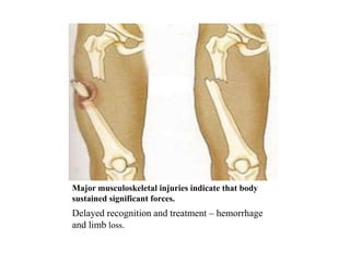
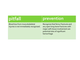
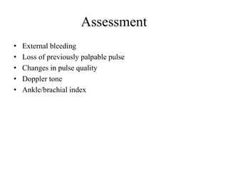
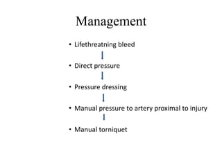
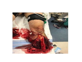
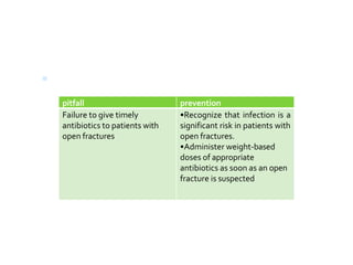
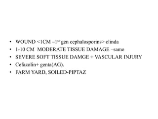
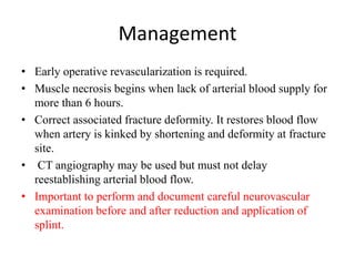
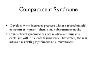
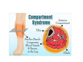
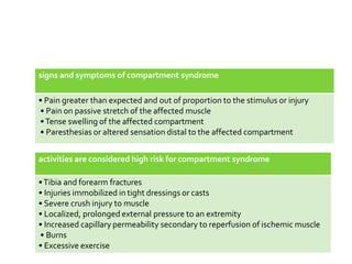
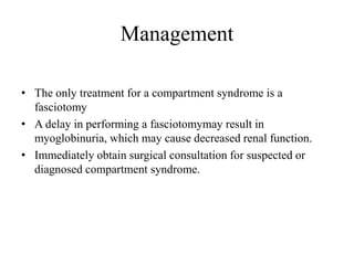
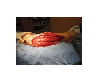
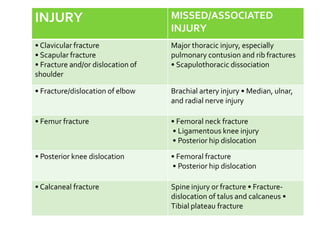
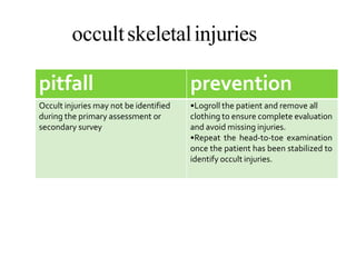
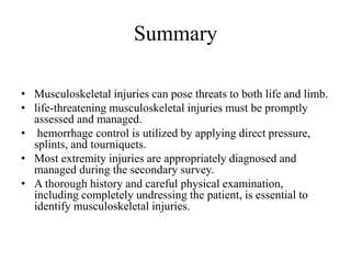
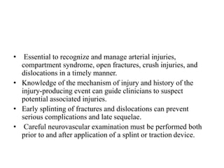
MUSCULOSKELETAL TRAUMA
Presenter – Dr.Mohit Garg
Major musculoskeletal injuries indicate that body
sustained significant forces.
Delayed recognition and treatment – hemorrhage
and limb loss.
Objectives
• Resuscitations of patients with extremities injury.
• Adjuncts to the primary survey.
• Secondary survey including history and physical examination.
• Principles of management of limb threatening musculoskeletal
injuries.
• Assessment and management of patients with
contusions,lacerations,joints ,fractures.
• Principles of proper immobilization of patients.
Primary Survey and Resuscitation
• Recognize and control hemorrhage.
• Potiential life threatning extremity injuries include
ļ‚§ Major arterial hemorrhage
ļ‚§ Traumatic Amputation
ļ‚§ Bilateral femoral fractures
ļ‚§ Crush Syndrome
• Hemorrhage control best achived with direct pressure.
pitfall prevention
Blood loss from musculoskeletal
injuries is not immediately recognized.
Recognize that femur fractures and
any open long-bone fractures with
major soft-tissue involvement are
potential sites of significant
hemorrhage
Assessment
• External bleeding
• Loss of previously palpable pulse
• Changes in pulse quality
• Doppler tone
• Ankle/brachial index
Management
• Lifethreatning bleed
• Direct pressure
• Pressure dressing
• Manual pressure to artery proximal to injury
• Manual torniquet
Bilateral Femur Fractures
• Patients with B/L femur fractures > Greater risk than U/L
fractures
• Higher risk for significant blood loss, pulmonary
complications, multiple organ failure and death
• Consider early transfer to a trauma center.
PITFALL PREVENTION
Delayed transfer to a trauma centre. •Transfer patients with vascular injury
and concomitant fracture to a trauma
center with vascular and orthopedic
surgical capabilities.
•Bilateral femur fractures result in a
significantly increased risk of
complications and death; these
patients benefit from early transfer to
a trauma centre.
Crush Syndrome
• Defined as systemic manifestations resulting from crush
injury.
• If left untreated, can lead to acute renal failure and shock.
• Sustained compression injury to significant muscle mass,
thigh or calf.
• The muscular insult is a combination of
– direct muscle injury
– muscle ischemia
– cell death with release of myoglobin.
Assessment
• Dark amber urine - hemoglobinuria.
• Amber-colored urine with serum creatine kinase of 10,000 U/L
or more is indicative of rhabdomyolysis when urine
myoglobinlevels are not available.
• Rhabdomyolysis- metabolic acidosis, hyperkalemia,
hypocalcemia, and DIC.
Management
• Intravenous fluid therapy
• Myoglobin-induced renal failure can be prevented with
– intravascular fluid expansion,
– alkalinization of the urine by intravenous administration of
bicarbonate, and osmotic diuresis.
Adjuncts to primary survey
• Includes fracture immobilization and xray examination.
• Goal
– realign the injured extremity in as close to anatomic
position as possible
– prevent excessive motion at the fracture site.
• Accomplished by applying inline traction and maintaining
traction with an immobilization device.
• Remove gross contamination and particulate matter from the
wound.
• Administer weight-based dosing of antibiotics.
• If reduction is unsuccessful, , splint the joint in the position in
which it was found.
• Resuscitation efforts must take priority over splint application.
X ray examination
• Based on
– patient’s initial and obvious clinical findings
– patient’s hemodynamic status
– mechanism of injury.
Secondary Survey
• Includes history and physical examination.
• Key aspects of history
– mechanism of injury
– environment
– preinjury status and predisposing factors
– prehospital observations and care.
Mechanism of Injury
• Patient located before the crash? type of injury- lateral # pelvis
from side impact collision
• After the crash—inside the vehicle or ejected?
• Was a seat belt or airbag in use?
• Was the vehicle’s exterior damaged, such as having its front
end deformed by a head-on collision?-suspect hip dislocation
• vehicle’s interior damaged, such as a deformed dashboard?
lower extremity injuries
• patient fall? distance of the fall, and how did the patient land?
–specturum of injuries.
• patient crushed by an object? If so, weight of the crushing
object, the site of the injury, and duration of weight applied to
the site.
• explosion occur? If so, magnitude of the blast, and what
was the patient’s distance from the blast?
– Primary injury from blast force
– Secondary injury from debris and other accelerated objects
– Tertiary injury- patient may be thrown to distant places.
• Was the patient involved in a vehicle-pedestrian collision?
Environment Questionare
• Ask following information about the post crash environment
– Did the patient sustain an open fracture in a contaminated
environment?
– Was the patient exposed to temperature extremes?
– Were broken glass fragments, which can also injure the
examiner, at the scene?
– Were there any sources of bacterial contamination, such as
dirt, animal feces, and fresh or salt water?
• This information can help the clinician anticipate potential
problems and determine the initial antibiotic treatment.
Preinjury status and predisposing
factors
• exercise tolerance
• activity level
• ingestion of alcohol and/or other drugs, emotional problems
or illnesses
• previous musculoskeletal injuries.
Prehospital observations and care
• Time of injury
• Position in which found
• Bleeding or pooling of blood at the scene - the estimated
amount
• Bone or fracture ends that may have been exposed
• Open wounds in proximity to obvious or suspected fractures
• Obvious deformity or dislocation
• Any crushing mechanism that can result in a crush syndrome
• Presence or absence of motor and/or sensory function in each
extremity
• Any delay in extrication procedures or transport.
• Changes in limb function, perfusion, or neurologic state.
• Dressings and splints applied, with special attention to
excessive pressure over bony prominences that can result in
peripheral nerve compression or compartment syndrome.
• Time of tourniquet placement.
Physical Examination
• The three goals for assessing the extremities are:
• 1. Identify life-threatening injuries (primary survey).
• 2. Identify limb-threatening injuries (secondary survey).
• 3. Conduct a systematic review to avoid missing any other
musculoskeletal injury.
Look and Ask
• Visual inspection helps identify sites of major external
bleeding.
• A pale or white distal extremity is indicative of a lack of
arterial inflow.
• Inspect the patient’s entire body for lacerations and abrasions.
• Open wounds may not be obvious on the dorsum of the body;
therefore, carefully logroll patients to assess for possible
hidden injuries.
• Observe the patient’s spontaneous extremity motor function to
help identify any neurologic and/or muscular impairment.
• If the patient is unconscious, absent spontaneous extremity
movement may be the only sign of impaired function.
JOINT DIRECTION DEFORMITY
Shoulder Anterior
Posterior
Squared of
Locked in internal
rotation
Elbow
Posterior Olecranon
prominent
posteriorly
Hip Anterior
Posterior
Extended, abducted,
externally rotated
Flexed, adducted,
internally rotated
Knee Anteroposterior Loss of normal
contour,
extended
*May
spontaneously
reduce prior to
Feel
• Palpate the extremities to determine sensation to the skin (i.e.,
neurologic function) and identify areas of tenderness, which
may indicate fracture
• Loss of sensation to pain and touch - spinal or peripheral nerve
injury.
• Joint stability can be determined only by clinical examination.
• Abnormal motion through a joint segment is indicative of a
tendon or ligamentous rupture.
Circulatory Evaluation
• Palpate the distal pulses in each extremity, and assess capillary
refill of the digits.
• The Doppler signal must have a triphasic quality to ensure no
proximal lesion.
• Arterial injury indications
– pulse discrepancies
– coolness, pallor
– paresthesia, and even motor function abnormalities.
• Knee dislocations can reduce spontaneously and may not
present with any gross external or radiographic anomalies until
a physical exam of the joint is performed and instability is
detected clinically.
• An ankle/brachial index of less than 0.9 indicates abnormal
arterial flow secondary to injury or peripheral vascular
disease.
X ray Examination
• The clinical examination of patients with musculoskeletal
injuries often suggests the need for x-ray examination.
• The only reason to forgo x-ray examination before treating a
dislocation or a fracture is the presence of vascular
compromise or impending skin breakdown. This condition is
commonly seen with fracture-dislocations of the ankle.
Limb threatening injuries
• open fractures and open joint injuries
• vascular injuries
• compartment syndrome
• neurological injury secondary to fracture or dislocation
Open fractures and open joint
injuries
• Open fractures and open joint injuries result from
communication between the external environment and the
bone or joint
• The diagnosis of an open fracture is based on a physical
examination of the extremity that demonstrates an open wound
on the same limb segment as an associated fracture. At no time
should the wound be probed.
Assessment
• Presence of an open joint injury may be identified using CT.
The presence of intra articular gas on a CT of the affected
extremity is highly sensitive and specific for identifying open
joint injury.
• If CT is not available, consider insertion of saline or dye into
the joint to determine whether the joint cavity communicates
with the wound.
Management
• Treat all patients with open fractures as soon as possible with
intravenous antibiotics using weight-based dosing.
ļ‚§ treat all patients with open fractures as soon
as possible with intravenous antibiotics using
weight-based dosing.
pitfall prevention
Failure to give timely
antibiotics to patients with
open fractures
•Recognize that infection is a
significant risk in patients with
open fractures.
•Administer weight-based
doses of appropriate
antibiotics as soon as an open
fracture is suspected
• WOUND <1CM –1st gen cephalosporins> clinda
• 1-10 CM MODERATE TISSUE DAMAGE –same
• SEVERE SOFT TISSUE DAMGE + VASCULAR INJURY
• Cefazolin+ genta(AG).
• FARM YARD, SOILED-PIPTAZ
Vascular injuries
• Limb initially appear viable because of collateral circulation.
• Non-occlusive – coolness and prolonged capillary refill in
distal part of extremity as well as dimnished peripheral pulses
and abormal ankle/brachial index.
• Occlusive – cold , pale and pulseless.
Management
• Early operative revascularization is required.
• Muscle necrosis begins when lack of arterial blood supply for
more than 6 hours.
• Correct associated fracture deformity. It restores blood flow
when artery is kinked by shortening and deformity at fracture
site.
• CT angiography may be used but must not delay
reestablishing arterial blood flow.
• Important to perform and document careful neurovascular
examination before and after reduction and application of
splint.
Compartment Syndrome
• Develops when increased pressure within a musculofascial
compartment causes ischemia and subsequent necrosis.
• Compartment syndrome can occur wherever muscle is
contained within a closed fascial space. Remember, the skin
acts as a restricting layer in certain circumstances.
• Delayed recognition and treatment of compartment syndrome
is catastrophic and can result in neurologic deficit, muscle
necrosis, ischemic contracture, infection, delayed healing of
fractures, and possible amputation.
signs and symptoms of compartment syndrome
• Pain greater than expected and out of proportion to the stimulus or injury
• Pain on passive stretch of the affected muscle
•Tense swelling of the affected compartment
• Paresthesias or altered sensation distal to the affected compartment
activities are considered high risk for compartment syndrome
•Tibia and forearm fractures
• Injuries immobilized in tight dressings or casts
• Severe crush injury to muscle
• Localized, prolonged external pressure to an extremity
• Increased capillary permeability secondary to reperfusion of ischemic muscle
• Burns
• Excessive exercise
Management
• The only treatment for a compartment syndrome is a
fasciotomy
• A delay in performing a fasciotomymay result in
myoglobinuria, which may cause decreased renal function.
• Immediately obtain surgical consultation for suspected or
diagnosed compartment syndrome.
pitfall prevention
Delayed diagnosis of compartment
syndrome
•Maintain a high index of suspicion for
compartment syndrome in any patient
with a significant musculoskeletal
injury.
•Be aware that compartment
syndrome can be difficult to recognize
in patients with altered mental status.
•Frequently reevaluate patients with
altered mental status for signs of
compartment syndrome.
Neurological injury secondary to
fracture or dislocation
• Neurologic injury due to the anatomic relationship and
proximity of nerves to bones and joint
• Sciatic nerve compression from posterior hip dislocation
• Axillary nerve injury from anterior shoulder dislocation
• Diffcult to assess nerve function initially. However,
assessment must be continually repeated, especially after the
patient is stabilized.
PERIPHERAL NERVE ASSESSMENT OF UPPER ExTREMITIES
NERVE MOTOR SENSATION INJURY
Ulnar Index and litle finger abduction Litle finger Elbow injury
Median distal Thenar contraction with opposition Distal tip of index finger Wrist fracture or dislocation
Median, anterior
interosseous
Index tip flexion None Supracondylar fracture of
humerus (children)
Musculocutaneous Elbow flexion Radial forearm Anterior shoulder dislocation
Radial Thumb, finger metocarpo-
phalangeal extension
First dorsal web space Distal humeral shaft, anterior
shoulder dislocation
Axillary Deltoid Lateral shoulder Anterior shoulder dislocation,
proximal humerus fracture
PERIPHERAL NERVE ASSESSMENT OF LOWER ExTREMITIES
NERVE MOTOR SENSATION INJURY
Femoral Knee extension Anterior knee Pubic rami fractures
Obturator Hip adduction Medial thigh Obturator ring fractures
Posterior tibial Toe flexion Sole of foot Knee dislocation
Superficial peroneal Ankle eversion Lateral dorsum of foot Fibular neck fracture,
knee dislocation
Deep peroneal Ankle/toe dorsiflexion Dorsal first to
second web space
Fibular neck fracture,
compartment syndrome
Sciatic nerve Ankle dorsiflexion or plantar
flexion
Foot Posterior hip dislocation
Superior gluteal Hip abduction Upper butocks Acetabular fracture
Inferior gluteal Gluteus maximus hip extension Lower butocks Acetabular fracture
Other extremity injuries
• Lacerations
– Require debridement and closure.
– If a laceration extends below the fascial level, it may
require operative intervention.
• Contusions
– recognized by pain, localized swelling, and tenderness
– treated by limiting function of the injured part and
applying cold packs.
• soft-tissue injuries are best evaluated by knowing the
mechanism of injury and by palpating the specific component
involved
• Soft-tissue avulsion can shear the skin from the deep fascia,
allowing for the significant accumulation of blood in the
resulting cavity (i.e., Morel-LavallƩelesion)
• drainage or debridement may be indicated
• Risk of tetanus is increased with
– wounds that are more than 6 hours old,
– contused or abraded
– more than 1 cm in depth
– from high-velocity missiles, due to burns or cold
– significantly contaminated, particularly wounds with
denervated or ischemic tissue .
Joint and ligament injuries
• Physical examination reveals tenderness throughout the
affected joint.
• A hemarthrosisis usually present unless the joint capsule is
disrupted and the bleeding diffuses into the soft tissues.
• Passive ligamentous testing of the affected joint reveals
instability.
• X-ray examination is usually negative, although some small
avulsion fractures from ligamentous insertions or origins may
be present radiographically.
Management
• Immobilize joint injuries, and serially reassess the vascular and
neurologic status of the limb distal to the injury.
• In a patient with a multiligament knee injury, a dislocation
may have occurred and placed the limb at risk for
neurovascular injury. Surgical consultation is usually required
for joint stabilization.
Fractures
• Examination of the extremity typically demonstrates pain,
swelling, deformity, tenderness, crepitus, and abnormal motion
at the fracture site.
• X-ray films taken at right angles to one another confirm the
history and physical examination findings of fracture.
• To exclude occult dislocation and concomitant injury, x-ray
films must include the joints above and below the suspected
fracture site.
Management
• Immobilization must include the joint above and below the
fracture. After splinting, be sure to reassess the neurologic and
vascular status of the extremity.
Principles of immobilization
• FEMORAL FRACTURES-Femoral fractures are
immobilized temporarily with traction splints.
• The traction splint’s force is applied distally at the ankle.
• Proximally, the post is pushed into the gluteal crease to apply
pressure to the buttocks, perineum, and groin.
• Hip fractures can be similarly immobilized with a traction
splint but are more suitably immobilized with skin traction or
foam boot traction with the knee in slight flexion.
Knee injuries
• Application of a commercially available knee immobilizer or a
posterior long-leg plaster splint is effective in maintaining
comfort and stability.
• Do not immobilize the knee in complete extension, but with
approximately 10 degrees of flexion to reduce tension on the
neurovascular structures.
Tibial fractures
• Immobilize tibial fractures to minimize pain and further soft-
tissue injury and decrease the risk of compartment syndrome.
• If readily available, plaster splints immobilizing the lower
thigh, knee, and ankle are preferred.
Upper extremity and hand injuries
• The hand may be temporarily splinted in an anatomic,
functional position with the wrist slightly dorsiflexed and the
fingers gently flexed 45 degrees at the
metacarpophalangealjoints.
• The forearm and wrist are immobilized flat on padded or
pillow splints.
• The elbow is typically immobilized in a flexed position, either
by using padded splints or by direct immobilization with
respect to the body using a sling-and-swath device.
• The upper arm may be immobilized by splinting it to the body
or applying a sling or swath, which can be augmented by a
thoracobrachial bandage.
• Shoulder injuries are managed by a sling-and-swath device or
a hook-and loop type of dressing.
Associated Injuries
• Certain musculoskeletal injuries are associated with other
injuries that are not immediately apparent may be missed.
• Steps to ensure recognition and management of these injuries
include
– Review the injury history especially mechanism of injury
– Thoroughly reexamine all extremities.
– Examine patient back including spine and pelvis.
– Document open injuries and closed soft tissue injuries
– Review x rays.
INJURY MISSED/ASSOCIATED
INJURY
• Clavicular fracture
• Scapular fracture
• Fracture and/or dislocation of
shoulder
Major thoracic injury, especially
pulmonary contusion and rib fractures
• Scapulothoracic dissociation
• Fracture/dislocation of elbow Brachial artery injury • Median, ulnar,
and radial nerve injury
• Femur fracture • Femoral neck fracture
• Ligamentous knee injury
• Posterior hip dislocation
• Posterior knee dislocation • Femoral fracture
• Posterior hip dislocation
• Calcaneal fracture Spine injury or fracture • Fracture-
dislocation of talus and calcaneus •
Tibial plateau fracture
occultskeletalinjuries
pitfall prevention
Occult injuries may not be identified
during the primary assessment or
secondary survey
•Logroll the patient and remove all
clothing to ensure complete evaluation
and avoid missing injuries.
•Repeat the head-to-toe examination
once the patient has been stabilized to
identify occult injuries.
Summary
• Musculoskeletal injuries can pose threats to both life and limb.
• life-threatening musculoskeletal injuries must be promptly
assessed and managed.
• hemorrhage control is utilized by applying direct pressure,
splints, and tourniquets.
• Most extremity injuries are appropriately diagnosed and
managed during the secondary survey.
• A thorough history and careful physical examination,
including completely undressing the patient, is essential to
identify musculoskeletal injuries.
• Essential to recognize and manage arterial injuries,
compartment syndrome, open fractures, crush injuries, and
dislocations in a timely manner.
• Knowledge of the mechanism of injury and history of the
injury-producing event can guide clinicians to suspect
potential associated injuries.
• Early splinting of fractures and dislocations can prevent
serious complications and late sequelae.
• Careful neurovascular examination must be performed both
prior to and after application of a splint or traction device.
THANK YOU

musculoskeletal trauma.pptx

  • 1.
  • 2.
    Major musculoskeletal injuriesindicate that body sustained significant forces. Delayed recognition and treatment – hemorrhage and limb loss.
  • 3.
    Objectives • Resuscitations ofpatients with extremities injury. • Adjuncts to the primary survey. • Secondary survey including history and physical examination. • Principles of management of limb threatening musculoskeletal injuries. • Assessment and management of patients with contusions,lacerations,joints ,fractures. • Principles of proper immobilization of patients.
  • 4.
    Primary Survey andResuscitation • Recognize and control hemorrhage. • Potiential life threatning extremity injuries include ļ‚§ Major arterial hemorrhage ļ‚§ Traumatic Amputation ļ‚§ Bilateral femoral fractures ļ‚§ Crush Syndrome • Hemorrhage control best achived with direct pressure.
  • 5.
    pitfall prevention Blood lossfrom musculoskeletal injuries is not immediately recognized. Recognize that femur fractures and any open long-bone fractures with major soft-tissue involvement are potential sites of significant hemorrhage
  • 6.
    Assessment • External bleeding •Loss of previously palpable pulse • Changes in pulse quality • Doppler tone • Ankle/brachial index
  • 7.
    Management • Lifethreatning bleed •Direct pressure • Pressure dressing • Manual pressure to artery proximal to injury • Manual torniquet
  • 9.
    Bilateral Femur Fractures •Patients with B/L femur fractures > Greater risk than U/L fractures • Higher risk for significant blood loss, pulmonary complications, multiple organ failure and death • Consider early transfer to a trauma center.
  • 10.
    PITFALL PREVENTION Delayed transferto a trauma centre. •Transfer patients with vascular injury and concomitant fracture to a trauma center with vascular and orthopedic surgical capabilities. •Bilateral femur fractures result in a significantly increased risk of complications and death; these patients benefit from early transfer to a trauma centre.
  • 11.
    Crush Syndrome • Definedas systemic manifestations resulting from crush injury. • If left untreated, can lead to acute renal failure and shock. • Sustained compression injury to significant muscle mass, thigh or calf. • The muscular insult is a combination of – direct muscle injury – muscle ischemia – cell death with release of myoglobin.
  • 12.
    Assessment • Dark amberurine - hemoglobinuria. • Amber-colored urine with serum creatine kinase of 10,000 U/L or more is indicative of rhabdomyolysis when urine myoglobinlevels are not available. • Rhabdomyolysis- metabolic acidosis, hyperkalemia, hypocalcemia, and DIC.
  • 13.
    Management • Intravenous fluidtherapy • Myoglobin-induced renal failure can be prevented with – intravascular fluid expansion, – alkalinization of the urine by intravenous administration of bicarbonate, and osmotic diuresis.
  • 14.
    Adjuncts to primarysurvey • Includes fracture immobilization and xray examination. • Goal – realign the injured extremity in as close to anatomic position as possible – prevent excessive motion at the fracture site. • Accomplished by applying inline traction and maintaining traction with an immobilization device.
  • 15.
    • Remove grosscontamination and particulate matter from the wound. • Administer weight-based dosing of antibiotics. • If reduction is unsuccessful, , splint the joint in the position in which it was found. • Resuscitation efforts must take priority over splint application.
  • 16.
    X ray examination •Based on – patient’s initial and obvious clinical findings – patient’s hemodynamic status – mechanism of injury.
  • 17.
    Secondary Survey • Includeshistory and physical examination. • Key aspects of history – mechanism of injury – environment – preinjury status and predisposing factors – prehospital observations and care.
  • 18.
    Mechanism of Injury •Patient located before the crash? type of injury- lateral # pelvis from side impact collision • After the crash—inside the vehicle or ejected? • Was a seat belt or airbag in use? • Was the vehicle’s exterior damaged, such as having its front end deformed by a head-on collision?-suspect hip dislocation
  • 19.
    • vehicle’s interiordamaged, such as a deformed dashboard? lower extremity injuries • patient fall? distance of the fall, and how did the patient land? –specturum of injuries. • patient crushed by an object? If so, weight of the crushing object, the site of the injury, and duration of weight applied to the site.
  • 20.
    • explosion occur?If so, magnitude of the blast, and what was the patient’s distance from the blast? – Primary injury from blast force – Secondary injury from debris and other accelerated objects – Tertiary injury- patient may be thrown to distant places. • Was the patient involved in a vehicle-pedestrian collision?
  • 22.
    Environment Questionare • Askfollowing information about the post crash environment – Did the patient sustain an open fracture in a contaminated environment? – Was the patient exposed to temperature extremes? – Were broken glass fragments, which can also injure the examiner, at the scene? – Were there any sources of bacterial contamination, such as dirt, animal feces, and fresh or salt water?
  • 23.
    • This informationcan help the clinician anticipate potential problems and determine the initial antibiotic treatment.
  • 24.
    Preinjury status andpredisposing factors • exercise tolerance • activity level • ingestion of alcohol and/or other drugs, emotional problems or illnesses • previous musculoskeletal injuries.
  • 25.
    Prehospital observations andcare • Time of injury • Position in which found • Bleeding or pooling of blood at the scene - the estimated amount • Bone or fracture ends that may have been exposed • Open wounds in proximity to obvious or suspected fractures • Obvious deformity or dislocation • Any crushing mechanism that can result in a crush syndrome
  • 26.
    • Presence orabsence of motor and/or sensory function in each extremity • Any delay in extrication procedures or transport. • Changes in limb function, perfusion, or neurologic state. • Dressings and splints applied, with special attention to excessive pressure over bony prominences that can result in peripheral nerve compression or compartment syndrome. • Time of tourniquet placement.
  • 27.
    Physical Examination • Thethree goals for assessing the extremities are: • 1. Identify life-threatening injuries (primary survey). • 2. Identify limb-threatening injuries (secondary survey). • 3. Conduct a systematic review to avoid missing any other musculoskeletal injury.
  • 28.
    Look and Ask •Visual inspection helps identify sites of major external bleeding. • A pale or white distal extremity is indicative of a lack of arterial inflow. • Inspect the patient’s entire body for lacerations and abrasions. • Open wounds may not be obvious on the dorsum of the body; therefore, carefully logroll patients to assess for possible hidden injuries.
  • 29.
    • Observe thepatient’s spontaneous extremity motor function to help identify any neurologic and/or muscular impairment. • If the patient is unconscious, absent spontaneous extremity movement may be the only sign of impaired function.
  • 30.
    JOINT DIRECTION DEFORMITY ShoulderAnterior Posterior Squared of Locked in internal rotation Elbow Posterior Olecranon prominent posteriorly Hip Anterior Posterior Extended, abducted, externally rotated Flexed, adducted, internally rotated Knee Anteroposterior Loss of normal contour, extended *May spontaneously reduce prior to
  • 31.
    Feel • Palpate theextremities to determine sensation to the skin (i.e., neurologic function) and identify areas of tenderness, which may indicate fracture • Loss of sensation to pain and touch - spinal or peripheral nerve injury. • Joint stability can be determined only by clinical examination. • Abnormal motion through a joint segment is indicative of a tendon or ligamentous rupture.
  • 32.
    Circulatory Evaluation • Palpatethe distal pulses in each extremity, and assess capillary refill of the digits. • The Doppler signal must have a triphasic quality to ensure no proximal lesion. • Arterial injury indications – pulse discrepancies – coolness, pallor – paresthesia, and even motor function abnormalities.
  • 33.
    • Knee dislocationscan reduce spontaneously and may not present with any gross external or radiographic anomalies until a physical exam of the joint is performed and instability is detected clinically. • An ankle/brachial index of less than 0.9 indicates abnormal arterial flow secondary to injury or peripheral vascular disease.
  • 34.
    X ray Examination •The clinical examination of patients with musculoskeletal injuries often suggests the need for x-ray examination. • The only reason to forgo x-ray examination before treating a dislocation or a fracture is the presence of vascular compromise or impending skin breakdown. This condition is commonly seen with fracture-dislocations of the ankle.
  • 35.
    Limb threatening injuries •open fractures and open joint injuries • vascular injuries • compartment syndrome • neurological injury secondary to fracture or dislocation
  • 36.
    Open fractures andopen joint injuries • Open fractures and open joint injuries result from communication between the external environment and the bone or joint • The diagnosis of an open fracture is based on a physical examination of the extremity that demonstrates an open wound on the same limb segment as an associated fracture. At no time should the wound be probed.
  • 37.
    Assessment • Presence ofan open joint injury may be identified using CT. The presence of intra articular gas on a CT of the affected extremity is highly sensitive and specific for identifying open joint injury. • If CT is not available, consider insertion of saline or dye into the joint to determine whether the joint cavity communicates with the wound.
  • 38.
    Management • Treat allpatients with open fractures as soon as possible with intravenous antibiotics using weight-based dosing.
  • 39.
    ļ‚§ treat allpatients with open fractures as soon as possible with intravenous antibiotics using weight-based dosing. pitfall prevention Failure to give timely antibiotics to patients with open fractures •Recognize that infection is a significant risk in patients with open fractures. •Administer weight-based doses of appropriate antibiotics as soon as an open fracture is suspected
  • 40.
    • WOUND <1CM–1st gen cephalosporins> clinda • 1-10 CM MODERATE TISSUE DAMAGE –same • SEVERE SOFT TISSUE DAMGE + VASCULAR INJURY • Cefazolin+ genta(AG). • FARM YARD, SOILED-PIPTAZ
  • 41.
    Vascular injuries • Limbinitially appear viable because of collateral circulation. • Non-occlusive – coolness and prolonged capillary refill in distal part of extremity as well as dimnished peripheral pulses and abormal ankle/brachial index. • Occlusive – cold , pale and pulseless.
  • 42.
    Management • Early operativerevascularization is required. • Muscle necrosis begins when lack of arterial blood supply for more than 6 hours. • Correct associated fracture deformity. It restores blood flow when artery is kinked by shortening and deformity at fracture site. • CT angiography may be used but must not delay reestablishing arterial blood flow. • Important to perform and document careful neurovascular examination before and after reduction and application of splint.
  • 43.
    Compartment Syndrome • Developswhen increased pressure within a musculofascial compartment causes ischemia and subsequent necrosis. • Compartment syndrome can occur wherever muscle is contained within a closed fascial space. Remember, the skin acts as a restricting layer in certain circumstances.
  • 45.
    • Delayed recognitionand treatment of compartment syndrome is catastrophic and can result in neurologic deficit, muscle necrosis, ischemic contracture, infection, delayed healing of fractures, and possible amputation.
  • 46.
    signs and symptomsof compartment syndrome • Pain greater than expected and out of proportion to the stimulus or injury • Pain on passive stretch of the affected muscle •Tense swelling of the affected compartment • Paresthesias or altered sensation distal to the affected compartment activities are considered high risk for compartment syndrome •Tibia and forearm fractures • Injuries immobilized in tight dressings or casts • Severe crush injury to muscle • Localized, prolonged external pressure to an extremity • Increased capillary permeability secondary to reperfusion of ischemic muscle • Burns • Excessive exercise
  • 47.
    Management • The onlytreatment for a compartment syndrome is a fasciotomy • A delay in performing a fasciotomymay result in myoglobinuria, which may cause decreased renal function. • Immediately obtain surgical consultation for suspected or diagnosed compartment syndrome.
  • 49.
    pitfall prevention Delayed diagnosisof compartment syndrome •Maintain a high index of suspicion for compartment syndrome in any patient with a significant musculoskeletal injury. •Be aware that compartment syndrome can be difficult to recognize in patients with altered mental status. •Frequently reevaluate patients with altered mental status for signs of compartment syndrome.
  • 50.
    Neurological injury secondaryto fracture or dislocation • Neurologic injury due to the anatomic relationship and proximity of nerves to bones and joint • Sciatic nerve compression from posterior hip dislocation • Axillary nerve injury from anterior shoulder dislocation • Diffcult to assess nerve function initially. However, assessment must be continually repeated, especially after the patient is stabilized.
  • 51.
    PERIPHERAL NERVE ASSESSMENTOF UPPER ExTREMITIES NERVE MOTOR SENSATION INJURY Ulnar Index and litle finger abduction Litle finger Elbow injury Median distal Thenar contraction with opposition Distal tip of index finger Wrist fracture or dislocation Median, anterior interosseous Index tip flexion None Supracondylar fracture of humerus (children) Musculocutaneous Elbow flexion Radial forearm Anterior shoulder dislocation Radial Thumb, finger metocarpo- phalangeal extension First dorsal web space Distal humeral shaft, anterior shoulder dislocation Axillary Deltoid Lateral shoulder Anterior shoulder dislocation, proximal humerus fracture
  • 52.
    PERIPHERAL NERVE ASSESSMENTOF LOWER ExTREMITIES NERVE MOTOR SENSATION INJURY Femoral Knee extension Anterior knee Pubic rami fractures Obturator Hip adduction Medial thigh Obturator ring fractures Posterior tibial Toe flexion Sole of foot Knee dislocation Superficial peroneal Ankle eversion Lateral dorsum of foot Fibular neck fracture, knee dislocation Deep peroneal Ankle/toe dorsiflexion Dorsal first to second web space Fibular neck fracture, compartment syndrome Sciatic nerve Ankle dorsiflexion or plantar flexion Foot Posterior hip dislocation Superior gluteal Hip abduction Upper butocks Acetabular fracture Inferior gluteal Gluteus maximus hip extension Lower butocks Acetabular fracture
  • 53.
    Other extremity injuries •Lacerations – Require debridement and closure. – If a laceration extends below the fascial level, it may require operative intervention. • Contusions – recognized by pain, localized swelling, and tenderness – treated by limiting function of the injured part and applying cold packs.
  • 54.
    • soft-tissue injuriesare best evaluated by knowing the mechanism of injury and by palpating the specific component involved • Soft-tissue avulsion can shear the skin from the deep fascia, allowing for the significant accumulation of blood in the resulting cavity (i.e., Morel-LavallĆ©elesion) • drainage or debridement may be indicated
  • 55.
    • Risk oftetanus is increased with – wounds that are more than 6 hours old, – contused or abraded – more than 1 cm in depth – from high-velocity missiles, due to burns or cold – significantly contaminated, particularly wounds with denervated or ischemic tissue .
  • 56.
    Joint and ligamentinjuries • Physical examination reveals tenderness throughout the affected joint. • A hemarthrosisis usually present unless the joint capsule is disrupted and the bleeding diffuses into the soft tissues. • Passive ligamentous testing of the affected joint reveals instability. • X-ray examination is usually negative, although some small avulsion fractures from ligamentous insertions or origins may be present radiographically.
  • 57.
    Management • Immobilize jointinjuries, and serially reassess the vascular and neurologic status of the limb distal to the injury. • In a patient with a multiligament knee injury, a dislocation may have occurred and placed the limb at risk for neurovascular injury. Surgical consultation is usually required for joint stabilization.
  • 58.
    Fractures • Examination ofthe extremity typically demonstrates pain, swelling, deformity, tenderness, crepitus, and abnormal motion at the fracture site. • X-ray films taken at right angles to one another confirm the history and physical examination findings of fracture. • To exclude occult dislocation and concomitant injury, x-ray films must include the joints above and below the suspected fracture site.
  • 59.
    Management • Immobilization mustinclude the joint above and below the fracture. After splinting, be sure to reassess the neurologic and vascular status of the extremity.
  • 60.
    Principles of immobilization •FEMORAL FRACTURES-Femoral fractures are immobilized temporarily with traction splints. • The traction splint’s force is applied distally at the ankle. • Proximally, the post is pushed into the gluteal crease to apply pressure to the buttocks, perineum, and groin. • Hip fractures can be similarly immobilized with a traction splint but are more suitably immobilized with skin traction or foam boot traction with the knee in slight flexion.
  • 61.
    Knee injuries • Applicationof a commercially available knee immobilizer or a posterior long-leg plaster splint is effective in maintaining comfort and stability. • Do not immobilize the knee in complete extension, but with approximately 10 degrees of flexion to reduce tension on the neurovascular structures.
  • 62.
    Tibial fractures • Immobilizetibial fractures to minimize pain and further soft- tissue injury and decrease the risk of compartment syndrome. • If readily available, plaster splints immobilizing the lower thigh, knee, and ankle are preferred.
  • 63.
    Upper extremity andhand injuries • The hand may be temporarily splinted in an anatomic, functional position with the wrist slightly dorsiflexed and the fingers gently flexed 45 degrees at the metacarpophalangealjoints. • The forearm and wrist are immobilized flat on padded or pillow splints. • The elbow is typically immobilized in a flexed position, either by using padded splints or by direct immobilization with respect to the body using a sling-and-swath device.
  • 64.
    • The upperarm may be immobilized by splinting it to the body or applying a sling or swath, which can be augmented by a thoracobrachial bandage. • Shoulder injuries are managed by a sling-and-swath device or a hook-and loop type of dressing.
  • 65.
    Associated Injuries • Certainmusculoskeletal injuries are associated with other injuries that are not immediately apparent may be missed. • Steps to ensure recognition and management of these injuries include – Review the injury history especially mechanism of injury – Thoroughly reexamine all extremities. – Examine patient back including spine and pelvis. – Document open injuries and closed soft tissue injuries – Review x rays.
  • 66.
    INJURY MISSED/ASSOCIATED INJURY • Clavicularfracture • Scapular fracture • Fracture and/or dislocation of shoulder Major thoracic injury, especially pulmonary contusion and rib fractures • Scapulothoracic dissociation • Fracture/dislocation of elbow Brachial artery injury • Median, ulnar, and radial nerve injury • Femur fracture • Femoral neck fracture • Ligamentous knee injury • Posterior hip dislocation • Posterior knee dislocation • Femoral fracture • Posterior hip dislocation • Calcaneal fracture Spine injury or fracture • Fracture- dislocation of talus and calcaneus • Tibial plateau fracture
  • 67.
    occultskeletalinjuries pitfall prevention Occult injuriesmay not be identified during the primary assessment or secondary survey •Logroll the patient and remove all clothing to ensure complete evaluation and avoid missing injuries. •Repeat the head-to-toe examination once the patient has been stabilized to identify occult injuries.
  • 68.
    Summary • Musculoskeletal injuriescan pose threats to both life and limb. • life-threatening musculoskeletal injuries must be promptly assessed and managed. • hemorrhage control is utilized by applying direct pressure, splints, and tourniquets. • Most extremity injuries are appropriately diagnosed and managed during the secondary survey. • A thorough history and careful physical examination, including completely undressing the patient, is essential to identify musculoskeletal injuries.
  • 69.
    • Essential torecognize and manage arterial injuries, compartment syndrome, open fractures, crush injuries, and dislocations in a timely manner. • Knowledge of the mechanism of injury and history of the injury-producing event can guide clinicians to suspect potential associated injuries. • Early splinting of fractures and dislocations can prevent serious complications and late sequelae. • Careful neurovascular examination must be performed both prior to and after application of a splint or traction device.
  • 70.