Download to read offline


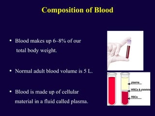

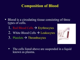

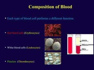


Blood is composed of plasma, red blood cells, white blood cells, and platelets. It transports oxygen, nutrients, waste, and helps remove toxins. Red blood cells contain hemoglobin and give blood its red color. White blood cells help fight infection. Platelets help with clotting to stop bleeding. The document provides detailed information on the composition and functions of these blood components.


