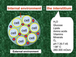
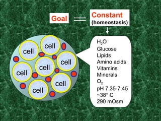
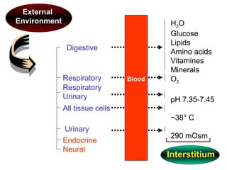
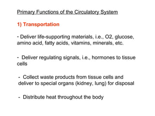
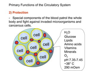
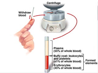
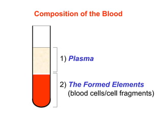
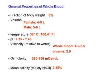
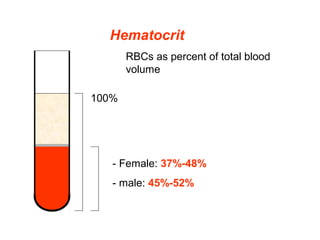
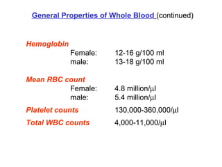
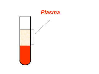
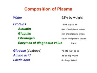
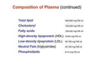
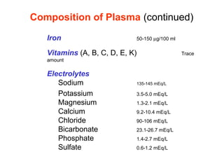
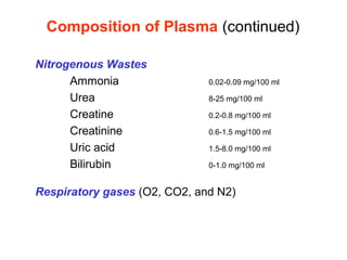
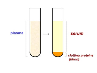
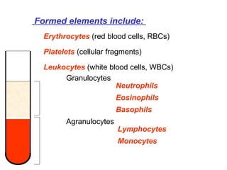
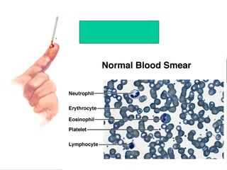
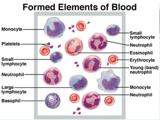
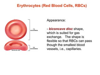
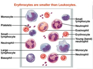
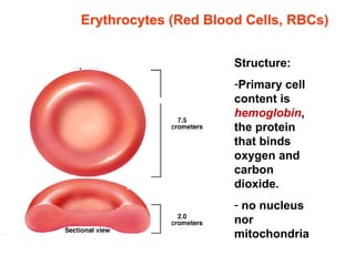
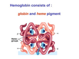
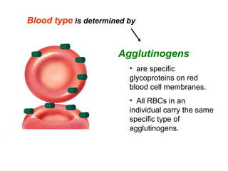
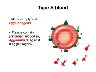
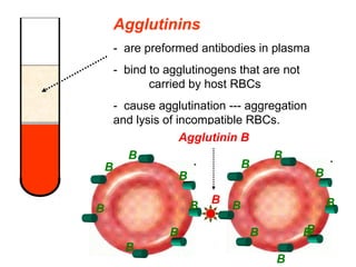
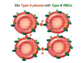
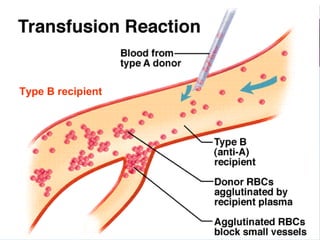
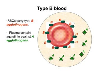
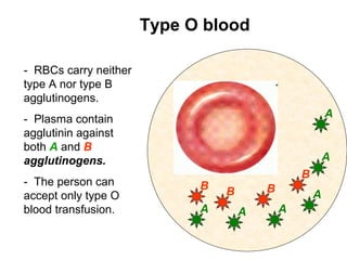
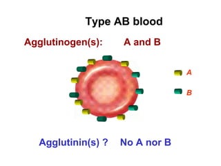
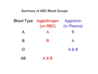
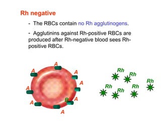
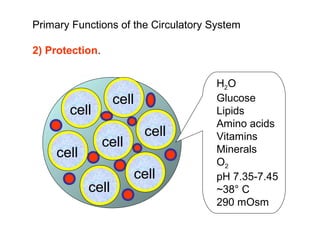
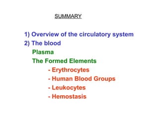
THE CIRCULATORY SYSTEM: BLOOD
TABLE OF CONTENT
1) Overview of the circulatory system
2) The blood
Plasma
The Formed Elements
- Erythrocytes
- Human Blood Groups
- Leukocytes
- Hemostasis
Overview of the
circulatory system
1) the blood (the circulating material)
2) the heart (pump)
3) blood vessels (conduit)
The circulatory system is composed of:
The circulatory system
Why is cardiovascular system needed?
Life evolved from oceans.
cell
Nutrients
cell
cell
cell
cell
cell
cell
cell
Nutrients
cell
cell
cell
cell
cell
cell
cell
Nutrients
cell
cell
cell
cell
cell
cell
cell
Internal environment
External environment
the interstitium
H2O
Glucose
Lipids
Amino acids
Vitamins
Minerals
O2
pH 7.35-7.45
~38° C
280-300 mOsm
cell
cell
cell
cell
cell
cell
cell
Goal
Constant
(homeostasis)
H2O
Glucose
Lipids
Amino acids
Vitamins
Minerals
O2
pH 7.35-7.45
~38° C
290 mOsm
H2O
Glucose
Lipids
Amino acids
Vitamines
Minerals
O2
pH 7.35-7.45
~38° C
290 mOsm
Digestive
Respiratory
Respiratory
Urinary
All tissue cells
Endocrine
Neural
Urinary
Blood
Interstitium
External
Environment
Primary Functions of the Circulatory System
1) Transportation
- Deliver life-supporting materials, i.e., O2, glucose,
amino acid, fatty acids, vitamins, minerals, etc.
- Deliver regulating signals, i.e., hormones to tissue
cells
- Collect waste products from tissue cells and
deliver to special organs (kidney, lung) for disposal
- Distribute heat throughout the body
Primary Functions of the Circulatory System
2) Protection
- Special components of the blood patrol the whole
body and fight against invaded microorganisms and
cancerous cells.
cell
cell
cell
cell
cell
cell
cell
H2O
Glucose
Lipids
Amino acids
Vitamins
Minerals
O2
pH 7.35-7.45
~38° C
290 mOsm
The Blood
1) Plasma
2) The Formed Elements
(blood cells/cell fragments)
Composition of the Blood
General Properties of Whole Blood
- Fraction of body weight
- Volume
- temperature
- pH 7.35 - 7.45
- Viscosity (relative to water)
- Osmolarity
- Mean salinity (mainly NaCl)
8%
Female: 4-5 L
Male: 5-6 L
38° C (100.4° F)
Whole blood: 4.5-5.5
plasma: 2.0
280-300 mOsm/L
0.85%
Hematocrit
RBCs as percent of total blood
volume
- Female: 37%-48%
- male: 45%-52%
100%
Hemoglobin
Female: 12-16 g/100 ml
male: 13-18 g/100 ml
Mean RBC count
Female: 4.8 million/µl
male: 5.4 million/µl
Platelet counts 130,000-360,000/µl
Total WBC counts 4,000-11,000/µl
General Properties of Whole Blood (continued)
Plasma
Water 92% by weight
Proteins Total 6-9 g/100 ml
Albumin 60% of total plasma protein
Globulin 36% of total plasma protein
Fibrinogen 4% of total plasma protein
Enzymes of diagnostic value trace
Glucose (dextrose) 70-110 mg/100 ml
Amino acid 33-51 mg/100 ml
Lactic acid 6-16 mg/100 ml
Composition of Plasma
Total lipid 450-850 mg/100 ml
Cholesterol 120-220 mg/100 ml
Fatty acids 190-420 mg/100 ml
High-density lipoprotein (HDL) 30-80 mg/100 ml
Low-density lipoprotein (LDL) 62-185 mg/100 ml
Neutral Fats (triglycerides) 40-150 mg/100 ml
Phospholipids 6-12 mg/100 ml
Composition of Plasma (continued)
Iron 50-150 µg/100 ml
Vitamins (A, B, C, D, E, K) Trace
amount
Electrolytes
Sodium 135-145 mEq/L
Potassium 3.5-5.0 mEq/L
Magnesium 1.3-2.1 mEq/L
Calcium 9.2-10.4 mEq/L
Chloride 90-106 mEq/L
Bicarbonate 23.1-26.7 mEq/L
Phosphate 1.4-2.7 mEq/L
Sulfate 0.6-1.2 mEq/L
Composition of Plasma (continued)
Nitrogenous Wastes
Ammonia 0.02-0.09 mg/100 ml
Urea 8-25 mg/100 ml
Creatine 0.2-0.8 mg/100 ml
Creatinine 0.6-1.5 mg/100 ml
Uric acid 1.5-8.0 mg/100 ml
Bilirubin 0-1.0 mg/100 ml
Respiratory gases (O2, CO2, and N2)
Composition of Plasma (continued)
plasma serum
clotting proteins
(fibrin)
The Formed Elements
(Blood Cells)
 Formed elements include:
Erythrocytes (red blood cells, RBCs)
Platelets (cellular fragments)
Leukocytes (white blood cells, WBCs)
Granulocytes
Agranulocytes
Neutrophils
Eosinophils
Basophils
Lymphocytes
Monocytes
Erythrocytes
(red blood cells)
Erythrocytes (Red Blood Cells, RBCs)
Appearance: 
- biconcave disc shape, 
which is suited for gas 
exchange.   The shape is 
flexible so that RBCs can pass 
though the smallest blood 
vessels, i.e., capillaries.
Erythrocytes are smaller than Leukocytes.
Erythrocytes (Red Blood Cells, RBCs)
Structure:
-Primary cell
content is
hemoglobin,
the protein
that binds
oxygen and
carbon
dioxide.
- no nucleus
nor
mitochondria
Hemoglobin consists of :
globin and heme pigment
Globin
- Consists of two α and two β subunits
- Each subunit binds to a heme group
Heme Group Structure
Heme Groups
carry four molecules of oxygen
Each heme group bears an atom of iron, which binds 
reversibly with one molecule of oxygen
Carbon monoxide competes with oxygen
for heme binding with a much higher
affinity. 
Problem:
Treatment:
deoxygenate hemoglobin
hyperbaric oxygen chamber
Oxyhemoglobin 
- bound with oxygen 
- red  
Deoxyhemoglobin 
- free of oxygen 
- dark red. 
Carbaminohemoglobin 
20% of carbon dioxide 
in the blood binds to the 
globin part of 
hemoglobin, which is 
called carbamino-
hemoglobin.
1) Primary Function
Transport oxygen from the lung to tissue cells
and carbon dioxide from tissue cells to the lung
Functions of Erythrocytes
2) Buffer blood pH
Production of Erythrocytes
Hematopoiesis 
refers to whole blood cell 
production.
Erythropoiesis 
refers specifically to red 
blood cell production.
All blood cells, including red and white, 
are produced in red bone marrow.
On average, one ounce, or 100 billion 
blood cells, are made each day.
Hematopoiesis
-The red bone marrow is a network of reticular 
connective tissue that borders on wide blood 
capillaries called blood sinusoids.  As 
hemocytoblasts mature, they migrate through 
the thin walls of the sinusoids to enter the blood. 
All of blood cells 
including red and 
white arise from 
the same type of 
stem cell, the 
hematopoietic
stem cell or 
hemocytoblast
Erythropoiesis
 Erythrocytes are produced throughout
whole life to replace dead cells.
-   regulated by renal 
oxygen content.
- Erythropoietin, a 
glycoprotein hormone, is 
produced by renal cells 
in response to a 
decreased renal blood 
O2 content.
-  Erythropoietin 
stimulates erythrocyte 
production in the red 
bone marrow.
 
Feedback Regulation of Erythropoiesis
 
A drop in renal blood oxygen level can
result from:
 
1)   reduced numbers of red blood cells due to 
hemorrhage or excess RBC destruction.
2) reduced availability of oxygen to the blood, 
as might occur at high altitudes or during 
pneumonia.
3)  increased demands for oxygen (common in 
those who are engaged in aerobic exercise). 
Ways to increase Red Blood Cell Count in Sports
Legal
    
Illegal
   
raise RBC count by training athletes at high
altitude
use erythropoietin, androgen, or their
analogs
Dietary Requirements for Erythropoiesis
Iron
vitamin B12
folic acid
More important to women due to the loss of 
blood during menstruation
The average 
life span of 
erythrocytes 
is 120 days.
Anemia 
is a condition in which the blood has an 
abnormally low oxygen-carrying capacity. 
 
Erythrocyte Disorders
Common causes of anemia include:
1) an insufficient number of red blood cells
2)  decreased hemoglobin content
3)  abnormal hemoglobin 
Two such examples are Thalassemias and 
Sickle-cell anemia, which are caused by 
genetic defects.
Common causes of polycythemia include:
1) Bone marrow cancer
2) A response to reduced availability of 
oxygen as at high altitudes
Polycythemia 
is an abnormal excess of erythrocytes that 
increases the viscosity of the blood, causing it to 
sludge or flow sluggishly. 
 
Erythrocyte Disorders - 2
Human Blood Groups
Human Blood Groups
-  were learned from tragedies (death) caused by 
mismatch during transfusion in ancient time.
-  ABO blood types were identified in 1900 by Karl 
Landstein (1930 Nobel laureate).
-  Other blood types were identified later.
Blood type is determined by
Agglutinogens
•  are specific 
glycoproteins on red 
blood cell membranes.
•  All RBCs in an 
individual carry the same 
specific type of 
agglutinogens.
ABO Blood Groups
Type A: RBCs carry agglutinogen A.
Type B: RBCs carry agglutinogen B.
Type O: RBCs carry no A nor B agglutinogens.
Type AB: RBCs carry both A and B agglutinogens.
A
 
- RBCs carry type A
agglutinogens.
-  Plasma contain 
preformed antibodies,
Agglutinin B, against 
B agglutinogens.
 
B
B
BB
B
Type A blood
A
A A
A
A
A A
Agglutinins
-  are preformed antibodies in plasma
-  bind to agglutinogens that are not 
carried by host RBCs
-  cause agglutination --- aggregation 
and lysis of incompatible RBCs.
B
B
B
BB
B
B
B
B
B
B
B
B
B B
B
B
Agglutinin B
B
B
B
B
B
B
B
B
B
B
B
B
B
B
B B
B
B
B
B
B
B
B
B
B
B B
B
B
B
B
B
B
BB
Mix Type A plasma with Type B RBCs
Type B recipient
-RBCs carry type B
agglutinogens.
-  Plasma contain 
agglutinin against A
agglutinogens.
 
Type B blood
A
A
A
AAA
B
B
B
B
BB
B B
B
-  RBCs carry neither 
type A nor type B 
agglutinogens.
-  Plasma contain 
agglutinin against 
both A and B
agglutinogens.
-  The person can 
accept only type O 
blood transfusion.
Type O blood
A
A
A
AAA
B
BB
B
Type AB blood
Agglutinogen(s) ?
Agglutinin(s) ?
Type AB blood
Agglutinogen(s): A and B
No A nor B
B
A
Agglutinin(s) ?
Blood Type Agglutinogen
(on RBC)
Agglutinin
(in Plasma)
A A B
B B A
O A & B
AB A & B
Summary of ABO Blood Groups
Blood Type Match
A B O AB
A Yes No Yes? No
B No Yes Yes? No
O No No Yes No
AB Yes? Yes? Yes? Yes
R
D
Case Study
A person lost 50% (3 liter) of his type-A blood.
There is only type-O blood available for 
transfusion.
Questions
1) Can transfusion with 3 liters of type O blood 
cause any problem?
2)  If can, what is the problem?
3)  How to solve the problem? A O
recipient donor
Rh positive
- RBCs contain Rh agglutinogens.
Rh Blood Groups
Classify blood groups based on Rh agglutinogens 
other than A/B agglutinogens
A
A
A
Rh
A
Rh
Rh
Rh
- The majority of human beings is Rh positive.
A
A A
A
B
A
A
A
A
Rh negative
-  The RBCs contain no Rh agglutinogens.
-  Agglutinins against Rh-positive RBCs are 
produced after Rh-negative blood sees Rh-
positive RBCs.
Rh
Rh Rh
Rh
Rh
Rh
The problem with a Rh-negative 
mother and her Rh-positive 
fetus. 
Protected by the
placenta-blood
barrier, the
mother is not
exposed to Rh
agglutinogens
until the time of
childbirth due to
placental tearing.
First Preganancy
no anti-Rh
no Rh
Generation of anti-
Rh agglutinins
anti-Rh agglutinins
no Rh
Born with severe anemia
Treatment:
use anti-Rh γ globulin to mask Rh agglutinogens
Leukocytes (White Cells)
 Leukocytes are grouped into two major categories: 
Granulocytes     
- contain specialized membrane-bound  
cytoplasmic granules
- include neutrophils, eosinophils, and 
basophils.
Agranulocytes
- lack obvious granules
- include lymphocytes and monocytes
Leukocytes (WBCs) Count
4,000-11,000 / µL
Function of Leukocytes:
defense against diseases
Leukocytes form a
mobile army that
helps protect the
body from damage
by bacteria,
viruses, parasites,
toxins and tumor
cells.
Primary Functions of the Circulatory System
 
2) Protection.    
cell
cell
cell
cell
cell
cell
cell
H2O
Glucose
Lipids
Amino acids
Vitamins
Minerals
O2
pH 7.35-7.45
~38° C
290 mOsm
Life span
- several hours to
several days for
the majority
- many years for a
few memory cells
Leukocytes circulate in the blood for
various length of time.
Neutrophils
-  40%-70% WBCs
-  Nucleus multilobed
-  Duration of development: 6-9 days
-  Life Span: 6 hours to a few days
-  Function: phagocytize bacteria
Eosinophils
-  1%-4% WBCs
-  Nucleus bilobed
-  Development:6-9 days
-  Life Span: 8-12 days
-  Function: 
1) Kill parasitic worms
2) destroy antigen-antibody complexes
3) inactivate some inflammatory chemical of allergy
 
Basophils
-  0.5% WBCs
-  Nucleus lobed
-  Development: 3-7 days
-  Life Span: a few hours to a few days
-  Function:
1) Release histamine and other 
mediators of inflammation
2) contain heparin, an anticoagulant
Lymphocytes
-  T cells and B cells
-  20%-45% WBCs
- Nucleus spherical or indented
-  Development: days to weeks
-  Life Span:  hours to years
-  Function
Mount immune response by direct cell attack 
(T cells) or via antibodies (B cells)
Monocytes
-  4%-8% WBCs
-  Nucleus U-shaped
-  Development: 2-3 days
-  Life Span: months
-  Function:
Phagocytosis
develop into macrophages in tissues
Blood Capillary
Leukocytes are deployed
in the infected areas
outside blood vessels via
3 steps.
1) Margination
2) Diapedesis
3) chemotaxis
Blood Capillary
slow down by cell
adhesion
molecules secreted 
by endothelial cells
Leukocytes are deployed
in the infected areas
outside blood vessels via
3 steps.
1) Margination
Blood Capillary
2) Diapedesis:
  Leukocytes slip 
out of the 
capillary blood 
vessels. 
Blood Capillary
3) Chemotaxis:
 Gather in large 
numbers at areas 
of tissue damage 
and infection by 
following the 
chemical trail of
molecules 
released by 
damaged cells or 
other leukocytes 
Blood Capillary
Phagocytosis
Destroy foreign 
substances or 
dead cells
Leukocyte Disorders
Normal Leukocyte Count:  4,000 – 11,000/µl
 
Leukopenia:  < 4,000/µl normal leukocytes
Leukocytosis: > 11,000/µl normal leukocytes
 
Leukopenia is one major side effect of chemotherapy.
-  Cancerous cells grow fast, which distinguish 
themselves from most of normal cells. 
-  Chemotherapy is designed to kill fast-growing 
cells by interrupting mitotic cell division.
- Chemotherapy also kills a few normal fast-
growing cells including:
Why Leukopenia during chemotherapy?
intestinal epithelial cells
leukocytes
hair
Leukemia
-  Leukemia refers to a group of cancerous 
conditions of white blood cells.
-  Descendants of a single stem cell in red 
bone marrow tend to remain unspecialized 
and mitotic, and suppress or impair normal 
bone marrow function. 
-  extraordinarily high number of abnormal 
(cancerous)  leukocytes
HEMOSTASIS
-HHemostasis refers to the stoppage of
bleeding.
Hemostatsis = Homeostasis
Maintaining balance
TThree phases occur in rapid sequence.
1) vascular spasms
2)   platelet plug formation
3)     blood clotting /
coagulation
PPlatelets  
Platelets are not cells but cytoplasmic fragments of 
extraordinarily large (up to 60 µm in diameter) cells 
called megakaryocytes. 
Normal Platelet Count: 130,000 – 400,000/µl
 
Function of Platelets
1) Secrete vasoconstrictors that cause vascular 
spasms in broken vessels
vascular spasms
2) Form temporary platelet plugs to stop bleeding
3) Secrete chemicals that attract neutrophils and 
monocytes to sites of inflammation
5) Dissolve blood clots that have outlast their usefulness
4) Secrete growth factors that stimulate mitosis in 
fibroblasts and smooth muscle and help maintain the 
linings of blood vessels
Coagulation (Clotting)
- Many clotting factors in 
plasma are involved in 
clotting.
- These factors are 
inactive in the blood. 
- They are activated when:
1) blood vessel is 
broken, or
2) blood flow slows 
down. 
- The sequential 
activation (reaction 
cascade) of the clotting 
factors finally leads to 
the formation of fibrin
meshwork.
-  Blood cells are 
trapped in fibrin 
meshwork to form a 
hard clot.  
Fibrinogen Fibrin Fibrin
Polymer
soluble monomer Insoluble filaments
Coagulation Disorders
 
Thrombosis  is  the  abnormal  clotting  of  blood  in 
an unbroken vessel.
 
Thrombus is  a  clot  that  attaches  to  the  wall  of 
blood vessel.
 
Embolus is a clot that comes off the wall of blood 
vessel and travel in the blood stream. 
 
Embolism is  the  blockage  of  blood  flow  by  an 
embolus that lodges in a small blood vessel.
Infarction  refers  to  cell  death  that  results  from 
embolism.    Infarction  is  responsible  for  most 
strokes and heart attacks.
1) Thrombocytopenia
-   the number of circulating platelets is deficient 
(<50,000/µl )
-  causes spontaneous bleeding from small blood 
vessels all over the body
 
2)  Deficiency of clotting factors due to impaired liver
function
 
3) Hemophilias 
hHereditary bleeding disorders due to deficiency of 
clotting factors
Bleeding Disorders
SUMMARY
1) Overview of the circulatory system
2) The blood
Plasma
The Formed Elements
- Erythrocytes
- Human Blood Groups
- Leukocytes
- Hemostasis

Blood