Downloaded 26 times

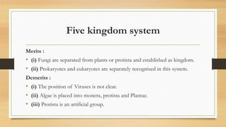
![4. Five kingdom system
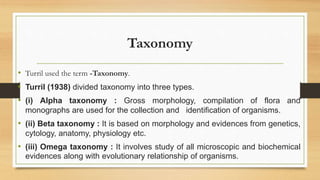
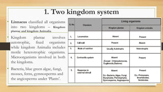
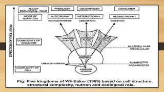
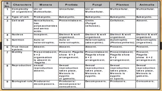
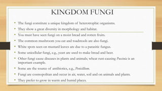
• It was proposed by Whittaker (1969). It is a phylogenetic system that is based on following
criteria.
• (i) Complexity of cell structure : Prokaryotes and eukaryotes.
• (ii) Complexity of organisms : i.e., thallus organisation (unicellular or multicellular organisms).
• (iii) Mode of nutrition : Autotrophic (holophytic) or heterotrophic [absorptive parasitic or
saprozoic ingestive (holozoic)]. It is major criteria of classification in this system.
• (iv) Ecological role of organism.
• (v) Phylogenetic relationship.
• The five kingdoms are Monera – Protista – Fungi – Plantae – Animalia.](https://image.slidesharecdn.com/chapter2biologicalclassificationmain-201002114610/85/Chapter-2-biological-classification-main-19-320.jpg)
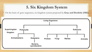
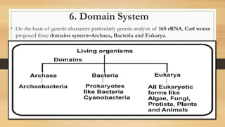


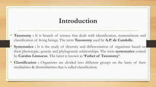
This document provides information on biological classification systems. It discusses taxonomy, systematics, and classification. It describes Turril's three types of taxonomy (alpha, beta, omega). It also summarizes classical and new systematics. The key classification systems discussed are the artificial, natural, and phylogenetic systems. It provides details on Linnaeus' artificial system and Bentham and Hooker's natural system. It concludes with descriptions of the two, three, four, five, and six kingdom classification systems.