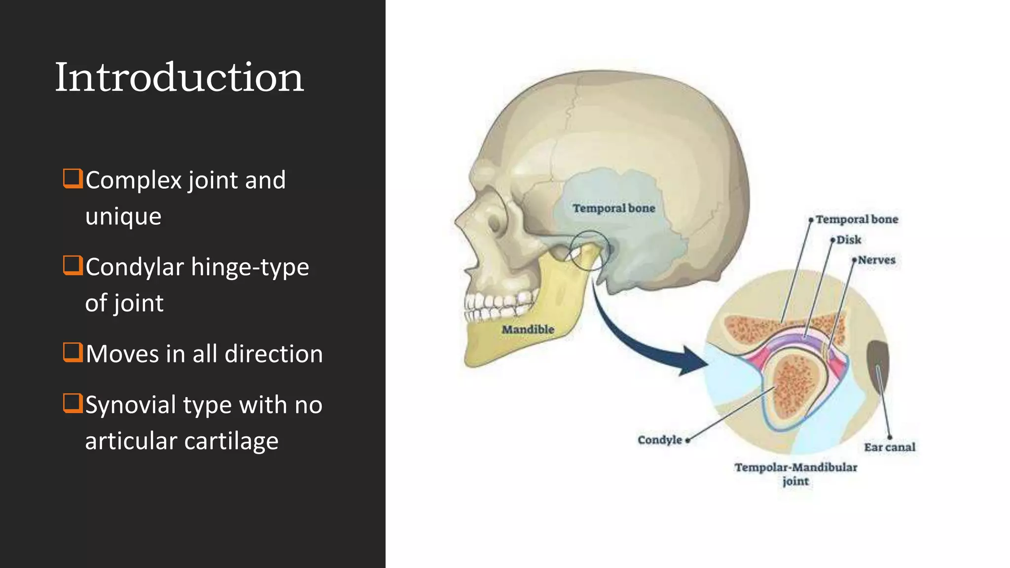
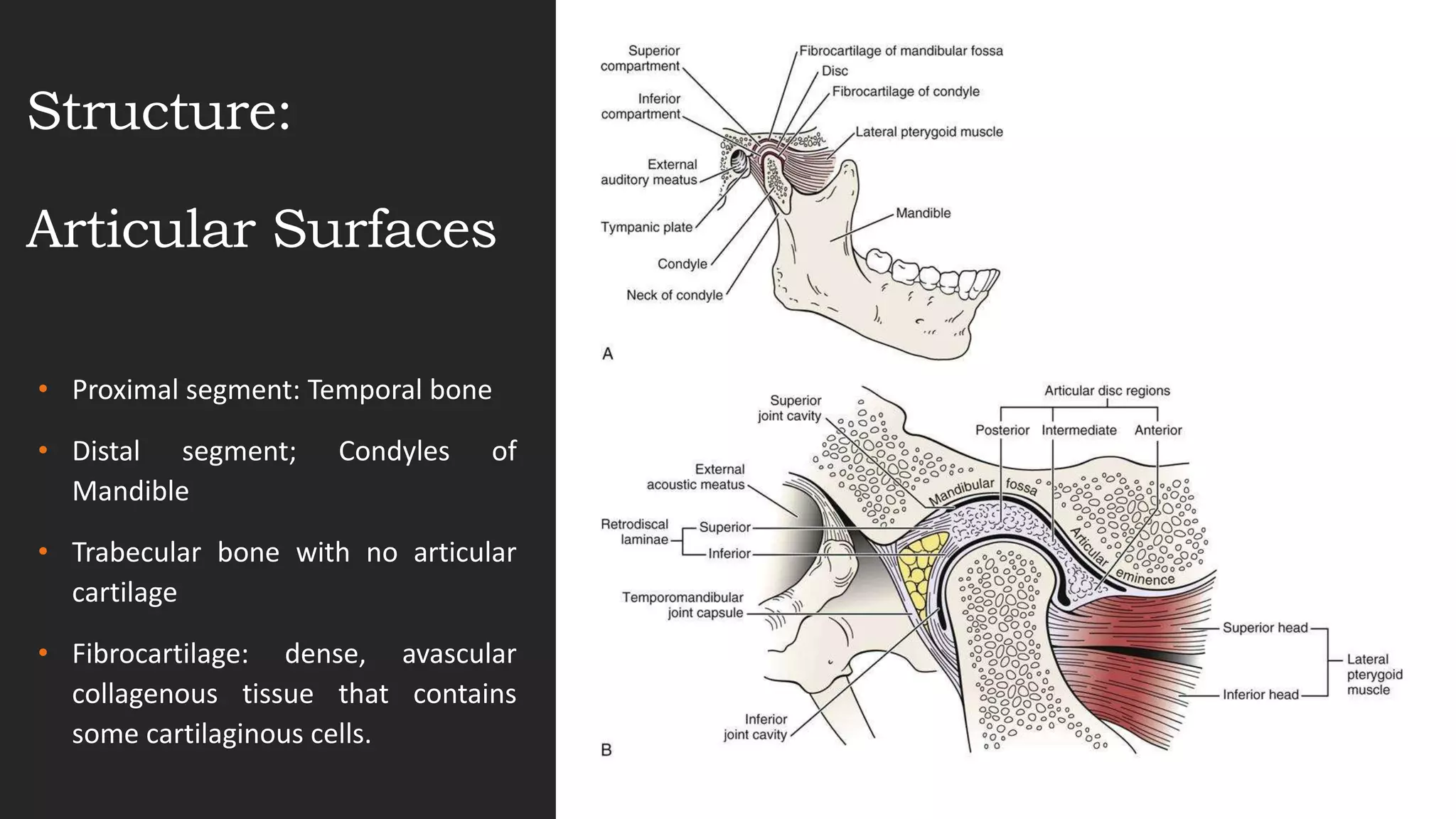
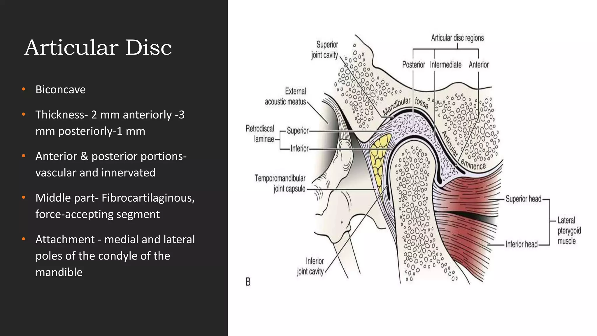
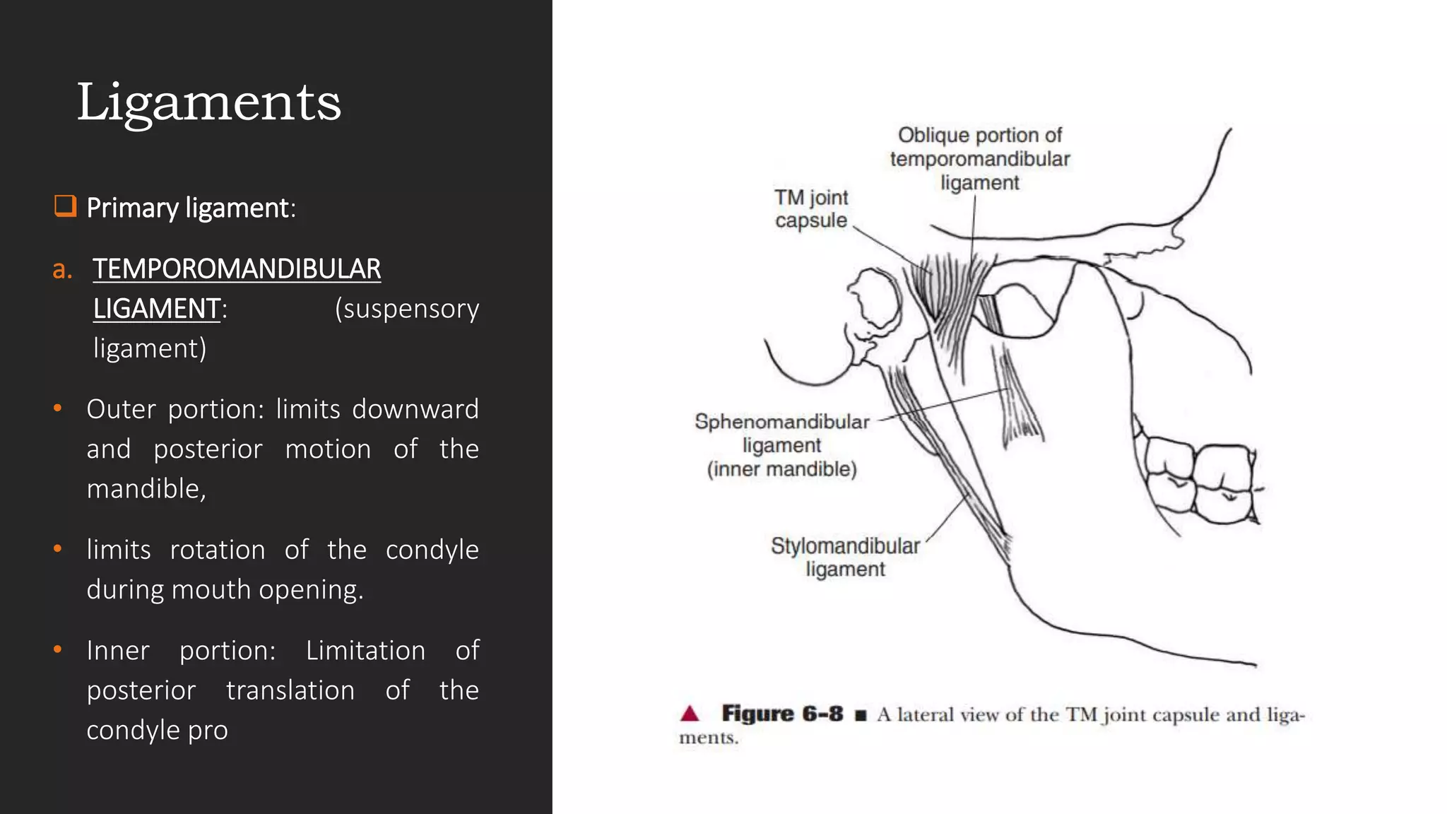
BIOMECHANICS:
TMJ
DR.QUAZI HUMA
MPT(NEUROSCIENCES)
ASST PROFESSOR
Objectives
Introduction
Structures: Articular Surfaces
Articular Disk Capsule and Ligaments
Upper and Lower Temporomandibular Joints
Function
Dysfunction
Introduction
Complex joint and
unique
Condylar hinge-type
of joint
Moves in all direction
Synovial type with no
articular cartilage
Structure:
Articular Surfaces
• Proximal segment: Temporal bone
• Distal segment; Condyles of
Mandible
• Trabecular bone with no articular
cartilage
• Fibrocartilage: dense, avascular
collagenous tissue that contains
some cartilaginous cells.
• Fibrocartilage - present in
areas, intended to withstand
repeated and high-level stress.
• For example – biting, chewing
• In closed mouth position, the
coronoid process sits under the
zygomatic arch, but it can be
palpated below the arch when
the mouth is open.
Articular Disc
• Biconcave
• Thickness- 2 mm anteriorly -3
mm posteriorly-1 mm
• Anterior & posterior portions-
vascular and innervated
• Middle part- Fibrocartilaginous,
force-accepting segment
• Attachment - medial and lateral
poles of the condyle of the
mandible
 Bilaminar retrodiskal pad-
1. Superior lamina – elastic in
nature
2. Inferior lamina – inelastic
 The superior lamina allows the
disk to translate anteriorly along
the articular eminence during
mouth opening ,its elastic
properties assist in repositioning
the disk posteriorly during mouth
closing.
 The inferior lamina simply serves
as a tether on the disk, limiting
forward translation
Capsule
• TM joint capsule is not as
well defined
• Anterior, medial, and
posterior capsule - quite
thin and loose
• Lateral aspect - stronger
and is reinforced with long
fibers
Ligaments
 Primary ligament:
a. TEMPOROMANDIBULAR
LIGAMENT: (suspensory
ligament)
• Outer portion: limits downward
and posterior motion of the
mandible,
• limits rotation of the condyle
during mouth opening.
• Inner portion: Limitation of
posterior translation of the
condyle pro
b. STYLOMANDIBULAR LIGAMENT:
• band of deep cervical fascia
• limitation to protrusion of the
the jaw
c.SPHENOMANDIBULAR
LIGAMENT:
• that it serves to suspend the
mandible
• to check the mandible from
excessive forward
translation.
Functions of Temporomandibular
Joint.
Most frequently used joints
Talking, chewing, and swallowing
Cartilage covering the articular surfaces is designed to tolerate repeated and high-
level stress.
Musculature is designed to provide both power and intricate control.
Speech requires fine control of the jaw, and the ability to chew requires great
strength.
Mandibular
Movements
i. Depression (mouth
opening)
ii. Elevation (mouth closing)
iii. Protrusion (jutting the chin
forward)
iv. Retrusion (sliding the teeth
backward)
v. Lateral deviation (sliding
the teeth to either side)
Muscles
• Mandibular depression –
Digastric muscle
• Mandibular elevation –
Temporalis, Masseter
• Protrusion -- bilateral action of
the masseter, medial pterygoid
and lateral pterygoid muscles
• Retrusion -- bilateral action of
the posterior fibers of the
temporalis muscles with
assistance of digastric muscle
Dysfunction of TMJ
Inflammatory
conditions
Capsulitis
Synovitis
Rheumatoid Arthrtis
Ank Spond
Capsular
Fibrosis
Overproduction of fibrous
connective tissue
Resulting in fibrosis
Articular disk
displacement
Osseous
Mobility
conditions
Hypermobility
Reciprocal click
Thank you!!!!!

BIOMECHANICS - Tmj.pptx

  • 1.
  • 2.
    Objectives Introduction Structures: Articular Surfaces ArticularDisk Capsule and Ligaments Upper and Lower Temporomandibular Joints Function Dysfunction
  • 3.
    Introduction Complex joint and unique Condylarhinge-type of joint Moves in all direction Synovial type with no articular cartilage
  • 4.
    Structure: Articular Surfaces • Proximalsegment: Temporal bone • Distal segment; Condyles of Mandible • Trabecular bone with no articular cartilage • Fibrocartilage: dense, avascular collagenous tissue that contains some cartilaginous cells.
  • 5.
    • Fibrocartilage -present in areas, intended to withstand repeated and high-level stress. • For example – biting, chewing • In closed mouth position, the coronoid process sits under the zygomatic arch, but it can be palpated below the arch when the mouth is open.
  • 6.
    Articular Disc • Biconcave •Thickness- 2 mm anteriorly -3 mm posteriorly-1 mm • Anterior & posterior portions- vascular and innervated • Middle part- Fibrocartilaginous, force-accepting segment • Attachment - medial and lateral poles of the condyle of the mandible
  • 7.
     Bilaminar retrodiskalpad- 1. Superior lamina – elastic in nature 2. Inferior lamina – inelastic  The superior lamina allows the disk to translate anteriorly along the articular eminence during mouth opening ,its elastic properties assist in repositioning the disk posteriorly during mouth closing.  The inferior lamina simply serves as a tether on the disk, limiting forward translation
  • 8.
    Capsule • TM jointcapsule is not as well defined • Anterior, medial, and posterior capsule - quite thin and loose • Lateral aspect - stronger and is reinforced with long fibers
  • 9.
    Ligaments  Primary ligament: a.TEMPOROMANDIBULAR LIGAMENT: (suspensory ligament) • Outer portion: limits downward and posterior motion of the mandible, • limits rotation of the condyle during mouth opening. • Inner portion: Limitation of posterior translation of the condyle pro
  • 10.
    b. STYLOMANDIBULAR LIGAMENT: •band of deep cervical fascia • limitation to protrusion of the the jaw c.SPHENOMANDIBULAR LIGAMENT: • that it serves to suspend the mandible • to check the mandible from excessive forward translation.
  • 11.
    Functions of Temporomandibular Joint. Mostfrequently used joints Talking, chewing, and swallowing Cartilage covering the articular surfaces is designed to tolerate repeated and high- level stress. Musculature is designed to provide both power and intricate control. Speech requires fine control of the jaw, and the ability to chew requires great strength.
  • 12.
    Mandibular Movements i. Depression (mouth opening) ii.Elevation (mouth closing) iii. Protrusion (jutting the chin forward) iv. Retrusion (sliding the teeth backward) v. Lateral deviation (sliding the teeth to either side)
  • 13.
    Muscles • Mandibular depression– Digastric muscle • Mandibular elevation – Temporalis, Masseter • Protrusion -- bilateral action of the masseter, medial pterygoid and lateral pterygoid muscles • Retrusion -- bilateral action of the posterior fibers of the temporalis muscles with assistance of digastric muscle
  • 14.
    Dysfunction of TMJ Inflammatory conditions Capsulitis Synovitis RheumatoidArthrtis Ank Spond Capsular Fibrosis Overproduction of fibrous connective tissue Resulting in fibrosis Articular disk displacement Osseous Mobility conditions Hypermobility Reciprocal click
  • 16.