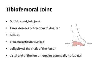
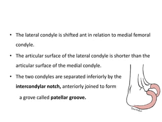
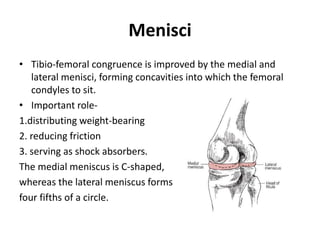
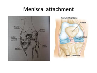
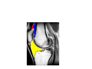
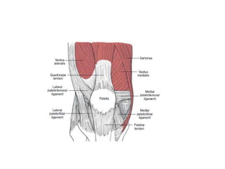
BIOMECHANICS OF KNEE JOINT
Dr.SHYAM D.GANVIR
PhD
The Knee Joint
 Joint Structure
• Joint functions
• Joint Kinematics
• Forces
The knee complex is composed of two distinct articulations
located within a single joint capsule: TFJ &PFJ
Tibiofemoral Joint
• Double condyloid joint
• Three degrees of freedom of Angular
• Femur-
• proximal articular surface
• obliquity of the shaft of the femur
• distal end of the femur remains essentially horizontal.
• The lateral condyle is shifted ant in relation to medial femoral
condyle.
• The articular surface of the lateral condyle is shorter than the
articular surface of the medial condyle.
• The two condyles are separated inferiorly by the
intercondylar notch, anteriorly joined to form
a grove called patellar groove.
Tibia
• The distal articular surface of the knee joint is formed by
asymmetrical medial and lateral tibial plateaus.
• Medial tibial plateau is longer in the anteroposterior direction
than is the lateral plateau.
• Lateral tibial articular cartilage is thicker
• proximal tibia is larger than the shaft.
• Overhang the shaft posteriorly (tibial plateau) 7-10 degree
• Inter-condylar tubercles – separates medial and lateral tibial
condyles.
• bony architecture of the tibial
plateaus does not match up well
with the convexity of the femoral
condyle, in such case menisci
are necessary to improve joint
congruency.
Tibio-femoral Alignment
and Weight-Bearing Forces
• Anatomic (longitudinal) axis of the femur is oblique, directed inferiorly and medially
from proximal to distal end.
• Anatomic axis of tibia is directed vertically, consequently the femoral and tibial
longitudinal axes normally form an angle medially at the knee joint of 180 to 185
degree.
• The femur is angled up to 5 degree off vertical, creating a slight physiologic (normal)
valgus angle at the knee
• (Abnormal condition )genu valgum- M-TF angle >185 degree(knock knees)
• (Abnormal condition)Genu varum - M-TF angle <185 degree(bow legs)
Tibio-femoral alignment &weight bearing forces
Alternative method of
measuring tibiofemoral
alignment is performed by
drawing a line from the
center of the femoral head
to the center of head of
talus-this line represents
the mechanical or
wt.bearing line.
Knock knee &Bow Leg
Corrections of mal-alignment
• Realignment procedure at the knee-called “high tibial osteotomy”
• Lessen the compressive forces on the damaged painful tibio-
femoral compartment.
• In case of genu-Varum or valgum –surgical fracture in the tibia or
femure is done to realign the limb in the neutral position.
• Less invasive method to diminish malalignment –medial or lateral
wedges or knee brace that shifts weight bearing to the uninvolved
compartment,so called “unloading Braces”
Menisci
• Tibio-femoral congruence is improved by the medial and
lateral menisci, forming concavities into which the femoral
condyles to sit.
• Important role-
1.distributing weight-bearing forces.
2. reducing friction
3. serving as shock absorbers.
The medial meniscus is C-shaped,
whereas the lateral meniscus forms
four fifths of a circle.
Menisci attachment
• .ANT.—Tranverse lig.
Both menisci are attached to patella-via-patello
meniscal ligament.
Menisci are connected to tibial condyle by –
coronary ligaments
Medial collateral lig.(MCL)
Ant.&Post.horns of medial menisci are attached
to ant.cruciate &post.cruciate lig.respectively.
Posteriorly ,lateral meniscus attached to PCL
&medial femoral condyle through
meniscofemoral ligament.
Meniscal attachment
Role of the menisci
• The strong attachment to the menisci prevent them from
being squeezed out during compression of tibial femoral joint.
• Allowing greater contact area between menisci &femur
• If femoral condyle sat directly on the relatively flat tibial
plateau,there would be little contact ,between bony surfaces.
• With the addition of the menisci ,the contact at the
tibiofemoral joint is increased and joint stress (force per unit
area)reduced on the joint articular.
Knee meniscal tear
• After removal of meniscus ,the contact area
in the tibio-femoral joint is decreased, which
increases joint stress.
• Removal of the menisci nearly doubles the
articular cartilage stress on the femurand
multiplies the force by six to seven times on
the tibial plateau.
• The increase in joint stress may contribute to
degenerative changes within the
tibiofemoral joint.
Joint stress
Meniscal Nutrition and Innervation
• During the first year of life, the meniscus contains blood
vessels throughout the meniscal body.
• Once Weight bearing initiated,reduces vascularity centrally, so
rely on diffusion of synovial fluid.
• Fluid diffusion for nutrition supply is dependent on joint
loading..
• Menisci are Innervated with free nerve endings (nociceptors)
and three different mechanoreceptors (Ruffini corpuscles,
pacinian corpuscles, and Golgi tendon organs).
Joint capsule
• Despite the congruency provided by the menisci, the joint
stability is heavily dependent on the surrounding structures.
• Stability and mobility varies in different position of joint.
• Congruency and ligament tautness is maximum in extension
(closed packed position).
• In flexion the periarticular structures in lax.
• Joint capsule encloses both tibiofemoral and patellofemoral
joint , it is large and lax.
• Consist of 2 layers, exterior or superficial layer and an thinner
internal synovial membrane.
• Attachment-
Weight bearing /non weight bearing
• Open chain /closed chain activities
Q-angle
The Q angle is formed between:
A line representing the resultant line of force
of the quadriceps, made by connecting a point
near the ASIS to the mid-point of the Patella.
The Q angle can be measured in laying or
standing.
Synovial layer of joint capsule
• The synovial membrane forms the inner lining in much of the
knee joint capsule.
• Synovial tissue secrete and absorb synovial fluid into the joint
for lubrication and to provide nutrition to avascular
structures, such as the menisci.
• Attachment –
• Fat pad-
Fibrous layer of joint capsule
• Superficial to the synovial lining of the knee joint lies the
fibrous joint capsule, which provides passive support for the
joint.
• The anterior portion of the knee joint capsule is called the
extensor retinaculum.
• Deep to this layer, the medial and lateral retinaculare
• The medial portion of the joint capsule is composed of the
deep and superficial portions of the MCL.
• Laterally, the joint capsule is composed superficially of the IT
band and its thick fascia lata.
• The capsule is reinforced posterolaterally by the arcuate
ligament and postero medially by the posterior oblique
ligament (POL)
Ligaments
• The roles of the ligaments are variously credited with resisting or
controlling:
1. excessive knee extension
2. varus and valgus stresses at the knee
3. anterior or posterior displacement of the tibia beneath the femur
4. medial or lateral rotation of the tibia beneath the femur
5. combinations of anteroposterior displacements and rotations of
the tibia, together known as rotatory stabilization of the tibia
• Medial Collateral Ligament
• Lateral Collateral Ligament
• Anterior Cruciate Ligament
• Posterior Cruciate Ligament
• Ligaments of the Posterior Capsule
Medial Collateral Ligament
• divided into a superficial portion and a deep
portion, separated by a bursa.
• The superficial portion of the MCL arises
proximally from the medial femoral
epicondyle and travels distally to insert into
the medial aspect of the proximal tibia.
• The deep portion originates from the inferior
aspect of the medial femoral condyle, and inserts
on the proximal aspect of the medial tibial
plateau.
• the deep portion of the MCL is rigidly affixed to
the medial border of medial meniscus.
• The MCL restraint to excessive abduction (valgus)
and lateral rotation stresses at the knee.
• The knee joint is best able to resist a valgus stress
at full extension.
• The MCL has the capacity to heal when
ruptured or damaged, because of its rich
blood supply.
• remodeling process can take up to a year.
Lateral Collateral Ligament
• Attaches Proximally from the lateral femoral
condyle.
• The LCL then travels distally to the fibular
head where it joins with the tendon of the
biceps femoris muscle to form the conjoined
tendon
• Its an extra capsular ligament.
• The LCL is responsible for checking varus stresses, and like the
MCL, limits varus motion most successfully at full extension.
Anterior Cruciate Ligament
Iliotibial band
• The IT band (or ITB) or IT tract is formed proximally from the fascia
investing the tensor fascia lata, the gluteus maximus, and the
gluteus medius muscles.
• The it band continues distally
to be inserted it to the
anterolateral tibial tubercle.
• It reinforces the anterolateral
aspect of the knee joint.
• Despite the muscular attachment proximally the IT band
remains a passive structure at the knee joint.
• Movement – anterior in extension, posterior to femoral
condyle in knee flexion.
• The IT band, remains consistently taut, regardless of the hip
or knee’s position.
• The fibrous connections of the IT band to the biceps femoris
and vastus lateralis muscles form a sling behind the lateral
femoral condyle, assisting the ACL in checking posterior
femoral (or anterior tibial) translation when the knee joint is
nearly full extension.
• With the knee in flexion, the combination of the IT band, the
LCL, and the popliteal tendon crossing over each other
increases the stability of the lateral side of the joint.
• Despite its lateral location, the IT band alone provides only
minimal resistance to lateral joint space opening.
• IT band also attaches to the patella via the lateral
patellofemoral ligament of the lateral retinaculum.
Bursa
• Bursae, prevent or limit degenerative forces.
• There are Three of the knee joint’s bursae that communicate
with the synovial capsule, the suprapatellar bursa, the
subpopliteal bursa, and the gastrocnemius bursa.
• suprapatellar bursa lies between the quadriceps tendon and
the anterior femur, superior to the patella.
• subpopliteal bursa lies between the tendon of the popliteus
muscle and the lateral femoral condyle, and the
gastrocnemius bursa lies between the tendon of the medial
head of the gastrocnemius muscle and the medial femoral
condyle.
• These three bursa allow the synovial fluid to move from one
end to other end of joint during flexion and extension.
• In extension synovial fluid is shifted anteriorly, in flexion fluid
is forced posteriorly.
• In semi flexed position the synovial is
under least amount of pressure.
• In Knee effusion following an injury
semi-flexed position relieves tension
in the capsule and, therefore,
minimizes discomfort.
• Other bursa associated with the knee
joint –
• suprapatellar bursa
• Subpopliteal bursa
• gastrocnemius bursa
• Prepatellar bursa
• Infrapatellar bursa
• deep infrapatellar bursa
• There are also several small bursae that are associated with the ligaments
of the knee joint.
• (between LCL and BF, MCL – Superficial and deep area, semitendinosus
and gracilis)
Tibiofemoral Joint Function
(joint kinematics)
• Motions present are F/ E (primary angular motion)
• Medial and lateral rotation(internal/external), valgus and varus
motion(adducton/abduction) also occur but to lesser extent.
• In addition to the angular motion, there is some amount antero-
posterior, medial and lateral translation.
• These translation are seen in normal knee joint because of joint
incongruency and ligamental laxity
• These joint motions are necessary for normal joint motions to
occur.
• If excessive they are considered abnormal.
Flexion/Extension
• This motion occurs along an immaginary horizontal line
passing through the femoral epicondyles.
• The articular surface of femur is large and that of the tibia is
comparatively small, now this creates a problem once the
femur begins to flex on fixed tibia.
• Now what problem does it creat?
• As flexion of femur occurs on the tibia 1st there is rolling of
femur on the tibia which is fallowed by an anterior glide.(that
is spin along with linear displacement after 25 degree of
flexion.)
• During extension the is reversal that is anterior rolling
fallowed by posterior gliding.
• Now all this was when the tibia was fixed…
• Now what if the femur is fixed…?
• the tibia both, rolls and glides
simultaneously.
Role of the Cruciate Ligaments and Menisci
in Flexion/Extension
• ACL becomes taut while posterior rolling
• PCL becomes taut while anterior polling
• Anterior gliding of the femur while knee flexion is facilitated by the
menisci.
• The wedge shape of the menisci posteriorly ,forces the femoral condyle to
roll “uphill” as the knee flexes.
• Posterior deformation occurs, this deformation allows menisci to stay in
place.
• On extension the menisci returns to neutral position.
• Failure of the menisci to distort in the proper direction can result in limitations of
joint motion and/or damage to the menisci
Oblique contact of the femur with the
wedge-shaped meniscus results in the
forces of meniscus on femur(MF) and
femur on meniscus(FM).
This can be resolved into vertical and
shear components .Shear 1 assist the
femur in its forward glide during
flexion and shear 2 assist in the
posterior migration of the menisci
that occur with knee flexion.
Flexion extension ROM
• Passive ROM of knee flexion is considered about 130 to 140.
• During squatting about 160
• Normal gait on level ground needs about 60 to 70 of knee
flexion.
• ascending stairs requires about 80.
• Sitting down and arising from the chair approx 90.
• 5 degree of hyperextension is considered under normal limits.
Medial and lateral rotation
• Medial and lateral rotation of the knee joint are angular
motions.
• These axial rotations of the knee joint occur about a
longitudinal axis that runs through or close to the medial tibial
intercondylar tubercle
• the medial condyle acts as the pivot point while the lateral condyles move
through a greater arc of motion, regardless of the direction of rotation.
• Axial rotation is permitted by articular incongruence and ligamentous
laxity.
• the range of knee joint rotation depends on the flexion/extension position
of the knee.
Valgus (Abduction)/Varus (Adduction)
• Frontal plane ROM is typically only 8 degree at full extension,
and 13 degree with 20 degree of knee flexion.
• Excessive frontal plane motion indicated ligament
insufficiency.
Coupled Motions
• Automatic or Locking Mechanism of the Knee
locking or screw home mechanism
Coupled motion
• Biplaner ,intraarticular motion can occur because of the
oblique orientation of the axes of motion with respect to the
bony levers.
• The true flexion /extension axis is not perpendicular to the
shafts of the femur and tibia.
• Therefore flexion and extension do not occur as pure saggital
plane motions but include frontal plane components termed
“coupled motion”
Automatic or locking mechanism of
the knee
• There is an obligatory lateral rotation of the tibia that
accompanies the final stages of knee extension that is not
voluntary or produced by muscular forces .
• This coupled motion lateral rotation with extension is referred
to as automatic or terminal rotation.
• Last 30 degree of non weight bearing knee extension ,the
shorter lateral tibial plateau /femoral condyle pair completes
its rolling and gliding before the longer articular surface do.
• As extension continues,longer medial plateu continues to roll
and to glide anteriorly after the lateral side of the plateu has
halted.
• Tibial tubercle now become lodged in the intercondylar notch
,the menisci are tightly interposed between the tibial and
femoral condyle and ligament are taut…..consequently
automatic rotation is also know as the “locking or screw home
mechanism”
Muscles
• muscles crossing the knee are called flexors or extensors, as
the primary function occurring at the knee is flexion and
extension.
• Each muscle has a MA which is capable of producing a frontal
and transverse plane mostions
summary

Knee joint biomechanics

  • 1.
    BIOMECHANICS OF KNEEJOINT Dr.SHYAM D.GANVIR PhD
  • 2.
    The Knee Joint Joint Structure • Joint functions • Joint Kinematics • Forces The knee complex is composed of two distinct articulations located within a single joint capsule: TFJ &PFJ
  • 3.
    Tibiofemoral Joint • Doublecondyloid joint • Three degrees of freedom of Angular • Femur- • proximal articular surface • obliquity of the shaft of the femur • distal end of the femur remains essentially horizontal.
  • 4.
    • The lateralcondyle is shifted ant in relation to medial femoral condyle. • The articular surface of the lateral condyle is shorter than the articular surface of the medial condyle. • The two condyles are separated inferiorly by the intercondylar notch, anteriorly joined to form a grove called patellar groove.
  • 5.
    Tibia • The distalarticular surface of the knee joint is formed by asymmetrical medial and lateral tibial plateaus. • Medial tibial plateau is longer in the anteroposterior direction than is the lateral plateau. • Lateral tibial articular cartilage is thicker • proximal tibia is larger than the shaft. • Overhang the shaft posteriorly (tibial plateau) 7-10 degree
  • 6.
    • Inter-condylar tubercles– separates medial and lateral tibial condyles. • bony architecture of the tibial plateaus does not match up well with the convexity of the femoral condyle, in such case menisci are necessary to improve joint congruency.
  • 7.
    Tibio-femoral Alignment and Weight-BearingForces • Anatomic (longitudinal) axis of the femur is oblique, directed inferiorly and medially from proximal to distal end. • Anatomic axis of tibia is directed vertically, consequently the femoral and tibial longitudinal axes normally form an angle medially at the knee joint of 180 to 185 degree. • The femur is angled up to 5 degree off vertical, creating a slight physiologic (normal) valgus angle at the knee • (Abnormal condition )genu valgum- M-TF angle >185 degree(knock knees) • (Abnormal condition)Genu varum - M-TF angle <185 degree(bow legs)
  • 8.
    Tibio-femoral alignment &weightbearing forces Alternative method of measuring tibiofemoral alignment is performed by drawing a line from the center of the femoral head to the center of head of talus-this line represents the mechanical or wt.bearing line.
  • 9.
  • 10.
    Corrections of mal-alignment •Realignment procedure at the knee-called “high tibial osteotomy” • Lessen the compressive forces on the damaged painful tibio- femoral compartment. • In case of genu-Varum or valgum –surgical fracture in the tibia or femure is done to realign the limb in the neutral position. • Less invasive method to diminish malalignment –medial or lateral wedges or knee brace that shifts weight bearing to the uninvolved compartment,so called “unloading Braces”
  • 11.
    Menisci • Tibio-femoral congruenceis improved by the medial and lateral menisci, forming concavities into which the femoral condyles to sit. • Important role- 1.distributing weight-bearing forces. 2. reducing friction 3. serving as shock absorbers. The medial meniscus is C-shaped, whereas the lateral meniscus forms four fifths of a circle.
  • 12.
    Menisci attachment • .ANT.—Tranverselig. Both menisci are attached to patella-via-patello meniscal ligament. Menisci are connected to tibial condyle by – coronary ligaments Medial collateral lig.(MCL) Ant.&Post.horns of medial menisci are attached to ant.cruciate &post.cruciate lig.respectively. Posteriorly ,lateral meniscus attached to PCL &medial femoral condyle through meniscofemoral ligament.
  • 13.
  • 14.
    Role of themenisci • The strong attachment to the menisci prevent them from being squeezed out during compression of tibial femoral joint. • Allowing greater contact area between menisci &femur • If femoral condyle sat directly on the relatively flat tibial plateau,there would be little contact ,between bony surfaces. • With the addition of the menisci ,the contact at the tibiofemoral joint is increased and joint stress (force per unit area)reduced on the joint articular.
  • 15.
    Knee meniscal tear •After removal of meniscus ,the contact area in the tibio-femoral joint is decreased, which increases joint stress. • Removal of the menisci nearly doubles the articular cartilage stress on the femurand multiplies the force by six to seven times on the tibial plateau. • The increase in joint stress may contribute to degenerative changes within the tibiofemoral joint.
  • 16.
  • 18.
    Meniscal Nutrition andInnervation • During the first year of life, the meniscus contains blood vessels throughout the meniscal body. • Once Weight bearing initiated,reduces vascularity centrally, so rely on diffusion of synovial fluid. • Fluid diffusion for nutrition supply is dependent on joint loading.. • Menisci are Innervated with free nerve endings (nociceptors) and three different mechanoreceptors (Ruffini corpuscles, pacinian corpuscles, and Golgi tendon organs).
  • 19.
    Joint capsule • Despitethe congruency provided by the menisci, the joint stability is heavily dependent on the surrounding structures. • Stability and mobility varies in different position of joint. • Congruency and ligament tautness is maximum in extension (closed packed position). • In flexion the periarticular structures in lax.
  • 21.
    • Joint capsuleencloses both tibiofemoral and patellofemoral joint , it is large and lax. • Consist of 2 layers, exterior or superficial layer and an thinner internal synovial membrane. • Attachment-
  • 22.
    Weight bearing /nonweight bearing • Open chain /closed chain activities
  • 23.
    Q-angle The Q angleis formed between: A line representing the resultant line of force of the quadriceps, made by connecting a point near the ASIS to the mid-point of the Patella. The Q angle can be measured in laying or standing.
  • 24.
    Synovial layer ofjoint capsule • The synovial membrane forms the inner lining in much of the knee joint capsule. • Synovial tissue secrete and absorb synovial fluid into the joint for lubrication and to provide nutrition to avascular structures, such as the menisci. • Attachment – • Fat pad-
  • 26.
    Fibrous layer ofjoint capsule • Superficial to the synovial lining of the knee joint lies the fibrous joint capsule, which provides passive support for the joint. • The anterior portion of the knee joint capsule is called the extensor retinaculum. • Deep to this layer, the medial and lateral retinaculare
  • 27.
    • The medialportion of the joint capsule is composed of the deep and superficial portions of the MCL. • Laterally, the joint capsule is composed superficially of the IT band and its thick fascia lata. • The capsule is reinforced posterolaterally by the arcuate ligament and postero medially by the posterior oblique ligament (POL)
  • 29.
    Ligaments • The rolesof the ligaments are variously credited with resisting or controlling: 1. excessive knee extension 2. varus and valgus stresses at the knee 3. anterior or posterior displacement of the tibia beneath the femur 4. medial or lateral rotation of the tibia beneath the femur 5. combinations of anteroposterior displacements and rotations of the tibia, together known as rotatory stabilization of the tibia
  • 30.
    • Medial CollateralLigament • Lateral Collateral Ligament • Anterior Cruciate Ligament • Posterior Cruciate Ligament • Ligaments of the Posterior Capsule
  • 31.
    Medial Collateral Ligament •divided into a superficial portion and a deep portion, separated by a bursa. • The superficial portion of the MCL arises proximally from the medial femoral epicondyle and travels distally to insert into the medial aspect of the proximal tibia.
  • 32.
    • The deepportion originates from the inferior aspect of the medial femoral condyle, and inserts on the proximal aspect of the medial tibial plateau. • the deep portion of the MCL is rigidly affixed to the medial border of medial meniscus. • The MCL restraint to excessive abduction (valgus) and lateral rotation stresses at the knee. • The knee joint is best able to resist a valgus stress at full extension.
  • 33.
    • The MCLhas the capacity to heal when ruptured or damaged, because of its rich blood supply. • remodeling process can take up to a year.
  • 34.
    Lateral Collateral Ligament •Attaches Proximally from the lateral femoral condyle. • The LCL then travels distally to the fibular head where it joins with the tendon of the biceps femoris muscle to form the conjoined tendon
  • 35.
    • Its anextra capsular ligament. • The LCL is responsible for checking varus stresses, and like the MCL, limits varus motion most successfully at full extension.
  • 36.
  • 37.
    Iliotibial band • TheIT band (or ITB) or IT tract is formed proximally from the fascia investing the tensor fascia lata, the gluteus maximus, and the gluteus medius muscles. • The it band continues distally to be inserted it to the anterolateral tibial tubercle. • It reinforces the anterolateral aspect of the knee joint.
  • 38.
    • Despite themuscular attachment proximally the IT band remains a passive structure at the knee joint. • Movement – anterior in extension, posterior to femoral condyle in knee flexion. • The IT band, remains consistently taut, regardless of the hip or knee’s position.
  • 39.
    • The fibrousconnections of the IT band to the biceps femoris and vastus lateralis muscles form a sling behind the lateral femoral condyle, assisting the ACL in checking posterior femoral (or anterior tibial) translation when the knee joint is nearly full extension.
  • 40.
    • With theknee in flexion, the combination of the IT band, the LCL, and the popliteal tendon crossing over each other increases the stability of the lateral side of the joint. • Despite its lateral location, the IT band alone provides only minimal resistance to lateral joint space opening. • IT band also attaches to the patella via the lateral patellofemoral ligament of the lateral retinaculum.
  • 41.
    Bursa • Bursae, preventor limit degenerative forces. • There are Three of the knee joint’s bursae that communicate with the synovial capsule, the suprapatellar bursa, the subpopliteal bursa, and the gastrocnemius bursa. • suprapatellar bursa lies between the quadriceps tendon and the anterior femur, superior to the patella.
  • 42.
    • subpopliteal bursalies between the tendon of the popliteus muscle and the lateral femoral condyle, and the gastrocnemius bursa lies between the tendon of the medial head of the gastrocnemius muscle and the medial femoral condyle. • These three bursa allow the synovial fluid to move from one end to other end of joint during flexion and extension.
  • 43.
    • In extensionsynovial fluid is shifted anteriorly, in flexion fluid is forced posteriorly.
  • 44.
    • In semiflexed position the synovial is under least amount of pressure. • In Knee effusion following an injury semi-flexed position relieves tension in the capsule and, therefore, minimizes discomfort. • Other bursa associated with the knee joint –
  • 45.
    • suprapatellar bursa •Subpopliteal bursa • gastrocnemius bursa • Prepatellar bursa • Infrapatellar bursa • deep infrapatellar bursa • There are also several small bursae that are associated with the ligaments of the knee joint. • (between LCL and BF, MCL – Superficial and deep area, semitendinosus and gracilis)
  • 47.
    Tibiofemoral Joint Function (jointkinematics) • Motions present are F/ E (primary angular motion) • Medial and lateral rotation(internal/external), valgus and varus motion(adducton/abduction) also occur but to lesser extent. • In addition to the angular motion, there is some amount antero- posterior, medial and lateral translation. • These translation are seen in normal knee joint because of joint incongruency and ligamental laxity • These joint motions are necessary for normal joint motions to occur. • If excessive they are considered abnormal.
  • 48.
    Flexion/Extension • This motionoccurs along an immaginary horizontal line passing through the femoral epicondyles. • The articular surface of femur is large and that of the tibia is comparatively small, now this creates a problem once the femur begins to flex on fixed tibia. • Now what problem does it creat?
  • 49.
    • As flexionof femur occurs on the tibia 1st there is rolling of femur on the tibia which is fallowed by an anterior glide.(that is spin along with linear displacement after 25 degree of flexion.)
  • 50.
    • During extensionthe is reversal that is anterior rolling fallowed by posterior gliding. • Now all this was when the tibia was fixed… • Now what if the femur is fixed…?
  • 51.
    • the tibiaboth, rolls and glides simultaneously.
  • 52.
    Role of theCruciate Ligaments and Menisci in Flexion/Extension • ACL becomes taut while posterior rolling • PCL becomes taut while anterior polling • Anterior gliding of the femur while knee flexion is facilitated by the menisci. • The wedge shape of the menisci posteriorly ,forces the femoral condyle to roll “uphill” as the knee flexes. • Posterior deformation occurs, this deformation allows menisci to stay in place. • On extension the menisci returns to neutral position.
  • 54.
    • Failure ofthe menisci to distort in the proper direction can result in limitations of joint motion and/or damage to the menisci Oblique contact of the femur with the wedge-shaped meniscus results in the forces of meniscus on femur(MF) and femur on meniscus(FM). This can be resolved into vertical and shear components .Shear 1 assist the femur in its forward glide during flexion and shear 2 assist in the posterior migration of the menisci that occur with knee flexion.
  • 55.
    Flexion extension ROM •Passive ROM of knee flexion is considered about 130 to 140. • During squatting about 160 • Normal gait on level ground needs about 60 to 70 of knee flexion. • ascending stairs requires about 80. • Sitting down and arising from the chair approx 90. • 5 degree of hyperextension is considered under normal limits.
  • 56.
    Medial and lateralrotation • Medial and lateral rotation of the knee joint are angular motions. • These axial rotations of the knee joint occur about a longitudinal axis that runs through or close to the medial tibial intercondylar tubercle
  • 57.
    • the medialcondyle acts as the pivot point while the lateral condyles move through a greater arc of motion, regardless of the direction of rotation. • Axial rotation is permitted by articular incongruence and ligamentous laxity. • the range of knee joint rotation depends on the flexion/extension position of the knee.
  • 58.
    Valgus (Abduction)/Varus (Adduction) •Frontal plane ROM is typically only 8 degree at full extension, and 13 degree with 20 degree of knee flexion. • Excessive frontal plane motion indicated ligament insufficiency.
  • 59.
    Coupled Motions • Automaticor Locking Mechanism of the Knee locking or screw home mechanism
  • 60.
    Coupled motion • Biplaner,intraarticular motion can occur because of the oblique orientation of the axes of motion with respect to the bony levers. • The true flexion /extension axis is not perpendicular to the shafts of the femur and tibia. • Therefore flexion and extension do not occur as pure saggital plane motions but include frontal plane components termed “coupled motion”
  • 61.
    Automatic or lockingmechanism of the knee • There is an obligatory lateral rotation of the tibia that accompanies the final stages of knee extension that is not voluntary or produced by muscular forces . • This coupled motion lateral rotation with extension is referred to as automatic or terminal rotation. • Last 30 degree of non weight bearing knee extension ,the shorter lateral tibial plateau /femoral condyle pair completes its rolling and gliding before the longer articular surface do.
  • 62.
    • As extensioncontinues,longer medial plateu continues to roll and to glide anteriorly after the lateral side of the plateu has halted. • Tibial tubercle now become lodged in the intercondylar notch ,the menisci are tightly interposed between the tibial and femoral condyle and ligament are taut…..consequently automatic rotation is also know as the “locking or screw home mechanism”
  • 64.
    Muscles • muscles crossingthe knee are called flexors or extensors, as the primary function occurring at the knee is flexion and extension. • Each muscle has a MA which is capable of producing a frontal and transverse plane mostions
  • 65.